FUSE Binding Protein 1 Facilitates Persistent Hepatitis C Virus
Replication in Hepatoma Cells by Regulating Tumor Suppressor p53
Updesh Dixit,aAshutosh K. Pandey,aZhihe Liu,a*Sushil Kumar,aMatthew B. Neiditch,aKenneth M. Klein,bVirendra N. Pandeya
Department of Microbiology, Biochemistry, and Molecular Genetics,aand Department of Pathology and Laboratory Medicine,bRutgers New Jersey Medical School, Rutgers, the State University of New Jersey, Newark, New Jersey, USA
ABSTRACT
Hepatitis C virus (HCV) is a leading cause of chronic hepatitis C (CHC), liver cirrhosis, and hepatocellular carcinoma (HCC).
Immunohistochemistry of archived HCC tumors showed abundant FBP1 expression in HCC tumors with the CHC background.
Oncomine data analysis of normal versus HCC tumors with the CHC background indicated a 4-fold increase in FBP1 expression
with a concomitant 2.5-fold decrease in the expression of p53. We found that FBP1 promotes HCV replication by inhibiting p53
and regulating BCCIP and TCTP, which are positive and negative regulators of p53, respectively. The severe inhibition of HCV
replication in FBP1-knockdown Huh7.5 cells was restored to a normal level by downregulation of either p53 or BCCIP.
Al-though p53 in Huh7.5 cells is transcriptionally inactive as a result of Y220C mutation, we found that the activation and DNA
binding ability of Y220C p53 were strongly suppressed by FBP1 but significantly activated upon knockdown of FBP1. Transient
expression of FBP1 in FBP1 knockdown cells fully restored the control phenotype in which the DNA binding ability of p53 was
strongly suppressed. Using electrophoretic mobility shift assay (EMSA) and isothermal titration calorimetry (ITC), we found no
significant difference in
in vitro
target DNA binding affinity of recombinant wild-type p53 and its Y220C mutant p53. However,
in the presence of recombinant FBP1, the DNA binding ability of p53 is strongly inhibited. We confirmed that FBP1
downregu-lates BCCIP, p21, and p53 and upregudownregu-lates TCTP under radiation-induced stress. Since FBP1 is overexpressed in most HCC
tu-mors with an HCV background, it may have a role in promoting persistent virus infection and tumorigenesis.
IMPORTANCE
It is our novel finding that FUSE binding protein 1 (FBP1) strongly inhibits the function of tumor suppressor p53 and is an
es-sential host cell factor required for HCV replication. Oncomine data analysis of a large number of samples has revealed that
overexpression of FBP1 in most HCC tumors with chronic hepatitis C is significantly linked with the decreased expression level
of p53. The most significant finding is that FBP1 not only physically interacts with p53 and interferes with its binding to the
tar-get DNA but also functions as a negative regulator of p53 under cellular stress. FBP1 is barely detectable in normal differentiated
cells; its overexpression in HCC tumors with the CHC background suggests that FBP1 has an important role in promoting HCV
infection and HCC tumors by suppressing p53.
H
epatitis C virus (HCV) infection is a leading cause of chronic
liver diseases. More than a decade after the identification of
HCV as the major causative agent of non-A, non-B hepatitis (
1
),
molecular strategies for complete eradication of HCV infection
are actively pursued. HCV is the major cause of chronic liver
dis-ease. According to new findings from the U.S. Centers for Disease
Control and Prevention (CDC), the number of individuals in the
U.S. living with chronic hepatitis C virus infection is about 2.7
million (
2
). Globally, the number of people with HCV is greater
than 185 million (
3
). During the past 3 years, the U.S. Food and
Drug Administration has approved four new medications
(boce-previr, tela(boce-previr, sofosbuvir, and simeprevir) for treatment of
HCV infection, and many new drugs are under development.
There has been a renewed effort by the CDC to prevent
HCV-associated complications by improving treatment. However, the
cost of HCV treatment is highly prohibitive; it costs $80,000 for a
three-month treatment course with the recently approved
sofos-buvir (Gilead Sciences, CA).
Although the majority of HCV-infected persons are unaware
of their infection (
4
), 15 to 25% of them clear the virus without
treatment, while the majority of infections persist, leading to
chronic hepatitis C (CHC), which is closely linked with the risk of
liver cirrhosis (LC) (
5
) and hepatocellular carcinoma (HCC). The
molecular mechanisms that establish persistent HCV infection
and its progression to LC and HCC are poorly understood. The
HCV genome is a positive-strand RNA containing highly
struc-tured 5
=
and 3
=
nontranslated regions (NTRs) with multiple
reg-ulatory elements essential for viral replication and translation. We
have identified many host cell factors associated with the viral
RNA genome (
6
,
7
); many of them were shown to be essential for
HCV replication. One of the host factors essential for HCV
repli-cation was FBP1 (
6
), which is known to interact with the
far-Received17 March 2015Accepted14 May 2015
Accepted manuscript posted online20 May 2015
CitationDixit U, Pandey AK, Liu Z, Kumar S, Neiditch M. 2015. FUSE binding protein 1 facilitates persistent hepatitis C virus replication in hepatoma cells by regulating tumor suppressor p53. J Virol 89:7905–7921.doi:10.1128/JVI.00729-15.
Editor:J.-H. J. Ou
Address correspondence to Virendra N. Pandey, [email protected].
*Present address: Zhihe Liu, Guangzhou Institute of Traumatic Surgery, Guangzhou Red Cross Hospital, Medical College, Jinan University, Guangzhou, China.
Copyright © 2015, American Society for Microbiology. All Rights Reserved.
doi:10.1128/JVI.00729-15
on November 7, 2019 by guest
http://jvi.asm.org/
Downloaded from
on November 7, 2019 by guest
http://jvi.asm.org/
Downloaded from
on November 7, 2019 by guest
http://jvi.asm.org/
upstream element (FUSE) of the c-
myc
proto-oncogene and
acti-vates its transcription (
8
,
9
). Earlier, we showed that FBP1
specifically interacts with HCV NS5A and the FUSE-like
poly(UC)-rich region in the HCV 3
=
NTR and promotes HCV
replication (
10
). Downregulation of FBP1 drastically inhibited
HCV replication in hepatic cells, whereas its overexpression
pro-motes robust viral replication (
10
). NS5A, which is
coimmuno-precipitated with FBP1 (
10
), also interacts with tumor suppressor
p53 (
11
), which significantly contributes to cellular antiviral
de-fense against HCV (
12
).
Recently, we showed that FBP1 coimmunoprecipitates p53
and antagonizes p53 activity in Huh7 cells in which FBP1 is
abun-dantly expressed (
13
). In approximately 80% of HCC tumors,
FBP1 is overexpressed, and its expression in tumor cells is linked
to poor patient survival (
14
,
15
). p53 in a Huh7-derived cell line
carries a mutation at codon 220 (Y220C). This mutation has been
attributed to inactivation of p53 due to the loss of DNA binding
activity (
16–18
). However, mutant p53
Y220Chas been shown to
display wild-type p53 activity in the yeast system (
19
), which could
be due to the absence of FBP1 homolog in the yeast system.
An-other study with truncated p53 containing the core domain has
shown that mutant p53
Y220Cdisplays 17% and 45% of the
wild-type DNA binding activity at physiological and subphysiological
temperatures, respectively (
20
).
In the crystal structure of the p53-DNA binary complex,
Tyr220 is 38 Å away from the DNA bound to the DNA binding
motif. Therefore, it is highly intriguing that Tyr
¡
Cys mutation at
this position negatively influenced the DNA binding function of
mutant p53. In this study, we have determined the DNA binding
affinity of the mutant and wild-type p53, and we explored the
mechanisms by which the activity of mutant p53
Y220Cin
Huh7-derived cells remained suppressed by FBP1 and how this
suppres-sion of p53 promotes cell survival and persistent HCV replication/
infection of hepatic cells.
MATERIALS AND METHODS
Immunohistochemistry of HCC tumors.We carried out immunohisto-chemistry on archived HCC tumors with a polyclonal FBP1 antibody and isotype IgG (Santa Cruz Biotechnology) at a 1:100 dilution. The archived HCC tissues embedded in paraffin were obtained from the university hospital. All of the archived tumor tissues were from patients who had undergone a liver transplant. The slides were cut in triplicate; one slide was processed for immunohistochemistry with FBP1 antibody, the sec-ond slide was processed with isotype IgG as a control (sc-2028; Santa Cruz Biotechnology), and the third slide was stained with hematoxylin-eosin and examined by a pathologist, Kenneth M. Klein, for marking the tumor area. All of the slides were examined using a Nikon Eclipse E800 micro-scope. An Institution Review Board (IRB) approval (IRB protocol 0120080317) was obtained prior to obtaining archived HCC tumors for immunohistochemistry.
Cell culture, preparation of cell lysates, preparation of nuclear and cytoplasmic extract, immunoprecipitation, Western blotting, and con-struction of stably transduced cells knocked down for targeted pro-teins.Cured MH14 and MH14 cells were gifts from Makoto Hijikata (Kyoto University, Japan). MH14 cells are a derivative of the Huh7 cell line, which carries stable HCV subgenomic replicons (21). Cured MH14 cells were prepared by treating MH14 cells with 5,000 IU/ml of alpha interferon (IFN-␣) for 2 weeks. Huh7.5 cells and cured MH14 cells were maintained in Dulbecco’s modified Eagle medium (DMEM; Sigma) sup-plemented with 10% fetal bovine serum (Sigma), 100 U/ml of nonessen-tial amino acids (Gibco), and 100g/ml of penicillin and streptomycin sulfate (Gibco) (21,22). MH14 cells were cultured in medium
supple-mented with 300g/ml of G418 (Calbiochem). Cells were grown at 37°C with 5% CO2. The HepG2 cells were maintained in RPMI 1640 medium supplemented with 10% fetal calf serum, 4 mML-glutamine, 100 U of penicillin, and 100g of streptomycin per ml at 37°C in 5% CO2
-contain-ing humidified air. The transfection of cells with short interfer-contain-ing RNA (siRNA), the preparation of cell lysates and nuclear and cytoplasmic ex-tracts, coimmunoprecipitation, and Western blotting were done as de-scribed earlier (10). Huh7.5, MH14, and HepG2 cells stably knocked down for either FBP1, p53, or BCCIP were constructed by transducing with a lentivirus vector encoding short hairpin RNA (shRNA) against the mRNA of the targeted protein (Santa Cruz Biotechnology) by following the manufacturer’s protocol.
Preparation of cell-free replication lysate.We prepared the replica-tive cytoplasmic fractions from MH14 cells by following the protocol described previously, with minor modifications (10,23, 24). In brief, MH14 cells were grown in 10-cm petri dishes and washed with ice-cold buffer containing 150 mM sucrose, 30 mM HEPES (pH 7.4), 33 mM ammonium chloride, 7 mM KCl, and 4.5 mM magnesium acetate. The washed cells first were treated with lysolecithin solution (250g/ml) in the washing buffer for 1 min, followed by washing with 3 ml of washing buffer. The cells were scraped from the plate after addition of 200l of replication buffer containing 100 mM HEPES (pH 7.4); 50 mM ammo-nium chloride; 7 mM KCl; 1 mM spermidine; 0.5 mM (each) ATP, GTP, UTP, and CTP; 1 mM dithiothreitol (DTT); and 10% glycerol. The cells were lysed gently by pipetting up and down several times, and then we centrifuged the lysed cells at 1,600 rpm for 5 min at 4°C. The supernatant fraction (replicative lysate) was stored at⫺80°C until use.
Endogenous HCV replication assay in cell-free replication lysate. For endogenous HCV replication assay, an aliquot of normalized cell-free replication lysate (equivalent to 100 g protein) containing 0.5 mM (each) four ribonucleoside triphosphates (rNTPs) and 10Ci of [␣
-32P]CTP (specific activity, 400 mCi/mmol) was incubated for 1 h at 30°C.
The reaction was terminated by adding 0.5% SDS in STE buffer (10 mM Tris-HCl, pH 7.5; 1 mM EDTA; 150 mM NaCl), and we extracted total RNA twice with phenol-chloroform-isoamyl alcohol (25:24:1) and twice with water-saturated ether. We precipitated the RNA with ethanol and dissolved it in diethyl pyrocarbonate (DEPC)-treated water. The total RNA from the reaction was purified and subjected to denatured agarose gel electrophoresis. The radioactive RNA products were visualized by au-toradiography.
Plasmids and oligonucleotides.Plasmids carrying the HCV sub-genomic replicon (pMH14) (25) were obtained from Makoto Hijikata (Kyoto University, Japan). Huh7.5 and pFL-J6/JFH were a gift from Charles Rice (22). FBP1 shRNA lentiviral particles and P53 siRNA sense (GCA UGA ACC GGA GGC CCA UTT) and antisense (5=-AUG GGC CUC CGG UUC AUG CTT-3=) primers were purchased from Santa Cruz Biotechnology. Lentiviral particles expressing shRNAs targeting FBP1, p53, or BCCIP were purchased from Santa Cruz Biotechnology (CA). Plasmids expressing green fluorescent protein (GFP) fused to the N ter-minus of human CD81 (pTRIP-GFP-hCD81) or expressing miR-122 (pTRIP-Puro-miR122) and their negative vector controls were a generous gift from Matthew J. Evans (26).
The primers for reverse transcription-PCR (RT-PCR) and real-time RT-PCR of the HCV 5=NTR (up, 5=-CGG GAG AGC CAT AGT GG-3=), HCV 5=NTR (down, 5=-AGT ACC ACA AGG CCT TTC G-3=), glyceral-dehyde-3-phosphate dehydrogenase (GAPDH) mRNA (up, 5=-CTCTGC TCCTCCTGTTCGAC-3=; down, 5=-ATG GGT GGA ATC ATA TTG GA AC-3=), actin mRNA (up, 5=-CAGGCACCAGGGCGTGATGG-3=; down, 5=-AGG CGT ACA GGG ATA GCA CA-3=), BCCIP mRNA (up, 5=-ATG GCG TCC AGG TCT AAG-3=; down, 5=-TTA GAA AAA GCT GCT GC-3=), p21 mRNA (up, 5=-TAC CCT TGT GCC TCG CTC AG-3=; down, 5=-CGG CGT TTG GAG TGG TAG-3=), TCTP mRNA (up, 5=-GAT CGC GGA CGG GTT GT-3=; down, 5=-TTC AGC GGA GGC ATT TCC-3=), and p53 mRNA (up, TCA ACA AGA TGT T TT GCC AAC-3=; down, 5=-ATG TGC TGT GAC TGC TTG TAG ATG-3=) were obtained from
on November 7, 2019 by guest
http://jvi.asm.org/
Sigma. Double-stranded 30-bp p53 target wild-type p53-activated frag-ment (WAF)-side DNA (5=-CGA GGA ACA TGT CCC AAC ATG TTG CTC GAG-3=and 5=-CTC GAG CAA CAT GTT GGG ACA TGT TCC TCG-3=) and p53 nontarget and nonspecific 30-bp double-stranded HIV-1 U5 PBS DNA (5=-CAG GGA CAA GCC CGC GGT GAC GAT CTC TAA-3=and 5=-TTA GAG ATC GTC ACC GCG GGC TTG TCC CTG-3=) also were obtained from Sigma (27).
Preparation of infectious HCV virions.Huh7.5 cells (2⫻106cells)
were grown overnight in 10-cm culture plates and then transfected with JFH1 HCV RNA transcribed from pJFH1 as described previously (22). Replication of HCV in cells was detected by RT-PCR for HCV RNA; release of infectious HCV virions in the culture supernatant was detected by Western blotting for HCV core protein. The culture supernatant was filtered through 0.45-m pores (Millipore, USA); the filtrate was concen-trated 10-fold by an Amicon Ultra-15 device, aliquoted, and stored at ⫺80°C.
Infection of Huh7.5 cells with infectious HCV virions.Huh7.5 cells grown overnight in 6-well plates were infected with 0.2 ml of concentrated culture supernatant in DMEM containing 5-g/ml Polybrene. After 3 h, cells were washed two times with phosphate-buffered saline (PBS) and supplemented with fresh medium and then grown for the indicated peri-ods of time.
Transient expression of FBP1 in FBP1-knockdown cells.The FBP1 shRNA lentiviral vector contains shRNA-targeting FBP1 codons 248 to 254 and 560 to 567. We constructed shRNA-resistant FBP1 expression clones (pCIA-CMV-FBP1SHR) by point mutations in the degenerate
codons without altering the amino acid sequence. The resistance to shRNA was confirmed by transient expression of FBP1 in FBP1-kd cells. Expression and purification of recombinant FBP1.A recombinant clone of His-FBP1 (pET28a-FBP1) was expressed inEscherichia coli Ro-setta (DE3) and purified by affinity chromatography using nickel-nitrilo-triacetic acid (Ni-NTA) and Hi-Trap heparin columns (Pharmacia). In brief, transformedE. coliRosetta (DE3) cells were grown at 37°C in Luria broth (LB) medium containing 30g/ml of kanamycin until an optical density at 595 nm (OD595) of 0.4 was achieved. The medium was cooled to
18°C, supplemented with 0.5 mM isopropyl--D-thiogalactopyranoside (IPTG), and further incubated at 18°C for 16 h with vigorous shaking. The cells were harvested, washed, and resuspended in a lysis buffer containing 20 mM Tris-HCl, pH 7.4, 200 mM NaCl, 1 mM-mercaptoethanol, 10% glycerol, 1% Triton-X 100, 5 mM imidazole, and 1⫻ProteoBlock pro-tease inhibitor cocktail (Fermentas) containing 2 mg/ml lysozyme. The suspension was sonicated and centrifuged. The clear supernatant was ap-plied to a Ni-NTA column preequilibrated with binding buffer (20 mM Tris-HCl, pH 7.4, 200 mM NaCl, 10% glycerol, 5 mM imidazole). The column was washed with the binding buffer containing 50 mM imidazole. His-FBP1 then was eluted with 200 mM imidazole in the same buffer. Fractions containing FBP1 were pooled and diluted 2-fold by adding an equal volume of a buffer containing 20 mM Tris-HCl (pH 7.5), 5% glyc-erol, 0.5% NP-40, and 1 mM-mercaptoethanol. The diluted fraction then was applied to the Hi-Trap heparin column (Pharmacia). The col-umn was washed extensively, and FBP1 was eluted with a linear gradient (0% to 80%) of 1 M KCl in the same buffer for 20 min (1 ml/min). Eluted fractions showing more than 95% purity on SDS-PAGE (8%) were pooled and dialyzed against buffer containing 50 mM Tris-HCl (pH 7.5), 2 mM DTT, 100 mM NaCl, and 50% glycerol.
Expression and purification of recombinant p53.The recombinant clone of GST-p53 (pET11GTK-p53) was expressed inE. coliBL21 and purified by affinity chromatography using glutathione-Sepharose beads (Pharmacia) and Hi-Trap heparin columns (Pharmacia). In brief,E. coli
cells transformed with pET11GTK-p53 plasmid were grown at 37°C in LB medium with 100g/ml ampicillin until the OD595was 0.5. The culture
medium was cooled to room temperature, supplemented with 0.4 mM IPTG, and further incubated at 25°C for 3 h with vigorous shaking. Cells were harvested, washed with 50 mM Tris-HCl containing 0.15 M NaCl, and resuspended in the lysis buffer (25 mM HEPES buffer, pH 7.6, 0.1 M
KCl, 2 mM EDTA, 20% glycerol, 2 mM DTT, 0.1% NP-40, and 1⫻ cock-tail of protease inhibitors) containing 100g/ml of lysozyme. Cells were subjected to three cycles of freezing at⫺80°C and thawing and then son-icated at an amplitude of 40 with three 15-s pulses. The lysed cells were centrifuged. The supernatant was mixed with glutathione-Sepharose beads (0.5 mg beads/ml), incubated at 4°C for 1 h, and placed in a column, which was extensively washed with a wash buffer containing 50 mM Tris-HCl (pH 8.0) and 5% glycerol. GST-p53 was eluted from the column with 10 mM reduced glutathione (Sigma-Aldrich) and diluted to 15-fold with a dilution buffer containing 20 mM HEPES (pH 7.5), 50 mM NaCl, 0.1 mM EDTA, 10 mM-mercaptoethanol, 10% glycerol, and protease cock-tail inhibitor (Roche) and then applied to the fast protein liquid chroma-tography (FPLC) Hi-Trap heparin column. The column was washed ex-tensively with the dilution buffer. p53 was eluted from the column at 20 min of application of a linear gradient (0% to 80%) of 1.5 M NaCl in the same buffer (1 ml/min). Eluted fractions showing greater than 95% purity on SDS-PAGE (8%) were dialyzed against isothermal titration calorime-try (ITC) buffer containing 50 mM sodium phosphate (pH 7.8) and 150 mM NaCl.
ITC of purified FBP1 with p53.Protein-protein interaction between purified FBP1 and p53 were measured by ITC using an ITC200 microcalo-rimeter (Microcal). Both of the purified proteins were dialyzed overnight in ITC buffer containing 20 mM sodium phosphate (pH 7.8) and 150 mM NaCl and then concentrated by a Centricon-30 (Millipore). The sample cell was filled with 8M FBP1 and then injected 20 times with 2l of 50 M p53 at 25°C. The ITC data then were applied to determine the binding isotherm of interaction between FBP1 and p53 using Origin7.
Streptavidin magnetic bead p53-DNA binding assay.We used the biotin-labeled top strand of 30-bp duplex WAF-side DNA as the target DNA sequence for p53 binding. For the DNA binding assay, we first in-cubated 20 pmol of biotin-labeled double-stranded WAF-side DNA and 6 g of nonspecific poly-d(AT) with the nuclear extract (equivalent to 200 g of protein) in a binding buffer containing 20 mM HEPES buffer (pH 7.5), 1 mM EDTA, 1 mM DTT, 10 mM ammonium sulfate, 10 mM KCl, and 0.2% Tween 20 in a final volume of 100l. The mixture was incu-bated at 37°C for 30 min with shaking. DNA-protein complexes were captured by adding 70g of streptavidin paramagnetic beads and then further incubated for 30 min at 37°C. The beads were magnetically sepa-rated, extensively washed with the binding buffer, and boiled in Laemmli buffer for 5 min. The beads were separated, after which the supernatant was resolved by SDS-PAGE and Western blotted for p53.
Electrophoretic mobility shift assay (EMSA) for p53-DNA binding. We used32P-labeled top-strand WAF side DNA annealed with its
com-plementary strand as the target DNA for p53 binding. An aliquot of nu-clear extract (20g protein) or purified recombinant p53 protein (0.12 to 10 nM) was incubated with the32P-labeled target WAF-side DNA (0.02
pmol; 40,000 cpm) at 37°C. We also used32P-labeled 30-bp nontarget
double-stranded HIV-1 U5-PBS DNA as the negative control. After incu-bation for 30 min at 37°C, the DNA-p53 complex was separated on 4% native polyacrylamide gel and visualized with a PhosphorImager. To mea-sure p53-bound DNA at different concentrations of p53, each gel-re-tarded band was excised and counted for radioactivity. The dissociation constant was determined by GraphPad using a nonlinear regression curve fit for one-site binding of DNA with tetrameric p53.
Isothermal titration calorimetry of interaction between recombi-nant p53 and its target sequence (WAF-side DNA).Interaction between purified p53 (wild-type and mutant p53) and WAF-side DNA was mea-sured by isothermal titration calorimetry. The purified recombinant pro-teins and WAF-side DNA were in ITC buffer containing 50 mM sodium phosphate (pH 7.8) and 150 mM NaCl. The sample cell, filled with 0.5M wild-type or mutant p53, was injected 20 times with 2l of 5M WAF-side DNA at 37°C. The ITC data then were applied to determine the binding isotherm of interaction between WAF-side DNA and wild-type or mutant Y220C p53 using Origin7.
FBP1 Facilitates Persistent HCV Replication
on November 7, 2019 by guest
http://jvi.asm.org/
Luciferase reporter gene assay.The reporter plasmids, p53-Luc pre-mixed with constitutively expressing Renilla luciferase (Qiagen), were transfected into Huh7.5 cells by following the manufacturer’s protocol. Transfected cells were grown for 48 h,␥irradiated (3 Gy), and, 6 h later, measured for luciferase activity.
Real-time quantitative RT-PCR.We isolated total RNA from cells using TRIzol reagent (Invitrogen) according to the manufacturer’s pro-tocol. One microgram of total RNA was used to synthesize cDNA corre-sponding to the mRNA of FBP1, p21, BCCIP, TCTP, p53, HCV 5=NTR, and GADPH by reverse transcription, as described earlier. We used 50 ng of cDNA in Fast SYBR green master mix (Applied Biosystems) with prim-ers directed to specific mRNA to do real-time quantitative PCR using the Fast Real PCR system (Applied Biosystems) as described previously (27, 28). Data were analyzed using 7500 software (Applied Biosystems), and the relative fold change in specific mRNA copies was calculated by nor-malizing the amount of GAPDH mRNA in each sample. All experiments were done in triplicate for each data point.
Assessment of cell migration by wound-healing assay.Control and FBP1-kd Huh7.5 cells were grown to 100% confluence in 6-well plates and then starved overnight in DMEM containing 0.1% fetal bovine serum (FBS). Cells then were washed with 1⫻PBS. Using a 200-l sterilized tip, a scratch was made across each well. Cells then were washed two times with PBS supplemented with 2 ml of medium with 10% FBS. Images were taken at 0, 16, and 32 h of incubation. A landmark was made on the scratches to maintain the same fields for all time points of imaging. Areas covered in the scratches were quantified by ImageJ software (29) and statistically tested by two-way analysis of variance (ANOVA) and Bonfer-roni’spost hoctests.
Real-time migration assay using the RTCA DP Xcelligence system. Real-time migration was done to test the migration dynamics of control and FBP1-kd Huh7.5 cells by a method described previously (30). Briefly, the cells were seeded in complete medium and grown to 70 to 80% con-fluence and then serum starved overnight in medium containing 0.5% FBS. After trypsinization, the cells were counted and resuspended in se-rum-free medium. In the lower chamber of the CIM plate, 1⫻DMEM containing 10% FBS was added as a chemoattractant. In the top chamber, 50l of 1⫻DMEM was added to all wells. Both chambers were assem-bled, loaded in the Xcelligence system, and run for 30 min for equilibra-tion or background readings. One hundred microliters of each cell line suspended in 1⫻DMEM (40,000 cells/100l) then was added in tripli-cates in upper chamber wells. Readings for changes in the cell index (CI) were taken every 15 min for 36 h. Finally, migration was shown as a change in cell index versus time.
Bioinformatics meta-analysis for expression of FBP1 in RNA se-quencing (RNA-seq) data from human cancers.To delineate the gene regulation pattern of FBP1 in different cancer types, we meta-analyzed next-generation sequencing data from various published studies. The data analyzed were obtained from the Cancer Genome Project, which was made available through cBioPortal for Cancer Genomics and Oncomine databases. Based on the data available from different cancer centers, the genomic and gene expression data were analyzed for copy number varia-tions and changes in mRNA expression levels in the FBP1 gene. The data sets that showedPvalues of less than 1E⫺4 were considered significant and were used for analysis. To eliminate bias arising from the experimen-tal conditions, such as sample preparation changes used in each data set, we did these analyses in multiple different data sets from different studies. The data for the final result of the analysis are represented in box-whisker plots and copy numbers, and mRNA expression values are presented on a log2scale.
Immunofluorescence and colocalization.Cells (1⫻105cells/well)
were grown on BD Falcon 8-chamber tissue culture slides at 37°C in a 5% CO2atmosphere for 24 h. Cultures were washed, fixed in 4%
paraformal-dehyde, permeabilized with 0.1% Triton X-100, and stained with primary monoclonal antibody as described before (10). Cells were washed with PBS containing 0.3% Triton X-100 and then incubated with anti-mouse
IgG antibody conjugated with Alexa Fluor 568 (Invitrogen) at a 1:500 dilution. After 1 h of incubation at room temperature, cells were washed, treated with rabbit monoclonal antibody against another target protein (Santa Cruz Biotechnology), and incubated with anti-rabbit IgG antibody conjugated with Alexa Fluor 488 (Invitrogen) as described above in the same chambers for 1 h at room temperature. Cells then were washed and stained with 4=,6-diamidino-2-phenylindole (DAPI; Sigma Chemical) for 10 min, washed four times with PBS, and air dried. The slides were mounted with mounting medium (ProLong antifade kit; Molecular Probes, Eugene, OR) and visualized under a multiphoton confocal micro-scope system (Nikon A1R). Images were processed using NIS software.
RESULTS
FBP1 is expressed in HCC tumors with a history of chronic
hep-atitis C.
Earlier, we showed that FBP1 is an essential cell factor for
HCV replication (
10
). In an immunochemical analysis of 109
HCC tumors, 83% were positive for FBP1 (
15
). However, it was
not known how many of these HCC tumors had a background of
chronic hepatitis C (CHC). We immunohistochemically stained
archived HCC tumors with anti-FBP1 antibody, finding that 7 of
these tumors had a history of CHC while 3 had an alcoholic
back-ground, and 2 were cryptogenic. FBP1 was specifically expressed
in all HCC tumors having a history of CHC but not in non-HCV
HCC tumors (
Fig. 1A
). The tumor area was confirmed by
hema-toxylin-eosin staining of the corresponding slide. An Oncomine
expression data analysis of 19 normal human livers versus
HCV-infected tumors, 58 of which were cirrhotic, and 38 HCC tumors
(
Fig. 1B
) demonstrated an average 4-fold increase in FBP1
expres-sion in LC and HCC samples compared to that of normal liver
samples (
31
). In contrast to the 4-fold increase in FBP1
expres-sion, there was a 2- to 2.5-fold decrease in the p53 mRNA level in
the same set of the HCV-infected cirrhotic liver and HCC tumors
(
Fig. 1C
). To determine whether increased FBP1 expression is
linked with an amplified FBP1 gene, we analyzed two different
data sets from Oncomine, one from TCGA and the other from
Guichard (
32
) (
Fig. 1D
). We found no significant difference in
amplification of the FBP1 gene in HCC tumors compared to that
in normal livers. These observations suggest that a high level of
expression of FBP1 in HCC tumors has implications concerning
HCV-associated tumorigenesis.
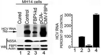
Endogenous HCV replication in cell-free replication lysates
is reduced in FBP1-kd cells but significantly enhanced on FBP1
overexpression.
Earlier, we demonstrated that HCV replication is
positively influenced by FBP1 expression level in MH14 cells (
10
).
Downregulation of FBP1 by FBP1-siRNA severely impaired HCV
replication, whereas overexpression of FBP significantly enhanced
HCV replication but had a negative influence on HCV translation
(
10
). In order to examine whether
in vitro
endogenous HCV
rep-lication in cell-free reprep-lication lysate also is affected by increased
or decreased levels of FBP1 in cells, we used replicative cell-free
lysates prepared from MH14 cells in which FBP1 was either stably
knocked down or overexpressed. The reaction products were
an-alyzed on a denaturing agarose gel and visualized by
autoradiog-raphy. We also Western blotted cell-free replication lysates for
FBP1, HCV NS5A, and actin after normalizing their protein
con-centrations. We found that
in vitro
endogenous HCV replication
in replicative lysates from FBP1-kd cells was only 15% (
Fig. 2
, lane
3) compared to that in control MH14 cells (lane 2). In contrast, a
3-fold increase in endogenous HCV replication activity occurred
in cell lysates in which FBP1 was overexpressed (lane 4). These
findings suggest that FBP1 facilitates HCV replication by
on November 7, 2019 by guest
http://jvi.asm.org/
ing with components of the HCV replication complex or by
abro-gating the inhibitory effect of some unknown cellular factor(s) on
HCV replication. We also Western blotted normalized cell-free
replication lysate for the expression levels of FBP1 and HCV
NS5A, as well as that of actin as the loading control. We found that
the level of NS5A in the replication lysate from control cells was
greater than that from cells in which FBP1 was overexpressed. This
is not surprising, since we showed earlier that the upregulation of
FBP1 enhanced HCV replication but inhibited HCV translation.
Since HCV is a positive-stranded RNA virus, its genomic RNA is
the template for both translation and replication complexes that
move in opposite directions on the RNA template. Thus, these two
processes must be mutually exclusive. In poliovirus, a switch from
translation to replication and vice versa has been demonstrated
(
33
). Therefore, it is possible that FBP is a key factor in regulating
this switch from translation to HCV replication.
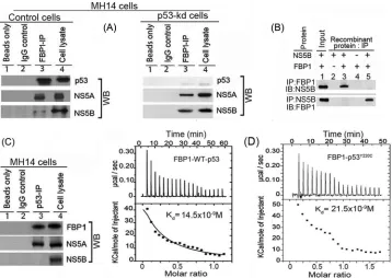
FBP1 interacts with p53 and viral proteins NS5A and NS5B.
FBP1 interacts with NS5A and stimulates HCV replication (
10
),
whereas NS5A interacts with p53 (
11
,
34
,
35
) and inhibits
p53-mediated apoptosis (
11
). We showed earlier that FBP1 interacts
with p53 and inhibits its transactivation activity in Huh 7 cells
(
13
). In this study, we found FBP1 immunoprecipitation (IP)
co-immunoprecipitates not only p53 and NS5A but also NS5B from
the cell lysates of control MH14 cells (
Fig. 3A
, lane 3, left). We
further confirmed that the interaction of FBP1 with viral proteins
NS5A and NS5B is independent of p53. FBP IP in lysate from
p53-kd cells also coprecipitated these viral proteins (
Fig. 3A
, lane
3, right), suggesting that it is an important component of the HCV
replication complex. Since HCV NS5A has been shown to be in a
complex with NS5B, it is possible that FBP1 IP pulls down NS5B
via its interaction with NS5A. To rule out this possibility, we
car-ried out immunoprecipitation on a mixture of recombinant
pu-rified FBP1 and NS5B and found that reciprocal IP coprecipitate
each other (
Fig. 3B
). FBP1 also contains ATP-dependent helicase
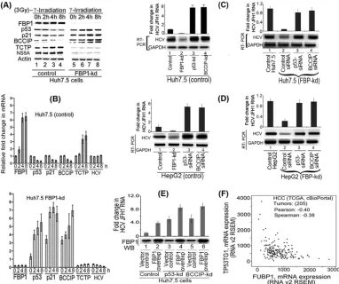
FIG 1FBP1 is specifically overexpressed in human HCC tumors with CHC background. (A) Immunohistochemistry was done on archived HCC tumors; seven had a history of chronic hepatitis (Hep) C, three had the alcoholic background, and two were cryptogenic. Based on the Edmondson and Steiner nuclear grading scheme (79), 4 of the HCC-HCV tumors were well differentiated (grade1), 2 were moderately differentiated (grade 2), and 1 was moderately to poorly differentiated (grades 2 to 3). All of the alcoholic and cryptogenic HCC were moderately differentiated grade 2 tumors. Representative pictures are shown of HCC tumors treated with a polyclonal antibody against FBP1 (left) or isotype IgG (middle). The hematoxylin-eosin (H&E) staining of the corresponding slides is shown on the right. The alcoholic and cryptogenic HCC slides show only the cancer area. (B and C) Oncomine data analysis for FBP1 and p53 expression in 19 normal human livers versus 58 cirrhotic livers and 38 HCC tumors with a CHC background (31). (D) Oncomine data analysis of FBP1 gene amplification in normal livers versus HCC liver tumors from two data sets, one from TCGA livers (TCGA 2012) and the other from Guichard livers (32).
FIG 2Endogenous HCV replication in cell-free replication lysates is severely reduced in FBP1-kd cells but increased in FBP1-overexpressing MH14 cells. An aliquot of normalized replication lysates from MH14 cells in which FBP1 was either knocked down or overexpressed was examined for endogenous HCV replication activity; the radiolabeled RNA products were analyzed by denaturing agarose gel electrophoresis and visualized by autoradiography. Normalized cell-free replication lysates also were Western blotted (WB) for the expression level of FBP1, HCV NS5A, and actin. (Left) Lane 1, cured MH14 cells without HCV replicons; lane 2, control MH14 cells with HCV replicons; lane 3, FBP1-kd MH14 cells; lane 4, MH14 cells in which FBP1 was overex-pressed. (Right) The percentage of endogenous replication products relative to those in controls.
FBP1 Facilitates Persistent HCV Replication
on November 7, 2019 by guest
http://jvi.asm.org/
[image:5.585.67.519.63.318.2] [image:5.585.79.249.520.612.2]activity, which can unwind both DNA/DNA and RNA/RNA
du-plexes (
36
). This could be one of the mechanisms by which FBP1
displays a stimulatory effect on HCV replication. The p53 IP also
coprecipitated FBP1 and NS5A but not NS5B (
Fig. 3C
, lane 3). We
confirmed earlier the direct interaction of FBP1 with p53 by
re-ciprocal IP on a mixture of purified recombinant proteins of FBP1
and p53 (
13
).
ITC of FBP1 interaction with wild-type p53 and mutant
p53
Y220C.
MH14 cells are derived from the Huh7 cell line, in which
p53 carries a Tyr
¡
Cys mutation at position 220. We examined
whether FBP1 binding to p53 is specific to mutant p53
Y220Cor also
can interact with wild-type p53. We used ITC to characterize the
interaction of recombinant FBP1 thermodynamically with Y220C
mutant and wild-type p53. A syringe of ITC containing purified
p53 was titrated into a cell containing purified FBP1. As the two
proteins interacted, the heat was absorbed in direct proportion to
the amount of binding that occurred. When FBP1 in the cell
be-came saturated with added p53, the heat signal diminished. The
absorbed heat was measured to determine the binding constant
(
K
) or dissociation constant (
37
) of interaction between FBP1 and
p53 (
Fig. 3D
). The top panel shows the calorimetric data; each
injection produced upward spikes indicating endothermic
inter-action between p53 and FBP1. The top panel displays a plot of the
total heat generated per injection as a function of the molar ratio
of p53 to FBP1. The amount of heat absorbed per mole of p53 is
shown as a function of the molar ratio of p53 to FBP1. The bottom
panel shows the best fit to the experimental data for the
one-binding-site model. We found that FBP1 interacted with both
wild-type and mutant p53 with nearly similar affinity, displaying
association constants of 1.72
⫻
10
7M
⫺1and 1.15
⫻
10
7M
⫺1,
respectively. With the standard state being equal to 1.0 M, the free
energy change for the binding of FBP1 to both wild-type and
mu-tant p53 was calculated (
⌬
G
°
⫽ ⫺
RT ln
K
a[where R is gas
con-stant, T is temperature in Kelvin, and
K
ais association constant])
to be
⫺
10.2 and
⫺
10.0 kcal mol
⫺1, respectively.
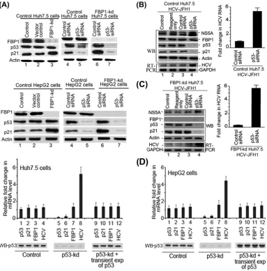
Downregulation of p53 abolishes the enhanced expression of
p21 in FBP1-kd cells of both Huh7.5 and HepG2.
We showed
earlier that FBP1-kd in Huh7.5 cells significantly enhanced p21
expression (
13
). Since the p21 expression is positively regulated by
p53, we postulated that the enhanced expression of p21 in
FBP1-kd was a result of the activation of p53 in the absence of
FBP1 (
13
). To examine this postulation, we used both Huh7.5
cells expressing mutant p53
Y220C(
Fig. 4A
, top) as well as HepG2
cells expressing wild-type p53 (
Fig. 4A
, bottom). We
downregu-lated p53 expression in control and FBP1-kd cells of both cell lines
and then analyzed the cell lysates for p21 expression. We found
that p21 expression was significantly enhanced in FBP-kd cells of
both cell lines (
Fig. 4A
, lane 3, top and bottom). We further
ob-served that enhanced expression of p21 in FBP1-kd cells was
nearly abolished upon downregulation of p53 in both cell lines
(
Fig. 4A
, lane 7, top and bottom). These results clearly indicate
that expression of p21 is under the positive control of p53 in both
FIG 3FBP1 physically interacts with p53 and HCV proteins NS5B and NS5A. (A) FBP1 IP was done on RNase-treated lysates from control MH14 cells (left) or p53-kd MH14 cells (right) and Western blotted for p53, NS5A, and NS5B. Lane 1, beads only; lane 2, isotype IgG control; lane 3, IP sample; lane 4, cell lysate. (B) FBP1 IP and NS5B IP on a mixture of purified proteins pull down each other. We used 2g each of purified recombinant FBP1 and NS5B and carried out reciprocal IP and Western blotting for either FBP1 or NS5A. (Upper) Lanes 2 to 5, FBP1 IP immunoblotted (IB) for NS5B. (Bottom) Lanes 2 to 5, NS5B IP immunoblotted for FBP1. Lane 1, input protein controls. (C) p53 IP on the lysates from control MH14 cells. Lane 1, beads only; lane 2, isotype IgG control; lane 3, IP sample; lane 4, cell lysate. (D) Isothermal titration calorimetry of FBP1 interaction with wild-type p53 (left) and Y220C mutant p53 (right). A syringe of ITC containing purified p53 was titrated into a cell containing purified FBP1 in ITC buffer containing 20 mM sodium phosphate buffer, pH 7.8, and 150 mM NaCl at 25°C. (Top) The calorimetric data on titration of FBP1 with p53 as a function of time. (Bottom) The integrated heat per injection versus the molar ratio of p53 to FBP1. The graph corresponds to the best fit of the experimental data to a one-site model, providing a dissociation constant (37) of 14.5 nM for wild-type p53 and 21.5 nM for the Y220C mutant.
on November 7, 2019 by guest
http://jvi.asm.org/
[image:6.585.114.472.66.320.2]Huh7-derived cells as well as in HepG2 cells expressing wild-type
p53. These findings also suggest that p53
Y220Cin Huh7.5 cells is
functional and active but strongly suppressed by FBP1.
Inhibition of HCV replication in FBP1-kd Huh7.5 cells is
re-versed by downregulation of p53.
We showed earlier that HCV
replication is severely impaired by downregulation of FBP1 (
10
).
Since p53 inhibits HCV replication (
12
), we postulated that
activa-tion of p53 causes the inhibiactiva-tion of HCV replicaactiva-tion in FBP1-kd cells.
To test this hypothesis, we transfected control and FBP1-kd Huh7.5
cells with p53-siRNA and then infected the cells with JFH1 HCV
virions. After 72 h of infection, cell lysates were Western blotted for
NS5A, FBP1, p53, and p21; quantitative real-time RT-PCR was done
on HCV RNA. We found that downregulation of p53 enhanced HCV
replication by 5-fold in control Huh7.5 cells compared to the level of
the siRNA control (
Fig. 4B
, lane 4, and right). Interestingly, the severe
reduction in HCV replication observed in FBP1-kd cells was
com-pletely reversed by downregulation of p53 (
Fig. 4C
, lane 4). The
quantitative RT-PCR data indicated that HCV replication in
FBP1-kd cells in which p53 was downregulated is enhanced 6-fold
compared to that of the FBP1-kd control (
Fig. 4C
, right). These
re-FIG 4Downregulation of p53 and FBP1 have opposite effects on p21 expression and HCV replication. (A) Upregulation of p21 in FBP1-kd Huh7.5 or HepG2 cells is abolished upon downregulation of p53. Control and FBP1-kd Huh7.5 cells (upper) or HepG2 cells (lower) were transfected with p53 siRNA and grown for 48 h. The normalized cell lysates were analyzed for the expression of FBP1, p53, p21, and actin by Western blotting. Lane 1, control cells; lane 2, vector control; lane 3, FBP1-kd cells; lanes 4 and 6, cells transfected with control siRNA; lanes 5 and 7, cells transfected with p53 siRNA. (B and C) Downregulation of p53 enhanced HCV replication in control and FBP1-kd Huh7.5 cells. Control Huh7.5 cells (B) and stable FBP1-kd Huh7.5 cells (C) were transfected with p53 siRNA and, 10 h later, infected with infectious HCV-JFH1 virions and then grown further for 72 h. Cell lysates were prepared, normalized for protein concentration, and Western blotted for the expression of NS5A, FBP1, p53, p21, and actin. Another set of cells was used for isolation of total RNA for quantitative real-time RT-PCR of JFH1 HCV RNA and GAPDH mRNA. (Left) Lane 1, control; lane 2, reagent control; lane 3, siRNA control; lane 4, p53 siRNA. (Right) Fold change in JFH1 HCV RNA concentration in control and FBP1-kd Huh7.5 cells quantified by real-time RT-PCR. (D) Transient (exp) expression of p53 suppresses HCV replication in p53-kd Huh7.5 and HepG2 cells. Stable p53-kd Huh7.5 cells (left) and HepG2 cells (right) were transfected with an shRNA-resistant expression clone of p53, and 10 h later, Huh7.5 cells were infected with HCV-JFH1 virions while HepG2 cells were transfected with MH14 HCV replicon RNA. Cells were grown for 72 h; total RNA was isolated. The quantitative real-time RT-PCR for HCV RNA and mRNA levels of p53, p21, and FBP1 were carried out with GAPDH mRNA as the internal control. The Western blot of p53 expression also is shown. Lanes 1 to 4, control cells; lane 5 to 8, p53-kd cells; lanes 9 to 12, p53-kd cells in which p53 was transiently expressed.
FBP1 Facilitates Persistent HCV Replication
on November 7, 2019 by guest
http://jvi.asm.org/
[image:7.585.102.485.67.456.2]sults strongly suggest that mutant p53
Y220Cin Huh7.5 cells displays an
inhibitory effect on HCV replication that is strongly suppressed in the
presence of FBP1.
Promotion of HCV replication in p53-kd cells is significantly
suppressed by transient expression of p53.
Since p53-kd cells are
highly permissive to HCV replication, we examined whether
re-storing the p53 expression in p53-kd cells could suppress the
en-hanced viral replication. Therefore, we constructed an
shRNA-resistant clone of p53 by point mutation in exon seven at codons
244 (GGC
¡
GGT) and 245 (GGC
¡
GGT) without altering the
codon usage and transfected this clone into p53-kd Huh7.5 and
HepG2 cells. The transfected cells were grown for 72 h, and the
levels of mRNA of p53, p21, and FBP1 as well as HCV RNA were
determined by quantitative RT-PCR. The results shown in
Fig. 4D
(left and right) indicate that enhanced HCV replication in p53-kd
cells in both cell lines (lane 8) was significantly suppressed and
restored to the control level upon transient expression of p53
(lane 12).
DNA binding and transactivation activities of mutant
p53
Y220Cin Huh7.5 cells are activated by knockdown of FBP1.
Mutant p53
Y220Cin Huh7 cells has been shown to be defective in
its ability to bind target DNA at physiological temperatures (
16–
18
). However, in yeast systems, mutant p53
Y220Chas been shown
to be transcriptionally active and displays wild-type target DNA
binding activity at subphysiological temperatures (
19
). In
con-trast, another study with the truncated p53
Y220Ccontaining only
the core domain showed that the DNA binding ability of mutant
p53
Y220Cis impaired at both physiological and subphysiological
temperatures (
20
).
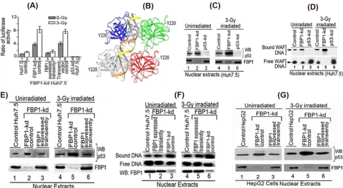
Our results indicated that p21 expression in Huh7.5 cells is
under the positive regulation of p53 that is strongly suppressed in
the presence of FBP1 (
Fig. 4
). This premise was further confirmed
using reporter p21-Luc activity in control and FBP1-kd Huh7.5
cells under radiation-induced stress. We also included p53-kd
Huh7.5 cells as negative controls. We found no activation of
re-porter activity in
␥
-irradiated control Huh7.5 cells compared to
that in unirradiated cells (
Fig. 5A
, lanes 1 and 2). In contrast, in
FBP1-kd Huh7.5 cells, reporter activity was significantly activated
in both unirradiated and irradiated cells (lanes 3 and 4). Reporter
activity was completely absent from p53-kd Huh7.5 cells (lanes 9
and 10).
These findings demonstrated that mutant p53
Y220Cin Huh 7.5
cells is transcriptionally active but strongly suppressed or
inhib-ited in the presence of FBP1. To confirm whether observed
acti-vation of mutant p53
Y220Cindeed was due to the absence of FBP1,
we transiently expressed FBP1 in FBP1-kd cells by transfecting the
shRNA-resistant FBP1 expression clone and examined the
activa-tion of p53 by measuring p21-Luc reporter activity. We found that
the activation of p53 in FBP1-kd cells was strongly suppressed
when FBP1 was transiently expressed (
Fig. 5A
, lanes 5 and 6) but
remained unaffected when transfected with empty vector alone
(lanes 7 and 8).
The residue Y220 in p53 is 38 Å away from the DNA binding
motif (
Fig. 5B
); it is intriguing that Tyr
¡
Cys mutation at this
position should affect the DNA binding function of p53. The
ob-served activation of mutant p53
Y220Cin irradiated FBP1-kd
Huh7.5 cells implies that the mutant p53 must be actively binding
to its target DNA in the absence of FBP1. To confirm this
possi-bility, we used a streptavidin-paramagnetic bead assay system to
examine the DNA binding ability of mutant p53
Y220Cin the
nu-clear extracts of
␥
-irradiated and unirradiated controls, FBP1-kd
cells, and p53-kd Huh7.5 cells. We incubated the normalized
nu-clear extracts with biotinylated 30-bp WAF-side DNA at 37°C,
captured the p53-DNA complex on streptavidin magnetic beads,
and analyzed the bound p53 by Western blotting. As shown in
Fig.
5C
, mutant p53 in the nuclear extract of both unirradiated and
irradiated control Huh7.5 cells displays only 20% to 25% of the
DNA binding ability (lanes 1 and 4) of the corresponding
FBP1-Kd cells (
Fig. 5C
, lanes 2 and 5). We did not detect any DNA
binding activity with the nuclear extract from both unirradiated
and irradiated p53-kd Huh7.5 cells (lanes 3 and 6).
We also did an electrophoretic mobility shift assay (EMSA) to
determine the DNA binding activity of mutant p53 in control and
FBP1-kd Huh7.5 cells. We incubated the normalized nuclear
ex-tracts with
32P-labeled 30-bp WAF-side DNA at physiological
temperature. The p53-bound DNA complexes were resolved by
EMSA. The results (
Fig. 5D
) indicated that mutant p53
Y220Cin
both unirradiated and irradiated Huh7.5 cells (lanes 1 and 4)
poorly binds WAF-side DNA compared to the enhanced DNA
binding observed in corresponding FBP1-kd cells (lanes 2 and 5).
The nuclear extract from p53-kd Huh7.5 cells did not show
bind-ing to the target WAF-side DNA (lanes 3 and 6). These results
confirm that the target DNA binding activity of mutant p53
Y220Cin Huh7.5 cells is strongly suppressed by FBP1.
Transient expression of FBP1 in FBP1-kd Huh7.5 cells
strongly suppresses DNA binding activity of mutant and
wild-type p53.
Our results strongly imply that the mutant p53
Y220Cin
Huh7.5 cells is significantly activated to bind target DNA in the
absence of FBP1. To ascertain this, we transiently expressed FBP1
in FBP1-kd Huh7.5 cells and examined DNA binding activity of
p53 in the nuclear extracts of both unirradiated and irradiated
cells. Results obtained with the streptavidin binding assay
indi-cated that enhanced DNA binding of p53
Y220Cobserved in the
nuclear extract from FBP1-kd cells (
Fig. 5E
, lanes 2 and 5) is
strongly inhibited by transient expression of FBP1 (
Fig. 5E
, lanes 3
and 6), and this inhibition was similar to that observed in control
Huh7.5 cells (lanes 1 and 4). We obtained similar results by EMSA
in the nuclear extracts from control, FBP1-kd, and FBP1-kd
Huh7.5 cells in which FBP1 was transiently expressed (
Fig. 5F
).
The enhanced DNA binding of mutant p53
Y220Cfrom the nuclear
extract of FBP1-kd cells (lanes 3 and 6) is strongly suppressed
when FBP1 is transiently expressed in the cells (lanes 2 and 5).
We also examined DNA binding activity of wild-type p53 in
the nuclear extract of irradiated and unirradiated control and
FBP1-kd HepG2 cells (
Fig. 5G
). We found significantly enhanced
DNA binding activity of wild-type p53 in the nuclear extract of
irradiated FBP-kd HepG2 cells (lane 5) compared to that of the
irradiated control cells (lane 4). Similar to Huh7.5 cells, the
en-hanced DNA binding activity of p53 in the nuclear extract from
FBP1-kd HepG2 cells (lanes 2 and 5) also was strongly inhibited by
transient expression of FBP1 (lanes 3 and 6). These findings
con-firm that DNA binding activity of both mutant and wild-type p53
is negatively influenced by the presence of FBP1.
DNA binding ability of the recombinant wild-type p53 and
its Y220C mutant derivative.
We also carried out EMSA with
purified recombinant wild-type p53 and mutant p53
Y220Cto
ex-amine their binding affinities to
32P-labeled 30-bp WAF-side
DNA (
Fig. 6A
) at physiological temperatures. The dissociation
constants determined from the EMSA data were 1.3 nM and 1.7
nM, respectively, for the wild-type and mutant p53, indicating no
on November 7, 2019 by guest
http://jvi.asm.org/
significant difference in their binding affinities for the target
WAF-side DNA. We also did ITC to determine the binding
affin-ity of wild-type and mutant p53 to the WAF-side DNA (
Fig. 6B
).
As p53 interacts with its target DNA, heat is absorbed in direct
proportion to the amount of binding that occurs. The top panel of
Fig. 6B
shows the calorimetric data. Each injection produced
downward spikes indicating exothermic interaction between p53
and its target DNA. The absorbed heat was measured to determine
the binding constant (
K
a) or dissociation constant (
37
) of
inter-action between p53 and the target DNA (
Fig. 6B
, lower). We
found that the wild-type and mutant p53 displayed similar
bind-ing affinities for the target DNA, with respective dissociation
con-stants of 5.5 nM and 7.2 nM. The free energy change for the
binding of p53 to target DNA was calculated to be
⫺
14.5 kcal
mol
⫺1for the wild-type p53 and
⫺
14.0 kcal mol
⫺1for the
Y220C mutant p53.
FBP1 blocks the binding of recombinant wild-type p53 to the
target WAF-side DNA.
Since recombinant proteins of both
p53
Y220Cand wild-type p53 bind WAF-side p21 DNA with similar
affinity, it is intriguing that basal levels of p21 expression in both
Huh7.5 and HepG2 cells were significantly enhanced upon
knock-down of FBP1. Since FBP1 physically interacts with p53, it is
pos-sible that, in the presence of FBP1, the binding of p53 to its target
WAF-side DNA is compromised. Thus, we examined the binding
FIG 5Transactivation activity and DNA binding ability of mutant p53Y220Cin Huh7.5 cells is activated by knockdown of FBP1 expression. (A) Transcription
activity of mutant p53Y220Cin control and FBP1-kd Huh7.5 cells under radiation-induced stress. The p21-luc reporter and pRL-SV40 plasmids were
cotrans-fected into control, FBP1-kd, and FBP1-kd cells in which FBP1 was transiently expressed via transfection of an shRNA-resistant FBP1-expressing clone (pCIA-cmv-FBP1SHR). Forty-eight hours later, cells were irradiated with a 3-Gy dose of gamma irradiation. Luciferase activity was measured after 6 h
postirra-diation. Experiments were done in triplicate; results are expressed as the ratio of firefly luciferase to Renilla luciferase activities in cell lysate. Lanes 1 and 2, control cells; lanes 3 and 4, FBP1-kd cells; lanes 5 and 6, FBP1 transiently expressed in FBP1-kd cells; lanes 7 and 8, FBP1-kd cells transfected with vector alone; lanes 9 and 10, p53 kd cells. (B) The three-dimensional structure of homotetrameric p53-DNA binary complex and position of Y220. Using Maestro molecular modeling software, version 9.3.5 (Schrodinger, Inc.), we downloaded the backbone structure of the p53-DNA binary complex from PDB entry4HJE(80). The three-dimensional crystal structure of DNA-bound p53 is displayed without any modification. The backbone of duplex DNA bound to the tetrameric p53 and the position of Y220 located far away from the bound DNA are shown. (C) Streptavidin magnetic bead DNA binding assay indicates enhanced DNA binding activity of mutant p53Y220Cin the nuclear extract from FBP1-kd Huh7.5 cells. Normalized nuclear extracts from unirradiated or 3-Gy-␥-irradiated control and FBP1-kd
cells were incubated with 30 bp biotinylated WAF-side DNA at 37°C. The nuclear extract from p53-kd cells was included as a negative control. DNA-protein complexes were captured on streptavidin paramagnetic beads, resolved on SDS-PAGE, and Western blotted for p53. The normalized nuclear extract also was Western blotted for FBP1. Lanes 1 to 3, unirradiated; lanes 4 to 6,␥-irradiated. (D) EMSA showing enhanced binding of 30 bp WAF-side DNA by p53Y220Cin
the nuclear extract from FBP1-kd Huh7.5 cells. Normalized nuclear extracts from unirradiated and 3-Gy-␥-irradiated control and FBP1-kd cells were incubated with32P-labeled WAF-side DNA and then subjected to EMSA on 4% native polyacrylamide gel. p53-kd Huh7.5 cells were included as a negative control. Lanes
1 to 3, unirradiated; lanes 4 to 6,␥-irradiated. (E and F) Transient expression of FBP1 in FBP1-kd cells strongly inhibits p53Y220Cbinding to its target DNA. DNA
binding activity of mutant p53 in normalized nuclear extract from unirradiated and irradiated control, FBP1-kd, and transiently FBP1 expressing FBP1-kd Huh7.5 cells was examined by streptavidin magnetic bead DNA binding assay (E) and EMSA (F). The normalized nuclear extract also was Western blotted for FBP1. Lanes 1 to 3, unirradiated; lanes 4 to 6, irradiated. (G) Transient expression of FBP1 in FBP1-kd HepG2 cells inhibits binding of wild-type p53 to its target DNA. DNA binding activity of wild-type p53 in a normalized nuclear extract from unirradiated and irradiated control, FBP1-kd, and transiently FBP1-expressing FBP1-kd HepG2 cells was examined by streptavidin magnetic bead DNA binding assay. The p53 bound to the target DNA was captured on streptavidin magnetic beads and Western blotted for p53. The normalized nuclear extract also was Western blotted for FBP1. Lanes 1 to 3 represent unirradiated control, FBP-kd, and FBP-kd cells, respectively, transiently expressing FBP1; lanes 4 to 6 represent irradiated control, FBP-kd, and FBP-kd cells, respectively, transiently expressing FBP1.
FBP1 Facilitates Persistent HCV Replication
on November 7, 2019 by guest
http://jvi.asm.org/
[image:9.585.45.540.65.337.2]FIG 6Target DNA binding affinity of recombinant wild-type p53 and mutant p53Y220C. (A) EMSA with purified wild-type p53 and mutant p53Y220Cshows
similar DNA binding affinity for the target DNA. A fixed concentration of32P-labeled 30-bp WAF-side DNA was incubated with increasing concentrations of
purified recombinant wild-type p53 (left) or mutant p53Y220C(right) at 37°C. The mixture was subjected to EMSA. The bound WAF-side DNA level versus the
tetrameric p53 concentration was plotted using GraphPad, and the dissociation constant was determined using a nonlinear regression curve fit for one-site binding of DNA with tetrameric p53. (B) ITC of 30-bp WAF-side DNA binding to the wild-type p53 (left) and mutant p53Y220C(right) shows similar binding
affinity. A syringe of ITC containing WAF-side DNA was titrated into a cell containing purified wild-type or mutant p53 in ITC buffer containing 20 mM sodium phosphate buffer, pH 7.8, and 150 mM NaCl at 37°C. (Top) The calorimetric data of titration of p53 with DNA as a function of time. (Bottom) The integrated heat per injection versus the molar ratio of tetrameric p53 to DNA. The graph corresponds to the best fit of the experimental data to the one-site model, providing a dissociation constant (37) of 5.5 nM for the wild-type p53 and 7.2 nM for the mutant p53Y220C. (C) Inhibition of DNA binding activity of wild-type p53 by
recombinant FBP1. The purified recombinant p53 (7 nM) was incubated with increasing concentrations of FBP1 (25 to 100 nM) in a final volume of 20l. After 30 min of incubation at room temperature, a fixed concentration of32P-labeled 30-bp WAF-side DNA (40,000 cpm) was added; the mixture was incubated for
30 min at 37°C. The p53-DNA complexes were resolved by EMSA. Lane 1, p53 binding to DNA in the absence of FBP1; lanes 2 through 5, p53 binding to DNA in the presence of 25, 50, 75, and 100 nM recombinant FBP1. (D) Binding of FBP1 alone to32P-labeled WAF-side DNA is not significant. Lanes 1 through 4,
increasing concentrations of FBP1 (25 to 100 nM) were incubated at room temperature with32P-labeled WAF-side DNA (40,000 cpm) in a final volume of 20 l for 30 min. The bound and unbound DNA were resolved by EMSA. (E) EMSA with a nontarget32P-labeled 30-bp HIV-1 U5 PBS DNA was used to determine
the target specificity of wild-type p53 and mutant p53Y220C.
on November 7, 2019 by guest
http://jvi.asm.org/
[image:10.585.125.460.67.545.2]of the wild-type p53 to the 30-bp WAF-side DNA in the absence
and presence of FBP1. We preincubated a fixed concentration of
tetrameric p53 (7 nM) with increasing concentrations of FBP1 (25
to 100 nM) and then incubated it with
32P-labeled WAF-side
DNA. In a parallel experiment, we incubated labeled WAF-side
DNA with increasing concentrations of FBP1 alone (25 to 100
nM). The binding of p53 to the target DNA was significantly
in-hibited in the presence of FBP1 (
Fig. 6C
). In the absence of p53,
the extent of FBP1 binding to the 30-bp WAF-side DNA was
in-significant (
Fig. 6D
). These results suggest a mechanism whereby
FBP1 blocks the binding of p53 to its target DNA sequence and
inhibits its transcription activity. We further confirmed that
bind-ing of both wild-type and mutant p53
Y220Cto the target WAF-side
DNA is highly specific, since they did not bind to a 30-bp
nontar-get double-stranded HIV-1 U5PBS DNA (
Fig. 6E
).
FBP1 promotes HCV replication by regulating p53
regula-tory proteins.
Although mutant p53
Y220Cbinds DNA as efficiently
as wild-type p53, it remained transcriptionally inactive in
Huh7-derived cells. Our results suggest that FBP1, which is
overex-pressed in Huh7.5 cells, is involved in suppressing the function of
p53 either directly or by regulating p53-regulatory proteins. The
translationally controlled tumor protein (TCTP; also known as
fortilin) is a negative regulator of p53 that functions as an
antiapo-ptotic factor by interacting with and destabilizing p53 (
38
,
39
).
BCCIP, an important cofactor for BRCA2 in tumor suppression,
is a positive regulator of p53 and modulates CDK2 kinase activity
by interacting with p21 (
40–42
). To examine whether FBP1 has
any influence on the expression of p53 and its regulatory proteins,
we first transfected control and FBP1-kd Huh7.5 cells with
JFH1-HCV virus; 72 h later, we irradiated cells with 3 Gy gamma
irra-diation and examined the expression of p21, p53, BCCIP, TCTP,
and HCV NS5A at both protein and mRNA levels. An aliquot of
the normalized cell lysates containing equivalent protein was
sub-jected to SDS-PAGE and Western blotted for the expression of
FBP1, p53, p21, BCCIP, and TCTP, as well as for HCV protein
NS5A. Real-time RT-PCR determined their mRNA levels.
We found that in control Huh7.5 cells, FBP1 expression was
only slightly increased by 4 h postirradiation. There was a
corre-sponding marginal decrease in the expression of p53 and p21
without any significant change in expression of the HCV protein
NS5A (
Fig. 7A
, left, lanes 3 and 4). In contrast, in FBP1-kd cells,
expression of both p53 and p21 was significantly enhanced at 2 to
8 h postirradiation (
Fig. 7A
, right, lanes 6 to 8), while the NS5A
level was drastically reduced at all time points. We further found
that TCTP, a negative regulator of p53, was upregulated in control
Huh7.5 cells at 4 to 8 h postirradiation (
Fig. 7A
, left, lanes 3 and 4),
while its expression level in FBP1-kd cells was drastically reduced
at all time points (
Fig. 7A
, right, lanes 5 to 8), suggesting that
TCTP is positively regulated by FBP1. In contrast, the expression
level of BCCIP, a positive regulator of p53, was strongly
sup-pressed in control Huh7.5 cells after irradiation but significantly
boosted in FBP1-kd cells (
Fig. 7A
, lanes 5 and 8). The upregulation
of BCCIP in FBP1-kd cells suggests that FBP1 is a negative
regu-lator of BCCIP. These results were confirmed by determining fold
changes in the mRNA levels of p21, p53, BCCIP, and TCTP, as
well as HCV RNA levels in irradiated control and FBP1-kd cells
(
Fig. 7B
). These results also confirmed that FBP1 suppresses the
p53-mediated response to cellular stress by regulating the
expres-sion of p53 and its regulatory factors, BCCIP and TCTP.
We then examined the level of HCV replication in control and
FBP1-kd cells of both Huh7.5 and HepG2 cells in which either p53
or BCCIP had been knocked down or downregulated (
Fig. 7C
and
D
). We found that control Huh7.5 cells are highly permissive to
HCV replication when knocked down for either p53 or BCCIP,
resulting in a relative 5-fold increase in the virus replication with
respect to the control (
Fig. 7C
, lanes 3 and 4, left). Interestingly,
the drastic reduction of HCV replication observed in FBP1-kd
Huh7.5 cells is completely restored to the control level by
down-regulation of either p53 or BCCIP (
Fig. 7C
, lanes 3 and 4, right).
We found similar results with HepG2 cells in which
downregula-tion of either p53 or BCCIP enhanced HCV replicadownregula-tion in control
cells (
Fig. 7D
, lanes 3 and 4, left) and restored the loss of HCV
replication in FBP1-kd HepG2 cells (
Fig. 7D
, lanes 3 and 4, right).
We further examined the effect of overexpression of FBP1 on
HCV replication in Huh7.5 cells stably knocked down for the
expression of p53 or BCCIP. We transfected control, p53-kd, and
BCCIP-kd cells with FBP1 overexpression plasmid or empty
vec-tor alone; 10 h later, the cells were infected with HCV JFH1 virions
and grown further for 72 h, and total RNA was isolated. The
rel-ative fold change in the level of HCV RNA was determined by
quantitative RT-PCR. As shown in
Fig. 7E
, a 5-fold increase in
HCV replication occurred in p53-kd cells (lane 3) with respect to
the control (lane 1), which was further increased to 8-fold upon
overexpression of FBP1 (lane 4). Similar results also were obtained
with BCCIP-kd cells in which FBP1 was overexpressed (lane 6).
These results clearly suggest that FBP1 promotes HCV replication
by suppressing the function of p53 and its positive regulator,
BCCIP. This contention also is supported by Oncomine
expres-sion data analysis for the expresexpres-sion of FBP1 and p53 in a large
number of normal human liver versus HCV-infected cirrhotic
livers and HCC tumors (
31
). In contrast to a 4-fold increase in
FBP1 expression (
Fig. 1B
), there was a 2.5-fold decrease in the p53
mRNA level in the same set of cirrhotic livers and HCC tumors
(
Fig. 1C
). That FBP1 suppresses p53 function also is supported by
the cBioPortal coexpression analysis (
43
,
44
) of data from 205
HCC tumors (TCGA, provisional), showing a significant decrease
in the p53-inducible gene, TP53TG1, with an increase in the FBP1
expression level (
Fig. 7F
). This observation also establishes an
in-verse correlation of FBP1 expression with the expression of
p53-targeted genes.
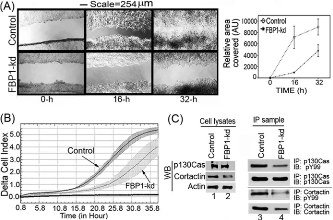
FBP1 enhances cell migration.
FBP1 has been shown to be
involved in tumor metastasis through the regulation of the
micro-tubule-destabilizing proteins Stathmin 1 and Stathmin 3 (
45
).
These two proteins have been suggested to be involved in cell
motility and metastasis, since they are abundantly expressed at the
invasion front in non-small-cell lung carcinoma (
45
,
46
). In a
wound-healing assay, we found that the migration of the FBP1-kd
Huh7.5 cells was drastically reduced compared to the level for the
control cells (
Fig. 8A
, left and right). In order to further confirm the
results obtained with the wound-healing assay, we also carried out
real-time migration dynamics of control and FBP1-kd Huh7.5 cells
using a real-time cell analyzer dual-plate RTCA DP xCELLigence
sys-tem (
30
). The real-time migration assay was done in triplicate for up
to 36 h, and changes in cell index were recorded at 15-min intervals.
We found a severe reduction in the migration of FBP1-kd cells
com-pared to that of control cells, as judged by the reduced cell index (
Fig.
8B
). We also analyzed the expression and phosphorylation levels of
cortactin and p130Cas, which are markers for cell migration and
in-vasion (
47–49
). Cortactin, involved in actin polymerization and cell
motility, is an important biomarker for many invasive cancers,
in-FBP1 Facilitates Persistent HCV Replication
on November 7, 2019 by guest
http://jvi.asm.org/
cluding HCC (
49
,
50
). P130Cas, also known as BCAR1, promotes
actin remodeling, actomyosin contraction, and cell migration (
51
).
Both cortactin and p130Cas are Src substrates and, in the
phosphor-ylated form, activate signaling events that are associated with cell
mi-gration (
49
,
52
). We determined the expression level of these markers
by Western blotting of cell lysates, and their phosphorylated form was
determined by immunoblotting of their IP samples using an antibody
against phosphotyrosine (pY99). The Western blot analysis of
p130Cas in cell lysates indicated no significant change in its
expres-sion level in control and FBP1-kd cells (
Fig. 8C
, lanes 1 and 2), but
compared to the control, the level of phosphorylated p130Cas was
significantly decreased in FBP1-kd cells (
Fig. 8C
, lanes 3 and 4). In
contrast, both cortactin expression and phosphorylation levels were
significantly decreased in FBP-kd cells (
Fig. 8C
, lanes 2 and 4). This
observation indicates that FBP1 not only promotes viral replication
but also facilitates migration and metastasis of HCC tumors.
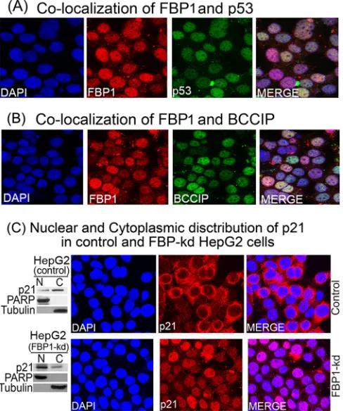
Colocalization of FBP1 with p53 and BCCIP in Huh7.5 cells.
FBP1, being a transcription factor, is localized mainly in the
nu-FIG 7FBP1 promotes HCV replication by regulating p53 and its regulatory proteins, TCTP and BCCIP. (A) FBP1 upregulates TCTP while it downregulates the expression of p53, p21, and BCCIP under cellular stress. The control and FBP1-kd Huh7.5 cells were transfected with JFH1-HCV virus; 72 h later, cells were irradiated with 3 Gy gamma irradiation and grown for the indicated times, and their cell lysates were examined for the expression of FBP1, p53, p21, BCCIP, TCTP, and NS5A by Western blotting. Lanes 1 to 4 (control Huh7.5 cells) and lanes 5 to 8 (FBP1-kd Huh7.5 cells) show results after growth for 0, 2, 4, and 8 h postirradiation. (B) Fold change in HCV RNA and mRNA level of FBP1, p53, p21, BCCIP, and TCTP in control (top) and FBP1-kd (bottom) Huh7.5 cells. Quantitative RT-PCR on total RNA isolated from another set of cells in the same experiment was done to determine relative fold changes in mRNA levels of FBP1, p53, p21, BCCIP, TCTP, and HCV RNA at 0, 2, 4, and 8 h postirradiation. (C, left) Control Huh7.5 cells knocked down for either p53Y220Cor BCCIP are highly
permissive to HCV replication. Lane 1,