Subunit Nup205 Is Required for Proper Viral Gene Expression
YiQing Lu,a,cThomas J. Kucharski,a,cIsabelle Gamache,aPaola Blanchette,cPhilip E. Branton,a,c,dJose G. Teodoroa,b,c
Goodman Cancer Research Centre, McGill University, Montréal, QC, Canadaa; Department of Microbiology and Immunology, McGill University, Montréal, QC, Canadab; Department of Biochemistry, McGill University, Montréal, QC, Canadac; Department of Oncology, McGill University, Montréal, QC, Canadad
ABSTRACT
Adenovirus type 5 E4orf4 is a multifunctional protein that regulates viral gene expression. The activities of E4orf4 are mainly
mediated through binding to protein phosphatase 2A (PP2A). E4orf4 recruits target phosphoproteins into complexes with
PP2A, resulting in dephosphorylation of host factors, such as SR splicing factors. In the current study, we utilized
immunopre-cipitation followed by mass spectrometry to identify novel E4orf4-interacting proteins. In this manner we identified Nup205, a
component of the nuclear pore complex (NPC) as an E4orf4 interacting partner. The arginine-rich motif (ARM) of E4orf4 was
required for interaction with Nup205 and for nuclear localization of E4orf4. ARMs are commonly found on viral nuclear
pro-teins, and we observed that Nup205 interacts with three different nuclear viral proteins containing ARMs. E4orf4 formed a
tri-molecular complex containing both Nup205 and PP2A. Furthermore, Nup205 complexed with E4orf4 was hypophosphorylated,
suggesting that the protein is specifically targeted for dephosphorylation. An adenovirus mutant that does not express E4orf4
(Orf4
ⴚ) displayed elevated early and reduced late gene expression relative to that of the wild type. We observed that knockdown
of Nup205 resulted in the same phenotype as that of the Orf4
ⴚvirus, suggesting that the proteins function as a complex to
regu-late viral gene expression. Furthermore, knockdown of Nup205 resulted in a more than a 4-fold reduction in the replication of
wild-type adenovirus. Our data show for first time that Ad5 E4orf4 interacts with and modifies the NPC and that Nup205-E4orf4
binding is required for normal regulation of viral gene expression and viral replication.
IMPORTANCE
Nuclear pore complexes (NPCs) are highly regulated conduits in the nuclear membrane that control transport of
macromole-cules between the nucleus and cytoplasm. Viruses that replicate in the nucleus must negotiate the NPC during nuclear entry, and
viral DNA, mRNA, and proteins must then be exported from the nucleus. Several types of viruses restructure the NPC to
facili-tate replication, and the current study shows that adenovirus type 5 (Ad5) utilizes a novel mechanism to modify NPC function.
We demonstrate that a subunit of the NPC, Nup205, is a phosphoprotein that is actively dephosphorylated by the Ad5-encoded
protein E4orf4. Moreover, Nup205 is required by Ad5 to regulate viral gene expression and efficient viral replication. Nup205 is a
nonstructural subunit that is responsible for the gating functions of the NPC, and this study suggests for the first time that the
NPC is regulated by phosphorylation both during normal physiology and viral infection.
E
arly region 4 (E4) of human adenovirus encodes seven
poly-peptides that regulate a variety of biological functions during
viral replication, including transcription, nuclear-cytoplasmic
transport of viral mRNAs, apoptosis, cell cycle, DNA repair, and
host cell shutoff (
1–5
). E4 open reading frame 4 (E4orf4) is
con-served among all eight species of human adenoviruses. In
adeno-virus type 2 (Ad2) and Ad5, E4orf4 encodes a 114-amino-acid
protein with no homology to eukaryotic proteins other than an
arginine-rich nucleolar targeting sequence (
6
,
7
). E4orf4 has no
intrinsic enzymatic activity and likely derives all of its reported
functions through interaction with the cellular serine/threonine
protein phosphatase 2A (PP2A) (
6
,
8–16
) or other
as-yet-uniden-tified polypeptides. E4orf4 interacts with the PP2A regulatory
subunit, B
␣
, and recruits target phosphoproteins into complexes
with PP2A, resulting in dephosphorylation of host and viral
pro-teins.
Early studies on E4orf4 demonstrated that it suppresses
tran-scriptional activity of the AP1 transcription factor by inducing its
dephosphorylation (
17
). In the same study, E4orf4 was also shown
to cause dephosphorylation of E1A and downregulate the E4
pro-moter activity in a negative-feedback mechanism (
17
). E4orf4 can
also downregulate the transcription of c-Myc in a
PP2A-depen-dent manner (
12
). E4orf4 has also been shown to downregulate E2
transcription by inhibiting E1A-mediated activation of the E2
promoter (
18
). A recent study identified the ACF
chromatre-modeling factor as interacting partner of E4orf4 that is also
in-volved in downregulation of early viral gene expression (
8
).
In addition to transcriptional regulation, the E4orf4-PP2A
complex also regulates posttranscriptional processes (
1
,
19
,
20
).
E4orf4 specifically interacts with hyperphosphorylated forms of
the cellular SR (serine/arginine) proteins SF2/ASF and SRp30c
and promotes PP2A-dependent dephosphorylation of these
pro-teins (
21
). During the early phase of infection, the alternative
splicing of the late message L1-IIIa pre-mRNA is repressed by the
SF2/ASF and SRp30c, which bind to an intronic repressor element
Received15 April 2014Accepted20 August 2014 Published ahead of print10 September 2014 Editor:M. J. Imperiale
Address correspondence to Jose G. Teodoro, [email protected].
Copyright © 2014, American Society for Microbiology. All Rights Reserved.
doi:10.1128/JVI.00933-14
on November 7, 2019 by guest
http://jvi.asm.org/
at the IIIa 3
=
splice site. E4orf4 enhances the alternative splicing
and production of the late viral mRNA L1-IIIa, thereby shifting
the viral gene expression from early to late phase (
1
).
Expressed by itself, E4orf4 also has the intriguing ability to
induce p53-independent death selectively in tumor cells (
22–24
).
This effect has been demonstrated in a wide range of cancer cells
and is also dependent upon the activity of PP2A (
13
,
14
,
24–26
). In
both mammalian cells and
Saccharomyces cerevisiae
,
E4orf4-ex-pressing cells become arrested or at least stalled in G
2/M, and a
significant amount of them become tetraploid and polyploid (
11
,
27
). Although the precise mechanism of E4orf4-dependent killing
remains to be identified, it is believed that E4orf4 exerts its toxicity
by inducing mitotic catastrophe in infected cells (
11
). E4orf4 can
interact with the cellular anaphase-promoting
complex/cyclo-some (APC/C), an important protein complex responsible for the
mitotic progression; it is believed that by recruiting PP2A into
complex with APC/C, E4orf4 can induce the premature activation
of the APC/C
cdc20form of the APC/C complex and arrest cells at
the G
2/M stage (
11
,
27
,
28
). E4orf4-induced toxicity has been
shown to be dose dependent and is believed to result from the
inhibition of PP2A activity (
10
,
16
). As the levels of E4orf4
re-quired to induce toxicity are considerably higher than those found
in adenovirus-infected cells, this E4orf4 effect is not believed to
play a role in furthering viral infection.
Although the primary function of E4orf4 during infection is to
target specific cellular phosphoproteins to PP2A, only a small
number of such host factors have been identified. In the current
study, we used a proteomic approach to identify novel E4orf4
interacting proteins. In this manner we found a subunit of the
nuclear pore complex (NPC), Nup205, which interacts with
E4orf4 and becomes dephosphorylated. We observe that Nup205
activity is required for proper viral gene expression and
replica-tion.
MATERIALS AND METHODS
Cell lines and viruses.H1299 (ATCC CRL-5803) and HeLa (ATCC
CCL-2) cell lines were maintained in Dulbecco’s modifed Eagle’s medium (DMEM; Wisent Inc.), supplemented with 10% (vol/vol) fetal bovine serum (HyClone), 100 U/ml of penicillin, and 100 mg/ml of streptomycin (Wisent Inc.) at 37°C under 5% CO2.
Wild-type (wt) Ad5 (H5pg4100), Orf4⫺ virus, and FLAG-tagged E4orf4 adenovirus (Ad-FLAG-E4orf4) have been previously described (6,
29,30). LacZ adenovirus (Ad-LacZ) was previously described (31). All viruses were amplified in 293 cells and purified by ultracentrifugation on a cesium chloride gradient, and titers were determined using the fluores-cence-forming units (FFU) method (29).
Plasmids and transfection.To generate FLAG-tagged Nup205, the
open reading frame (gift from Douglass Forbes, UCSD) was amplified using PCR with the addition of flanking NotI and BamHI sites. The re-sulting product was ligated into the plasmid p3XFlag-myc-CMV-26 (Sigma). Similarly, Nup205 truncation mutants were generated by PCR from the full-length template. HA-E4orf4 and all E4orf4 mutants have been described previously (14). The green fluorescent protein (GFP) fu-sion of E4orf4 has been described previously (7), as has been the GFP fusion with HIV Rev (32). Myc-PK was previously described in reference
33and was a gift of Gideon Dreyfuss, and myc-PK-E4ARM was previously described in reference7. FLAG-PP2A-B55␣was previously described in reference14. V5-PP2A-B55␣was generated by amplifying the B55␣open reading frame using PCR with the addition of flanking KpnI (5=) and EcoRI (3=) sites and subsequent ligation into pcDNA4/V5-His A (Invit-rogen). The N-terminal hemagglutinin (HA)-tagged and C-terminal GFP-tagged large T antigen (LgT) constructs were made by amplifying the
large T DNA sequence using PCR and ligating it into the vectors pCDNA3 HA (Invitrogen) and pEGFP-C1 (Clonetech), respectively. The source of the simian virus 40 (SV40) sequence was pBABE-zeo largeT (gift from Robert Weinberg). All constructs were confirmed by DNA sequencing.
Cells were grown in 35-mm (for immunofluorescence) or 100-mm (for immunoprecipitation) dishes to about 70% confluence and trans-fected with Lipofectamine 2000 (Invitrogen) according to the manufac-turer’s protocols.
siRNA transfections.Cells were grown in 24-well, 12-well, 35-mm or
100-mm dishes to about 50% confluence and transfected with Lipo-fectamine 2000 (Life Technologies) to deliver small interfering RNAs (siRNAs) according to the manufacturer’s protocols. Experiments were conducted after 42 h posttransfection. siRNA duplexes used to knock-down Nup205 were as follows: Nup205 siRNA 1, 5=-CUGCGUCAGUUU AAAUUUCAA-3=(Qiagen; SI02665257); Nup205 siRNA 2, 5=-CUGACA GGAAUUAUAAGUAAA-3=(Qiagen; SI02665264); and nonsilencing control, 5=-AAUUCUCCGAACGUGUCACGU-3=(Qiagen; 1022076).
Antibodies, immunoprecipitation, and Western blotting.Affinity
purification of E4orf4 binding proteins was performed exactly as previ-ously described (34). Rabbit anti-FLAG followed and protein G beads (Sigma) were used to immunoprecipitate FLAG-tagged Nup205 in the Nup205-E4orf4-PP2A-B␣tri-molecular complex experiment. EZview Red anti-FLAG M2 affinity gel (Sigma; M2) was used to pull down FLAG-tagged proteins in all other immunoprecipitation (IP) experiments.
Whole-cell extracts were prepared by lysing cells in buffer X (50 mM Tris [pH 8.5], 250 mM NaCl, 1 mM EDTA, 1% NP-40, protease inhibitor minitablet [Roche]). Equal amounts of protein (lysate or immunoprecipi-tation samples) were separated by SDS-PAGE and transferred to 0.45-m nitrocellulose membranes (Bio-Rad). Membranes were incubated with primary antibodies followed by appropriate horseradish peroxidase-cou-pled secondary antibodies (anti-rabbit or anti-mouse from Jackson Im-munoResearch or TrueBlot anti-mouse from eBioscience). Western Lightning Plus enhanced chemiluminescence substrate (PerkinElmer) was used to visualize proteins on autoradiography film.
E1A was detected with M73 monoclonal antibody (35) collected from ascites fluid. The anti-E4orf6 rabbit polyclonal antibody 1807 was de-scribed in reference36, anti-E4orf4 rabbit polyclonal antibody 2419 was described in reference24, and anti-Ad capsid polyclonal antibody L133 was described in reference37(gift from T. Dobner). Anti-Nup205 rabbit polyclonal was a gift from Douglass Forbes. Antitubulin monoclonal an-tibody was clone B-7 (Santa Cruz Biotechnology).
In vivolabeling.Two million H1299 cells were seeded on 10-cm dishes 24 h prior to transfection. Cells were transfected with 6g of FLAG-tagged Nup205 and 4g of wt HA-E4orf4 together using Lipo-fectamine 2000 (Life Technologies) in 7.5 ml of Opti-MEM. The me-dium was changed 4 h after transfection and then again at 24 h post-transfection. Forty-eight hours posttransfection, the medium was changed to phosphate-free DMEM for 15 min. The medium then was changed for medium containing [32P]orthophosphate (PerkinElmer)
at a concentration of 250Ci/ml for 1 h at 37°C. The cells were then washed twice with cold phosphate-buffered saline (PBS), centrifuged, and then lysed in 1 ml of cold RIPA buffer (50 mm HEPES [pH 7.5], 150 mm NaCl, 1% Triton X-100, 0.1% SDS, and 0.2% sodium deoxy-cholate plus 1 complete miniprotease tablet and 1 PhosSTOP phos-phatase inhibitor tablet per 10 ml [Roche]). The cells were lysed on ice for 30 min and then centrifuged for 15 min at 14,000⫻gat 4°C. The lysate was then precleared with 10l of protein A agarose beads for 30 min. Finally, the lysate was incubated with 10l of anti-FLAG affinity agarose for 2 h on a rotating platform at 4°C. The beads were then washed 6 times with RIPA buffer. Bound proteins were eluted by boil-ing in 1⫻Laemmli buffer for 5 min. Half of the sample was resolved on a 6% SDS-PAGE gel and then dried and exposed to film.
Viral replication assays.To measure the replication ability of
ade-novirus in H1299 cells with or without Nup205 knockdown, cells were infected with adenovirus at a multiplicity of infection (MOI) of 5 and
on November 7, 2019 by guest
http://jvi.asm.org/
harvested at 42 h postinfection (p.i.) by scraping. They were then lysed by three cycles of freeze-thaw, and the cell lysates were serially diluted in medium for infection of H1299 cells. Replication was measured by the virus yield as determined using a FFU assay at 40 h after infection (29,38).
Fluorescence microscopy.HeLa cells were cultured on glass
cover-slips in 6-well plates and were⬃60% confluent at the time of transfec-tion with GFP fusion plasmids. Constructs were expressed for no more than 14 h and fixed with methanol (except for human immunodefi-ciency virus [HIV] Rev constructs). For GFP-Rev, fixation was per-formed with 3.2% formaldehyde for 5 min. Coverslips were counter-stained with DAPI (4=,6-diamidino-2-phenylindole) and mounted on slides using DABCO (Sigma). Cells were then viewed using a Zeiss Axiovision 3.1 microscope equipped with Axiocam HR (Zeiss, Thorn-wood, NY) digital camera.
qPCR.RNA was isolated from cells using TRIzol reagent (Invitrogen)
according to the manufacturer’s protocol. DNase treatment and cDNA synthesis were performed with the QuantiTect reverse transcription kit (Qiagen) using 1g of RNA as a template according to the manufacturer’s protocol. Quantitative real-time PCR (qPCR) was performed using a Re-alplex-2 Mastercycler (Eppendorf) and QuantiFast SYBR green master mix (Qiagen) supplemented with 1g of cDNA. Fold changes of viral transcripts were calculated using the threshold cycle (⌬⌬CT) method rel-ative to the housekeeping gene 18S rRNA. Primer sequences used for PCR were as follows: E1A forward primer, 5=-GTGCCCCATTAAACCAG TTG-3=; E1A reverse primer, 5=-GGCGTTTACAGCTCAAGTCC-3=; E4orf6 forward primer, 5=-GCTGGTTTAGGATGGTGGTG-3=; E4orf6
reverse primer, 5=-CCCTCATAAACACGCTGGAC-3=; tripartite leader forward primer, 5=-CGCTGTCTGCGAGGGCCAG-3=; tripartite leader reverse primer, 5=-GGCGGCGGAGTACCGTTCG-3=; 18S forward primer, 5=-CGGCTACCACATCCAAGGAA-3=; and 18S reverse primer, 5=-GCTGGAATTACCGCGGCT-3=.
RESULTS
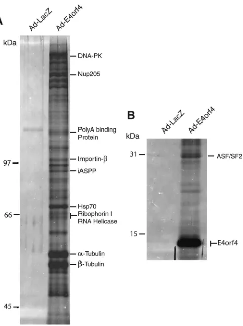
Identification of novel E4orf4-interacting proteins.
To identify
interacting partners of E4orf4, H1299 cells were infected with
ad-enoviral vectors expressing FLAG-tagged E4orf4
(Ad-FLAG-E4orf4) (
6
) or control

-galactosidase (Ad-LacZ) (
31
).
Immuno-precipitation (IP) using an anti-FLAG monoclonal antibody was
used to purify E4orf4 complexes from cell extracts.
Figure 1
shows
typical silver-stained SDS-PAGE of immunoprecipitates
ob-tained. Major bands on the gels were excised and submitted for
analysis by mass spectrometry. In total, 10 proteins were identified
in complexes with E4orf4. One of the proteins identified was
ser-ine/arginine splicing factor 1 (SRSF1, also known as ASF/SF2),
which has been previously reported as an E4orf4-associated
pro-tein (
20
,
21
). We also identified 9 other novel proteins interacting
with E4orf4, including DNA-PK, Nup205, poly(A) binding
pro-tein, importin-

, iASPP, Hsp 70, p68 DEAD Box RNA helicase,
ribophorin I, and
␣
/

tubulin. The observation that E4orf4
inter-acts with Nup205, a subunit of the nuclear pore complex (NPC),
FIG 1Identification of E4orf4 binding proteins. (A) Silver-stainedSDS-PAGE (8% polyacrylamide) of immunoprecipitated FLAG-E4orf4 complexes from H1299 cells infected with Ad-E4orf4. Cells were infected with Ad-LacZ as a negative control. (B) 15% polyacrylamide silver-stained gel of the same sam-ples as in panel A. The position of E4orf4 is indicated.
FLAG-Nup205
EV E4orf4 R69/70/72-75AR73/74/75AF84A R81A/F84AK88A L51A/L54A
IP HA-E4orf4
WCE HA-E4orf4
IP Flag-Nup205
FLAG-Nup205
EV myc-PK myc-PK -ARM
IP myc-PK
WCE myc-PK
IP FLAG-Nup 205
*
A
B
FIG 2The arginine-rich motif (ARM) of E4orf4 is required for interaction with Nup205. (A) FLAG-Nup205 was cotransfected in H1299 cells with wild-type or the indicated point mutants of E4orf4. Immunoprecipitation (IP) from cell extracts was performed using anti-FLAG antibody. IP com-plexes were analyzed by Western blotting to detect E4orf4 (anti-HA) and Nup205 (anti-FLAG). Anti-HA Western blotting of whole-cell extract (WCE) was used to confirm expression of FLAG-Nup205 (middle por-tion). (B) H1299 cells were cotransfected with FLAG-Nup205 and empty vector (EV), myc-pyruvate kinase (myc-PK), or myc-PK fused to the E4orf4 ARM (myc-PK-ARM). IP from cell extracts was performed using anti-FLAG. IP complexes were analyzed by Western blotting to detect myc-PK and myc-PK-ARM (anti-myc). myc immunoblotting of WCE is used to confirm expression of PK fusions. The asterisks indicates a back-ground band with anti-myc antibody.
on November 7, 2019 by guest
http://jvi.asm.org/
[image:3.585.46.286.64.385.2] [image:3.585.316.523.65.308.2]was particularly intriguing considering that interactions with the
NPC have been shown to regulate gene expression in many diverse
types of virus (reviewed in reference
39
). Further studies were
therefore performed to characterize the E4orf4-Nup205
interac-tion and its potential role in viral replicainterac-tion.
The ARM of E4orf4 is required for interaction with Nup205.
In order to determine the binding site for Nup205 on E4orf4, a
series of previously characterized point mutants along the length
of E4orf4 were utilized (
14
). Deletion mutations of E4or4 render
the protein highly labile and necessitate the use of discrete point
mutations for structure/function studies. In a previous study,
these E4orf4 mutants were characterized for ability to bind to
PP2A and activity for inducing cell death (
14
). Phenotypes of the
E4orf4 mutants fell into two classes. Class I mutants were defective
for interactions with PP2A and cell killing activity, and class II
mutants interacted with PP2A but had disrupted killing activity. A
subset of class I and II mutants were tested for interaction with
Nup205 by cotransfecting wild-type or mutant HA-E4orf4 and
FLAG-Nup205, followed by immunoprecipitation using
anti-FLAG antibody.
Figure 2A
shows that only E4orf4 point
muta-tions of the arginine-rich motif (ARM) between residues 69 and
75 (R69/70/72–75A) disrupted binding with Nup205. We then
determined if the ARM of E4orf4 was sufficient for interaction
with Nup205.
Figure 2B
shows that fusion of the ARM of E4orf4 to
pyruvate kinase (PK) conferred Nup205 binding activity on the
resulting fusion protein.
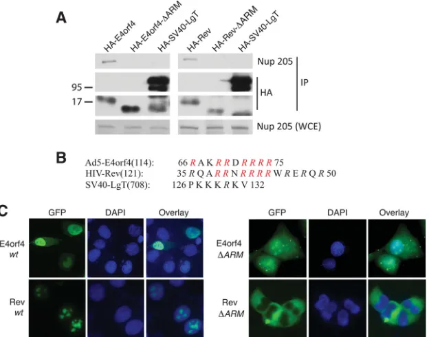
The ARM is required for nuclear localization of E4orf4 and
other nuclear viral proteins.
The observation that the ARM of
E4orf4 was necessary and sufficient for E4orf4 interaction with
Nup205 is intriguing considering that this sequence is also
re-quired to localize the protein to the nucleus (
7
). Several nuclear
viral proteins also contain an ARM. For example, apoptin from
chicken anemia virus (CAV Ap) and Rev from human
immuno-deficiency virus (HIV) are nuclear viral proteins that contain
ARMs.
Figure 3A
shows that HIV Rev is also able to interact with
endogenous Nup205 and point mutation of the ARM
(HA-Rev-⌬
ARM) abolished binding as observed with E4orf4. As a negative
control, we tested SV40 large T antigen (LgT), a nuclear viral
pro-tein that utilizes a conventional nuclear localization signal (NLS),
and showed that this protein does not bind Nup205. Point
muta-tion of the ARM of both HIV Rev and E4orf4 abolished nuclear
localization, indicating that interaction with Nup205 plays a role
in nuclear import of these proteins (
Fig. 3C
).
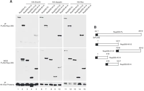
A common region on Nup205 is required for interaction with
viral ARMs.
A series of deletion mutants of Nup205 was
con-structed to determine the binding site of E4orf4. A schematic of
the FLAG-tagged Nup205 constructs is shown in
Fig. 4B
.
Figure
4A
shows that a central region from amino acids 419 to 1017 of
Nup205 was minimally required to interact with E4orf4 (lane 5).
Interestingly, the same region was also required to interact with
the two other ARM-containing viral proteins, CAV apoptin (lane
10) and HIV Rev (lane 15). These data show that the E4orf4 and
other ARM-containing proteins bind to Nup205 via the same
re-gion in the protein. Amino acid sequences rich in arginine are
highly positively charged and can potentially interact with a
suit-ably negatively charged region on Nup205. Analysis of the amino
acid sequence of Nup205 between residues 419 and 1017 did not
reveal any stretch of negatively charged amino acids, suggesting
FIG 3Nup205 associates with HIV Rev via the ARM domain and is required for nuclear localization. (A) H1299 cells were transfected with HA-E4orf4 or HA-Rev or mutants that lack the ARM (⌬ARM). HA-SV40-LgT is included as a negative control. Immunoprecipitation from cell extracts was performed using anti-HA. IP complexes were analyzed by Western blotting to detect endogenous Nup205 and HA-tagged viral proteins. Anti-Nup205 Western blotting of whole-cell extract was used to confirm expression of Nup205 (lower portion). (B) Schematic showing the ARMs of E4orf4 and HIV Rev. The nuclear localization sequence of SV40 is shown for comparison. Mutated residues in the⌬ARM mutants are indicated in red. (C) Epifluorescence microscopy showing localization of EGFP-fused E4orf4, Rev, and⌬ARM mutants.on November 7, 2019 by guest
http://jvi.asm.org/
[image:4.585.138.450.68.313.2]that interaction with the ARM may be a structural property of
Nup205 rather than the primary amino acid sequence.
Nup205 is required for the nuclear localization of HIV Rev
but not E4orf4.
Having identified that the ARM is required for
E4orf4 to interact with Nup205 and for E4orf4 to localize into the
nucleus, we tested whether the nuclear localization was dependent
upon Nup205. RNAi was used to knockdown expression of
Nup205 in HeLa cells. The Western blot in
Fig. 5A
shows that
siRNA targeting of Nup205 was able to silence protein expression
very efficiently relative to a nonsilencing control siRNA.
En-hanced GFP (EGFP)-tagged versions of E4orf4, HIV Rev, and LgT
were then transfected into Nup205 knockdown and control cells.
Figure 5B
shows that knockdown of Nup205 resulted in only a
slight increase in cytoplasmic levels of E4orf4. In contrast, nuclear
localization of HIV Rev was mostly inhibited after depletion of
Nup205. LgT, which includes a canonical NLS sequence, was not
affected by Nup205 knockdown. These data suggest that although
Nup205 may play a minor role in E4orf4 nuclear localization,
other factors must be primarily involved, since only a modest
localization defect was observed after Nup205 knockdown.
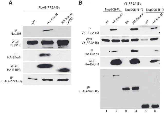
E4orf4 forms a trimolecular complex with PP2A-B
␣
and
Nup 205.
E4orf4 itself has no intrinsic enzymatic activity, and
most of its functions are mediated through interaction with PP2A.
E4orf4 can recruit target phosphoproteins into complexes with
PP2A, resulting in dephosphorylation of host and viral proteins
such as the SR splicing factors and the AP1 transcription factor
(
16
,
21
,
40
). We therefore determined if E4orf4 could form a
tri-molecular complex with Nup205 and PP2A as was observed with
other targets. A cDNA expressing a FLAG-tagged PP2A-B
␣
was
cotransfected with those expressing HA-E4orf4 or a mutant form
lacking the ARM (HA-E4orf4
⌬
ARM). As shown in
Fig. 6A
,
en-dogenous Nup205 coimmunoprecipitated with PP2A-B
␣
only in
the presence of wt E4orf4, indicating that the three proteins exist
as a trimolecular complex. Performing the inverse experiment,
transfected FLAG-Nup205 also coimmunoprecipitated PP2A-B
␣
,
but again, only in the presence of wt E4orf4 (
Fig. 6B
, lanes 1 and
2). As expected, even the deletion mutants of Nup205 that
main-tained binding to E4orf4 could also coimmunoprecipitate
PP2A-B
␣
with E4orf4 (
Fig. 6B
, lanes 3 to 6). Taken together, these
data show that E4orf4 is able to bring PP2A into a complex with
the NPC through Nup205.
E4orf4 modulates the phosphorylation state of Nup205.
The
above-described results show that E4orf4 may function by
bringing PP2A into a complex with Nup205. This mechanism
is similar to how E4orf4 modulates the splicing of viral late
mRNAs by inducing PP2A-dependent dephosphorylation of
the splicing factor ASF/SF2 (
20
). We therefore determined
whether E4orf4 could similarly modulate the phosphorylation
state of Nup205. There have been no studies examining
phos-phorylation of Nup205, although serine/threonine
phosphor-HA-E4orf4
Nup205-FLNup205-N1/2Nup205-C1/2
IP FLAG-Nup 205
WCE FLAG-Nup 205
IP HA-Viral Proteins
Nup205-A1/4Nup205-B1/4
HA-Rev HA-Apoptin
A
B
3xFLAG
aa 1 2012
1 1017
1 418
1018 2012
419 1017
Nup205-FL
Nup205-N1/2
Nup205-C1/2
Nup205-A1/4
Nup205-B1/4
Nup205-FLNup205-N1/2Nup205-C1/2Nup205-A1/4Nup205-B1/4Nup205-FLNup205-N1/2Nup205-C1/2Nup205-A1/4Nup205-B1/4
1 2 3 4 5 6 7 8 9 10 11 12 13 14 15
FIG 4ARM-containing viral proteins associated with the same domain on Nup205. (A) H1299 cells were cotransfected with wild-type or deletion mutants of FLAG-Nup205 and HA-tagged E4orf4, HA-apoptin, or HIV Rev. Immunoprecipitation from cell extracts was performed using anti-HA. IP complexes were analyzed by Western blotting to detect Nup205 and deletion mutants (anti-FLAG) and HA-tagged proteins (lower portion). Anti-FLAG Western blot of whole-cell extract (WCE) was used to confirm expression of FLAG-Nup205 and deletion mutants (middle portion). (B) Schematic of FLAG-Nup205 deletion constructs.
on November 7, 2019 by guest
http://jvi.asm.org/
[image:5.585.44.545.69.384.2]ylation other NPC components has been shown to regulate
nuclear envelope breakdown at mitosis (
41
). The
bioinformat-ics tool NetPhos 2.0 predicts 48 serine and 16 threonine
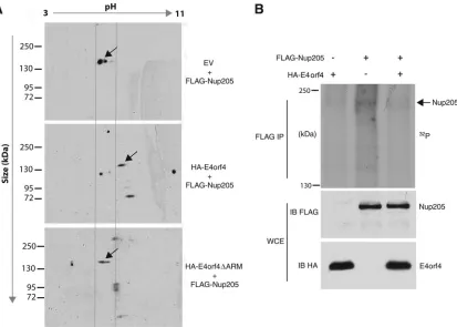
puta-tive phosphorylation sites on Nup205. Two-dimensional gel
electrophoresis was performed to analyze Nup205 in the
pres-ence or abspres-ence of E4orf4. FLAG-Nup205 was cotransfected in
H1299 cells with either wt E4orf4, E4orf4
⌬
ARM, or a vector
control.
Figure 7A
shows that in the presence of wt E4orf4, the
migration of Nup205 in the first dimension (pH) migrated with
a higher isoelectric point (pI) relative to that of the vector
control or the E4orf4
⌬
ARM mutant. Since phosphorylated
proteins generally have a lower pI value due to the negatively
charged phosphate groups, a lower pI value suggests that the
protein is hyperphosphorylated. Therefore, the increased pI
FIG 5Nup205 is required for nuclear localization of HIV Rev but not E4orf4. (A) Western blot demonstrating knockdown of Nup205 in HeLa cells. A tubulin blot is included as loading control. (B) Epifluorescence microscopy showing localization of EGFP-fused SV40-LgT, E4orf4, and HIV Rev in HeLa cells after transfection with nonsilencing or Nup205 siRNAs.FLAG-PP2A-Bα
EV HA-E4orf4 HA -E4orf4
ΔARM
IP Nup205
WCE Nup205
IP HA-E4orf4
WCE HA-E4orf4
IP FLAG-PP2A-Bα
V5-PP2A-Bα
IP V5-PP2A-Bα
WCE V5-PP2A-Bα
IP HA-E4orf4
WCE HA-E4orf4
IP FLAG-Nup205
A
B
EV HA -E4orf4
Nup205-FL Nup205-N1/2 Nup205-B1/4
EV HA -E4orf4
EV HA -E4orf4
1 2 3 4 5 6
FIG 6E4orf4 forms a trimolecular complex with PP2A-B␣and Nup205. (A) H1299 cells were cotransfected with FLAG-PP2A-B␣and wild-type HA-E4orf4, ⌬ARM mutant, or empty vector. Immunoprecipitation from cell extracts was performed using anti-FLAG to pull down FLAG-PP2A-B␣. IP complexes were analyzed by Western blotting to detect endogenous Nup205 (top), HA-E4orf4, and FLAG-PP2A-B␣. Western blotting of whole-cell extracts was used to confirm expression of endogenous Nup205 and HA-E4orf4. (B) H1299 cells were cotransfected with V5-PP2A-B␣, wild-type HA-E4orf4, or EV and FLAG-Nup205 or the indicated deletion mutants. IP from cell extracts was performed to using anti-FLAG to pull down FLAG-Nup205. IP complexes were analyzed by Western blotting to detect V5-PP2A-B␣(top), HA-E4orf4, and FLAG-Nup205 (bottom). Anti-HA Western blot of WCE is shown to confirm expression of E4orf4 and anti-V5 to confirm expression of PP2A-B␣.
on November 7, 2019 by guest
http://jvi.asm.org/
[image:6.585.48.540.63.271.2] [image:6.585.123.460.425.657.2]value of Nup205 in the presence of wt E4orf4 suggests that the
viral protein is mediating the dephosphorylation of Nup205.
As a more direct demonstration that E4orf4 can modulate the
phosphorylation state of Nup205, we performed metabolic
label-ing uslabel-ing [
32P]orthophosphate in H1299 cells cotransfected with
E4orf4 or an empty vector control.
Figure 7B
, lane 2, shows that
FLAG-Nup205 immunoprecipitated from
32P-labeled cell
ex-tracts migrated on SDS-PAGE as a major band at approximately
200 kDa, the correct molecular mass of Nup205. Furthermore, in
the presence of E4orf4 the intensity of the band is reduced
consid-erably (
Fig. 6B
, lane 2). These data show for the first time that
Nup205 exists as a phosphoprotein and that E4orf4 can reduce the
phosphorylation of Nup205.
E4orf4 and Nup205 regulate viral gene expression.
Previous
studies have demonstrated that E4orf4 regulates viral gene
ex-pression during infection primarily by downregulation of early
promoters.
Figure 8A
shows that a viral mutant that lacks
ex-pression of E4orf4 displays elevated exex-pression of early protein
production (E1A and E4orf6), consistent with previous studies
(
8
,
17
,
18
). In contrast, we observe that late protein production
as measured by Western blot detection of capsid proteins is
markedly reduced in the Orf4
⫺virus relative to the wt. In order
to determine if Nup205 plays a role in E4or4 gene regulation,
Nup205 mRNA expression was knocked down in H1299 cells
and subsequently infected with wt virus.
Figure 8B
shows that
cells with Nup205 knockdown displayed a pattern of gene
ex-pression similar to that of the E4orf4
⫺virus, with elevated early
gene expression and low late gene expression. The efficiency of
Nup205 knockdown is shown in
Fig. 8C
. In order to determine if
the alterations in viral gene expression were at the level of mRNA,
real-time RT-PCR analysis was performed on E1A and E4orf6 at
7.5 h postinfection. Late viral gene expression was assessed at 23 h
postinfection by analyzing the abundance of the tripartite leader
containing mRNAs. We observed that late mRNAs were reduced
relative to the nonsilencing control (
Fig. 8D
), whereas early viral
mRNAs were elevated (
Fig. 8E
), which is in agreement with results
obtained by Western blot analysis. The similar phenotypes of
E4orf4
⫺virus and cells with knockdown of Nup205 suggest that
these proteins act as a complex to regulate gene viral expression at
the mRNA level.
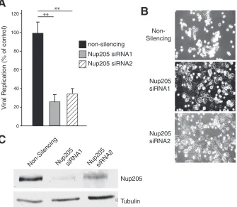
Knockdown of Nup205 impairs viral replication and delays
the cytopathic effect.
Since Nup205 was required for proper viral
gene expression, we then determined if it is also required for
ef-fective viral replication. Replication of wt Ad5 was measured in
H1299 cells following siRNA knockdown of Nup205 or a
nonsi-lencing control. Virus yields were quantitatively determined using
the FFU assay (
29
,
38
). H1299 cells with knockdown of Nup205
did not exhibit major deleterious effects and appeared similar to
nonsilencing control cells during the course of the experiment
(data not shown). However, analysis of viral replication in
Fig. 9A
FIG 7E4orf4 modulates the phosphorylation state of Nup205. (A) H1299 cells were cotransfected with FLAG-Nup205 and empty vector, HA-E4orf4, or HA-E4orf4⌬ARM. Immunoprecipitation from cell extracts was performed using anti-FLAG. IP complexes were analyzed by Western blotting (anti-FLAG) after 2-dimensional gel electrophoresis to detect FLAG-Nup205. Isoelectric focusing was performed in thexaxis on a pH gradient from 3 to 11, followed by size separation on theyaxis. Arrows indicate the position of FLAG-Nup205, which migrates at approximately 200 kDa. (B) H1299 cells were transfected with HA-E4orf4, FLAG-Nup205, or both. Forty-eight hours posttransfection, cells were labeled with [32P]orthophosphate and immunoprecipitation was performed on cell extracts using anti-FLAG. Immunoprecipitates were resolved on SDS-PAGE to detect32P-labeled proteins (top). Western blotting was performed on whole-cell extracts to confirm expression of Nup205 (anti-FLAG) and E4orf4 (anti-HA).on November 7, 2019 by guest
http://jvi.asm.org/
[image:7.585.88.501.65.360.2]showed that knockdown of Nup205 using two different siRNA
duplexes resulted in replication of 30% and 35% relative to the
nonsilencing control. In addition, 30 h postinfection, most of the
H1299 cells treated with nonsilencing siRNA displayed a rounded
and swollen appearance characteristic of viral cytopathic effect
(CPE), whereas in cells treated with the two Nup205 siRNAs, only
half had such morphology (
Fig. 9B
). The efficiency of Nup205
knockdown is shown in
Fig. 9C
. Taken together, these data show
that adenovirus replication is significantly compromised in cells
with reduced levels of Nup205.
DISCUSSION
Many types of viruses, including adenoviruses, interact with
the NPC in order to facilitate entry into the nucleus during
initial infection (reviewed in reference
39
). Our study has
dem-onstrated for the first time a mechanism by which adenoviral
proteins interact with the nuclear pore complex during viral
replication to regulate viral gene expression. We propose a
mechanism by which the E4orf4 protein recruits the PP2A
phosphatase into a trimolecular complex with Nup205 and
thereby causes dephosphorylation of the protein and alters its
function. The NPC is an enormous complex (approximately 60
MDa) composed of about 30 distinct nucleoporins (Nups)
known to be extensively regulated by phosphorylation. For
ex-ample, phosphorylation of NPC components during mitosis
mediates the disassembly of the NPC (
41
).
The function of Nup205 within the NPC remains poorly
un-derstood. The Nups can be broadly categorized as being
compo-Nup 205
Tubulin
Non-Silencing Nup205siRNA2
Non-Silencing
0
E4orf6
E1A
Capsid
(Late)
14
21 28 48
Nup205 siRNA2
A
B
H5pg4100 (wt)
E4orf4-E1A
E4orf4
E4orf6
0
24
48
72
Capsid
(Late)
0 14
21 28 48
H5pg4100 (wt)
0
24
48
72
Relativ
e mRNA e
xpression
Late Message (tripartite leader)
non-silencing
Nup205 siRNA1
Nup205 siRNA2
0 0.25 0.50 0.75 1.0 1.25 1.50
*
C
Relativ
e mRNA e
xpression
0 2.0 4.0 6.0
E1A E4orf6
*
*
*
*
D
E
h.p.i
h.p.i
FIG 8E4orf4 and Nup205 regulate viral gene expression. (A) Western blot showing expression of indicated viral proteins at various hours postinfection (h.p.i) in H1299 cells infected with wild-type (H5pg400) Ad5 or the E4orf4⫺virus. (B) Western blot showing expression of viral proteins at indicated h.p.i. in H1299 cells transfected with nonsilencing or siRNA targeting Nup205. (C) Western blot demonstrating knockdown of Nup205 in H1299 cells. A tubulin blot is included as loading control. (D) Quantitative RT-PCR of late viral gene expression (mRNAs with tripartite leader) at 23 hours postinfection in H1299 cells transfected with nonsilencing or siRNAs targeting Nup205. (E) qRT-PCR of early gene expression (E1A and E4orf6) at 7.5 hours postinfection.
on November 7, 2019 by guest
http://jvi.asm.org/
[image:8.585.48.539.67.503.2]nents of the outer structural skeleton of the NPC or the central
pore that mediates import/export of cargoes across the nuclear
membrane. Nup205 is one of the components of the central pore
that is responsible for the gating functions of the NPC and is not
required for formation or integrity of the NPC (
42
). Interestingly,
knockdown of Nup205 in
Caenorhabditis elegans
was shown to
increase the size limit of nonnuclear macromolecules that could
passively enter the nucleus (
43
). These changes occurred without
affecting the active transport of proteins into the nucleus.
There-fore, E4orf4 interactions with Nup205 may alter the dynamics of
viral and/or host cell proteins across the nuclear membrane as a
means to enhance replication. In late stages of adenovirus
infec-tion, export of viral mRNAs from the nucleus is enhanced,
whereas host mRNAs are blocked through an unknown
mecha-nism (reviewed in references
3
and
44
). Since we observed that
Nup205 knockdown caused reduced late gene expression but
ap-peared to enhance early gene expression, E4orf4 targeting of
Nup205 may provide a molecular explanation for the pattern of
adenovirus gene expression during late phases.
E4orf4 stimulated the dephosphorylation of Nup205 as it does
other cellular targets, such as SR splicing factors, which suggests
that the activity of Nup205 is itself regulated by phosphorylation
under normal physiological conditions. However, no study to
date has mapped phosphorylation sites on Nup205 or determined
how these modifications may affect activities such as transcription
and mRNA transport. Judging from the large number of potential
phosphorylation sites that are present on Nup205, it is likely that
the protein is dynamically regulated by signaling and/or cell
cycle-dependent kinases. Further studies into the phosphorylation of
Nup205 may therefore provide additional insights into the
regu-lation of gene expression in normal physiology as well as during
viral infection. In addition to inducing dephosphorylation of
Nup205, another possible function for the E4orf4-Nup205
inter-action may be to localize PP2A to the nuclear pore, where it may,
in turn, dephosphorylate host factors as they are transported. For
example, the phosphorylation states of E1A, SR proteins, myc, and
AP1 have all been shown to be reduced in the presence of E4orf4
(
8
,
17
,
21
). E4orf4 complexes associated with Nup205 may
there-fore dephosphorylate such proteins as they are transported
through the NPC.
Our results suggest that activity of Nup205 is required for
ad-enoviral replication. Interestingly, Nup205 may be required for
replication of other viruses. For example, a recent study describing
a genome-wide screen for host factors required for replication of
influenza virus also identified Nup205 (
45
). In addition, our data
also suggest that nuclear localization of HIV Rev is dependent on
Nup205. It would be interesting to determine if Nup205
knock-down also has inhibitory effects on HIV replication as it does with
adenovirus and influenza virus. In addition to adenoviruses,
sev-eral other virus types have been shown to alter the properties of the
NPC during the course of replication (reviewed in reference
39
).
For example, herpesvirus (
46
), picornavirus (
47–49
), and HIV
(
50
) have all been shown to induce reorganization of NPC
sub-units without affecting the overall integrity of the pore. Although
Nup205 siRNA1
Nup205 siRNA2
Nup205
Tubulin
A
B
C
Non-SilencingNup205
siRNA1 Nup205 siRNA2
Non-Silencing
Vir
al Replication (% of control)
non-silencing
Nup205 siRNA1 Nup205 siRNA2
0 20 40 60 80 100
120
**
**
FIG 9Knockdown of Nup205 impairs viral replication and delays the cytopathic effect. (A) Replication of wild-type Ad5 (H5pg400) in H1299 cells transfected with nonsilencing or two separate siRNAs targeting Nup205. (B) Light micrographs showing appearance of cells infected with wild-type Ad4 under conditions of Nup205 knockdown or control. (C) Western blot demonstrating knockdown of Nup205 in H1299 cells. A tubulin blot is included as a loading control.
on November 7, 2019 by guest
http://jvi.asm.org/
[image:9.585.127.462.72.364.2]these viral interactions with the NPC differ greatly in their specific
mechanisms, they may act as conserved strategies to control the
transport of macromolecules during replication and regulate both
viral and cellular gene expression.
ACKNOWLEDGMENTS
We thank Sabrina Schreiner for insightful discussions and Bob Weinberg, Thomas Dobner, and Douglass Forbes for reagents.
This work was supported by grants from the Canadian Institutes of Health Research (CIHR), MOP-179122 (J.G.T.), MOP-93753 (P.E.B. and J.G.T.), and the Natural Science and Engineering Research Council of Canada (J.G.T.). T.J.K. was supported by a doctoral studentship from the CIHR. Y.L. was supported by a studentship from the FRSQ.
REFERENCES
1.Täuber B, Dobner T.2001. Molecular regulation and biological function of adenovirus early genes: the E4 ORFs. Gene278:1–23.http://dx.doi.org
/10.1016/S0378-1119(01)00722-3.
2.Imperiale MJ, Akusjnarvi G, Leppard KN. 1995. Post-transcriptional control of adenovirus gene expression. Curr. Top. Microbiol. Immunol. 199(Part 2):139 –171.
3.Dobner T, Kzhyshkowska J.2001. Nuclear export of adenovirus RNA. Curr. Top. Microbiol. Immunol. 259:25–54.http://dx.doi.org/10.1007
/978-3-642-56597-7_2.
4.Leppard KN.1997. E4 gene function in adenovirus, adenovirus vector and adeno-associated virus infections. J. Gen. Virol.78(Part 9):2131– 2138.
5.Weinberg DH, Ketner G.1986. Adenoviral early region 4 is required for efficient viral DNA replication and for late gene expression. J. Virol.57: 833– 838.
6.Miron MJ, Blanchette P, Groitl P, Dallaire F, Teodoro JG, Li S, Dobner T, Branton PE. 2009. Localization and importance of the adenovirus E4orf4 protein during lytic infection. J. Virol.83:1689 –1699.http://dx.doi
.org/10.1128/JVI.01703-08.
7.Miron MJ, Gallouzi IE, Lavoie JN, Branton PE.2004. Nuclear localiza-tion of the adenovirus E4orf4 protein is mediated through an arginine-rich motif and correlates with cell death. Oncogene23:7458 –7468.http:
//dx.doi.org/10.1038/sj.onc.1207919.
8.Brestovitsky A, Sharf R, Mittelman K, Kleinberger T.2011. The adeno-virus E4orf4 protein targets PP2A to the ACF chromatin-remodeling fac-tor and induces cell death through regulation of SNF2h-containing com-plexes. Nucleic Acids Res.39:6414 – 6427.http://dx.doi.org/10.1093/nar
/gkr231.
9.Zhang Z, Mui MZ, Chan F, Roopchand DE, Marcellus RC, Blanch-ette P, Li S, Berghuis AM, Branton PE. 2011. Genetic analysis of B55alpha/Cdc55 protein phosphatase 2A subunits: association with the adenovirus E4orf4 protein. J. Virol.85:286 –295.http://dx.doi.org
/10.1128/JVI.01381-10.
10. Li S, Brignole C, Marcellus R, Thirlwell S, Binda O, McQuoid MJ, Ashby D, Chan H, Zhang Z, Miron MJ, Pallas DC, Branton PE.2009. The adenovirus E4orf4 protein induces G2/M arrest and cell death by blocking protein phosphatase 2A activity regulated by the B55 subunit. J. Virol.83:8340 – 8352.http://dx.doi.org/10.1128/JVI.00711-09.
11. Li S, Szymborski A, Miron MJ, Marcellus R, Binda O, Lavoie JN, Branton PE.2009. The adenovirus E4orf4 protein induces growth arrest and mitotic catastrophe in H1299 human lung carcinoma cells. Oncogene 28:390 – 400.http://dx.doi.org/10.1038/onc.2008.393.
12. Ben-Israel H, Sharf R, Rechavi G, Kleinberger T. 2008. Adenovirus E4orf4 protein downregulates MYC expression through interaction with the PP2A-B55 subunit. J. Virol.82:9381–9388.http://dx.doi.org/10.1128
/JVI.00791-08.
13. Shtrichman R, Sharf R, Kleinberger T.2000. Adenovirus E4orf4 protein interacts with both Balpha and B’ subunits of protein phosphatase 2A, but E4orf4-induced apoptosis is mediated only by the interaction with Balpha. Oncogene19:3757–3765.http://dx.doi.org/10.1038/sj.onc.1203705. 14. Marcellus RC, Chan H, Paquette D, Thirlwell S, Boivin D, Branton
PE.2000. Induction of p53-independent apoptosis by the adenovirus E4orf4 protein requires binding to the Balpha subunit of protein phos-phatase 2A. J. Virol.74:7869 –7877.http://dx.doi.org/10.1128/JVI.74
.17.7869-7877.2000.
15. Kleinberger T, Shenk T.1993. Adenovirus E4orf4 protein binds to pro-tein phosphatase 2A, and the complex down regulates E1A-enhanced junB transcription. J. Virol.67:7556 –7560.
16. Mui MZ, Kucharski M, Miron MJ, Hur WS, Berghuis AM, Blanchette P, Branton PE.2013. Identification of the adenovirus E4orf4 protein binding site on the B55alpha and Cdc55 regulatory subunits of PP2A: Implications for PP2A function, tumor cell killing and viral replication. PLoS Pathog. 9:e1003742.http://dx.doi.org/10.1371/journal.ppat.1003742.
17. Müller U, Kleinberger T, Shenk T.1992. Adenovirus E4orf4 protein reduces phosphorylation of c-Fos and E1A proteins while simultaneously reducing the level of AP-1. J. Virol.66:5867–5878.
18. Mannervik M, Fan S, Strom AC, Helin K, Akusjarvi G.1999. Adeno-virus E4 open reading frame 4-induced dephosphorylation inhibits E1A activation of the E2 promoter and E2F-1-mediated transactivation inde-pendently of the retinoblastoma tumor suppressor protein. Virology256: 313–321.http://dx.doi.org/10.1006/viro.1999.9663.
19. Kanopka A, Muhlemann O, Akusjarvi G.1996. Inhibition by SR proteins of splicing of a regulated adenovirus pre-mRNA. Nature381:535–538.
http://dx.doi.org/10.1038/381535a0.
20. Kanopka A, Muhlemann O, Petersen-Mahrt S, Estmer C, Ohrmalm C, Akusjarvi G.1998. Regulation of adenovirus alternative RNA splicing by dephosphorylation of SR proteins. Nature393:185–187.http://dx.doi.org
/10.1038/30277.
21. Estmer Nilsson C, Petersen-Mahrt S, Durot C, Shtrichman R, Krainer AR, Kleinberger T, Akusjarvi G.2001. The adenovirus E4-ORF4 splicing enhancer protein interacts with a subset of phosphorylated SR proteins. EMBO J.20:864 – 871.http://dx.doi.org/10.1093/emboj/20.4.864. 22. Marcellus RC, Teodoro JG, Wu T, Brough DE, Ketner G, Shore GC,
Branton PE.1996. Adenovirus type 5 early region 4 is responsible for E1A-induced p53-independent apoptosis. J. Virol.70:6207– 6215. 23. Shtrichman R, Kleinberger T.1998. Adenovirus type 5 E4 open reading
frame 4 protein induces apoptosis in transformed cells. J. Virol.72:2975– 2982.
24. Lavoie JN, Nguyen M, Marcellus RC, Branton PE, Shore GC.1998. E4orf4, a novel adenovirus death factor that induces p53-independent apoptosis by a pathway that is not inhibited by zVAD-fmk. J. Cell Biol. 140:637– 645.http://dx.doi.org/10.1083/jcb.140.3.637.
25. Shtrichman R, Sharf R, Barr H, Dobner T, Kleinberger T.1999. Induc-tion of apoptosis by adenovirus E4orf4 protein is specific to transformed cells and requires an interaction with protein phosphatase 2A. Proc. Natl. Acad. Sci. U. S. A.96:10080 –10085.http://dx.doi.org/10.1073/pnas.96.18
.10080.
26. Branton PE, Roopchand DE.2001. The role of adenovirus E4orf4 protein in viral replication and cell killing. Oncogene20:7855–7865.http://dx.doi
.org/10.1038/sj.onc.1204862.
27. Kornitzer D, Sharf R, Kleinberger T.2001. Adenovirus E4orf4 protein induces PP2A-dependent growth arrest in Saccharomyces cerevisiae and interacts with the anaphase-promoting complex/cyclosome. J. Cell Biol. 154:331–344.http://dx.doi.org/10.1083/jcb.200104104.
28. Mui MZ, Roopchand DE, Gentry MS, Hallberg RL, Vogel J, Branton PE.2010. Adenovirus protein E4orf4 induces premature APCCdc20 acti-vation in Saccharomyces cerevisiae by a protein phosphatase 2A-dependent mechanism. J. Virol.84:4798 – 4809.http://dx.doi.org/10.1128
/JVI.02434-09.
29. Groitl P, Dobner T.2007. Construction of adenovirus type 5 early region 1 and 4 virus mutants. Methods Mol. Med.130:29 –39.http://dx.doi.org
/10.1385/1-59745-166-5:29.
30. Tollefson AE, Kuppuswamy M, Shashkova EV, Doronin K, Wold WS. 2007. Preparation and titration of CsCl-banded adenovirus stocks. Meth-ods in molecular medicine. 130:223–235. http://dx.doi.org/10.1385/1
-59745-166-5:223.
31. Bacchetti S, Graham F.1993. Inhibition of cell-proliferation by an ade-novirus vector expressing the human wild type-p53 protein. Int. J. Oncol. 3:781–788.
32. Heilman DW, Teodoro JG, Green MR.2006. Apoptin nucleocytoplas-mic shuttling is required for cell type-specific localization, apoptosis, and recruitment of the anaphase-promoting complex/cyclosome to PML bod-ies. J. Virol.80:7535–7545.http://dx.doi.org/10.1128/JVI.02741-05. 33. Siomi H, Dreyfuss G.1995. A nuclear localization domain in the hnRNP
A1 protein. J. Cell Biol.129:551–560.http://dx.doi.org/10.1083/jcb.129.3
.551.
34. Teodoro JG, Heilman DW, Parker AE, Green MR. 2004. The viral protein Apoptin associates with the anaphase-promoting complex to
on November 7, 2019 by guest
http://jvi.asm.org/
duce G2/M arrest and apoptosis in the absence of p53. Genes Dev.18: 1952–1957.http://dx.doi.org/10.1101/gad.1198404.
35. Harlow E, Franza BR, Jr, Schley C.1985. Monoclonal antibodies specific for adenovirus early region 1A proteins: extensive heterogeneity in early region 1A products. J. Virol.55:533–546.
36. Boivin D, Morrison MR, Marcellus RC, Querido E, Branton PE.1999. Analysis of synthesis, stability, phosphorylation, and interacting polypep-tides of the 34-kilodalton product of open reading frame 6 of the early region 4 protein of human adenovirus type 5. J. Virol.73:1245–1253. 37. Kindsmüller K, Groitl P, Hartl B, Blanchette P, Hauber J, Dobner T.
2007. Intranuclear targeting and nuclear export of the adenovirus E1B-55K protein are regulated by SUMO1 conjugation. Proc. Natl. Acad. Sci. U. S. A.104:6684 – 6689.http://dx.doi.org/10.1073/pnas.0702158104. 38. Philipson L.1961. Adenovirus assay by the fluorescent cell-counting procedure.
Virology15:263–268.http://dx.doi.org/10.1016/0042-6822(61)90357-9. 39. Le Sage V, Mouland AJ. 2013. Viral subversion of the nuclear pore
complex. Viruses5:2019 –2042.http://dx.doi.org/10.3390/v5082019. 40. Müller U, Roberts MP, Engel DA, Doerfler W, Shenk T.1989. Induction
of transcription factor AP-1 by adenovirus E1A protein and cAMP. Genes Dev.3:1991–2002.http://dx.doi.org/10.1101/gad.3.12a.1991.
41. Laurell E, Beck K, Krupina K, Theerthagiri G, Bodenmiller B, Horvath P, Aebersold R, Antonin W, Kutay U.2011. Phosphorylation of Nup98 by multiple kinases is crucial for NPC disassembly during mitotic entry. Cell144:539 –550.http://dx.doi.org/10.1016/j.cell.2011.01.012. 42. Theerthagiri G, Eisenhardt N, Schwarz H, Antonin W. 2010. The
nucleoporin Nup188 controls passage of membrane proteins across the nuclear pore complex. J. Cell Biol.189:1129 –1142.http://dx.doi.org/10
.1083/jcb.200912045.
43. Galy V, Mattaj IW, Askjaer P.2003. Caenorhabditis elegans nucleo-porins Nup93 and Nup205 determine the limit of nuclear pore complex size exclusion in vivo. Mol. Biol. Cell14:5104 –5115.http://dx.doi.org/10
.1091/mbc.E03-04-0237.
44. Flint SJ, Gonzalez RA.2003. Regulation of mRNA production by the adenoviral E1B 55-kDa and E4 Orf6 proteins. Curr. Top. Microbiol. Im-munol.272:287–330.http://dx.doi.org/10.1007/978-3-662-05597-7_10. 45. Karlas A, Machuy N, Shin Y, Pleissner KP, Artarini A, Heuer D, Becker
D, Khalil H, Ogilvie LA, Hess S, Maurer AP, Muller E, Wolff T, Rudel T, Meyer TF.2010. Genome-wide RNAi screen identifies human host factors crucial for influenza virus replication. Nature463:818 – 822.http:
//dx.doi.org/10.1038/nature08760.
46. Copeland AM, Newcomb WW, Brown JC. 2009. Herpes simplex virus replication: roles of viral proteins and nucleoporins in capsid-nucleus attach-ment. J. Virol.83:1660 –1668.http://dx.doi.org/10.1128/JVI.01139-08. 47. Gustin KE, Sarnow P.2001. Effects of poliovirus infection on
nucleo-cytoplasmic trafficking and nuclear pore complex composition. EMBO J. 20:240 –249.http://dx.doi.org/10.1093/emboj/20.1.240.
48. Gustin KE, Sarnow P.2002. Inhibition of nuclear import and altera-tion of nuclear pore complex composialtera-tion by rhinovirus. J. Virol.76: 8787– 8796.http://dx.doi.org/10.1128/JVI.76.17.8787-8796.2002. 49. Park N, Katikaneni P, Skern T, Gustin KE.2008. Differential targeting of
nuclear pore complex proteins in poliovirus-infected cells. J. Virol.82: 1647–1655.http://dx.doi.org/10.1128/JVI.01670-07.
50. Monette A, Pante N, Mouland AJ.2011. HIV-1 remodels the nuclear pore complex. J. Cell Biol.193:619 – 631.http://dx.doi.org/10.1083/jcb
.201008064.
on November 7, 2019 by guest
http://jvi.asm.org/