0022-538X/95/$04.0010
Copyrightq1995, American Society for Microbiology
Point Mutations in the Herpes Simplex Virus Type 1 Vmw110 RING
Finger Helix Affect Activation of Gene Expression, Viral Growth,
and Interaction with PML-Containing Nuclear Structures
ROGER EVERETT,1* PETER O’HARE,2DELIA O’ROURKE,2PAUL BARLOW,3
ANDANNE ORR1
Medical Research Council Virology Unit, Glasgow G11 5JR, Scotland,1and Department of
Biochemistry, University of Oxford, Oxford OX1 3QU,3and Marie Curie Research
Institute, Oxted, Surrey RH8 0TL,2England
Received 24 May 1995/Accepted 28 July 1995
Herpes simplex virus type 1 immediate-early protein Vmw110 (also known as ICP0) has been implicated in the control of the balance between the lytic and latent states, but the precise mechanisms by which it exerts its
effects are unknown. Vmw110 includes a characteristic zinc binding domain, termed the C3HC4domain or
RING finger, which is essential for its function. The solution structure of a related herpesvirus RING finger domain suggested that an amphipathic alpha helix might be an important functional component of the RING finger. In this paper, we show that the equivalent region of Vmw110 is important for virus growth in tissue culture and for the normal interaction of Vmw110 with nuclear structures which include the PML protein.
Herpes simplex virus type 1 (HSV-1) is an important human pathogen with the intriguing ability to replicate lytically in the periphery and to establish a latent infection in sensory neu-rons. The virus encodes at least 75 distinct genes (27, 28) which are regulated differentially during lytic growth and latency. During the lytic cycle, all viral genes are expressed in a tem-poral cascade in which the five immediate-early (IE) genes are transcribed first, followed by the early and late classes (see reference 30 for a review). During latency, only the latency-associated transcripts, whose function remains obscure, have been detected (17, 33). An understanding of the regulatory mechanisms which distinguish the lytic and latent states of the virus is of both clinical and biological importance.
Several of the five IE gene products have significant or essential roles in the regulation of lytic viral gene expression. Of these, the product of IE gene 1 (IE-1) (Vmw110 or ICP0) appears to be involved in both lytic growth and latency and is able to activate gene expression in transfection assays (see references 8 and 15 for reviews). Viruses with lesions in Vmw110 have multiplicity-dependent and cell-type-dependent defects in the onset of the lytic cycle (10, 32, 34) and are unable to reactivate latent virus in an in vitro latency system (20, 31). Such viruses also reactivate inefficiently in mouse latency mod-els (3, 4, 23). Mutational analysis of Vmw110 has established that a RING finger zinc binding domain is very important for Vmw110 activity (9, 10, 18–20). The solution structure of the RING finger domain of a related herpesvirus protein illus-trates that it comprises a triple antiparallel beta sheet, on one side of which is embedded an amphipathic alpha helix (1, 12). Two zinc atoms are coordinated at either end of the helix, which extends basic and polar side chains into solvent. We surmised that these hydrophilic side chains would be required for the function of the herpesvirus RING finger. This predic-tion was supported by the observapredic-tion that amino acid replace-ments in the potential helix region reduced Vmw110 activity in
transfection assays, when acting in synergy with HSV-1 IE protein Vmw175 (1).
To extend these studies, we have investigated whether mu-tations in the potential Vmw110 helix region disrupt its ability to activate gene expression in the absence of Vmw175, and we have constructed recombinant viruses which express the mu-tant Vmw110 proteins. Since Vmw110 becomes associated with nuclear structures which contain the PML protein (re-ferred to herein as ND10) (6, 14, 21, 22, 25, 26, 36), we have also investigated the interactions between the mutant Vmw110 proteins and ND10.
Mutations in the potential RING finger helix reduce
activa-tion of gene expression by Vmw110.The RING finger domain
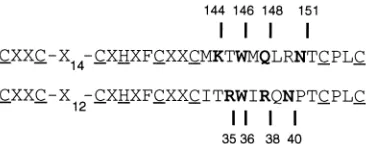
of the equine herpesvirus type 1 (EHV-1) gene 63 protein includes an amphipathic alpha helix with exposed polar and basic side chains (1). Comparison of primary sequences shows that charged and polar residues are present in the Vmw110 RING finger at positions similar to those in the EHV gene 63 RING finger helix (Fig. 1). In all the herpesvirus members of the protein family there is a trytophan residue 4 residues from the zinc-coordinating cysteine at the start of the potential helix region. We have shown that mutation of Vmw110 residues lysine 144, glutamine 148, and asparagine 151 affects the ability of the protein to activate gene expression in transfection assays in the presence of the major HSV-1 transactivator, Vmw175 (1). We wished to determine what effect these mutations had on the ability of Vmw110 to activate gene expression when acting alone; in addition, the effect of mutating the conserved tryptophan was investigated.
Vmw110 strongly activates expression of genes which in-clude the HSV-1 UL39 promoter in both transfection assays and during virus infection (5). Therefore, we used a plasmid with the UL39 promoter linked to chloramphenicol acetyl-transferase to assay the effects of substitution mutations in the Vmw110 RING finger. Surprisingly, mutation of the trypto-phan at position 146 to an alanine residue had little effect on the activity of Vmw110 (Fig. 2). Presumably, structural stability can be maintained without the interactions which W-146 makes with the core of the domain. The K-to-E mutation at position 144 (K144E) had the most substantial effect, similar to
* Corresponding author. Fax: 44 141 337 2236. Electronic mail ad-dress: [email protected].
7339
on November 9, 2019 by guest
http://jvi.asm.org/
that of the RING finger deletion mutant p110FXE. The N151D mutation also had a significant effect, while the activity of Q148E was reduced only about twofold (Fig. 2). We con-clude that mutations in the potential RING finger helix region reduce the intrinsic ability of Vmw110 to activate gene expres-sion, but the extent of the effect depends on the nature of the mutation.
These results differ slightly from those obtained with the same Vmw110 mutant plamids acting on a gD promoter re-porter in the presence of Vmw175 (1). The Q148E mutant was less active in the latter system, while the K144E mutant was more active. Variations in the details of results obtained with different transfection protocols have been well established (15); it is interesting that the results obtained with the UL39 reporter construct reported in this paper reflect more closely the effects of the mutations on virus growth than those ob-tained with the gD reporter in the presence of Vmw175.
Mutations in the potential RING finger helix of Vmw110
affect growth of HSV-1 virus in tissue culture.Infectious viral
dl1403 DNA, which carries a deletion of the majority of the
Vmw110 coding region (34), was cotransfected into cells with plasmids containing the mutant IE-1 genes. Progeny plaques were screened for the presence of a reconstituted IE-1 region, and clones were purified as previously described (10). Silent coding changes were introduced into the K144E, Q148E, and N151D viruses to form an NsiI site for screening purposes. However, the actual mutagenic base changes could not be detected by restriction enzyme analysis. To prove that the viruses encoded the mutant proteins, their complete RING finger coding regions were amplified by PCR and the pro-ducts were sequenced directly. In all cases, the expected mu-tation was detected and there was no evidence of other alter-ations.
HSV-1 viruses which fail to express Vmw110, or which ex-press Vmw110 proteins with an inactivated RING finger, have a 1- to 2-log reduction in growth efficiency in single-step growth curves (10). The extent of the reduction correlates inversely with the growth rate of the indicator cells (11). The single-step growth kinetics of viruses K144E, W146A, Q148E, and N151D in BHK cells were determined in comparison with wild-type virus and the mutant virus FXE, from which the Vmw110 RING finger has been deleted (10). The results clearly show that the K144E and N151D mutations were as deleterious for virus growth in BHK cells as removal of the entire RING finger domain (Fig. 3). The W146A and Q148E mutations had little, if any, significant effect. Therefore, the potential helix region of the Vmw110 RING finger is important for HSV-1 growth in tissue culture. Interestingly, the effects of the individual muta-tions on virus growth mirror their effects on activation of the UL39 promoter in transfected HeLa cells (Fig. 2).
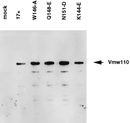
All of the mutant viruses expressed normal amounts of full-length Vmw110, which suggests that the single-residue substi-tutions had not caused gross conformational alteration or in-stability of the mutant proteins (Fig. 4).
Mutations in the potential RING finger helix affect the
in-teraction of Vmw110 with discrete nuclear domains.At early
times of HSV-1 infection, wild-type Vmw110 is located in part in discrete nuclear structures (ND10) which contain PML, sp100, and other uncharacterized cellular proteins. At later times of infection, the cellular ND10 structures are disrupted, an effect which requires a functional Vmw110 RING finger domain (25, 26). Similarly, in transfected cells which express wild-type Vmw110, detection of PML in ND10 becomes much more difficult, indicating either that PML is being dispersed or that conformational changes occur so as to occlude the epitope recognized by the 5E10 anti-PML monoclonal antibody (14). When the Vmw110 RING finger domain is deleted, PML and mutant Vmw110 stably colocalize in transfected cells (14).
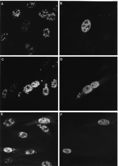
[image:2.612.87.270.71.143.2]To assess the effects of the RING finger helix mutations on the interactions between Vmw110 and ND10, BHK cells were transfected with p111, p110FXE, and plasmids expressing Vmw110 proteins with substitution mutations in the potential RING finger helix region. The cells were stained for Vmw110 and PML 16 h later; representative results from several hun-dred Vmw110-positive cells are presented in Fig. 5. Wild-type Vmw110 drastically reduced the detection of PML (Fig. 5A and B). In fact, PML was undetectable in many cells expressing wild-type Vmw110, but a field where faint PML fluorescence remains visible has been selected so as to enable comparison between the images at the two different wavelengths. In con-trast, the RING finger mutant p110FXE stably colocalized with the ND10 structures (Fig. 5K and L). The RING finger helix mutations exhibited a range of phenotypes. Mutant K144E was similar to FXE, with extensive colocalization of mutant Vmw110 and PML in ND10 structures (Fig. 5C and D). Mutant N151D also failed to disperse PML and colocalized at ND10 (Fig. 5E and F). However, mutant Q148E exhibited intermediate phenotypes, with some cells which expressed the mutant Vmw110 protein retaining PML in ND10, while in
FIG. 1. An alignment of the RING finger helix regions of the HSV-1 Vmw110 (upper line) and EHV-1 gene 63 (lower line). The residues that are relevant to this study are shown in boldface type, and their coordinates within the parent proteins are indicated. The coordinating cysteine and histidine residues are underlined. Residues within the helix and adjacent regions and other con-served residues are indicated by the single-letter code. Nonconcon-served residues are indicated by X.
FIG. 2. Single amino acid substitutions in the HSV-1 Vmw110 RING finger helix affect its ability to activate gene expression. Gene expression from the UL39 promoter in HeLa cells was activated by increasing amounts of p111, expressing wild-type Vmw110 (29), and the single-point mutant plasmids are compared with p110FXE, which lacks the RING finger region (9). Transfections and chloram-phenicol acetyltransferase assays were performed exactly as previously described (7, 9). In all experiments, four amounts of each test plasmid were used, and positive (p111) and negative (p110FXE) control titrations were included. The results presented are the averages from four independent repeat experiments of each complete titration.
on November 9, 2019 by guest
http://jvi.asm.org/
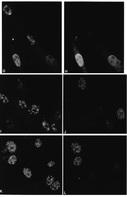
[image:2.612.318.554.485.640.2]other cells PML staining was much reduced (compare the amounts of PML detected in the two Vmw110-positive cells; Fig. 5G and H). The W146A mutant protein exhibited a local-ization phenotype closer to that of the wild type, but even with this mutation, retention of PML seemed to occur in some cells (Fig. 5I and J; compare the relative Vmw110 and PML signals in the two cells at the bottom right of panel J). Similar exper-iments were conducted with virus-infected cells, with equiva-lent results (data not shown).
It is interesting that the degree of abnormality of the mutant proteins in this assay correlates directly with their effects on the activation of gene expression and the growth of the mutant viruses (Fig. 2 and 3). This suggests that ND10 structures constitute an important part of the mechanism of action of Vmw110. However, it is not possible at this stage to determine if Vmw110 actually functions at these sites or whether the disruption of ND10 releases factors which operate elsewhere. It is also possible that ND10 structures in some way inhibit viral gene expression and that their disruption by Vmw110 overcomes this effect.
What is the function of the RING finger domain?The RING
finger family of proteins includes a wide range of members from evolutionarily diverse sources (18, 19). The domain in-cludes a core sequence of CXHXXCXXC, which comprises two metal coordinating doublets separated by a short beta strand (1, 2). Given that the spacing of this sequence is invari-ant in the family, it is likely that the positions of the two zinc atoms are also highly conserved. Immediately following this core region in EHV-1 gene 63 lies an alpha helix. Examination of the corresponding sequences of other members of the family indicates that many are also likely to form an alpha helix at this point (1). However, even among the herpesvirus proteins, the charged residues do not occupy exactly the same positions within the primary sequence of the predicted helix (1), which implies significantly different spatial positions of the side chains. When the predicted helix regions of other members of the RING finger family are considered, the diversity of the positions and the nature of the polar and charged residues (1) strongly suggest that, if RING finger domains bind to other macromolecules, their individual targets may differ consider-ably. Molecular modelling based on the topological similarity between part of the RING finger structure and a TFIIIA-type zinc finger (1) has shown that the EHV-1 gene 63 RING domain could not bind in the major groove of normal DNA because of extreme steric clashing between other parts of the RING domain and the DNA helix (24).
[image:3.612.326.551.460.674.2]The results presented here and elsewhere (14, 25, 26) indi-cate that the Vmw110 RING finger is required for modifica-tion of ND10 nuclear structures, suggesting that the role of the Vmw110 RING finger concerns complex interactions between the components of ND10. Since ND10 structures have been found to be closely associated with the nuclear matrix (35), these interactions may include not only the contacts between the various protein constituents of ND10 but also the interac-tions between these proteins and the nuclear scaffold. The mutants described in this paper may provide useful tools with which to try to dissect these connections.
FIG. 3. Single-step growth curves of wild-type HSV-1 (strain 171) and the Vmw110 mutant viruses in BHK cells. The cells were infected at a multiplicity of infection of 1 PFU per cell, and parallel plates were harvested at 4, 8, 16, and 24 h after infection. Titers of progeny virus were determined by using BHK cells. The results represent the averages from two independent experiments.
FIG. 4. Expression of Vmw110 by wild-type and mutant viruses. BHK cells were infected with the viruses as indicated at a multiplicity of infection 5 PFU per cell and harvested 16 h later. Samples were analyzed by sodium dodecyl sulfate-gel electrophoresis and Western blotting with monoclonal antibody 11060 (13). The position of full-length Vmw110 protein (as previously determined with respect to other characterized marker proteins) is indicated.
on November 9, 2019 by guest
http://jvi.asm.org/
FIG. 5. Intracellular localization of wild-type and mutant forms of Vmw110 16 h after transfection (16) of BHK cells. The left-hand panels show staining of representative fields of cells with monoclonal antibody 5E10 (35) to detect PML (6), while the right-hand panels show the same fields of cells stained with rabbit r95 serum (13) to detect Vmw110. (A and B) p111 (wild-type Vmw110); (C and D) p110K144E; (E and F) p110N151D; (G and H) p110Q148E; (I and J) p110W146A; (K and L) p110FXE (RING domain deletion). The methods used were exactly as described previously (14).
on November 9, 2019 by guest
http://jvi.asm.org/
FIG. 5—Continued.
on November 9, 2019 by guest
http://jvi.asm.org/
The continued interest and support of John H. Subak-Sharpe are much appreciated. We thank Gerd Maul for stimulating discussions on the nature of ND10, Ben Luisi for structural advice, J. Barklie Clem-ents and Stuart Simpson for pSS80, Roel van Driel for monoclonal 5E10, and Dairena Gaffney for PCR amplification of mutant herpes-virus DNA.
This work was supported by the Medical Research Council Virology Unit (R.D.E. and A.O.), by the Marie Curie Research Foundation (D.O. and P.O.), and by grants from the Medical Research Council and the Wellcome Trust (P.B.).
REFERENCES
1. Barlow, P. N., B. Luisi, A. Milner, M. Elliott, and R. D. Everett. 1994. Structure of the C3HC4 domain by 1H-nuclear magnetic resonance spec-troscopy. J. Mol. Biol. 237:201–211.
2. Borden, K. L. B., M. N. Boddy, J. Lally, N. J. O’Reilly, S. Martin, K. Howe, E. Solomon, and P. S. Freemont.1995. The solution structure of the RING finger domain from the acute promyelocytic leukaemia proto-oncoprotein PML. EMBO J. 14:1532–1541.
3. Cai, W., T. L. Astor, L. M. Liptak, C. Cho, D. M. Coen, and P. A. Schaffer. 1993. The herpes simplex virus type 1 regulatory protein ICP0 enhances virus replication during acute infection and reactivation from latency. J. Virol. 67: 7501–7512.
4. Clements, G. B., and N. D. Stow. 1989. A herpes simplex virus type 1 mutant containing a deletion within immediate-early gene 1 is latency competent in mice. J. Gen. Virol. 70:2501–2506.
5. Desai, P., R. Ramakrishnan, Z. W. Lin, B. Osak, J. Glorioso, and M. Levine. 1993. The RR1 gene of herpes simplex virus type 1 is uniquely transactivated by ICP0 during infection. J. Virol. 67:6125–6135.
6. Dyck, J. A., G. G. Maul, W. H. Miller, J. D. Chen, A. Kakizuka, and R. M. Evans.1994. A novel macromolecular structure is a target of the promyelo-cytic-retinoic acid receptor oncoprotein. Cell 76:333–343.
7. Everett, R. D. 1986. A detailed mutational analysis of Vmw110, a trans-acting transcriptional activator encoded by herpes simplex virus type 1. EMBO J. 6:2069–2076.
8. Everett, R. D. 1987. The regulation of transcription of viral and cellular genes by herpesvirus immediate-early gene products. Anticancer Res. 7:589–604. 9. Everett, R. D. 1988. Analysis of the functional domains of herpes simplex
virus type 1 immediate-early polypeptide Vmw110. J. Mol. Biol. 202:87–96. 10. Everett, R. D. 1989. Construction and characterisation of herpes simplex virus type 1 mutants with defined lesions in immediate-early gene 1. J. Gen. Virol. 70:1185–1202.
11. Everett, R. D. Unpublished observations.
12. Everett, R. D., P. N. Barlow, A. Milner, B. Luisi, A. Orr, R. G. Hope, and D. Lyon.1993. A novel arrangement of zinc binding residues and secondary structure in the C3HC4 motif of an alpha herpes virus protein family. J. Mol. Biol. 234:1038–1047.
13. Everett, R. D., A. Cross, and A. Orr. 1993. A truncated form of herpes simplex virus type 1 immediate-early protein Vmw110 is expressed in a cell-type dependent manner. Virology 197:751–756.
14. Everett, R. D., and G. G. Maul. 1994. HSV-1 IE protein Vmw110 causes redistribution of PML. EMBO J. 13:5062–5069.
15. Everett, R. D., C. M. Preston, and N. D. Stow. 1991. Functional and genetic analysis of the role of Vmw110 in herpes simplex virus replication. In E. K. Wagner (ed.), Herpes virus transcription and its regulation. CRC Press, Inc., Boca Raton, Fla.
16. Felgner, P. L., T. R. Gadek, M. Holm, R. Roman, H. W. Chan, M. Wenz, J. P. Northrop, G. M. Ringold, and M. Danielson.1987. Lipofection: a highly efficient, lipid-mediated DNA-transfection procedure. Proc. Natl. Acad. Sci. USA 84:7413–7417.
17. Fraser, N. W., T. M. Block, and J. G. Spivack. 1992. The latency associated
transcripts of herpes simplex virus: RNA in search of a function. Virology 191:1–8.
18. Freemont, P. S. 1993. The RING finger. A novel protein sequence related to the zinc finger. Ann. N. Y. Acad. Sci. 684:174–192.
19. Freemont, P. S., I. M. Hanson, and J. Trowsdale. 1991. A novel cysteine-rich motif. Cell 64:483–484.
20. Harris, R. A., R. D. Everett, X. Zhu, S. Silverstein, and C. M. Preston. 1989. Herpes simplex virus type 1 immediate-early protein Vmw110 reactivates latent herpes simplex virus type 2 in an in vitro latency system. J. Virol. 63:3513–3515.
21. Kastner, P., A. Perez, Y. Lutz, C. Rochette-Egly, M. P. Gaub, B. Durand, M. Lanotte, R. Berger, and P. Chambon.1992. Structure, localisation and tran-scriptional properties of two classes of retinoic acid receptor alpha fusion proteins in acute promyelocytic leukaemia APL: structural similarities with a new family of oncoproteins. EMBO J. 11:629–642.
22. Koken, M. H. M., F. Puvion-Dutilleul, M. C. Guillemin, A. Viron, G. Lin-ares-Cruz, N. Stuurman, L. de Jong, C. Szostecki, F. Calvo, C. Chomienne, L. Degos, E. Puvion, and H. de The.1994. The t15;17 translocation alters a nuclear body in a retinoic acid reversible fashion. EMBO J. 13:1073–1083. 23. Lieb, D. A., D. M. Coen, C. L. Bogard, K. A. Hicks, D. R. Yager, D. M. Knipe, K. L. Tyler, and P. A. Schaffer.1989. Immediate-early regulatory gene mutants define different stages in the establishment and reactivation of herpes simplex virus latency. J. Virol. 63:759–768.
24. Luisi, B. Personal communication.
25. Maul, G. G., and R. D. Everett. 1994. The nuclear location of PML, a cellular member of the C3HC4 zinc binding domain protein family, is rearranged during herpes simplex virus infection by the C3HC4 viral protein ICP0. J. Gen. Virol. 75:1223–1233.
26. Maul, G. G., H. H. Guldner, and J. G. Spivack. 1993. Modification of discrete nuclear domains induced by herpes simplex virus type 1 immediate-early gene 1 product ICP0. J. Gen. Virol. 74:2679–2690.
27. McGeoch, D. J., B. C. Barnett, and C. A. MacLean. 1993. Emerging functions of alphaherpesvirus genes. Semin. Virol. 4:125–134.
28. McGeoch, D. J., M. A. Dalrymple, A. J. Davison, A. Dolan, M. C. Frame, D. McNab, L. J. Perry, J. E. Scott, and P. Taylor.1988. The complete nucle-otide sequence of the long unique region in the genome of herpes simplex virus type 1. J. Gen. Virol. 69:1531–1574.
29. Perry, L. J., F. J. Rixon, R. D. Everett, M. C. Frame, and D. J. McGeoch. 1986. The IE110 gene of herpes simplex virus type 1: characterisation by mRNA mapping, DNA sequence, oligopeptide antiserum and mutational analysis. J. Gen. Virol. 67:2365–2380.
30. Roizman, B., and A. E. Sears. 1990. Herpes simplex viruses and their repli-cation, p. 1795–1842. In B. N. Fields, D. M. Knipe, et al. (ed.), Virology, 2nd ed. Raven Press, New York.
31. Russell, J., N. D. Stow, E. C. Stow, and C. M. Preston. 1987. Herpes simplex virus genes involved in latency in vitro. J. Gen. Virol. 68:3009–3018. 32. Sacks, W. R., and P. A. Schaffer. 1987. Deletion mutants in the gene
encod-ing the herpes simplex virus type 1 immediate-early protein ICP0 exhibit impaired growth in cell culture. J. Virol. 61:829–839.
33. Stevens, J. G., E. K. Wagner, G. B. Devi-Rao, M. L. Cook, and L. T. Feldman. 1987. RNA complimentary to a herpes virus gene mRNA is prominent in latently infected neurones. Science 235:1056–1059.
34. Stow, N. D., and E. C. Stow. 1986. Isolation and characterisation of a herpes simplex virus type 1 mutant containing a deletion within the gene encoding the immediate-early polypeptide Vmw110. J. Gen. Virol. 67:2571–2585. 35. Stuurman, N., A. DeGraaf, A. Josso, B. Humbel, L. DeYong, and A. van
Driel.1992. A monoclonal antibody recognising nuclear matrix associated nuclear bodies. J. Cell Sci. 101:773–784.
36. Weis, K., S. Rambaud, C. Lavau, J. Jansen, T. Carvalho, M. Carmo-Fonseca, A. Lamond, and A. Dejean.1994. Retinoic acid regulates aberrant nuclear localization of PML-RAR alpha in acute promyelocytic leukaemia cells. Cell 76:345–356.