0022-538X/95/$04.0010
Copyrightq1995, American Society for Microbiology
The NS2 Protein of Hepatitis C Virus Is a
Transmembrane Polypeptide
ELISA SANTOLINI, LAURA PACINI, CRISTINA FIPALDINI,
GIOVANNI MIGLIACCIO,ANDNICOLA LA MONICA*
Istituto di Ricerche di Biologia Molecolare ‘‘P. Angeletti,’’ Pomezia 00040, Italy
Received 6 July 1995/Accepted 5 September 1995
The NS2 protein of hepatitis C virus (HCV) is released from its polyprotein precursor by two proteolytic cleavages. The N terminus of this protein is separated from the E2/p7 polypeptide by a cleavage thought to be mediated by signal peptidase, whereas the NS2-3 junction located at the C terminus is processed by a viral protease. To characterize the biogenesis of NS2 encoded by the BK strain of HCV, we have defined the minimal region of the polyprotein required for efficient cleavage at the NS2-3 site and analyzed the interaction of the mature polypeptide with the membrane of the endoplasmic reticulum (ER). We have observed that although cleavage can occur in vitro in the absence of microsomal membranes, synthesis of the polyprotein precursor in the presence of membranes greatly increases processing at this site. Furthermore, we show that the membrane dependency for efficient in vitro processing varies among different HCV strains and that host proteins located on the ER membrane, and in particular the signal recognition particle receptor, are required to sustain efficient proteolysis. By means of sedimentation analysis, protease protection assay, and site-directed mutagen-esis, we also demonstrate that the NS2 protein derived from processing at the NS2-3 site is a transmembrane polypeptide, with the C terminus translocated in the lumen of the ER and the N terminus located in the cytosol.
The molecular cloning of the cDNA of hepatitis C virus (HCV), the most common cause of non-A non-B hepatitis (4, 29), has opened new avenues to the study of viral replication of this important human pathogen. The viral genome consists of a positive-sense RNA of about 9.5 kb (5, 27, 44), and the mature enveloped virion has a diameter of approximately 60 nm (26). Sequence analysis of the cDNA isolated from differ-ent sources has allowed the iddiffer-entification of at least five major genotypes and accompanying subtypes and has indicated that the virus distantly resembles human flaviviruses and animal pestiviruses (3, 34). Thus, HCV has been classified as a sepa-rate genus in the Flaviviridae family (13).
The genetic organization of the HCV genome has been characterized by means of transient expression of HCV cDNA in transfected cells (1, 17, 42, 45) and by cell-free translation studies of in vitro-synthesized transcripts (20). The putative structural proteins core, E1, and E2 are found in the N-termi-nal quarter of the 3,010-residue polyprotein that represents the primary translation product of the viral RNA. Core, a basic protein with RNA binding activity (41), is thought to be the nucleocapsid component of the virion, whereas E1 and E2 are glycoproteins that are probably associated with the viral enve-lope. The E2 glycoprotein exists in two different forms: as a fully processed polypeptide or fused to the adjacent down-stream p7 protein, which may be an additional structural com-ponent of the viral particle (30, 35). The nonstructural poly-peptides NS2, NS3, NS4a, NS4b, NS5a, and NS5b are localized within the remainder of the polyprotein and are likely to be involved in processing of the viral polypeptide and possibly in the replication of viral RNA (24). The N terminus of all pro-cessed HCV polypeptides has been determined by direct se-quencing of proteins expressed in vitro and in transfected cells (15, 16, 23, 31, 36, 39).
Previous studies have shown that the putative structural polypeptides of HCV are released from the polyprotein pre-cursor by host protease, most likely the endoplasmic reticulum (ER) signal peptidase, which is responsible for the removal of a hydrophobic domain at the C terminus of the core polypep-tide (20, 41) and catalyzes the primary cleavages at the E1-E2, E2-p7, and p7-NS2 sites (20, 30, 36). However, although a hydrophobic domain located upstream of the N terminus of NS2 could function as signal or signal-anchor sequences (35), processing at the p7-NS2 junction and the overall membrane topology of NS2 are not clear, particularly in view of the posttranslational nature of the cleavage at the N terminus of this viral polypeptide (9). The HCV-encoded serine protease, localized in the N-terminal region of the NS3 protein, mediates in conjunction with the NS4a polypeptide processing of the putative nonstructural proteins by cleaving at the following sites: NS3-NS4a, NS4a-NS4b, NS4b-NS5a, and NS5a-NS5b (1, 2, 10, 12, 15, 31, 45). Cleavage at the N terminus of the NS3 protein is mediated by yet another virus-encoded protease, which includes the NS2 region and the serine protease domain of the NS3 protein (16, 21). However, the molecular mecha-nism of the proteolytic processing at the NS2-3 site is poorly understood.
To further characterize the biogenesis of the NS2 protein of HCV and to define the membrane topology of this polypep-tide, we have examined the processing and membrane inter-action of this viral protein in in vitro translation studies utiliz-ing the cDNA of the BK type of HCV. We have observed that efficient cleavage at the NS2-3 site is dependent on the syn-thesis of the protein precursor in the presence of functional microsomal membranes. Furthermore, there is an apparent difference in cleavage efficiency between the HCV-BK and -J precursors in the absence of membranes. Also, we show that processing at the NS2-3 site resulted in the integration of the NS2 protein into the ER membrane as a bona fide transmem-brane polypeptide.
* Corresponding author. Phone: 93443. Fax: 39-6-910-93225. Electronic mail address: [email protected].
7461
on November 9, 2019 by guest
http://jvi.asm.org/
MATERIALS AND METHODS
Construction of recombinant plasmids.Clones expressing HCV E2-NS2-NS3 sequences are derived from cDNA fragments of the HCV-BK strain (44) from nucleotides (nt) 2496 to 5303 (amino acid [aa] residues 722 to 1657) unless otherwise specified. Cloning of the cDNA fragments in the desired expression vectors was achieved by PCR amplification of the area of interest, using synthetic oligonucleotides with the appropriate restriction sites, or by standard DNA protocols.
Plasmid pCITE(722-1325) was obtained by cloning into the MscI-XbaI sites of the expression vector pCITE (Novagen) (11) nt 2496 to 4307 to which the MscI and XbaI sites were engineered by PCR. The plasmid contains the HCV cDNA fragment downstream of a T7 promoter and of the 59untranslated region of encephalomyocarditis virus (25). pN(722-1325) was provided by K. Shimotohono and has already been described (21). It contains nt 2496 to 4307 of the HCV-J strain downstream of a T7 promoter.
pCD(810-1615) contains HCV sequences from an NcoI site at position 2758 to a BamHI site engineered at position 5177. The cDNA fragment was cloned in the expression vector pCDNA3 (Invitrogen) downstream of a T7 promoter and of the 59untranslated region of theb-globin gene of Xenopus laevis (28).
Plasmids containing specific mutations were obtained by oligonucleotide-di-rected mutagenesis, and the mutations were confirmed by sequence analysis. Mutations are denoted by the parental amino acid residue and its position in the HCV-BK strain, followed by the substituted residue. pCD(810-1615; C993A) was created by inserting the mutation in the PCR primers that were used to generate the mutant DNA fragment according to the procedure of Higuchi et al. (19). The mutant DNA fragment was cloned into the parent plasmid by using flanking restriction sites. The triplet coding for cysteine 993, TGT, was replaced by GCT, which codes for alanine. pGEM(810-1615; L1026DQ) was created by changing the HCV nucleotide sequence starting at position 3402 from CGACTCCTC GCG into CGACTGCAG according to the procedure of Higuchi et al. (19). The amplified DNA fragment derived from pCITE(810-1615) was inserted into plas-mid pGEMBP1 (6).
Site-directed mutagenesis of aa residues 983 and 988 was carried out by inserting the mutations in the PCR primers that were used to generate the mutant DNA clone pGEM(810-1026G) according to the procedure of Hemsley et al. (18). Plasmid pGEM(810-1026) (see below) was used as the template for PCR amplification of the target DNA. The triplets coding for isoleucine 983, ATC, and alanine 988, GCA, were replaced by AAC, which codes for asparagine. To construct plasmid pGEM(810-1615G), pGEM(810-1026G) was cleaved with
SacII and EcoRI and a SacII-EcoRI DNA fragment derived from
pCD(810-1615) containing nt 3307 to 5177 (aa residue 992 to pCD(810-1615) was inserted into the plasmid.
pCITE(849-1615) was obtained by cloning into the pCITE vector digested with
MscI and XbaI an SspI-XbaI cDNA fragment derived from plasmid
pCITE(623-1615) (7). The construct contains nt 2872 to 5177 downstream of a T7 promoter. pCITE(849-1325) was obtained by subcloning an SspI-PstI fragment derived from plasmid pN(722-1325) into the pCITE vector digested with MscI and PstI. The construct contains nt 2872 to 4307 of the HCV-J strain.
pCITE(923-1615) was constructed by cloning into the pCITE vector digested with BstXI and PstI an SphI-PstI fragment derived from plasmid pCITE(849-1615) after treatment of both DNAs with T4 DNA polymerase. The plasmid contains nt 3098 to 5177 downstream of a T7 promoter.
pCITE(992-1615) was constructed by cloning a SacII-PstI fragment derived from plasmid pCITE(849-1615) into the pCITE vector digested with BstXI and
PstI. Prior to ligation, both DNAs were treated with T4 DNA polymerase. The
plasmid contains nt 3301 to 5177.
pCITE(960-1615) was derived from PCR amplification of nt 3208 to 5177 with sequence-specific primers using pCITE(849-1615) as the template. The amplified DNA was digested with PvuII, whose site had been included within the 59end primer, and BstXI and inserted into the pCITE vector that had been digested with MscI and BstXI.
pGEM(810-1026) was obtained by PCR amplification of nt 2760 to 3410 (aa residues 810 to 1026) with sequence-specific primers. The amplified DNA frag-ment derived from pCITE(810-1615) was digested with XbaI and inserted into plasmid pGEMBP1.
pT7(1027-1657) contains nt 3410 to 5303 (7).
In vitro transcription and translation.All plasmids used as templates for in vitro RNA synthesis were linearized with the appropriate restriction enzyme and transcribed in vitro with either T7 or SP6 RNA polymerase as previously de-scribed (45). Transcripts encoding aa residues 849 to 1083, 849 to 1115, 849 to 1137, 849 to 1237, 849 to 1355, 849 to 1401, and 849 to 1462 were obtained by runoff transcription of pCITE(849-1615) digested with BstXI (nt 3578), SmaI (nt 3675), AatII (nt 3744), ScaI (nt 4041), BstEII (nt 4394), SacI (nt 4536), and SalI (nt 4716), respectively (44).
In vitro translation reactions (25ml of mixture) were performed for 60 min at 308C with nuclease-treated reticulocyte lysate (Promega). Unless stated other-wise, canine rough microsomes (RM) were used at a concentration of 1 eq/25ml. After translation, samples were either diluted in sample buffer and directly analyzed by sodium dodecyl sulfate-polyacrylamide gel electrophoresis (SDS-PAGE) or treated as described below. Incorporation of radioactivity into specific bands was determined by direct analysis of the dried gel with a PhosphorImager
(Molecular Dynamics). The processing efficiency was estimated on the basis of the incorporation of radioactivity in translation products according to the fol-lowing formula:
% of total cpm5NSNS~a/b~a/b! 1! P (1)
where NS is counts per minute of the processed protein, a is the number of methionine residues present in the precursor, b is the number of methionine residues present in the processed protein, and P is the counts per minute of the unprocessed precursor.
Preparation of microsomes and of SRP receptor fragment.Canine pancreatic RM were prepared as described elsewhere (37) and washed with 0.5 M potas-sium acetate (KOAc) and 10 mM EDTA. KOAc-EDTA-washed membranes were subjected to limited proteolysis after resuspension in buffer A (0.25 M sucrose, 50 mM triethanolamine [pH 7.5], 5 mM CaCl2, 5 mM MgCl2, 2 mM dithiothreitol) to a concentration of 1 eq/ml. Trypsin (0.5 mg/ml in buffer A) was added to a final concentration of 5mg/ml, and the microsome suspension was incubated for 60 min on ice. Proteolysis was terminated by the addition of ethylene glycol-bis(b-aminoethyl ether)-N, N, N9, N9-tetraacetic acid (EGTA) and phenylmethylsulfonyl fluoride (PMSF) to final concentrations of 20 and 5 mM, respectively. KOAc-EDTA-washed membranes at a concentration of 1 eq/ml in buffer A were alkylated for 30 min at 228C in the presence of 2.5 mM
N-ethylmaleimide (NEM). Alkylation reactions were quenched by the addition
of dithiothreitol to a final concentration of 20 mM. Trypsinized microsomes (TRM) and alkylated microsomes (NRM) were collected by centrifugation through a cushion of 0.5 M sucrose–50 mM triethanolamine, pH 7.5, for 20 min at 65,000 rpm in a TL 100 rotor (Beckman Instruments Inc.) and resuspended in 0.25 M sucrose–50 mM triethanolamine, pH 7.5, to a concentration of 1 eq/ml. Under these conditions, the cytoplasmic domain of the signal recognition particle (SRP) receptor is either removed (TRM) or inactivated (NRM) and the micro-some fraction is translocation incompetent. Control membranes were mock treated and processed similarly (37).
The 52-kDa cytoplasmic fragment of the SRP receptor (37) was prepared as follows: 20 ml of KOAc-EDTA-washed membrane suspension, at a concentra-tion of 1 eq/ml in buffer A, was supplemented with 10 U of aprotinin (Boehringer Mannheim) per ml and digested with elastase (Boehringer Mannheim), at a final concentration of 1mg/ml, for 60 min on ice. After addition of PMSF and KOAc to concentrations of 1 mM and 0.5 M, respectively, microsomes were collected by centrifugation at 48C for 35 min at 40,000 rpm in a 50.2 Ti rotor (Beckman Instruments Inc.). The proteolytic digestion was repeated, and the supernatants from the two treatments were combined and dialyzed versus buffer B (50 mM Tris-HEPES [N-2-hydroxyethylpiperazine-N9-2-ethanesulfonic acid; pH 7.4], 150 mM KOAc, 2 mM dithiothreitol). The dialyzed fraction was centrifuged for 1 h at 45,000 rpm in a 50.2 Ti rotor to remove the precipitated material and applied to a 1-ml column of CM Sephadex equilibrated in buffer B. The column was washed with 10 column volumes of buffer B and 10 column volumes of buffer B containing 0.25 M KOAc. The 52-kDa SRP receptor fragment was eluted with buffer B containing 0.425 M KOAc and concentrated by ultrafiltration with Centricon 30 (Amicon Corp., Danvers, N.H.).
Sedimentation of membrane-associated proteins on sucrose step gradients and urea extraction.Ten microliters of the translation reaction mixtures was supplemented with an equal volume of TKM buffer (20 mM Tris-Cl [pH 7.5], 100 mM KOAc, 2 mM MgCl2); after 10 min of incubation on ice, samples were overlaid on an 80-ml cushion of 0.4 M sucrose and centrifuged at 60,000 rpm for 8 min in a Beckman TLA-100 rotor at 48C. Alternatively, 10ml of the translation reaction mixtures was supplemented with 90ml of 5 M urea–11.1 mM Tris-Cl (pH 7.5)–55.5 mM KOAc–1.1 mM MgCl2; after 10 min at room temperature samples were overlaid on 100ml of 0.25 M sucrose containing 4.5 M urea and centrifuged at 70,000 rpm in a Beckman TLA-100 rotor for 15 min at 208C. The supernatants and cushion fractions were precipitated with 2 volumes of saturated ammonium sulfate, washed with 5% trichloroacetic acid, and resuspended in sample buffer. Pellet fractions were directly resuspended in sample buffer and analyzed by SDS-PAGE.
Protease digestion.Five microliters of the translation mixtures was diluted with 5ml of solution I (6 mM tetracaine), II (6 mM tetracaine, 0.4 mg of proteinase K [PK] per ml), or III (6 mM tetracaine, 0.4 mg of PK per ml, 2% Triton-X) and incubated for 60 min on ice. Proteolysis was terminated by adding 1ml of 100 mM PMSF and incubating the samples for 10 min on ice. Samples were then diluted with sample buffer and analyzed by SDS-PAGE. For protease protection assay of in vivo-synthesized protein, HeLa cells were infected and transfected and labeled with [35
S]Promix (Amersham, Little Chalfont, United Kingdom) as described below. After metabolic labeling, cells (approximately 23 106
) were resuspended in 400ml of H buffer (20 mM Tris-Cl [pH 7.5], 250 mM sucrose, 50 mM NaCl, 1.5 mM MgCl2, 0.5 mM EDTA, 0.5 mM EGTA) and homogenized with a 0.5-ml glass Teflon homogenizer. The homogenate was centrifuged for 4 min at 1,0003g in a Haereus bench top centrifuge. Aliquots
(100ml) of the postnuclear supernatant were supplemented with an equal vol-ume of either solution I, II, or III and incubated for 1 h on ice. Proteolysis was terminated by adding PMSF and SDS to final concentrations of 10 mM and 2%, respectively, and by incubating the samples for 5 min at 958C. Samples were diluted with 1.5 ml of 20 mM Tris-Cl (pH 7.5)–150 mM NaCl–0.5 mM EDTA–0.5
on November 9, 2019 by guest
http://jvi.asm.org/
mM EGTA–1.25% Triton X-100 and immunoprecipitated as described previ-ously (45).
Cell transfections.HeLa cell monolayers were grown in Dulbecco’s modified Eagle’s medium containing 10% fetal calf serum. Cells were infected with vac-cinia virus vTF7-3 and subsequently transfected by calcium phosphate DNA precipitation as previously described (45). Immunoprecipitations of transfected cell lysates were performed with HCV-specific rabbit polyclonal antisera as previously described (45).
Endoglycosidase H treatment.Eighty microliters of transfected cell lysate was immunoprecipitated with NS2-specific rabbit polyclonal antiserum (45), and the pelleted samples were resuspended in 20ml of buffer C (10 mM Tris-Cl [pH 7.5], 150 mM NaCl, 1% SDS, and 0.5 mM PMSF) and denatured at 958C for 5 min. The samples were then diluted with 80ml of buffer D (50 mM sodium acetate [pH 5.2], 1.25% Triton X-100 and 0.5 mM PMSF). Deglycosylation was carried out by adding 2 mU of endoglycosidase H (Boehringer Mannheim), and the mixture was incubated at 378C for 4 h. The protein samples were then precipi-tated for 1 h on ice with 10% trichloroacetic acid and processed for SDS-PAGE analysis.
RESULTS
Effect of deletions and point mutations on NS2-3 cleavage.
To determine the HCV sequence requirements for cleavage at
the NS2-3 site, a series of 59and 39deletions were introduced
in the cDNA encoding the HCV polyprotein and used for cell-free translation studies. Figure 1 shows a diagram of all transcripts used in this study. To define the importance of membranes for cleavage efficiency, translations were per-formed in the absence or the presence of canine pancreatic microsomal membranes. The results of in vitro processing as-says using transcripts spanning the NS2 and most of the NS3
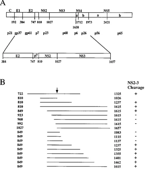
polypeptides are shown in Fig. 2. The transcript encoding aa 810 to 1615 yielded an 88-kDa unprocessed precursor as well as two proteins of 65 and 23 kDa corresponding to the cleaved NS3 and NS2 proteins, respectively (Fig. 2A, lane 2). Process-ing was clearly evident when the translation reaction was car-ried out in the presence of microsomal membranes, whereas the cleaved products were almost undetectable when transla-tion was performed in the absence of membranes (Fig. 2A, lane 1). The N-terminal boundary of the NS2 portion required for efficient processing appears to be located between aa 849 and 923, since polyproteins starting at or downstream of resi-due 923 did not yield cleaved NS2 and NS3 polypeptides (Fig. 2A, lanes 3 to 10). Similarly, the C-terminal boundary of the NS3 portion required for processing appears to be located between aa 1137 and 1237 because proteins terminating be-yond aa 1237 were properly cleaved (Fig. 2B, lanes 7 to 16), whereas precursors terminating at or upstream of aa 1137 were not processed at the NS2-3 site (Fig. 2B, lanes 1 to 6). Inter-estingly, efficient processing at the NS2-3 site could not be reestablished by extending the truncated NS2-3 precursor that comprised aa 849 to 1083 with aa 2885 to 3010 derived from the NS5b region (data not shown), suggesting that the NS3 sequences located downstream of the NS2-3 junction play an active role in the reaction and are therefore specifically re-quired for proper cleavage.
The results described above could be interpreted as indica-tive of a signal peptidase-mediated cleavage of the NS2-3 pre-cursor that requires microsomal membranes and thus is dis-tinct from viral proteolytic processing at the NS2-3 site. To ascertain that the cleavage observed in the cell-free translation of the NS2-3 polyprotein was indeed due to the viral protease, we introduced by site-directed mutagenesis a cysteine-to-ala-nine substitution at residue 993. This mutation inactivates the HCV NS2-3 protease encoded by the H and J strains of HCV (16, 21). Furthermore, to confirm the substrate specificity of
the BK NS2-3 protease, mutant L1026DQ, in which the amino
acid sequence LLAP spanning residues 1025 to 1028 and in-cluding the cleavage site (16, 21) was mutated into LQP with a net deletion of residue 1026 and an alanine-to-glutamine sub-stitution at residue 1027, was constructed. Mutations were introduced into a plasmid encoding aa 810 to 1615, and the translation products of the in vitro-transcribed mutant RNAs were compared with those of the parental transcript. As shown in Fig. 2C, both mutations resulted in the lack of processing at the NS2-3 junction and did not yield processed NS2 and NS3 polypeptides. Thus, these data are in agreement with the in-terpretation of a viral protease-mediated cleavage at the NS2-3 site and also confirm the observation that amino acid changes in the NS2-3 junction have a significant effect on the processing efficiency (23).
Membrane dependence for in vitro NS2-3 cleavage varies among different HCV cDNAs. The results described above indicate that efficient in vitro processing of the NS2-3 site requires a specific segment of the HCV polyprotein, in agree-ment with recent published data (1, 16, 21). However, it ap-peared that in vitro cleavage of the NS2-3 site of the HCV-BK strain was heavily dependent on the presence of microsomal membranes in the translation reaction, more than what has been described by other groups that have utilized a cDNA clone of HCV strains other than HCV-BK. Specifically, Hi-jikata et al., using the cDNA clones of the HCV-J strain, have clearly shown that the processing of the NS2-3 site in vitro could occur efficiently in the absence of the microsomal mem-branes (21).
[image:3.612.61.297.74.349.2]To ascertain whether this different requirement for micro-somal membranes is due to a real diversity between HCV
FIG. 1. Schematic representation of the HCV genome and of the transcripts used in this study. (A) Diagram of the HCV polyprotein, showing the cleavage products. The amino acid residues representing the N termini and the molecular sizes of the processed viral proteins are indicated (16, 20, 21). The lower diagram indicates the NS2 coding sequence and its surrounding region. (B) Transcripts expressing the HCV polyprotein. Lines describe the HCV protein expressed in a variety of constructs and are drawn to scale and oriented with respect to the lower diagram in panel A. Numbers refer to the first and last amino acids of the HCV polyprotein expressed in each transcript; the arrow indicates NS2-3 cleav-age site. Processing at the NS2-3 junction is indicated at the right.
on November 9, 2019 by guest
http://jvi.asm.org/
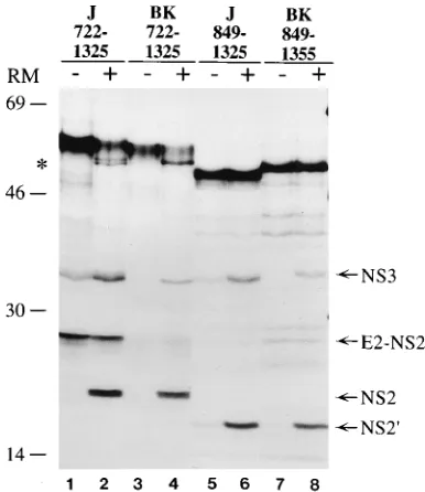
strains rather than to distinct experimental procedures, we compared the effects of microsomal membranes on the pro-cessing efficiency of the NS2-3 protein encoded by the HCV-J and HCV-BK strains. The transcripts utilized in this compar-ison comprise part of the E2 glycoprotein as well as most of the NS2-3 region. Thus, to generate a mature NS2 protein, the polyprotein precursor encoded by these RNAs must be pro-cessed at the E2-NS2 junction, presumably by signal peptidase, and at the NS2-3 site by the viral protease (21). Translation of the HCV-J aa 722 to 1325 RNA in the absence of microsomal membranes yielded a major precursor band of approximately 66 kDa as well as two smaller proteins of 32 and 26 kDa corresponding to the NS3 and E2-NS2 proteins, respectively (Fig. 3, lane 1). The 26-kDa band decreased in intensity when translation was carried out in the presence of microsomal membranes with the concomitant appearance of the 23-kDa protein, indicating that the NS2 product had been released from the E2 protein (21, 36). Furthermore, an additional 56-kDa protein running as a doublet was also observed. We in-terpret this polypeptide to be derived by the signal
peptidase-mediated cleavage at the E2-p7 and/or p7-NS2 sites, releasing the unprocessed NS2-3 precursor from the truncated E2 polypeptide (Fig. 3, lane 2). The identity of each of the trans-lation products was confirmed by immunoprecipitation with specific antisera (data not shown). In contrast, translation of the HCV-BK aa 722 to 1325 transcript in the absence of mi-crosomal membranes yielded a major precursor band of 66 kDa, but little or no processed NS3 and E2-NS2 could be detected (Fig. 3, lane 3). However, when translation was car-ried out in the presence of microsomal membranes, processing of the NS2-3 site as well as at the E2-NS2 junction was evident. Notably, the 56-kDa unprocessed precursor migrated as a sin-gle band, suggesting that cleavage at the N terminus of NS2 also varies between different HCV strains (Fig. 3, lane 4). The differential requirement of microsomal membranes was also observed when shorter transcripts of the two strains were an-alyzed. Translation of the HCV-J transcript encoding aa 849 to 1325 yielded a 53-kDa unprocessed precursor as well as two proteins of 32 and 19 kDa corresponding to the cleaved NS3
and to the truncated NS2 (NS29) proteins, respectively. The
[image:4.612.90.530.73.505.2]processed NS2 and NS3 polypeptides were more visible when translation was carried out in the presence of microsomal membranes in the reaction mixture, suggesting that processing of the HCV-J strain was also affected, at least in part, by the presence of membranes (Fig. 3, lanes 5 and 6). Additionally, translation of the HCV-BK transcript encoding aa 849 to 1355 in the absence of membranes yielded only a 53-kDa precursor, and the NS2 and NS3 proteins could be detected only when translation was carried out in the presence of microsomal
FIG. 2. Genetic analysis of NS2-3 proteinase. (A) A series of N-terminal deletion mutants encompassing aa 810 to 1615 were used as templates for in vitro transcription, and the resulting RNAs were translated with a rabbit reticulocyte lysate in the absence (lanes2) or presence (lanes1) of canine pancreas RM. (B) In vitro-transcribed RNAs of C-terminal deletion mutants encompassing aa 849 to 1615 were translated in vitro in the absence (lanes2) or presence (lanes1) of microsomal membranes. (C) RNAs of the mutants C993A (lane 2) and L1026DQ (lane 3) were translated in vitro in the presence of microsomal mem-branes, and their translation patterns were compared with that of the parental transcript encoding aa 810 to 1615 (lane 1). Translation products labeled with35S were analyzed on an SDS–11% polyacrylamide gel. The cleaved products derived from the NS2 and NS3 coding sequences are indicated by arrows and asterisks, respectively. Positions of the molecular mass standards (in kilodaltons) are
in-dicated.
on November 9, 2019 by guest
http://jvi.asm.org/
membranes (Fig. 3, lanes 7 and 8). These results demonstrate that, independent of the length of the transcript, the HCV-J polyprotein precursor is less dependent on microsomal mem-branes than is that of HCV-BK for efficient in vitro cleavage at the NS2-3 site. This finding confirms the observation that dif-ferent strains of HCV have, at least in vitro, difdif-ferent biological requirements for processing of the NS2-3 junction (1, 8, 16, 21).
Role of membranes in the NS2-3 cleavage.To investigate the role of microsomal membranes in the in vitro cleavage at the NS2-3 site, translation reactions were carried out with the HCV-BK transcript encoding aa 849 to 1615 in the presence of increasing amounts of microsomal membranes. As shown in Fig. 4A, translation in the absence of membranes produced an 84-kDa unprocessed precursor and little or no 65- and 19-kDa protein product, corresponding to the cleaved NS3 and NS2 polypeptides, could be detected. However, an increase in pro-cessed NS2 and NS3 proteins could be detected as a function of the microsomal membrane concentration in the translation reaction. Although we cannot rule out the possibility that the presence of microsomal membranes in the translation reaction had a stabilizing effect on the processed polypeptides, it is apparent that membranes contributed to an increase in the processing efficiency at the NS2-3 site, as indicated by a de-creased amount of the unprocessed precursor concomitant with the increase in the amounts of cleaved NS3 and NS2 (Fig. 4). The yield of processed NS2 and NS3 reached a plateau at
around 2 membrane equivalents per 25ml of translation
mix-ture.
To determine whether the increase in efficiency of cleavage at the NS2-3 site observed in the presence of microsomal membranes was merely due to the hydrophobic interaction between the precursor protein and the membranes or whether
a specific host factor contributed to the processing at the NS2-3 junction, we examined the effects on the cleavage at the NS2-3 site of trypsin treatment of the microsomal membranes. This treatment was chosen on the basis of its capacity to inactivate protein components involved in protein translocation (37). Also, we compared the effect of membrane treatment on the processing efficiency of the NS2-3 protein encoded by both the HCV-J and HCV-BK strains.
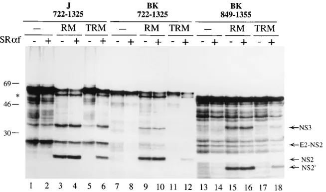
As shown in Fig. 5, translation of the HCV-BK transcript encoding aa 722 to 1325 in the presence of TRM yielded only the 66-kDa precursor (lane 11) whereas translation in the presence of mock-treated membranes produced the 66-kDa precursor, the 56-kDa partially processed polypeptide, and the NS2 and NS3 proteins of 23 and 32 kDa, respectively (lane 9). Similarly, translation of the HCV-BK transcript encoding aa 849 to 1355 was also affected by the trypsin treatment of the membranes, since no processed products could be detected (Fig. 5, lane 17). Thus, trypsin treatment of microsomal mem-branes completely inhibited processing at all sites of the HCV-BK precursor. As expected, membrane inactivation re-sulted in the loss of processing at the E2-p7 and p7-NS2 sites of HCV-J precursor comprising aa 722 to 1325, whereas cleav-age at the NS2-3 site showed a similar level of processing efficiency compared with translation reactions carried out in the absence of microsomal membranes (Fig. 5, lanes 1 and 5), in agreement with the observation that cleavage at NS2-3 junc-tion of the HCV-J strain is less membrane dependent than that in HCV-BK.
A similar inactivation of efficient cleavage of the HCV-BK NS2-3 precursor was obtained when membranes (NRM) were treated with the sulfhydryl-directed alkylating agent NEM or when transcripts encoding aa 810 to 1615 were utilized in the translation reactions (data not shown). This result indicated that both treatments impaired the ability of microsomal mem-branes to contribute to the increased processing efficiency at the NS2-3 site independently of the transcript length utilized in the experiment, suggesting that the stimulating activity re-quired a proteinaceous component located on the cytosolic side of the ER membrane and containing at least one free sulfhydryl group. Furthermore, addition of the cytoplasmic
fragment of theasubunit of the SRP receptor to the
transla-tion reactransla-tions restored, at least in part, the processing-stimu-lating activity of both TRM and NRM (Fig. 5, lanes 6, 12, and 18, and data not shown). Conversely, addition of the SRP
receptora subunit to reactions performed in the absence of
membranes or in the presence of mock-treated membranes (Fig. 5, lanes 2, 4, 8, 10, 14, and 16) did not change the protein processing pattern, indicating that the SRP receptor was nec-essary but not sufficient to stimulate processing at the NS2-3 site. These results indicate that the nascent NS2-3 protein is targeted to the ER membrane by the SRP-SRP receptor tar-geting machinery and that this tartar-geting event results in an increased processing efficiency at the NS2-3 site.
[image:5.612.80.273.71.294.2]Membrane interaction of the NS2 protein. The data re-ported above established that the cleavage of the NS2-3 site is enhanced by the interaction between the newly synthesized NS2-3 protein and the ER membrane, possibly by determining a proper folding and/or correct localization on the membrane of the polyprotein precursor that ultimately results in the in-creased processing efficiency at the NS2-3 site. To define whether the interaction between the nascent HCV protein and the ER membrane resulted in a stable association between the NS2 polypeptide and the ER membrane, the sedimentation profiles of the in vitro translation products of HCV-BK tran-scripts encoding aa 810 to 1615 (NS2-3), 810 to 1026 (NS2), and 1027 to 1657 (NS3) were analyzed by ultracentrifugation
FIG. 3. Comparison of in vitro processing efficiencies at the NS2-3 site be-tween the HCV-BK and HCV-J polyproteins. Transcripts encoding the indicated amino acids of the HCV-J strain (J) and the HCV-BK strain (BK) were trans-lated in vitro with a rabbit reticulocyte lysate in the absence (lanes2) or presence (lanes1) of RM. Translation products labeled with35
S were analyzed on an SDS–11% polyacrylamide gel. The positions of the processed NS3, E2-NS2, E2-NS2, and NS29polypeptides are indicated. The asterisk indicates the par-tially processed NS2-3 precursor. Positions of the molecular mass standards (in kilodaltons) are indicated.
on November 9, 2019 by guest
http://jvi.asm.org/
on sucrose step gradients. The effect of cleavage at the NS2-3 site on the sedimentation of the HCV proteins was determined by comparing the sedimentation of the NS2 and NS3 polypep-tides derived from processing at the 2-3 site with that of the same proteins translated from two separate transcripts. The translations were performed in the absence or in the presence of microsomal membranes. Furthermore, the influence of mem-brane components on the sedimentation profile of the HCV proteins was assessed by translating their corresponding RNAs in the presence of microsomal membranes that had been treated with trypsin or NEM.
NS2-3 precursor protein synthesized in the absence of mi-crosomal membranes or in the presence of pretreated
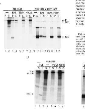
mem-branes was found mostly in the supernatant, whereas it was recovered in equal amounts in the pellet and supernatant when the translation was carried out in the presence of untreated microsomal membranes (Fig. 6A, lanes 1 to 8). Independent of the presence of biologically active or pretreated membranes, the majority of the NS3 protein derived from processing of the NS2-3 precursor or from the translation of the RNA encoding aa 1027 to 1657 was recovered in the supernatant, although a portion of this protein was present also in the pellet (Fig. 6A, lanes 3, 4, and 9 to 16). In contrast, the NS2 polypeptide derived from processing of the NS2-3 precursor was mostly found in the pellet fraction (Fig. 6A, lanes 3 and 4) whereas the same 23-kDa protein when expressed as the polypeptide
FIG. 4. Effect of microsomal membrane concentration on NS2-3 processing efficiency. Transcript encoding HCV-BK aa 849 to 1615 was translated in vitro with a rabbit reticulocyte lysate in the presence of different amounts of microsomal membrane (membrane equivalents). (A) Translation products labeled with35
[image:6.612.145.472.484.677.2]S were analyzed on an SDS–11% polyacrylamide gel. Unprocessed precursor protein (P) and cleaved NS2 and NS3 proteins are indicated. (B) Percentage of processing was calculated as described in Materials and Methods and plotted as a function of membrane equivalents present in the translation reaction.
FIG. 5. The microsomal membrane-dependent enhancement of NS2-3 processing efficiency requires an active SRP receptor. Transcripts encoding the indicated amino acids of the HCV-J strain (J) and the HCV-BK strain (BK) were translated in vitro with a rabbit reticulocyte lysate either in the absence of microsomal membranes (lanes 1, 2, 7, 8, 13, and 14) or in the presence of mock-treated membranes (lanes RM) or trypsinized membranes (lanes TRM). Where indicated (1) the 52-kDa cytoplasmic fragment of theasubunit of the SRP receptor (SRaf) was present at a concentration of 3mg/ml. The positions of the processed NS3, E2-NS2, NS2, and NS29polypeptides are indicated. The asterisk indicates the partially processed NS2-3 precursor. Positions of molecular mass standards (in kilodaltons) are indicated.
on November 9, 2019 by guest
http://jvi.asm.org/
encompassing aa 810 to 1026 in the presence of active mem-branes was equally distributed between the pellet and super-natant fractions (Fig. 6A, lanes 11 and 12). However, the 23-kDa polypeptide sedimented mostly in the supernatant when synthesized in the absence or presence of treated membranes (Fig. 6A, lanes 9, 10, and 13 to 16). These data indicated that, unlike the NS2-3 precursor and the NS3 protein, which did not show a strong association with the ER membrane, the NS2 polypeptide was intimately associated with the ER membrane. Interestingly, the association of NS2 with membranes was more pronounced for the protein derived from processing of the aa 810 to 1615 precursor than for the aa 810 to 1026 polypeptide.
To analyze whether the association with the ER membrane of the NS2 peptide was indicative of its integration in the bilayer, we performed sedimentation analysis of the in vitro-translated protein after urea extraction, as this treatment em-pirically distinguishes peripheral membrane proteins from transmembrane integral proteins. Translation reactions were carried out with untreated membranes or membranes inacti-vated with trypsin or NEM. The aa 810 to 1615 precursor sedimented mostly in the supernatant independently of the presence of biologically active or pretreated membranes (Fig. 6B, lanes 1 to 8). Similarly, the NS3 protein synthesized from a transcript encoding either aa 810 to 1615 or 1027 to 1657 was found in the supernatant independently of the presence of and the type of membranes used in the translation reaction (Fig.
6B, lanes 1, 2, and 9 to 16). In contrast, the NS2 protein derived from processing of the aa 810 to 1615 precursor sedi-mented almost entirely in the pellet (Fig. 6B, lanes 1 and 2), whereas the 23-kDa polypeptide derived from the translation of the transcript encoding aa 810 to 1026 was mostly found in the supernatant independently of the presence of membranes (Fig. 6B, lanes 9 to 16). These results suggest that the NS2 protein derived from processing of the aa 810 to 1615 precur-sor is integrated in the membrane in a manner indistinguish-able from that of a conventional integral membrane protein and that the integration event is preceded by SRP receptor-mediated targeting of the nascent polypeptide to the ER mem-brane and processing of the precursor protein.
[image:7.612.65.346.369.729.2]Orientation of the NS2 protein with respect to the ER mem-brane.To characterize the topology of the NS2 protein in the membrane, we investigated the sensitivity to PK digestion of the NS2 and NS3 polypeptides synthesized in the presence or in the absence of microsomal membranes. As shown in Fig. 7A, PK digestion of the aa 810 to 1615 protein synthesized in the presence of membranes yielded a 17-kDa undigested peptide (lane 5). This peptide derives from NS2 since it could be im-munoprecipitated with anti-NS2 antibodies (data not shown). No protected bands could be observed when translation was carried out in the absence of microsomal membranes (Fig. 7A, lane 2). The resistance to PK treatment was abolished by treatment with detergents (Fig. 7A, lane 6), suggesting that the NS2 protein has a 17-kDa portion that was protected from PK digestion by virtue of its interaction with the microsomal mem-brane. In view of the observation that the association of NS2 with membranes was dependent on the cleavage at the NS2-3 site, we determined whether any correlation existed between processing at the NS2-3 site and translocation across the mem-branes, as judged by sensitivity to PK digestion. Translation of a series of NS2-3 proteins that started from aa 849 and con-tained C-terminal deletions (Fig. 1) followed by PK digestion showed that only those polypeptides that terminated at or beyond aa 1237, and therefore were processed, gave rise to the 17-kDa protected band. In contrast, those precursors that were
FIG. 6. Membrane association of HCV NS2 and NS3 proteins synthesized in vitro. Transcripts encoding aa 810 to 1615 (NS2-3), 810 to 1026 (NS2), and 1027 to 1657 (NS3) were translated in vitro in a reticulocyte lysate system in the absence (lanes2) or presence of either untreated RM, TRM, or NRM. Aliquots of the translation reaction mixture were extracted as described in Materials and Methods with physiological buffer (A) or 4.5 M urea (B), separated into super-natant (lanes S) and pellet (lanes P) fractions, and analyzed on an SDS–11% polyacrylamide gel. Unprocessed precursor (P) and the polypeptides derived from the NS2 and NS3 coding sequences are indicated.
on November 9, 2019 by guest
http://jvi.asm.org/
not cleaved did not yield any PK-resistant band (data not shown). This result was confirmed by PK treatment of NS2 and NS3 proteins synthesized from separate transcripts. In fact, when HCV-BK aa 810 to 1026 and 1027 to 1657 transcripts were cotranslated in the presence of microsomal membranes, no protein resistant to PK digestion could be observed (Fig. 7A, lanes 8 and 11). Thus, these data indicate that in spite of their ability to partially bind to microsomes, neither the un-cleaved precursors nor the NS2 protein derived from the aa 810 to 1026 transcript could be efficiently inserted in the mem-brane and thus protected from PK digestion.
To determine what domain of NS2 was inserted in the mem-brane and to ascertain that the NS2 protein of the HCV-J strain behaved in a fashion similar to that of the HCV-BK protein, the PK protection patterns of the HCV-J translation products spanning aa 722 to 1325 and 849 to 1325 were com-pared with those of the HCV-BK polypeptides spanning aa 722 to 1325 and 849 to 1355 (Fig. 7B). Translation of all transcripts in the presence of microsomal membranes yielded a PK-resis-tant fragment of 17 kDa (Fig. 7B, lanes 5, 11, 17, and 23). The 17-kDa fragment comigrates with the protected fragment ob-tained with the precursor spanning aa 810 to 1615 (data not shown). Taken together, these data indicate that the NS2 translocation across the ER membrane is a common charac-teristic of both the HCV-BK and HCV-J strains and that it can be observed independently of the transcript length as long as processing at the NS2-3 junction is preserved. Furthermore, the observation that transcripts containing progressive N-ter-minal deletions of NS2 yield a protected fragment of the same size suggests that the N-terminal region of NS2 is exposed, at least in part, to the cytosol.
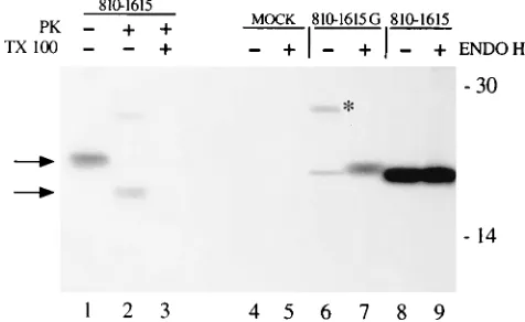
To determine whether the integration of NS2 in the ER membranes was also observed in vivo, a plasmid encoding aa 810 to 1615 was transiently expressed in HeLa cells by the T7-vaccinia virus system (14) and the PK protection assay was performed on the transfected cell lysate. As shown in Fig. 8, the 17-kDa protein band resistant to PK digestion was immu-noprecipitated from the lysate (lane 2) and resistance to the PK digestion was abolished if detergents were included during proteolysis (lane 3). A 12-kDa polypeptide resistant to PK digestion was also detected in transfected cells, although as a minor species (Fig. 8, lane 2). The exact nature of this protein species is not clear and is currently being investigated. A sim-ilar PK protection pattern was observed upon expression in transfected cells of a full-length cDNA copy of the viral ge-nome (data not shown). Thus, these data indicate that in trans-fected cells the NS2 protein is inserted in the ER membrane with an orientation similar to that observed in vitro.
To investigate if the NS2 peptide was indeed a true trans-membrane protein, we tested whether glycosylation signals in-troduced at the C-terminal region of the NS2 protein were recognized by the oligosaccharyl-transferase located in the ER lumen. To that end, we introduced by site-directed mutagen-esis two Asn residues at positions 983 and 988 of NS2, thus obtaining the Asn-X-Thr consensus sequence required for N-linked glycosylation. Plasmid pGEM(810-1615G) bearing the two Asn substitutions was transfected, and the NS2 profile was compared with that of the wild-type clone expressing the same polypeptide. Figure 8 shows that besides the 23-kDa band corresponding to mature NS2, a protein of 28 kDa was immu-noprecipitated with anti-NS2 antiserum from cells transfected with pGEM(810-1615G) and not with pCD(810-1615) (Fig. 8, compare lanes 6 and 8). The size of this polypeptide is consis-tent with that of the mature NS2 bearing two N-linked oligo-saccharide chains. Furthermore, this protein was sensitive to endoglycosidase H treatment (Fig. 8, lanes 7 and 9), indicating
that high-mannose sugars had been attached to the polypep-tide backbone. Thus, these results confirm the cell-free trans-lation data and indicate that the NS2 protein spans the ER membrane at least once, with the C-terminal region located in the lumen and the N-terminal domain exposed in the cytosol.
DISCUSSION
We have examined by cell-free protein synthesis and trans-fection studies the biogenesis and membrane localization of the NS2 protein of HCV with particular emphasis on the C-terminal processing event. The results obtained indicate that the NS2 polypeptide is a transmembrane protein and that cleavage at the NS2-3 junction involves a cellular compo-nent(s) as a cofactor(s) and has important implications for the localization of the viral polypeptide.
The data reported here indicate that in analogy to what has been described for the HCV-J and -H strains (16, 21), process-ing of the NS2-3 junction of the BK strain is mediated by the viral autoprotease that spans most of the NS2 polypeptide and the entire NS3 serine protease domain. Deletion analysis of the NS2-3 region shows that efficient cleavage at the NS2-3 site of HCV-BK requires the expression of aa 849 to 1237. Also, substitution of Cys-993 of the BK polyprotein abolishes the NS2-3 cleavage similarly to what has been described for the HCV-J and -H strains (16, 21) (Fig. 2). Thus, although the biochemical requirements for efficient in vitro cleavage at this site appear to differ from one strain to another (see below), it is reasonable to conclude that cleavage at the NS2-3 site of these three HCV strains is mediated by the same autoproteo-lytic activity. Furthermore, although the role of the NS3 do-main in the processing reaction is not clear, our data demon-strate that NS3 sequences cannot be substituted by other segments of the HCV polyprotein.
The HCV-J and -BK strains display distinct degrees of mem-brane dependency for efficient in vitro cleavage at the NS2-3 site. Direct comparison of the in vitro synthesis of the NS2-3 polyproteins of these two strains clearly shows that, indepen-dently of the transcript length, the BK strain requires the presence of membranes during translation for efficient process-ing at the NS2-3 junction (Fig. 3). However, translation in the presence of membrane and expression of the NS2-3 autopro-tease in transfected cells (data not shown) indicate that the overall processing efficiencies of these two strains are not dis-similar. The contribution of the microsomal membranes to BK NS2-3 processing is not limited to a stabilizing effect on the reaction products but rather causes an enhancement of cleav-age (Fig. 4). Also, our data indicate that the membrane-stim-ulating activity of the NS2-3 protease requires targeting of the nascent precursor to the membrane via the SRP-SRP receptor machinery (Fig. 5). Thus, although the exact role of mem-branes in the cleavage reaction remains to be defined, it is conceivable that they affect the processing at the NS2-3 junc-tion by assisting the proper folding of the nascent precursor. Thus, it is likely that the differences in amino acid sequence of the HCV-BK and -J strains (27, 44) influence the capacity of the nascent protein precursor to properly fold in the absence of membranes, rather than compromising its intrinsic protease activity.
It is not clear what the biological significance of the distinct in vitro requirements for cleavage at the NS2-3 site of the BK and J strains is. Interestingly, the processing at the p7-NS2 junction appears also to differ in efficiency between the HCV-J and -BK strains (Fig. 3). Furthermore, the kinetics of associa-tion between the E1 and E2 polypeptides derived from the BK strain has been shown to differ from that of the H strain (9),
on November 9, 2019 by guest
http://jvi.asm.org/
suggesting that observed differences among HCV strains are not limited to processing at the NS2-3 junction but are a more general phenomenon. It is tempting to speculate that the above-described differences influence the replication capacity of the HCV strains and affect their pathogenic properties. Alternatively, the observed differences in enzymatic activity and processing kinetics may be the results of the changes in the protein sequence introduced during cloning of the cDNAs of these viruses and therefore may not be representative of their actual biological properties.
The results presented in this study reveal that the NS2 pro-tein is a transmembrane polypeptide with the C-terminal re-gion translocated in the ER lumen and the N-terminal rere-gion
exposed, at least in part, in the cytoplasm. Several lines of evidence support this conclusion. Resistance to urea extraction of the processed NS2 protein is consistent with the sedimen-tation profile of an integral membrane polypeptide (Fig. 6). Also, the NS2 protein is protected by the membranes from digestion by exogenously added PK both in cell-free translation studies and in transfected cells (Fig. 7 and 8). Furthermore, the C terminus of the NS2 protein is available for N-linked glyco-sylation in transfected cells once the appropriate signals are introduced in the protein backbone (Fig. 8).
Interestingly, although NS2 targeting to the ER membrane appears to involve SRP and its receptor, much like all known secretory and transmembrane polypeptides (40), the mode of interaction with the targeting machinery as well as the mech-anism of insertion across the ER bilayer present some peculiar features. In fact, analysis of the amino acid sequence of the NS2 polypeptide revealed no long stretches of hydrophobic residues that could be identified as a signal sequence(s) and/or transmembrane domain(s). However, the inefficient targeting to the membrane of the aa 810 to 1026 NS2 precursor suggests that the functional signal sequence responsible for proper tar-geting is located across or downstream from the cleavage site, at the C terminus of the translocated polypeptide. This obser-vation is in line with the data presented by Hijikata et al. (20, 21), in which the authors failed to observe an NS2 protein species resistant to PK digestion when they expressed
trun-FIG. 7. PK protection assay of HCV proteins synthesized in vitro. (A) Tran-scripts encoding aa 810 to 1615 and aa 810 to 1026 and 1027 to 1657 were translated in vitro in the absence or presence of RM. (B) Transcripts encoding the indicated amino acids of the HCV-J strain (J) and the HCV-BK strain (BK) were translated in vitro with a rabbit reticulocyte lysate either in the absence or in the presence of microsomal membranes. Aliquots of the translation mixtures were treated with PK or with PK and Triton X-100 (Tx100) as described in Materials and Methods and subsequently were analyzed on an SDS–11% poly-acrylamide gel. The positions of the processed NS3, E2-NS2, NS2, and NS29 polypeptides are indicated. Asterisks indicate the partially processed NS2-3 pre-cursor. Arrowheads indicate the protected NS2 protein. Positions of molecular mass standards (in kilodaltons) are indicated.
on November 9, 2019 by guest
http://jvi.asm.org/
[image:9.612.57.552.262.726.2]cated forms of the NS2 polypeptide that did not require pro-cessing at the C terminus for correct maturation.
Although our results do not address whether the NS2 pro-tein spans the bilayer more than once, they indicate that at least one membrane-spanning domain is located in the N-terminal third of the protein, possibly between residues 50 and 80 (Fig. 7). However, the localization of the extreme N termi-nus of NS2 as proposed by Mizushima et al. (36) cannot be substantiated by our experiments. Furthermore, our data pro-vide epro-vidence that cleavage at the NS2-3 site is a prerequisite for NS2 translocation. In fact, the perfect correlation between the ability of the various deletion mutants to be processed at the NS2-3 site and their translocation competence strongly suggests that only the NS2 protein derived from the processed NS2-3 precursor can be efficiently translocated into the ER membrane. This conclusion is strengthened by the observation that the NS3 polypeptide is neither protected nor associated with the membrane (Fig. 6 and 7) and that the C993A and
L1026DQ mutants, which are no longer processed at the NS2-3
site, have also lost their translocation competence into the membrane bilayer (data not shown). The inference that pro-cessing at its C terminus precedes NS2 insertion in the mem-brane has important implications for the mechanism by which this protein is translocated. In fact, it confirms previous obser-vations that targeting to and translocation across the ER mem-brane are distinct events (37, 38) and indicates that at variance with the majority of mammalian secretory and membrane pro-teins, the NS2 polypeptide is translocated entirely posttransla-tionally. This conclusion raises the intriguing possibility that proper topogenesis of NS2 involves folding of the polyprotein precursor into an active autoprotease and subsequent unfold-ing of the cleaved NS2 polypeptide to allow translocation across the ER membrane.
The functional role of the NS2-3 protease as well as that of the NS2 protein in the life cycle of HCV remains to be eluci-dated. The NS3 serine protease of the J, H, and BK strains appears to be largely insensitive to cleavage at the NS2-3 site, since inactivation of the NS2-3 protease by site-directed
mu-tagenesis does not perturb the processing capacity of the serine protease (16, 21; data not shown). Thus, it appears that cleav-age at the NS2-3 site has a regulatory role in the biogenesis of the HCV proteins limited to the processing and membrane topology of the NS2 polypeptide and does not affect the func-tionality of the serine protease. However, recent data have indicated that the presence of uncleaved NS2 sequences may severely interfere with the NS3-mediated proteolysis at down-stream sites in the polyprotein of a United Kingdom HCV isolate (8). These conflicting observations need further inves-tigation but could be the result of dissimilar experimental ap-proaches utilized to investigate this problem or, alternatively, may reflect differential dependence of various HCV strains on NS2-3 cleavage for NS3 activity.
The finding that NS2 is a transmembrane peptide is in agree-ment with the observation that in cells expressing the entire polyprotein of HCV the NS2 polypeptide is associated with the E1 and E2 glycoproteins, as judged by coimmunoprecipitation with monospecific antibodies (32, 43; data not shown). These data prompt the provocative speculation that NS2 is a struc-tural rather than a nonstrucstruc-tural viral component (43). Pro-tease protection experiments suggest that both HCV glyco-proteins do not contain a cytoplasmic tail suitable for a specific interaction with the viral nucleocapsid (20, 33, 35). It is thus conceivable that NS2 functions during the assembly process as a structural bridge between the envelope glycoproteins and the nucleocapsid. However, the NS2 polypeptide has been also immunoprecipitated with NS5A and NS5B, which are the pu-tative components of the replication complex (22). Obviously, the definitive classification of NS2 as a structural component awaits a precise biochemical characterization of the HCV virion.
ACKNOWLEDGMENTS
We thank K. Shimotohno for providing the HCV-J subclone pN(722-1325) and R. De Francesco for plasmids pCITE(623-1615) and pT7(1027-1657). We also thank S. Bonatti, J. Jiricny, and C. Stein-ku¨hler for critical review; M. Emili for graphics; and P. Neuner for oligonucleotide synthesis.
REFERENCES
1. Bartenschlager, R., L. Ahlborn-Laake, J. Mous, and H. Jacobsen. 1993. Nonstructural protein 3 of hepatitis C virus encodes a serine-type proteinase required for cleavage at the NS3/4 and NS4/5 junctions. J. Virol. 67:3835–3844. 2. Bartenschlager, R., L. Ahlborn-Laake, J. Mous, and H. Jacobsen. 1994. Kinetic and structural analyses of hepatitis C virus polyprotein processing. J. Virol. 68:5045–5055.
3. Cha, T.-A., E. Beall, B. Irvine, J. Kolberg, D. Chien, G. Kuo, and M. S.
Urdea.1992. At least five related, but distinct, hepatitis C virus viral geno-types exist. Proc. Natl. Acad. Sci. USA 89:7144–7148.
4. Choo, Q.-L., G. Kuo, A. J. Weiner, L. R. D. W. Bradley, and M. Houghton. 1989. Isolation of a cDNA clone derived from a blood-borne non-A non-B viral hepatitis genome. Science 244:359–362.
5. Choo, Q.-L., K. H. Richman, J. H. Han, K. Berger, C. Lee, C. Dong, C.
Gallegos, D. Coit, A. Medina-Selby, P. J. Barr, A. J. Weiner, D. W. Bradley, G. Kuo, and M. Houghton.1991. Genetic organization and diversity of the hepatitis C virus. Proc. Natl. Acad. Sci. USA 88:2451–2455.
6. Connolly, T., and R. Gilmore. 1986. Formation of a functional ribosome-membrane junction during translocation requires the participation of a GTP-binding protein. J. Cell Biol. 103:2253–2261.
7. De Francesco, R. Unpublished data.
8. D’Souza, E. D. A., E. O’Sullivan, E. M. Amphlett, D. J. Rowlands, D. V.
Sangar, and B. E. Clarke.1994. Analysis of NS3-mediated processing of the hepatitis C virus non-structural region in vitro. J. Gen. Virol. 75:3469–3476. 9. Dubuisson, J., H. H. Hsu, R. C. Cheung, H. B. Greenberg, D. G. Russel, and
C. M. Rice.1994. Formation and intracellular localization of hepatitis C virus envelope glycoprotein complexes expressed by recombinant vaccinia virus and Sindbis virus. J. Virol. 68:6147–6160.
10. Eckart, M. R., M. Selby, F. Maisiarz, C. Lee, K. Berger, K. Crawford, C.
[image:10.612.58.296.73.219.2]Kuo, G. Kuo, M. Houghton, and Q.-L. Choo.1993. The hepatitis C virus encodes a serine protease involved in processing of the putative nonstruc-tural proteins from the viral polyprotein precursor. Biochem. Biophys. Res. Commun. 192:399–406.
FIG. 8. NS2 is translocated across the ER membrane in transfected cells. Recombinant plasmids pCD(810-1615) and pGEM(810-1615G) were transfected into vaccinia virus vT7F-3-infected HeLa cells as previously described (45). Lysates were immunoprecipitated with anti-NS2 antiserum (45). Prior to immu-noprecipitation, lysates from cells transfected with pCD(810-1615) were sub-jected to a PK protection assay as described in Material and Methods (lanes 1 to 3). The immunoprecipitates were digested with endoglycosidase H (lanes 5, 7, and 9) and analyzed on an SDS–12.5% polyacrylamide gel. Mock-transfected cell lysate was also treated.1and2, digested and mock digested, respectively. Arrows indicate unglycosylated and protected NS2 protein. The asterisk indi-cates glycosylated NS2 polypeptide. Positions of molecular mass standards (in kilodaltons) are shown.
on November 9, 2019 by guest
http://jvi.asm.org/
11. Elroy-Stein, O., T. R. Fuerst, and B. Moss. 1989. Cap-independent transla-tion of mRNA conferred by encephalomyocarditis virus 59sequence im-proves the performance of vaccinia/bacteriophage T7 hybrid system. Proc. Natl. Acad. Sci. USA 86:6126–6130.
12. Failla, C., L. Tomei, and R. De Francesco. 1994. Both NS3 and NS4a are required for processing of hepatitis C virus polyprotein. J. Virol. 68:3753–3760. 13. Francki, R. I., C. M. Fauquet, D. L. Knudson, and F. Brown. 1991.
Classi-fication and nomenclature of viruses. Arch. Virol. S2:223–233.
14. Fuerst, T. R., E. G. Niles, F. W. Studier, and B. Moss. 1986. Eukaryotic transient-expression system based on recombinant vaccinia virus that syn-thesizes bacteriophage T7 RNA polymerase. Proc. Natl. Acad. Sci. USA
83:8122–8126.
15. Grakoui, A., D. W. McCourt, C. Wychowski, S. Feinstone, and C. M. Rice. 1993. Characterization of hepatitis C virus-encoded serine proteinase: de-termination of proteinase-dependent polyprotein cleavage sites. J. Virol. 67: 2832–2843.
16. Grakoui, A., D. W. McCourt, C. Wychowski, S. M. Feinstone, and C. R. Rice. 1993. A second hepatitis C virus-encoded proteinase. Proc. Natl. Acad. Sci. USA 90:10583–10587.
17. Grakoui, A., C. Wychowski, C. Lin, S. M. Feinstone, and C. M. Rice. 1993. Expression and identification of hepatitis C virus polyprotein products. J. Virol. 67:1385–1395.
18. Hemsley, A., N. Arnheim, M. D. Toney, G. Cortopassi, and D. J. Galas. 1989. A simple method for site-directed mutagenesis using the polymerase chain reaction. Nucleic Acids Res. 17:6545–6551.
19. Higuchi, R., B. Krummel, and R. S. Saiki. 1988. A general method of in vitro preparation and specific mutagenesis of DNA fragments: study of protein and DNA interaction. Nucleic Acids Res. 16:7351–7367.
20. Hijikata, M., N. Kato, Y. Ootsuyama, M. Nakagawa, and K. Shimotohno. 1991. Gene mapping of the putative structural region of the hepatitis C virus genome by in vitro processing analysis. Proc. Natl. Acad. Sci. USA 88:5547– 5551.
21. Hijikata, M., H. Mizushima, T. Akagi, S. Mori, N. Kakiuchi, N. Kato, T.
Tanaka, K. Kimura, and K. Shimotohno.1993. Two distinct proteinase activities required for the processing of a putative nonstructural precursor protein of hepatitis C virus. J. Virol. 67:4665–4675.
22. Hijikata, M., H. Mizushima, T. Akagi, Y. Tanji, Y. Komoda, Y. Hirowatari,
T. Akagi, N. Kato, K. Kimura, and K. Shimotohno.1993. Proteolytic pro-cessing and membrane association of putative nonstructural proteins of hepatitis C virus. Proc. Natl. Acad. Sci. USA 90:10773–10777.
23. Hirowatari, Y., M. Hijikata, Y. Tanji, H. Nyunoya, H. Mizushima, K.
Kimura, T. Tanaka, N. Kato, and K. Shimotohno.1993. Two proteinase activities in HCV polypeptide expressed in insect cells using baculovirus vector. Arch. Virol. 133:349–356.
24. Houghton, M., A. Weiner, J. Han, G. Kuo, and Q.-L. Choo. 1991. Molecular biology of the hepatitis C viruses: implication for diagnosis, development and control of viral disease. Hepatology 14:381–388.
25. Jang, S. K., M. V. Davies, R. J. Kaufman, and E. Wimmer. 1989. Initiation of protein synthesis by internal entry of ribosomes into the 59nontranslated region of encephalomyocarditis virus RNA in vivo. J. Virol. 63:1651–1660. 26. Kaito, M., S. Watanabe, K. Tsukiyama-Kohara, D. Yamaguchi, Y.
Koba-yashi, M. Konishi, M. Yokoi, S. Ishida, S. Suzuki, and M. Kohara.1994. Hepatitis C virus particle detected by immunoelectron microscopic study. J. Gen. Virol. 75:1755–1760.
27. Kato, N., M. Hijikata, Y. Ootsuyama, M. Nakagawa, S. Ohkoshi, T.
Sug-imura, and K. Shimotohno.1990. Molecular cloning of human hepatitis C virus genome from Japanese patients with non-A non-B hepatitis. Proc. Natl. Acad. Sci. USA 87:9524–9528.
28. Krieg, P. A., and D. A. Melton. 1984. Functional messenger RNAs are produced by SP6 in vitro transcription of cloned cDNAs. Nucleic Acids Res.
12:7057–7070.
29. Kuo, G., Q.-L. Choo, H. J. Alter, G. L. Gitnick, A. G. Redecker, R. H. Purcell,
T. Myamura, J. L. Dienstag, M. J. Alter, C. E. Syevens, G. E. Tagtmeier, F. Bonino, M. Colombo, W.-S. Lee, C. Kuo, K. Berger, J. R. Shister, L. R. Overby, D. W. Bradley, and M. Houghton.1989. An assay for circulating antibodies to a major etiologic virus of human non-A non-B hepatitis. Sci-ence 244:362–364.
30. Lin, C., B. D. Lindenbach, B. M. Pra´gai, D. W. McCourt, and C. M. Rice.
1994. Processing in the hepatitis C virus E2-NS2 region: identification of p7 and two distinct E2-specific products with different termini. J. Virol. 68: 5063–5073.
31. Lin, C., B. M. Pra´gai, A. Grakoui, J. Xu, and C. M. Rice.1994. Hepatitis C virus NS3 serine proteinase: trans-cleavage requirement and processing ki-netics. J. Virol. 68:8147–8157.
32. Matsuura, Y., T. Suzuki, R. Suzuki, M. Sato, H. Aizaki, I. Sato, and T.
Miyamura.1994. Processing of E1 and E2 glycoproteins of hepatitis C virus expressed in mammalian and insect cells. Virology 205:141–150. 33. Migliaccio, G. Unpublished data.
34. Miller, R. H., and R. H. Purcell. 1990. Hepatitis C virus shares amino acid sequence similarity with pestiviruses and flaviviruses as well as members of two plant virus subgroups. Proc. Natl. Acad. Sci. USA 87:2057–2061. 35. Mizushima, H., M. Hijikata, S.-I. Asabe, M. Hirota, K. Kimura, and K.
Shimotohno.1994. Two hepatitis C virus glycoproteins E2 products with different C termini. J. Virol. 68:6215–6222.
36. Mizushima, H., M. Hijikata, Y. Tanji, K. Kimura, and K. Shimotohno. 1994. Analysis of N-terminal processing of hepatitis C virus nonstructural protein 2. J. Virol. 68:2731–2734.
37. Nicchitta, C. V., and G. Blobel. 1989. Nascent secretory chain binding and translocation are distinct processes: differentiation by chemical alkylation. J. Cell Biol. 108:789–795.
38. Nicchitta, C. V., G. Migliaccio, and G. Blobel. 1991. Biochemical fraction-ation and assembly of the membrane components that mediate nascent chain targeting and translocation. Cell 65:587–598.
39. Pizzi, E., A. Tramontano, L. Tomei, N. La Monica, C. Failla, M. Sardana, T.
Wood, and R. De Francesco.1994. Molecular modeling of the specificity pocket of the hepatitis C virus protease: implications for substrate recogni-tion. Proc. Natl. Acad. Sci. USA 91:888–892.
40. Rapoport, T. A. 1992. Transport of protein across the endoplasmic reticulum membrane. Science 258:931–936.
41. Santolini, E., G. Migliaccio, and N. La Monica. 1994. Biosynthesis and bio-chemical properties of the hepatitis C virus core protein. J. Virol. 68:3631–3641. 42. Selby, M. J., Q.-L. Choo, K. Berger, G. Kuo, E. Glazer, M. Eckart, C. Lee, D.
Chien, C. Kuo, and M. Houghton.1993. Expression, identification and sub-cellular localization of the proteins encoded by the hepatitis C virus. J. Gen. Virol. 74:1103–1113.
43. Selby, M. J., E. Glazer, F. Masiarz, and M. Houghton. 1994. Complex processing and protein:protein interactions in the E2:NS2 region of HCV. Virology 204:114–122.
44. Takamizawa, A., C. Mori, I. Fuke, S. Manabe, S. Murakami, J. Fujita, E.
Onoshi, T. Andoh, I. Yoshida, and H. Okayama.1991. Structure and organ-ization of the hepatitis C virus genome isolated from human carriers. J. Virol. 65:1105–1113.
45. Tomei, L., C. Failla, E. Santolini, R. De Francesco, and N. La Monica. 1993. NS3 is a serine protease required for processing of hepatitis C virus polypro-tein. J. Virol. 67:4017–4026.