Copyright © 1999, American Society for Microbiology. All Rights Reserved.
An Intact TAR Element and Cytoplasmic Localization Are
Necessary for Efficient Packaging of Human
Immunodeficiency Virus Type 1 Genomic RNA
C. HELGA-MARIA,† MARIE-LOUISE HAMMARSKJO
¨ LD,
ANDDAVID REKOSH*
Myles H. Thaler Center for AIDS and Human Retrovirus Research and Department of Microbiology,
University of Virginia, Charlottesville, Virginia 22908
Received 5 November 1998/Accepted 1 February 1999
Although most reports defining the human immunodeficiency virus type 1 (HIV-1) genomic RNA packaging
signal have focused on the region downstream of the major 5
*
splice site, others have suggested that sequences
upstream of the splice site may also play an important role. In this study we have directly examined the role
played by the HIV-1 TAR region in RNA packaging. For these experiments we used a proviral expression
system that is largely independent of Tat for transcriptional activation. This allowed us to create constructs
that efficiently expressed RNAs carrying mutations in TAR and to determine the ability of these RNAs to be
packaged. Our results indicate that loss of sequences in TAR significantly reduce the ability of a viral RNA to
be packaged. The requirement for TAR sequences in RNA packaging was further examined by using a series
of missense mutations positioned throughout the entire TAR structure. TAR mutations previously shown to
influence Tat transactivation, such as G31U in the upper loop region or UCU to AAG in the bulge (nucleotides
[nt] 22 to 24), failed to have any effect on RNA packaging. Mutations which disrupted the portion of the TAR
stem immediately below the bulge also had little effect. In contrast, dramatic effects on RNA packaging were
observed with constructs containing mutations in the lower portion of the TAR stem. Point mutations which
altered nt 5 to 9, 10 to 15, 44 to 49, or 50 to 54 all reduced RNA packaging 11- to 25-fold. However,
compensatory double mutations which restored the stem structure were able to restore packaging. These
results indicate that an intact lower stem structure, rather than a specific sequence, is required for RNA
packaging. Our results also showed that RNA molecules retained within the nucleus cannot be packaged,
unless they are transported to the cytoplasm by either Rev/Rev response element or the Mason-Pfizer monkey
virus constitutive transport element.
While a detailed understanding of the mechanism for RNA
packaging in human immunodeficiency virus (HIV) is lacking,
it is widely believed that the HIV genome contains a packaging
region that interacts with a viral factor to mediate the
incor-poration of full-length genomic RNA into virus particles (for a
review, see reference 6). RNA mapping studies have shown
that important determinants for RNA packaging map
through-out the entire 5
9
-end region of the HIV type 1 (HIV-1) RNA,
up to the start of gag (8, 37, 38, 43, 46, 48, 56, 63). However,
most attention has focused on the region of viral RNA
map-ping between nucleotides 230 and 352 (4, 12, 32). This region
has been called the HIV-1
C
region, in analogy to the
pack-aging region of murine leukemia virus (MLV) RNA (44, 62).
The HIV-1
C
region forms four stem-loop structures termed
SL1 through SL4 (4, 12, 32). SL1 contains the primary site of
RNA dimer formation (14, 40, 45, 49, 50, 53, 59), and SL3
contains a specific binding site for HIV-1 nucleocapsid protein
(NC) (5, 17, 42). Most data suggest that there are determinants
in both SL1 and SL3 that are essential for efficient packaging.
Since the major 5
9
splice site is contained within SL2 (58)
(which maps between SL1 and SL3), only unspliced RNA
contains both the SL1 and SL3 determinants. This explains, at
least in part, why only unspliced HIV RNA is efficiently
pack-aged.
Despite the progress that has been made in defining the
HIV-1 packaging signal, there is no unifying model to explain
how this complex, multipartite signal is actually recognized by
the assembling virus particle. The viral factor involved in this
process appears to be NC (1, 7, 9, 25, 55, 64), but the details of
the mechanism by which NC provides specificity remain
elu-sive. For example, even though the three-dimensional
struc-ture of complex of NC and SL3 has recently been described
(17), we do not understand why only two molecules of RNA
are packaged into a particle that contains about 1,800 NC
molecules and thus 1,800 SL3 binding sites. We also have little
understanding of the role played by sequences mapping
up-stream of SL1.
The HIV-1 genome contains at least two other cis-acting
sequence elements whose interactions with viral proteins have
been well defined. The HIV-1 TAR element consists of a
57-nucleotide sequence that forms a stem-loop. It is found at
both ends of HIV-1 genomic RNA, forming the first part of the
“R” repeat present in all retroviral RNAs. The element
regu-lates transcription through its interaction with the HIV-1 Tat
protein and cellular factors (for reviews, see references 34 and
36). Parts of the TAR stem-loop not involved with the Tat
interaction have also been shown to be essential for viral
rep-lication, though the reasons for their importance are much less
well defined (31, 39). For example, elegant genetic studies have
implicated the lower stem region of TAR as an essential
ele-ment for viral infectivity (39).
Another RNA element in the HIV genome that is involved
* Corresponding author. Mailing address: Myles H. Thaler Center
for AIDS and Human Retrovirus Research, Department of
Microbi-ology, HSC Box 441 University of Virginia, Charlottesville, VA 22908.
Phone: (804) 982-1599. Fax: (804) 982-1590. E-mail: dr4u@virginia
.edu.
† Present address: Institute for Experimental Pathology, Keldur,
University of Iceland, IS-112 Reykjavik, Iceland.
4127
on November 9, 2019 by guest
http://jvi.asm.org/
in the binding of a viral specific protein is the Rev response
element (RRE). This 248-nucleotide element is located within
the coding region of env and is present in full-length and singly
spliced HIV RNAs. Rev binding is needed to mediate the
export of the RRE-containing RNA from the nucleus to
cyto-plasm (for reviews, see references 26 and 54). An element
functionally equivalent to the RRE, termed the constitutive
transport element (CTE), has been identified in type D
retro-viruses (10, 65). The CTE mediates the nuclear export of
intron containing RNA (20) by interacting exclusively with
factors that are encoded by the cell.
The present study examines determinants of HIV-1 RNA
packaging by introducing deletion and cluster mutations into
the RNA and analyzing their effects on encapsidation. We
have focused our effort mainly on the region mapping
up-stream of
C
and, in particular, on the HIV TAR region. Our
data strongly suggest that a distinct packaging determinant
exists within the bottom part of the TAR stem. Our results
suggest that it is the structure, rather than the sequence, of the
TAR stem that is important. We also describe in this report
other experiments that demonstrate that an RNA which is
dependent on Rev for transport must interact with Rev (and
presumably exit the nucleus) before it can be packaged.
MATERIALS AND METHODS
HIV-1 proviral strain and numbering system. The HIV-1 proviral clone (pHR969) and its derivative plasmids are derived from HIV-1 HxB2 (GenBank accession no. K03455, M38432, and g327742). Sequences from pNL4-3 (Gen-Bank accession no. M1992) and BH10 (Gen(Gen-Bank accession no. M15654, K02008, K02009, and K02010) were also used. The HIV-1 numbering system used is derived from HxB2 and places nucleotide 1 at the start of R in the 59long terminal repeat (LTR).
Plasmids. Plasmid pGAGPOL-RRE-r (pHR146) has been previously de-scribed (60). It contains the coding regions for the HIV-1 proteins Gag, Pol, Vif, and truncated Vpr, followed by the RRE and a rabbit b-globin intron and poly(A) site. Expression is directed by the simian virus 40 (SV40) late promoter. pHR969 contains the full-length HIV-1 genome from the HIV-1 HxB2 pro-viral construct with the exception of a 330-bp in-frame deletion in pol making it noninfectious. The plasmid also contains cellular flanking sequences on each side of the viral genome as well as the SV40 origin of replication in the vector backbone.
pHR1256 was derived directly from pHR969. It has a deletion in the midpor-tion of the genome corresponding to the central pormidpor-tion of the HIV-1 genome that is deleted in pHR146. Details of its construction are available on request. pHR1486 was derived from sequences present in the vectors pHR1256 and RRE-r through a series of intermediate constructions. It contains a 220-bp
SacI-BssHII fragment from the HIV-1 proviral clone pNL4-3 between the SacI
site at position 28 and the BssHII site at position 248 in place of HxB2 sequences. Thus, it lacks the SacI site normally found at position 228 in HxB2.
pHR1573 was made by linearizing vector pHR1486 at the unique SacI site in TAR, repairing the 4-bp 39overhangs with T4 DNA polymerase and religation. pHR1512, pHR1679, pHR1487, pHR1582, pHR1583, pHR1584, pHR1585, pHR1586, pHR1815, pHR1816, pHR1817, pHR1819, pHR1820, and pHR1821 are derivatives of pHR1486 which were made by PCR-based mutagenesis. De-tails of their construction are available on request.
pHR1152 (previously described [10]) was derived from pNL4-3. It produces a nonfunctional Rev protein due to a stop codon at amino acid 23 (TAT3TAA) in the first coding exon of rev. It also has the Mason-Pfizer monkey virus (MPMV) CTE (MPMV bp 8007 to 8240) inserted into a unique XhoI site at the start of the nef gene. Prior to insertion of the CTE, 150 bp were deleted from nef, moving the XhoI site just downstream of the nef start codon.
pCMVrev (pHR30), previously described as pRev1 (60), contains the HIV-1 Rev coding region under the control of simian cytomegalovirus immediate-early promoter-enhancer (35). pCMVtat (pHR136) is analogous to pCMVrev. It con-tains the Tat coding sequences from pCV1 (3) under the control of the simian cytomegalovirus immediate-early promoter-enhancer.
Plasmid pHR1290 was constructed by deleting the 2.3-kb BssHII-EcoRV frag-ment from the previously described pGem-11Zf(2)HIV (pHR243) (61) and religating the vector fragment. This plasmid was used for in vitro transcription to make the RNA standards for reverse transcription (RT)-PCR.
Cell culture and transfections.Maintenance of and transfection CMT3-COS cells (23) have been previously described. In a standard packaging experiment, 15
mg of test plasmid and 10mg of pCMVrev per 150-mm-diameter plate were transfected by using DEAE-dextran as previously described (28). In some
ex-periments, plasmid pCMVtat was used for cotransfections as well (10m g/150-mm-diameter plate).
In some instances, A5.6 cells were substituted for CMT3-COS cells to over-come the need for pCMVrev cotransfection. A5.6 cells are derived from CMT3-COS by stable transfection with pCMVrev and a hygromycin B resistance gene (pHR392). They constitutively express the HIV-1 Rev protein and are similar to the previously described cell line A5.9 (19).
Cellular and virion RNA isolation [total poly(A) selection].Total poly(A)1 RNA was isolated from cells as previously described (27). To prepare virion RNA, virus particles were collected from the medium of transfected cells. To do this, medium was first cleared of cells and debris by centrifugation two times at 2,500 rpm for 15 min at 4°C in a tabletop centrifuge. The cleared medium was then subjected to ultracentrifugation through a 6-ml cushion of 20% sucrose for 1 h at 4°C and 27,000 rpm (100,0003g), using a SW28 rotor. The pelleted virus
particles were resuspended in 100ml of stock buffer (200 mM NaCl, 200 mM Tris-HCl [pH 7.5], 1.5 mM MgCl2). To prepare RNA, 3 ml of lysis buffer (200
mM NaCl, 200 mM Tris-HCl [pH 7.5], 1.5 mM MgCl2, 2% sodium dodecyl
sulfate, 200mg of proteinase K [Sigma] per ml) together with 200mg of yeast tRNA (GibcoBRL) as a carrier was added to the virions. RNA was then pre-pared by the procedure used to prepare cellular RNA.
DNase I treatment of RNA.After ethanol precipitation, the RNA samples were resuspended in 20 mM Tris-HCl (pH 8.4)–2 mM MgCl2–125 mM KCl–40
U of rRNasin (Pharmacia)–20 U of DNase I (Boehringer Mannheim) and incubated at 37°C for 1 h. The RNA was then extracted with phenol-CHIASM (chloroform-isoamyl alcohol; 24:1) and reprecipitated.
In vitro transcription.pHR1290 digested with EcoRI (20 U/ml; New England Biolabs) was used as a template to produce RNA in vitro for a standard curve. This plasmid produces RNA of the same sense as the HIV genome when transcription is driven by T7 RNA polymerase. The in vitro transcription reac-tions were performed according to instrucreac-tions from Promega.
Oligonucleotides.The sense-strand oligonucleotide used to detect HIV-1
gag-pol RNA was oligonucleotide 304, which hybridized to gag-pol at positions 3167 to
3188 and had the sequence 59-AGGGGTGCCCACACTAATGATG-39. The antisense oligonucleotide used for this RNA was oligonucleotide 305, which hybridized to pol at positions 3664 to 3685 and had the sequence 59-GGCTAC ATGAACTGCTACCAGG-39.
To detect HIV-1 rev RNA, the sense-strand oligonucleotide used was oligo-nucleotide 298, which hybridized to the start of the first exon of rev at positions 5186 to 5206 and had the sequence 59-ATGGCAGGAAGAAGCGGAGAC-39. The antisense oligonucleotide used, 190, hybridized to the nef gene at positions 8137 to 8161) and had the sequence 59-GCTGCTGTGTTGCTACTTGTGATT G-39.
32P 5*-end labeling of oligonucleotides.Oligonucleotides were labeled by using
polynucleotide kinase and [g-32P]ATP (6,000 Ci/mmol) as previously described
(27). The specific activity of the labeled oligonucleotides was usually in the range 23108to 43108cpm/mg of oligonucleotide.
RT and cDNA synthesis.The RT-PCR procedure of Arrigo et al. was fol-lowed, with some modifications (2). Both cellular and virion poly(A)-selected RNA was used as template to make cDNA for quantitative PCR. Each cellular RNA sample was a yield from a 150-mm-diameter cell culture plate; one such plate usually gave the yield of 4 to 8mg of total poly(A)-selected cellular RNA. The cellular RNA samples were resuspended in 200ml of diethylpyrocarbonate-treated H2O, and usually 1/200 (20 to 40 ng of RNA) was used per RT reaction.
The virion RNA samples (from the medium of a 150-mm-diameter cell culture plate) were resuspended in 100ml of H2O, and the yield from 1/10 of a plate was
used per RT reaction. Each RT reaction was performed in a final volume of 50
ml containing 1ml (40 U) of rRNasin, 10ml of 53RT buffer (250 mM Tris-HCl [pH 8.3], 375 mM KCl, 15 mM MgCl2, 50 mM dithiothreitol), 1ml (100 ng/ml) of
antisense oligonucleotide primer, 5ml (1mg/ml) of bovine serum albumin (New England Biolabs), 2.5ml 10 mM each deoxynucleoside triphosphate, 5ml (500
mg/ml) actinomycin D (Sigma), and 1ml of M-Moloney murine leukemia virus reverse transcriptase (200 U/ml; GibcoBRL). To detect potential DNA contam-ination in the RNA samples, the same amounts of cellular and virion RNA samples were treated exactly the same way as the RT samples except that no reverse transcriptase was added to the tubes. Known amounts of serially diluted RNA standards which were made by in vitro transcription were also used as controls to measure the amounts of HIV-1-specific RNA in the test samples and to ensure the linearity of the RT-PCRs; they were treated the same way as other RT reactions. The reactions were carried out at 37°C for 2 h, after which 950ml of Tris-EDTA buffer was added to each tube to give the volume of 1 ml. The cDNA was stored at275°C until used for quantitative PCR.
Quantitative PCR.For each quantitative PCR, 10ml of cDNA (from the total volume of 1 ml) was used. To that was added 15ml containing 15 ng of32
P-labeled 39oligonucleotide (specific activity, 23108to 43108cpm/mg), 15 ng of
unlabeled 39oligonucleotide, 100 ng 59oligonucleotide, 2.5ml of 103modified PCR buffer (250 mM Tris pH 8.0, 50 mM MgCl2, 500 mM NaCl, 2.5 mM dATP,
2.5 mM dTTP, 2.5 mM dCTP, 2.5 mM dGTP, 1mg of bovine serum albumin per
ml), and 0.5ml of Amplitaq DNA polymerase (5 U/ml; Perkin-Elmer), so that the final volume was 25ml per reaction. The reactions were performed in 0.5-ml Eppendorf tubes; the reaction products were overlaid with 1 drop of mineral oil and placed in a DNA thermal cycler. The reactions were performed at 94°C for 1 min and 60°C for 2 min for 22 cycles.
on November 9, 2019 by guest
http://jvi.asm.org/
Following the quantitative RT-PCRs, 5ml of each sample was mixed with 4ml of H2O and 1ml of 103restriction stop buffer; the mixture was loaded on a 2%
agarose gel, which was subjected to electrophoresis in 0.53Tris-acetate-EDTA buffer at 150 V (at 0.25-A setting) for about an hour. The agarose gel was overlaid with Saran Wrap, dried, and quantitated with a phosphorimager.
p24 enzyme-linked immunosorbent assay (ELISA) on HIV-1 virions. The concentrated virus particles were diluted and assayed at the dilutions 1/62,500 and 1/312,500 to keep the test samples within the linear range of the assay. The assay was performed according to instructions from the manufacturer (Cellular Products Inc.). A molecular weight of 25,390 was used to convert the amount of p24 to moles.
Calculation of the virion RNA/virion p24 ratio.The amount of virion RNA present in a sample was determined from the relative intensity of the RT-PCR product by using an RNA standard curve which was generated from in vitro-transcribed RNA. This amount was then multiplied by the appropriate dilution factor to calculate the total amount of virion RNA produced per plate. For virion p24, the value obtained in the ELISA by using the p24 standard curve was also multiplied by an appropriate dilution factor. A ratio of millimoles of virion RNA per mole of virion p24 was then calculated. For most experimental samples, a normalized value for this ratio was obtained by multiplying the value by 100 and dividing by the ratio obtained from the control transfection.
Calculation of the percentage of total RNA packaged.Total virion RNA RI (relative intensity) units per plate were determined from the relative intensity of the RT-PCR product by multiplying the value obtained from the phosphorim-ager by the appropriate dilution factor for each test sample. To calculate the cellular plasmid-derived RNA RI units per plate, total cellular RNA was pre-pared and quantitated by optical density. Usually 20 ng of this RNA was tested by RT-PCR for plasmid-derived RNA content. The RI units obtained from this sample were then multiplied by the appropriate dilution factor to obtain the total RI units per plate. Since the yield of total RNA from each plate varied some-what, it was assumed that the plate that gave the highest yield of total cellular RNA in each experiment had a yield closest to 100%, and the dilution factor was determined from this value. Yields usually did not vary more than twofold. Finally, the total RI units from both cellular and virion samples were added together and the percentage of the RNA which was found in the virions was calculated.
RESULTS
Packaging of RNA expressed from transient expression
vec-tors.
We have previously described an SV40 late replacement
vector, pGAGPOL-RRE-r, and its derivatives, that were used
to study assembly interactions between the Pr55
gagand
Pr160
gag-polprecursors (60, 61). In those studies we
demon-strated that virus-like particles efficiently formed and budded
when this plasmid and a plasmid that expressed Rev were
transfected into appropriate cells. However, it was never
de-termined whether the virus-like particles produced packaged
viral RNA.
The HIV RNA produced by pGAGPOL-RRE-r lacks the
first 228 nucleotides found in a normal HIV genome but
re-tains sequences further downstream known to be involved in
RNA packaging; in addition, sequences 3
9
of the SalI site in the
middle of the HIV-1 genome are replaced with a removable
rabbit
b
-globin intron and poly(A) signal (Fig. 1). Thus, it was
of interest to determine whether RNA produced by this
plas-mid could be packaged into particles, since this would shed
light on the sequence requirements for RNA packaging by
HIV.
In the first experiment, we compared the packaging
effi-ciency of pGAGPOL-RRE-r to those of the proviral clone
pHR969 and its derivative pHR1256. pHR969 is a full-length
viral clone, derived from HxB2, that contains a small in-frame
deletion in the RNase H domain of pol. This renders it
non-infectious. The plasmid also contains an SV40 origin of
repli-cation which allows Tat-independent expression in cells
con-taining SV40 T antigen (data not shown). pHR1256 is derived
from pHR969 and was used for comparison since it shares a
common deletion in the central portion of the genome with
pGAGPOL-RRE-r but contains an intact 5
9
LTR. Both
pHR1256 and pGAGPOL-RRE-r lack a functional rev gene
and require cotransfection with a Rev-expressing plasmid to
obtain cytoplasmic RNA expression. All three plasmids are
diagrammed in Fig. 1.
CMT3-COS cells were transfected with the various plasmids
by using DEAE-dextran, and the cells and medium were
quan-titatively harvested at 65 h posttransfection. Both cellular and
particle-derived RNA were prepared. Quantitative RT-PCR
was then performed to determine the amount of RNA in each
fraction (see Materials and Methods). The results of this RNA
packaging analysis are presented in Fig. 2 and Table 1.
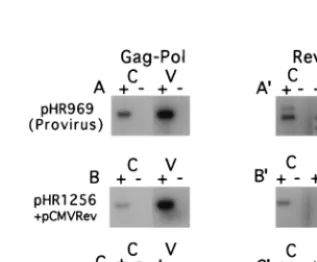
Figure 2 shows the PCR products obtained from the RNA
derived from both cells and pelleted virus particles. Both
Gag-Pol and Rev RNAs were analyzed by using specific primer
pairs. The figure shows that in all cases, there was no signal
when reverse transcriptase was omitted from the reaction
mix-ture, indicating that the RNA preparations were free from
contaminating DNA. Experiments treating the virus particles
with micrococcal nuclease before RNA extraction yielded
es-sentially the same results, indicating that the signal from the
particles was derived exclusively from packaged RNA (data
not shown). The intensity of each band in Fig. 2 was measured
with a phosphorimager and corrected for the dilution factor (see
Materials and Methods); the results are tabulated in Table 1.
FIG. 1. Schematic representation of HIV-1 sequences present in six plasmids used in this study. The upper portion shows a map of the entire HIV-1 genome, with a few key restriction enzyme sites marked. The two KpnI sites in pol were used to create a 330-bp in-frame deletion in the proviral clone pHR969 in order to make it noninfectious. Plasmids pHR1256 and pHR1486 were derived from pHR969 and hence contain the same deletion. These plasmids, as well as pGAGPOL-RRE-r, also contain a deletion in the midportion of the viral genome (between SalI and BglII) shown as a dashed line. Expression of pGAGPOL-RRE-r is directed by the SV40 late promoter rather than the 59LTR. pGAGPOL-RRE-r also lacks 228 bp at the 59end of the viral RNA. pGAGPOL-RRE and pHR1486 contains an intron and poly(A) signal derived from the rabbitb-globin gene.on November 9, 2019 by guest
http://jvi.asm.org/
The data in Table 1 demonstrate that in the case of the
full-length proviral clone pHR969, nearly 16% of the Gag-Pol
RNA produced was packaged into particles. For pHR1256
cotransfected with pCMVrev, this value reached nearly 42%.
On the other hand, for pGAGPOL-RRE-r cotransfected with
pCMVrev, only about 1.5% of the vector RNA was
incorpo-rated into particles. For the Rev mRNA, incorporation into
virus particles was in all cases less than 1% of the total,
whether it was derived from the proviral clone (Fig. 2A
9
) or
pCMVrev (Fig. 2B
9
and C
9
). It is interesting that in this
ex-periment, as well as in several others (data not shown), Rev
RNA produced from pCMVrev was packaged significantly less
well than the Rev RNA produced from the proviral clone. One
reason for this could be that a portion of the HIV packaging
signal mapping upstream of the 5
9
splice site (see below) is
retained in the Rev mRNA that is derived from the provirus
but is missing in Rev mRNA from pCMVRev and that this
sequence promotes a low level of packaging.
In a second experiment using the same combinations of
plasmids, we analyzed RNA packaging by measuring the
amount of RNA found in virus particles in comparison to the
amount of capsid protein (CA) p24. This analysis is shown in
Table 2. In this experiment, both pHR969 and pHR1256
pro-duced RNAs that were packaged at a level of between 1.6 and
1.9 mmol of RNA/mol of p24, or more than 100 times more
efficiently than the RNA produced from pGAGPOL-RRE-r.
Interestingly, this value was within twofold of the theoretical
value of 1.06 mmol of RNA/mol of p24, which can be
calcu-lated by assuming that every viral particle contained the
theo-retical 2 molecules of RNA per 1,890 molecules of CA p24
(52). Our data thus clearly show that in comparison to
pHR1256 and pHR969, pGAGPOL-RRE-r lacks a critical
de-terminant for RNA packaging.
The 5
*
TAR element is a determinant for RNA packaging.
pGAGPOL-RRE-r differs from pHR1256 in several ways. It
lacks sequences from both the extreme 5
9
and 3
9
ends of the
HIV genome, and RNA derived from it is spliced to remove an
intron near its 3
9
end (Fig. 1). To determine which of these
differences led to the differential in RNA packaging between
pGAGPOL-RRE-r and pHR1256, we made a series of
pHR1256 derived constructs and tested them for the ability to
produce packageable RNA. Our results showed that sequences
from the 3
9
end of the genome were not needed for packaging
from these constructs and that splicing of the RNA did not
prevent it from being packaged (data not shown). These results
confirmed previous studies by others working with slightly
dif-ferent systems (13, 37, 46, 48). Thus, it seemed likely that
sequences present in pHR1256 at the extreme 5
9
end of the
genome, but lacking in pGAGPOL-RRE-r, contained
packag-ing determinants.
To determine whether sequences at the 5
9
end of the
ge-nome played a role in RNA packaging, we constructed
plas-mids which had a series of deletions within the TAR element,
as shown in Fig. 3A. For these experiments, A5.6 cells were
used in place of CMT3-COS cells. A5.6 cells are a clone of
CMT-COS created by stable transfection with pCMVrev and a
plasmid that confers hygromycin resistance. These cells
consti-tutively express the Rev protein at levels similar to those
ex-pressed by the previously described (19) A5.9 cell line. Using
these cells allowed us to omit plasmid pCMVrev from the
transfection.
Packaging efficiency for each construct was measured as a
ratio of Gag-Pol RNA to CA p24 in pelleted particles. For
comparison, these values were normalized to the ratio found in
the pHR1486 control. pHR1486 produces an RNA which is
identical to the RNA produced from pGAG-POL-RRE-r
ex-cept for sequences at the 5
9
end of the genome (Fig. 1). The
results are shown in Fig. 3B. To assess possible differences in
expression levels, which could complicate the interpretation of
the results, the total p24 found in the medium and total
plas-mid-specific RNA were also measured in each case and
nor-malized to the value for the pHR1486 control.
[image:4.612.54.550.84.182.2]Figure 3B shows that the deletions in pHR1679 and
TABLE 1. RNA packaging measured as a percentage of total RNA produced (quantitative analysis of the data shown in Fig. 2)
Fig. 2 panel, plasmid RNA
Total intensity/plate
% RNA in virions Cellular RNAa Virion
RNAb
A, pHR969(provirus)
Gag-Pol
7.10
3
10
81.33
3
10
815.7
A
9
, pHR969(provirus)
Rev
8.70
3
10
77.12
3
10
50.80
B, pHR1256
Gag-Pol
1.68
3
10
81.20
3
10
841.7
B
9
, pCMVrev
Rev
3.62
3
10
7,
10
,
0.00027
C, pGAGPOL-RRE-r
Gag-Pol
9.25
3
10
71.37
3
10
61.46
C
9
, pCMVrev
Rev
3.55
3
10
75.93
3
10
40.16
[image:4.612.94.253.479.610.2]aThe measured intensities of the bands in the lanes marked C in Fig. 2 were multiplied by a correction factor of 357 as described in Materials and Methods. bThe measured intensities of the bands in the lanes marked V in Fig. 2 were multiplied by a correction factor of 10 as described in Materials and Methods.
FIG. 2. RT-PCR analysis of total poly(A)-selected cellular (C) and virion RNA (V) isolated from CMT3-COS cells transiently transfected with the indi-cated plasmids. (A to C) PCR products derived from the Gag-Pol RNA; (A9to C9) PCR products derived from Rev-specific RNA. The reactions for all RNA samples were performed in the presence (1) and absence (2) of reverse tran-scriptase. Both primers used to detect the Gag-Pol RNA anneal in pol (primers 304 and 305), while the primers used to detect Rev RNA anneal in the first exon of rev and in nef (primers 190 and 298). The same 39primers were used for the RT reaction and the PCR. The upper band in panel A9is likely derived from the Tev mRNA, which is not produced by the other plasmids. The RT-PCR products derived from the proviral clone pHR969 (A and A9) and pHR12561pCMVrev (B and B9) were exposed for 5 h on the phosphorimager; the products from pGAGPOL-RRE-r plus pCMVrev (C and C9) were exposed for 36 h. Quanti-tation of the data is given in Table 1.
on November 9, 2019 by guest
http://jvi.asm.org/
pHR1487 seemed to have a slight (two- to threefold) effect on
RNA stability, as judged by the amount of total RNA found in
the cells compared to pHR1486. However, the effect of the
deletions on RNA packaging was much greater. The deletion
in pHR1573, on the other hand, had no effect on stability and
led to an RNA that was efficiently packaged. The failure of
pHR1679 and pHR1487 to produce packageable RNA
sug-gested that the TAR stem was required for packaging.
The structure, but not the sequence, of the TAR stem is
essential for RNA packaging.
To further evaluate the role of
TAR in RNA packaging, we created a series of site-directed
mutations throughout the TAR region (Fig. 3A). These
muta-tions were made in the background of pHR1486, and the
resulting plasmids were subjected to an RNA packaging
anal-ysis similar to the ones described above. The results are shown
in Fig. 3B.
All of the constructs produced levels of particle p24 and
total RNA that were within twofold of the control level,
indi-cating that there was no gross effect of any of the mutations on
RNA stability or translation. pHR1584 and pHR1583
pro-duced RNA that was packaged at wild-type levels, indicating
that the TAR bulge and loop are not determinants for RNA
packaging even though they are binding sites for Tat and other
factors required for transactivation. The mutations in pHR1582
and pHR1585 destroyed the upper part of the TAR stem but
had only a modest (twofold) effect on packaging that was fully
restored by the stem-restoring compensatory mutations in
pHR1586.
Much more dramatic effects on RNA packaging were
ob-served with mutations that destroyed the lower portions of the
TAR stem. The mutations in pHR1815, pHR1816, pHR1819,
and pHR1817 all reduced RNA packaging significantly (10- to
25-fold). Interestingly, the compensatory mutations in pHR1820
and pHR1821 which restored the TAR stem structure fully
restored RNA packaging. These results allow us to conclude
that the lower portion of the TAR stem must remain base
paired for efficient RNA packaging. However, the actual
se-quence of the stem does not seem to matter.
Cytoplasmic localization of the Gag-Pol RNA, mediated by
either Rev-RRE or the MPMV CTE, is necessary for RNA
packaging.
It has been suggested that retroviral RNA
packag-ing may utilize a special pool of unspliced Gag-Pol RNA that
is not translated (reviewed in reference 6; 41). One way of
separating the putative packaging pool from the pool of RNA
that is to be translated could be selection of the RNA for
packaging in the nucleus, before it has a chance to be
trans-ported by Rev and interact with the translational machinery.
Consistent with this notion are several reports that
demon-strate the presence of Gag precursors from various retroviruses
in the nucleus (18, 24, 51, 57) and others that show that the
HIV-1 Pr55
gagand Pr160
gag-polprecursors contain putative
nu-clear localization signals (11, 21, 22).
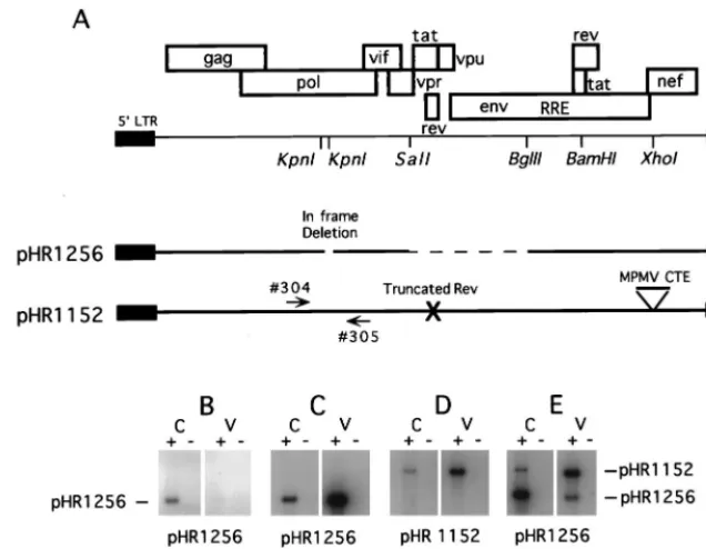
Transfection of cells with pHR1256 alone yields an nuclear
form of Gag-Pol RNA that can be detected in total cellular
RNA preparations despite the fact that no p24 is produced
(Fig. 4B). To determine if this RNA could be packaged in the
absence of Rev, we cotransfected pHR1256 together with
pHR1152, which has been previously described as pRev(
2
)
MPMV (10). pHR1152 lacks a functional rev gene but
ex-presses Gag and Gag-Pol due to the presence of the MPMV
CTE. Since pHR1256 has a 330-bp deletion in pol but
pHR1152 does not, RNA produced from the two plasmids can
be readily distinguished by using PCR primers which span the
deleted region (Fig. 4A).
Analysis of cellular and viral particle-derived RNA from the
cotransfection of pHR1256 and pHR1152 is shown in Fig. 4E.
As controls, cells were also transfected with pHR1256 alone
(Fig. 4B), with pHR1256, pCMVrev and pCMVtat (Fig. 4C),
or with pHR1152 alone (Fig. 4D). Quantitation of the results
of this experiment is given in Table 3.
The control experiments demonstrated that RNA derived
from pHR1256 could be packaged as efficiently as RNA
de-rived from a proviral construct when Rev was supplied in trans
from a second plasmid. About 17% of the pHR1256-specific
RNA was packaged, compared to 11% of the RNA produced
in the pHR1152 transfection. However, when pHR1256 and
pHR1152 were cotransfected, RNA produced from pHR1152
was efficiently packaged, while RNA derived from pHR1256
was not. In this case, only 0.2% of the pHR1256 RNA was
packaged, compared to 9.4% for the pHR1152-derived RNA.
Since RNA from pHR1256 remains in the nucleus under these
conditions (27), we can conclude that an RNA residing in the
nucleus because of a block to its transport cannot be packaged.
DISCUSSION
[image:5.612.57.549.84.140.2]Our results demonstrate that the HIV TAR element is
re-quired for efficient encapsidation of HIV RNA into virus
par-ticles. The region of TAR necessary for packaging maps to the
lower portion of the TAR stem. It appears that the integrity of
this stem is the important factor, as a series of compensatory
TABLE 2. RNA packaging measured as a ratio of RNA to p24
Plasmid(s) pmol of particlep24/plate fmol of particleRNA/plate mmol of particle RNA/mol of particle p24 NormalizedRNA/p24
pHR969 (provirus)
9.1
14.3
1.6
100
pHR1256
1
pCMVrev
3.9
7.6
1.9
119
pGAGPOL-RRE-r
1
pCMVrev
12.2
0.2
0.02
1.3
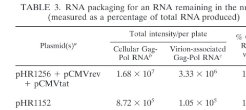
TABLE 3. RNA packaging for an RNA remaining in the nucleus
(measured as a percentage of total RNA produced)
Plasmid(s)a
Total intensity/per plate % Gag-Pol RNA in
virions Cellular
Gag-Pol RNAb Virion-associatedGag-Pol RNAc
pHR1256
1
pCMVrev
1
pCMVtat
1.68
3
10
7
3.33
3
10
616.6
pHR1152
8.72
3
10
51.05
3
10
510.7
pHR1152
1
pHR1256
1.46
3
10
6
1.52
3
10
59.4
1.74
3
10
73.46
3
10
40.2
aValues for pHR1256 alone were not determined since no virion particles
were produced.
bThe measured intensities of the bands in the lanes marked C in Fig. 4B were
multiplied by a correction factor of 360 as described in Materials and Methods.
cThe measured intensities of the bands in the lanes marked V in Fig. 4B were
multiplied by a correction factor of 5 as described in Materials and Methods.
on November 9, 2019 by guest
http://jvi.asm.org/
[image:5.612.310.550.572.679.2]on November 9, 2019 by guest
http://jvi.asm.org/
double mutants, with changed stem sequence but unaltered
two-dimensional structure, were still packaged. In contrast,
mutants with changes in regions of TAR which are critically
important for transactivation (the TAR bulge or loop) had
little effect on packaging. These results are consistent with a
previously published genetic study that demonstrated the
ne-cessity of base pairing in the lower portion of the TAR stem for
optimal HIV replication (39). That study did not define the
actual role played by the lower portion of the stem, although it
did suggest that the lower stem structure might be necessary
for transcription, RNA packaging, or reverse transcription.
Other genetic studies have also suggested functions of TAR
that go beyond its role in transactivation (15, 29–31).
Further-more, a recent report demonstrated decreased (about 3.5-fold)
packaging in an HIV vector mutant containing a total deletion
of TAR (48).
Previously published reports from other laboratories suggest
that additional packaging determinants other than TAR are
present in R-U5 (16, 47, 48, 63). The data suggest that at least
two distinct stem loop structures exist in the R-U5 region and
that both of these structures are needed for efficient
encapsi-dation of HIV RNA. These results, together with our present
data, and data showing the importance of regions mapping
downstream of the primer binding site (SL1 to SL4) in RNA
packaging, explain our previous observation that at least 1 kb
of HIV RNA must be included in an HIV-1 vector for efficient
packaging (8).
[image:7.612.142.460.73.320.2]Mutational studies, such as the ones presented here, that
measure incorporation of RNA into particles as an endpoint
are subject to several general interpretations. Each identified
packaging element could be a unique domain for the binding
of a host or viral factor that somehow mediates the
encapsi-dation of the RNA. Alternatively, all of the elements would not
be involved in the binding to specific factors. Many would
provide only the correct structural context for RNA folding,
thus exposing the true packaging element(s) (i.e., the one[s]
that binds the important host or viral factor[s]). It could also be
that the TAR mutants somehow prevent the RNA from being
delivered to the correct packaging compartment, though this
seems less likely since none of the TAR mutants affect p24
production or RNA stability to any significant extent. We
therefore cannot conclude that the TAR stem is necessary for
the binding of a specific host or viral packaging factor. The
stem structure might simply facilitate the formation of a
cor-rect structure elsewhere in the RNA. However, binding of NC
to SL3 does not require other viral RNA sequences (5, 17, 42).
A recently published paper describes a novel model for the
structure of the 5
9
end of HIV RNA (33). The model is based
FIG. 4. Lack of RNA packaging from a plasmid that expresses RNA which remains in the nucleus. (A) Schematic representation of the plasmids used in this study. pHR1256 contains a small in-frame deletion in pol as well as a larger deletion in the midportion of the genome. Because of the latter deletion, Rev coexpression is required for the nucleocytoplasmic transport of the Gag-Pol mRNA. pHR1152 is a full-length proviral clone with a mutated nonfunctional rev gene. The plasmid also contains the MPMV CTE inserted into the XhoI site of the nef gene. Gag-Pol expression from this plasmid is rev independent. Gag-Pol RNA from the two plasmids can be readily distinguished by using PCR primers 304 and 305 (arrows), which flank the KpnI sites in pol. (B) RT-PCR analysis of total poly(A)-selected cellular (C) and virion RNA (V) isolated from CMT3-COS cells transiently transfected with the indicated plasmids. The reactions for all RNA samples were performed in the presence (1) and the absence (2) of reverse transcriptase. Both primers used to detect the Gag-Pol RNA anneal in pol as shown in panel A. Quantitation of the data is given in Table 3.FIG. 3. Packaging from plasmids expressing RNA with nucleotide changes in TAR. (A) Schematic representations of the TAR regions from 12 plasmids. Each mutant plasmid was derived from pHR1486. The changed nucleotides are boxed, and each arrow points to the new sequence. (B) Quantitation of RNA packaging. The
rev-containing cell line A5.6 was transfected with either pHR1486 as a control or with one of the indicated plasmids. Total RNA, total p24, or particleRNA/particle
24 (packaging) was determined for each transfection as described in Materials and Methods. The measured values were normalized to the values obtained for the control, pHR1486, which were set to 100. The combined results from multiple experiments are shown.
on November 9, 2019 by guest
http://jvi.asm.org/
primarily on electron microscopic data examining dimer RNA
extracted from HIV-1 particles. In contrast to RNA extracted
from other retroviruses, the results in this report show the
dimer linkage region of HIV-1 RNA as a loop of about 325
nucleotides. The 5
9
ends of the RNA are not free. It is thus
proposed that the dimer linkage region consists of two parts:
(i) an intramolecular base pairing of SL1 and (ii) an
intramo-lecular base pairing of the R-U5 region and/or TAR which
creates the circular loop between the 5
9
end and SL1. If this
model is correct, then the role of the bottom part of the TAR
stem in packaging could be to help create this novel bipartite
dimer linkage which may be necessary for efficient packaging.
We have also shown that HIV RNAs that are normally
retained within the nucleus in the absence of Rev cannot be
packaged into particles unless Rev is provided in trans or the
CTE from MPMV is provided in cis. Since the CTE can fully
substitute for the RRE to enable RNA packaging, it is unlikely
that the RRE contains a direct packaging determinant as has
been proposed in some earlier studies (56). Rather, Rev/RRE
or the CTE is probably necessary to allow the RNA to exit
from the nucleus so that it can be recognized by the packaging
factors which presumably reside in the cytoplasm.
RNA export mediated by Rev/RRE or CTE may utilize
export pathways different from the pathways that normally
mediate the export of spliced cellular mRNA (see reference 26
for a review). It will therefore be of interest to determine if
transport utilizing these elements is a critical factor in
packag-ing or if RNA exported by the normal mRNA export pathway
can also be packaged. Since all of the RNAs examined in the
present study required Rev/RRE or CTE for export, it will be
necessary to create other substrates before this question can be
answered.
ACKNOWLEDGMENTS
We thank Joy Niesen for expert tissue culture assistance.
This work was supported by NIH grants AI34721 to M.-L.H. and
AI38186 to D.R. and by the Charles H. Ross, Jr., and Myles H. Thaler
Endowments at the University of Virginia.
REFERENCES
1. Aldovini, A., and R. Young. 1990. Mutations of RNA and protein sequences involved in human immunodeficiency virus type 1 packaging result in pro-duction of noninfectious virus. J. Virol. 64:1920–1926.
2. Arrigo, S. J., S. Weitsman, J. A. Zack, and I. S. Chen. 1990. Characterization and expression of novel singly spliced RNA species of human immunodefi-ciency virus type 1. J. Virol. 64:4585–4588.
3. Arya, S. K., C. Guo, S. F. Josephs, and F. Wong-Staal. 1985. Trans-activator gene of human T-lymphotropic virus type III (HTLV-III). Science 229:69– 73.
4. Baudin, F., R. Marquet, C. Isel, J. L. Darlix, B. Ehresmann, and C. Ehres-mann.1993. Functional sites in the 59region of human immunodeficiency virus type 1 RNA form defined structural domains. J. Mol. Biol. 229:382– 397.
5. Berglund, J. A., B. Charpentier, and M. Rosbash. 1997. A high affinity binding site for the HIV-1 nucleocapsid protein. Nucleic Acids Res. 25:1042– 1049.
6. Berkowitz, R., J. Fisher, and S. P. Goff. 1996. RNA packaging, p. 177–218. In H.-G. Krausslich (ed.), Morphogenesis and maturation of retroviruses. Springer, Berlin, Germany.
7. Berkowitz, R. D., and S. P. Goff. 1994. Analysis of binding elements in the human immunodeficiency virus type 1 genomic RNA and nucleocapsid pro-tein. Virology 202:233–246.
8. Berkowitz, R. D., M. L. Hammarskjold, C. Helga Maria, D. Rekosh, and S. P. Goff.1995. 59regions of HIV-1 RNAs are not sufficient for encapsida-tion: implications for the HIV-1 packaging signal. Virology 212:718–723. 9. Berkowitz, R. D., A. Ohagen, S. Hoglund, and S. P. Goff. 1995. Retroviral
nucleocapsid domains mediate the specific recognition of genomic viral RNAs by chimeric Gag polyproteins during RNA packaging in vivo. J. Virol. 69:6445–6456.
10. Bray, M., S. Prasad, J. W. Dubay, E. Hunter, K. T. Jeang, D. Rekosh, and M. L. Hammarskjo¨ld.1994. A small element from the Mason-Pfizer monkey virus genome makes human immunodeficiency virus type 1 expression and
replication Rev-independent. Proc. Natl. Acad. Sci. USA 91:1256–1260. 11. Bukrinsky, M. I., S. Haggerty, M. P. Dempsey, N. Sharova, A. Adzhubel, L.
Spitz, P. Lewis, D. Goldfarb, M. Emerman, and M. Stevenson.1993. A nuclear localization signal within HIV-1 matrix protein that governs infec-tion of non-dividing cells. Nature 365:666–669.
12. Clever, J., C. Sassetti, and T. G. Parslow. 1995. RNA secondary structure and binding sites for gag gene products in the 59packaging signal of human immunodeficiency virus type 1. J. Virol. 69:2101–2109.
13. Clever, J. L., and T. G. Parslow. 1997. Mutant human immunodeficiency virus type 1 genomes with defects in RNA dimerization or encapsidation. J. Virol. 71:3407–3414.
14. Clever, J. L., M. L. Wong, and T. G. Parslow. 1996. Requirements for kissing-loop-mediated dimerization of human immunodeficiency virus RNA. J. Virol. 70:5902–5908.
15. Das, A. T., B. Klaver, and B. Berkhout. 1998. The 59and 39TAR elements of human immunodeficiency virus exert effects at several points in the virus life cycle. J. Virol. 72:9217–9223.
16. Das, A. T., B. Klaver, B. I. Klasens, J. L. van Wamel, and B. Berkhout. 1997. A conserved hairpin motif in the R-U5 region of the human immunodefi-ciency virus type 1 RNA genome is essential for replication. J. Virol. 71: 2346–2356.
17. De Guzman, R. N., Z. R. Wu, C. C. Stalling, L. Pappalardo, P. N. Borer, and M. F. Summers.1998. Structure of the HIV-1 nucleocapsid protein bound to the SL3 psi-RNA recognition element. Science 279:384–388.
18. Delchambre, M., D. Gheysen, D. Thines, C. Thiriart, E. Jacobs, E. Verdin, M. Horth, A. Burny, and F. Bex.1989. The GAG precursor of simian immunodeficiency virus assembles into virus-like particles. EMBO J. 8:2653– 2660.
19. Dundr, M., G. H. Leno, M.-L. Hammarskjo¨ld, D. Rekosh, C. Helga-Maria, and M. O. J. Olson.1995. The roles of nuclear structure and function in the subcellular location of the HIV-1 Rev protein. J. Cell Sci. 108:2811–2823. 20. Ernst, R., M. Bray, D. Rekosh, and M.-L. Hammarskjo¨ld. 1997. A structured
retroviral RNA element that mediates nucleocytoplasmic export of intron-containing RNA. Mol. Cell. Biol. 17:135–144.
21. Freed, E. O., G. Englund, and M. A. Martin. 1995. Role of the basic domain of human immunodeficiency virus type 1 matrix in macrophage infection. J. Virol. 69:3949–3954.
22. Gallay, P., S. Swingler, C. Aiken, and D. Trono. 1995. HIV-1 infection of nondividing cells: C-terminal tyrosine phosphorylation of the viral matrix protein is a key regulator. Cell 80:379–388.
23. Gerard, R. D., and Y. Gluzman. 1985. New host cell system for regulated simian virus 40 DNA replication. Mol. Cell. Biol. 5:3231–3240.
24. Gheysen, D., E. Jacobs, F. de Foresta, C. Thiriart, M. Francotte, D. Thines, and M. De Wilde.1989. Assembly and release of HIV-1 precursor Pr55gag virus-like particles from recombinant baculovirus-infected insect cells. Cell 59:103–112.
25. Gorelick, R. J., S. M. Nigida, J. R. Bess, L. O. Arthur, L. E. Henderson, and A. Rein.1990. Noninfectious human immunodeficiency virus type 1 mutants deficient in genomic RNA. J. Virol. 64:3207–3211.
26. Hammarskjo¨ld, M.-L. 1997. Regulation of retroviral RNA export. Semin. Cell Dev. Biol. 8:83–90.
27. Hammarskjo¨ld, M.-L., J. Heimer, B. Hammarskjo¨ld, I. Sangwan, L. Albert, and D. Rekosh.1989. Regulation of human immunodeficiency virus env expression by the rev gene product. J. Virol. 63:1959–1966.
28. Hammarskjo¨ld, M.-L., S.-C. Wang, and G. Klein. 1986. High-level expres-sion of the Epstein-Barr virus EBNA1 protein in CV1 cells and human lymphoid cells using a SV40 late replacement vector. Gene 43:41–50. 29. Harrich, D., C. Hsu, E. Race, and R. B. Gaynor. 1994. Differential growth
kinetics are exhibited by human immunodeficiency virus type 1 TAR mu-tants. J. Virol. 68:5899–5910.
30. Harrich, D., G. Mavankal, S. A. Mette, and R. B. Gaynor. 1995. Human immunodeficiency virus type 1 TAR element revertant viruses define RNA structures required for efficient viral gene expression and replication. J. Vi-rol. 69:4906–4913.
31. Harrich, D., C. Ulich, and R. B. Gaynor. 1996. A critical role for the TAR element in promoting efficient human immunodeficiency virus type 1 reverse transcription. J. Virol. 70:4017–4027.
32. Harrison, G. P., and A. M. Lever. 1992. The human immunodeficiency virus type 1 packaging signal and major splice donor region have a conserved stable secondary structure. J. Virol. 66:4144–4153.
33. Hoglund, S., A. Ohagen, J. Goncalves, A. T. Panganiban, and D. Gabuzda. 1997. Ultrastructure of HIV-1 genomic RNA. Virology 233:271–279. 34. Jeang, K. T. 1998. Tat, Tat-associated kinase, and transcription. J. Biomed.
Sci. 5:24–27.
35. Jeang, K. T., D. R. Rawlins, P. J. Rosenfeld, J. H. Shero, T. J. Kelly, and G. S. Hayward.1987. Multiple tandemly repeated binding sites for cellular nuclear factor 1 that surround the major immediate-early promoters of simian and human cytomegalovirus. J. Virol. 61:1559–1570.
36. Jones, K. A. 1997. Taking a new TAK on tat transactivation. Genes Dev. 11:2593–2599. (Comment.)
37. Kaye, J. F., J. H. Richardson, and A. M. Lever. 1995. cis-acting sequences
on November 9, 2019 by guest
http://jvi.asm.org/
involved in human immunodeficiency virus type 1 RNA packaging. J. Virol. 69:6588–6592.
38. Kim, H.-J., K. Lee, and J. J. O’Rear. 1994. A short sequence upstream of the 59major splice site is important for encapsidation of human immunodefi-ciency virus type 1 genomic RNA. J. Virol. 198:336–340.
39. Klaver, B., and B. Berkhout. 1994. Evolution of a disrupted TAR RNA hairpin structure in the HIV-1 virus. EMBO J. 13:2650–2659.
40. Laughrea, M., and L. Jette. 1994. A 19-nucleotide sequence upstream of the 59major splice donor is part of the dimerization domain of human immu-nodeficiency virus 1 genomic RNA. Biochemistry 33:13464–13474. 41. Levin, J. G., P. M. Grimley, J. M. Ramseur, and I. K. Berezesky. 1974.
Deficiency of 60 to 70S RNA in murine leukemia virus particles assembled in cells treated with actinomycin D. J. Virol. 14:152–161.
42. Lochrie, M. A., S. Waugh, J. D. Pratt, J. Clever, T. G. Parslow, and B. Polisky.1997. In vitro selection of RNAs that bind to the human immuno-deficiency virus type-1 gag polyprotein. Nucleic Acids Res. 25:2902–2910. 43. Luban, J., and S. P. Goff. 1994. Mutational analysis of cis-acting packaging
signals in human immunodeficiency virus type 1 RNA. J. Virol. 68:3784– 3793.
44. Mann, R., R. C. Mulligan, and D. Baltimore. 1983. Construction of a retro-virus packaging mutant and its use to produce helper-free defective retrovi-rus. Cell 33:153–159.
45. Marquet, R., J. C. Paillart, E. Skripkin, C. Ehresmann, and B. Ehresmann. 1994. Dimerization of human immunodeficiency virus type 1 RNA involves sequences located upstream of the splice donor site. Nucleic Acids Res. 22:145–151.
46. McBride, M. S., and A. T. Panganiban. 1996. The human immunodeficiency virus type 1 encapsidation site is a multipartite RNA element composed of functional hairpin structures. J. Virol. 70:2963–2973.
47. McBride, M. S., and A. T. Panganiban. 1997. Position dependence of func-tional hairpins important for human immunodeficiency virus type 1 RNA encapsidation in vivo. J. Virol. 71:2050–2058.
48. McBride, M. S., M. D. Schwartz, and A. T. Panganiban. 1997. Efficient encapsidation of human immunodeficiency virus type 1 vectors and further characterization of cis elements required for encapsidation. J. Virol. 71: 4544–4554.
49. Mujeeb, A., J. L. Clever, T. M. Billeci, T. L. James, and T. G. Parslow. 1998. Structure of the dimer initiation complex of HIV-1 genomic RNA. Nat. Struct. Biol. 5:432–436.
50. Muriaux, D., P. Fosse, and J. Paoletti. 1996. A kissing complex together with a stable dimer is involved in the HIV- 1Lai RNA dimerization process in vitro. Biochemistry 35:5075–5082.
51. Nash, M. A., M. K. Meyer, G. L. Decker, and R. B. Arlinghaus. 1993. A subset of Pr65gagis nucleus associated in murine leukemia virus-infected
cells. J. Virol. 67:1350–1356.
52. Nermut, M. V., and D. J. Hockley. 1996. Comparative morphlogy and struc-tural classification of retroviruses, p. 1–24. In H.-G. Krausslich (ed.), Mor-phogenesis and maturation of retroviruses. Springer, Berlin, Germany. 53. Paillart, J. C., R. Marquet, E. Skripkin, B. Ehresmann, and C. Ehresmann.
1994. Mutational analysis of the bipartite dimer linkage structure of human immunodeficiency virus type 1 genomic RNA. J. Biol. Chem. 269:27486– 27493.
54. Pollard, V. W., and M. H. Malim. 1998. The HIV-1 Rev protein. Annu. Rev. Microbiol. 52:491–532.
55. Poon, D. T., G. Li, and A. Aldovini. 1998. Nucleocapsid and matrix protein contributions to selective human immunodeficiency virus type 1 genomic RNA packaging. J. Virol. 72:1983–1993.
56. Richardson, J. H., L. A. Child, and A. M. Lever. 1993. Packaging of human immunodeficiency virus type 1 RNA requires cis-acting sequences outside the 59leader region. J. Virol. 67:3997–4005.
57. Schliephake, A. W., and A. Rethwilm. 1994. Nuclear localization of foamy virus Gag precursor protein. J. Virol. 68:4946–4954.
58. Schwartz, S., B. K. Felber, E. M. Fenyo, and G. N. Pavlakis. 1990. Env and Vpu proteins of human immunodeficiency virus type 1 are produced from multiple bicistronic mRNAs. J. Virol. 64:5448–5456.
59. Skripkin, E., J. C. Paillart, R. Marquet, B. Ehresmann, and C. Ehresmann. 1994. Identification of the primary site of the human immunodeficiency virus type 1 RNA dimerization in vitro. Proc. Natl. Acad. Sci. USA 91:4945–4649. 60. Smith, A. J., M. I. Cho, M. L. Hammarskjo¨ld, and D. Rekosh. 1990. Human immunodeficiency virus type 1 Pr55gagand Pr160gag-polexpressed from a
simian virus 40 late replacement vector are efficiently processed and assem-bled into viruslike particles. J. Virol. 64:2743–2750.
61. Smith, A. J., N. Srinivasakumar, M. L. Hammarskjo¨ld, and D. Rekosh. 1993. Requirements for incorporation of Pr160gag-polfrom human
immunodefi-ciency virus type 1 into virus-like particles. J. Virol. 67:2266–2275. 62. Sorge, J., D. Wright, V. D. Erdman, and A. E. Cutting. 1984. Amphotropic
retrovirus vector system for human cell gene transfer. Mol. Cell. Biol. 4:1730–1737.
63. Vicenzi, E., D. S. Dimitrov, A. Engelman, T. S. Migone, D. F. Purcell, J. Leonard, G. Englund, and M. A. Martin.1994. An integration-defective U5 deletion mutant of human immunodeficiency virus type 1 reverts by elimi-nating additional long terminal repeat sequences. J. Virol. 68:7879–7890. 64. Zhang, Y., and E. Barklis. 1995. Nucleocapsid protein effects on the
speci-ficity of retrovirus RNA encapsidation. J. Virol. 69:5716–5722. (Erratum, 71:5712, 1997.)
65. Zolotukhin, A. S., A. Valentin, G. N. Pavlakis, and B. K. Felber. 1994. Continuous propagation of RRE(2) and Rev(2)RRE(2) human immuno-deficiency virus type 1 molecular clones containing a cis-acting element of simian retrovirus type 1 in human peripheral blood lymphocytes. J. Virol. 68:7944–7952.
on November 9, 2019 by guest
http://jvi.asm.org/