Copyright © 2000, American Society for Microbiology. All Rights Reserved.
Increased Expression and Immunogenicity of Sequence-Modified
Human Immunodeficiency Virus Type 1
gag
Gene
JAN
ZURMEGEDE, MIN-CHAO CHEN, BARBARA DOE, MARY SCHAEFER, CATHERINE E. GREER,
MARK SELBY, GILLIS R. OTTEN,
ANDSUSAN W. BARNETT*
Chiron Corporation, Emeryville, California 94608
Received 2 September 1999/Accepted 20 December 1999
A major challenge for the next generation of human immunodeficiency virus (HIV) vaccines is the induction
of potent, broad, and durable cellular immune responses. The structural protein Gag is highly conserved
among the HIV type 1 (HIV-1) gene products and is believed to be an important target for the host
cell-mediated immune control of the virus during natural infection. Expression of Gag proteins for vaccines has
been hampered by the fact that its expression is dependent on the HIV Rev protein and the Rev-responsive
element, the latter located on the
env
transcript. Moreover, the HIV genome employs suboptimal codon usage,
which further contributes to the low expression efficiency of viral proteins. In order to achieve high-level
Rev-independent expression of the Gag protein, the sequences encoding HIV-1
SF2p55
Gagwere modified
extensively. First, the viral codons were changed to conform to the codon usage of highly expressed human
genes, and second, the residual inhibitory sequences were removed. The resulting modified
gag
gene showed
increases in p55
Gagprotein expression to levels that ranged from 322- to 966-fold greater than that for the
native gene after transient expression of 293 cells. Additional constructs that contained the modified
gag
in
combination with modified
protease
coding sequences were made, and these showed high-level Rev-independent
expression of p55
Gagand its cleavage products. Density gradient analysis and electron microscopy further
demonstrated that the modified
gag
and
gagprotease
genes efficiently expressed particles with the density and
morphology expected for HIV virus-like particles. Mice immunized with DNA plasmids containing the modified
gag
showed Gag-specific antibody and CD8
ⴙcytotoxic T-lymphocyte (CTL) responses that were inducible at
doses of input DNA 100-fold lower than those associated with plasmids containing the native
gag
gene. Most
importantly, four of four rhesus monkeys that received two or three immunizations with modified
gag
plasmid
DNA demonstrated substantial Gag-specific CTL responses. These results highlight the useful application of
modified
gag
expression cassettes for increasing the potency of DNA and other gene delivery vaccine
ap-proaches against HIV.
The induction of long-lasting, potent humoral and cellular
immune responses will be important for an effective human
immunodeficiency virus (HIV) vaccine. Data from
HIV-in-fected patients, and in particular from long-term
nonprogres-sors, have shown that viral structural genes can elicit
substan-tial immune responses. Gag-specific CD8
⫹cytotoxic T
lymphocytes (CTL) have been shown to be important in
con-trolling virus load during acute infection (4, 21) as well as
during the asymptomatic stages of the infection (20, 24).
More-over, a strong Gag-specific CTL response appears to correlate
inversely with the viral load of HIV-1-infected patients (7). In
addition, studies of exposed but uninfected prostitutes indicate
that Gag-specific CTL may be involved in protection against
the establishment of a persistent HIV type 1 (HIV-1) infection
(28). Combined, these studies provide convincing evidence
that immune responses directed against HIV Gag proteins may
be an important component of an effective HIV vaccine. The
usefulness of Gag immunogens for vaccines is further indicated
by the fact that the protein is relatively conserved among
di-verse HIV strains and subtypes, and cross-clade CTL
recogni-tion directed against Gag-specific targets has been well
docu-mented (2, 3, 11, 23).
Immunization with naked DNA or recombinant virus
in-duces both antibody and CTL responses and has been shown to
be an efficient method of eliciting protective immune
re-sponses against a broad range of pathogens in animal studies
(10). However, the potency of current gene delivery methods
such as naked-DNA and viral vectors must be improved to
induce adequately robust responses for protection in primates
(1). One means to achieve this may be through increasing the
expression efficiency of encoded HIV antigens. The poor
ex-pression of the HIV structural genes in recombinant vectors is
caused by a strong Rev dependency that allows efficient
ex-pression only in the presence of the viral Rev protein (25, 30).
The translation efficiency and stability of
gag
transcripts are
further decreased by the presence of a relatively high AU
content and destabilizing AUUUA motifs (inhibitory
se-quences [INS]). In previous studies, inactivation of these INS
enabled the Rev-independent expression of HIV-1
gag
(29),
but these modifications reduced the approximate AT content
of the
gag
gene only from 56 to 50%. Elevated percentages of
AU in human mRNAs have been shown to result in instability,
increased turnover, and low expression levels (15). These
find-ings suggest that further reductions of the AT content of the
gag
gene could result in improved mRNA stability and
in-creased protein expression. In support of this, it has been
shown that highly expressed human genes employ codon usage
patterns different from those used by HIV genomes. For highly
expressed genes, G or C is generally preferred over A or T.
Furthermore, changes in the codon usage of HIV-1
env
to
those employed by highly expressed human codons resulted in
increased Rev-independent expression (14).
In order to achieve high-level Rev-independent expression
* Corresponding author. Mailing address: Chiron Corporation,
Mailstop 4.3, 4560 Horton St., Emeryville, CA 94608. Phone: (510)
923-7565. Fax: (510) 923-2586. E-mail: [email protected].
2628
on November 9, 2019 by guest
http://jvi.asm.org/
ditionally, the inclusion of modified protease-coding sequences
in the modified
gag
resulted in high-level Rev-independent
expression, processing of the Gagprotease polyprotein, and the
production of virus-like particles (VLP) with the morphologies
of both immature and mature HIV-1 virions.
MATERIALS AND METHODS
gagandgagproteaseplasmids.The native sequences coding for the 502 amino acids (aa) of HIV-1SF2p55Gag(GenBank accession no. K02007) were modified to change the codon usage to that utilized by highly expressed human genes as described recently for HIV-1MNgp120 (14). In addition, regions with INS were further inactivated without altering the reading frame for the p55Gagnucleic acid sequence. The resulting modified HIV-1SF2gagencoded a p55Gagprotein with three amino acid changes (Asn377Thr, Ile403Thr, and Lys405Arg); the resulting amino acid sequence conformed to the sequences for other HIV-1 subtype B Gag proteins in the HIV sequence database (Los Alamos National Laboratory; http://hiv-web.lanl.gov/cgi-bin/hivDB3/public/wdb/ssampublic) (GenBank acces-sion no. AF201927). To further enhance the translation efficiency of the modified gag, an optimal consensus sequence for the initiation of translation (GCCACC AUGG) was employed (22). The resulting 1,527-bp gene cassette included the SalI andEcoRI cloning sites and was constructed synthetically by the Midland Certified Reagent Company (Midland, Tex.). This modifiedgagsequence was cloned into theSalI andEcoRI restriction sites of the eukaryotic expression vector pCMVKm2 that employs the cytomegalovirus (CMV) immediate-early en-hancer/promoter and bGH terminator (Chiron Corporation, Emeryville, Calif.) (6), resulting in the plasmid pCMVKm2.GagMod.SF2. For the comparison of expression efficiencies between the modified and the native HIV-1SF2gag ex-pression cassettes, three different vectors containing the native p55Gagcoding se-quence were used, pCMV6ap55GagPRE, pCMVKm2p55GagPRE, and pCMVLinkPREp55Gag (Chiron). The pCMVLink plasmid differs from pCM-VKm2 only in its multiple cloning site. All of these use the CMV immediate-early enhancer/promoter and include the hepatitis B virus posttranscriptional regula-tory element (PRE) (9, 16–18) to partially overcome the Rev dependency ofgag. This was demonstrated by transfection experiments using the native HIV-1SF2
gaggene with and without PRE. The expression of p55Gagwas clearly improved using PRE over that using thegaggene only (S. W. Barnett, unpublished data). For the construction of thegagproteaseexpression cassettes, modifications were made in the same manner as that described forgagup to the⫺1 frameshift region of thepolgene. The sequence from there to thegaggene’s stop codon was unaltered. The sequences from thegagstop codon to the codons for first 26 aa of the reverse transcriptase were codons either optimized with subsequent INS inactivation as described above (GP1; GenBank accession no. AF202464) or modified by INS inactivation alone (GP2; GenBank accession no. AF202465). Both versions of thegagproteasecassette were cloned into the pCMVKm2 vector as described above for the modifiedgagto yield the plasmids pCMVKm2. GagProtMod.SF2 (GP1) and pCMVKm2.GagProtMod.SF2 (GP2).
In vitro expression assays.Plasmid DNA was purified using endotoxin-free columns (Qiagen, Valencia, Calif.). African green monkey kidney (COS-7; Eu-ropean Culture Collections Organization no. 87021302), human kidney (293; American Type Tissue Collection [ATCC; Atlanta, Ga.] no. 45504), and human rhabdomyosarcoma (RD; ATCC no. CCL-136) cells were plated 1 day prior to transfection at a density of 5⫻105cells per 35-mm-diameter well (Corning). For the transfections, 2g of each plasmid DNA was mixed with the Mirus TransIT-LT1 polyamine transfection reagent (PanVera, Madison, Wis.). The green flu-orescent protein (GFP) reporter gene vector pEGFP (Clontech, Palo Alto, Calif.) was used as a transfection efficiency control in co- and parallel transfec-tions. The cells were incubated with 2 ml of medium per well (for 293 cells, Iscove’s modified Dulbecco’s medium, 10% fetal calf serum [FCS]; for COS-7 and RD cells, Dulbecco’s modified Eagle medium, 10% FCS; Gibco, Rockville, Md.). To estimate the transfection efficiency, GFP-expressing cells were analyzed quantitatively by flow cytometry (Becton Dickinson Immunocytometry Systems,
were collected at 24 and 48 h posttransfection, filtered through a 0.2- m-pore-size filter, and concentrated by ultracentrifugation through a 20% (wt/wt) sucrose cushion for 2 h at 140,000⫻g(24,000 rpm) using a Beckman SW28 rotor. The pellets were then suspended in PBS, loaded on a 20 to 60% sucrose gradient, and centrifuged at 285,000⫻g(40,000 rpm) for 2 h in a Beckman SW41ti rotor. Each gradient was fractionated into 1-ml aliquots, and 10-l aliquots of each fraction were electrophoresed on an SDS–8 to 16% polyacrylamide gel electrophoresis gel (Novex). In addition, 2.5l of the concentrated gradient preload material was also analyzed. The proteins were then transferred to Immobilon P membranes (Millipore) and probed with mouse anti-p24 MAb 76C.5EG at a dilution of 1:2,000.
Electron microscopy.COS-7 or 293 cells (4⫻106) were transfected in 100-mm-diameter dishes (Corning), and cells were harvested at 24 or 48 h posttrans-fection. Cells transfected with vector DNA alone served as negative controls. After two washes with PBS the cells were fixed in 2% glutaraldehyde (Sigma), incubated for 20 min at room temperature, gently scraped from the plate, and transferred into a 15-ml polypropylene tube. The fixed cells were then stained with uranium acetate and lead citrate. Electron microscopy was carried out using a transmission electron microscope (Zeiss; 10c) at⫻50,000 and⫻100,000 mag-nifications.
Animal studies.Female BALB/c and CB6F1 mice, 6 to 8 weeks old, were used for immunogenicity studies. For the first experiment (Fig. 5), four groups of BALB/c mice (n⫽4) were immunized with either modifiedgagplasmid DNA (pCMVKm2.GagMod.SF2) or the native gagplasmid DNA (pCMV Link.Gag.SF2.PRE). The plasmid DNA doses for the different groups were 20, 2, 0.2, and 0.02g in 100l of sterile endotoxin-free saline (Sigma). pCMVKm2 vector DNA was used to maintain the total concentration of DNA in each dose at 20g/100l to control for effects due to the lower concentration of plasmid DNA (2-, 0.2-, and 0.02-g doses). For experiments 2 and 3 shown in Fig. 6 and 7, the mouse strain employed was CB6F1. For experiment 3, plasmid DNA doses were further diluted to include doses as low as 0.0002g.
For the DNA immunization study with rhesus monkeys (Macaca mulatta), four animals were immunized bilaterally in the quadriceps muscles with 1-mg doses of pCMVKm2.GagMod.SF2 plasmid DNA in saline at weeks 0, 4, and 8 and bled at weeks 0, 4, 6, 8, and 10. Animals were maintained at the Southwest Foundation for Biomedical Research (San Antonio, Tex.).
Measurements of antibody responses.Ninety-six-well plates (Corning) were coated with 100l of recombinant HIV-1SF2p24 antigen at a concentration of 2 g per ml in 50 mM borate buffer, pH 9. Sera were diluted 1:25, followed by threefold serial dilutions in dilution buffer containing 1% casein as the blocking reagent. Pooled anti-p24 antibody-positive mouse sera served as both a positive control and an assay standard. The sera were incubated for 50 min at 37°C, washed, and incubated with a 1:22,000 dilution of goat anti-mouse immunoglob-ulin G (IgG)-IgM peroxidase conjugate (Pierce, Rockford, Ill.) for an additional 50 min at 37°C. After the plates were washed, the tetramethylbenzone substrate (Pierce) was added to each well, and the reaction was stopped after 30 min by the addition of 2 N H2SO4. The plates were read on an ELISA reader (312e; Bio-Tek, Winooski, Vt.) at 450 nm with a reference wavelength of 620 nm. The calculated titers are the reciprocal of the dilution of serum at a cutoff optical density of 0.4.
Recombinant vaccinia virus challenge of immunized mice.The recombinant vaccinia virus containing the HIV-1SF2gagandpolgenes (rVVgag-pol) has been described previously (8). Nine (experiment 2, Fig. 6) and 5 (experiment 3, Fig. 7) weeks followinggagDNA immunization, mice were challenged with an intra-peritoneal injection of 107PFU of rVVgag-pol. Five days later spleens were harvested and tested directly for cytolytic activity against Gag peptide-pulsed, 51Cr-labeled tumor target cells or were stimulated with Gag peptide and then stained for intracellular gamma interferon (IFN-␥), as described below. This rVVgag-polchallenge model provides a quantitative measure of CD8⫹T-cell function (G. Otten, unpublished data).
CTL assays.Spleen cells were tested for cytolytic activity in a 4-h51Cr release assay using51Cr-labeled SVBALB (H-2d) or RMA (H-2b) tumor target cells (5,000 targets per well) that had been pulsed for 1 h with a 1-g/ml concentration
on November 9, 2019 by guest
of the H-2Kd-binding HIV-1 Gag peptide p7g (8) or the control HIV-1 Gag peptide pgagb(12, 26). After 4 h of incubation, 50l of culture supernatants was transferred to Lumaplates (Packard, Meriden, Conn.), dried, and counted in a Microbeta scintillation counter (Wallac, Gaithersburg, Md.). Percent specific 51Cr release was determined from the formula percent specific51Cr release⫽ (mean experimental release⫺mean spontaneous release)/(maximum release⫺ spontaneous release)⫻100%, where spontaneous release⫽mean counts per minute released from target cells in the absence of spleen cells and maximum release⫽mean counts per minute released from target cells in the presence of 0.1% Triton X-100.
Measurement of Gag-specific IFN-␥-producing CD8ⴙlymphocytes.Spleens were taken 5 days post-rVVgag-polchallenge. Erythrocyte-depleted single cell suspensions were prepared by treatment with Tris-buffered NH4Cl (Sigma). Nucleated spleen cells (1⫻106to 2⫻106) were cultured in duplicate at 37°C in the presence or absence of 10g of p7g peptide/ml. Monensin (Pharmingen, San Diego, Calif.) was added to block cytokine secretion. After 3 to 5 h cells were washed, incubated with anti-CD16/32 (Pharmingen) to block Fc␥receptors, and fixed in 2% (wt/vol) paraformaldehyde and stored overnight at 4°C. The follow-ing day cells were treated with 0.5% (wt/vol) saponin (Sigma) and then incubated with a phycoerythrin (Pharmingen)-conjugated mouse IFN-␥MAb in the pres-ence of 0.1% (wt/vol) saponin. Cells were then washed free of saponin, stained with fluorescein isothiocyanate-conjugated CD8 MAb (Pharmingen), washed, and then analyzed on a FACSCalibur flow cytometer (Becton Dickinson Immu-nocytometry Systems). Samples were cultured and stained in duplicate.
Peptide pools.A set of 51 Gag peptides 20 residues long, overlapping by 10 aa and spanning residues 1 to 496 of HIV-1SF2p55Gag, was synthesized (Chiron Mimotopes, Clayton, Australia). Eight pools were made by mixing 5 to 7 over-lapping peptides. Gag amino acid sequences spanned by the pools were as follows: aa 1 to 80, pool 1; aa 71 to 144, pool 2; aa 135 to 203, pool 3; aa 194 to 263, pool 4; aa 254 to 323, pool 5; aa 314 to 365, pool 6; aa 351 to 430, pool 7; aa 421 to 496, pool 8. A pool of six 20-aa overlapping peptides representing HIV-1SF2Env served as a negative-control pool.
Purification of rhesus macaque PBMC and derivation of B-LCL.Rhesus macaque peripheral blood mononuclear cells (PBMC) were separated from heparinized whole blood on Percoll gradients (5) and cultured at 3⫻106to 3.5⫻ 106per well in 1.5 ml in 24-well plates for 8 days in AIM-V–RPMI 1640 (50:50) culture medium (Gibco) supplemented with 10% FCS. Gag-specific cells were stimulated by the addition of either a Gag peptide pool (13.3g of total peptide/ ml) or autologous PBMC that had been infected with rVVgag-poland cultured in 24-well plates. Recombinant human interleukin-7 (IL-7; 15 ng/ml; R&D Sys-tems, Minneapolis, Minn.) was added at the initiation of culture. Human recom-binant IL-2 (20 IU/ml; Proleukin; Chiron) was added on days 1, 3, and 6. For the derivation of stable rhesus B-lymphoblastoid cell lines (B-LCL), PBMC were exposed to herpesvirus papio-containing culture supernatant from the S594 cell line (13, 27) in the presence of 1g of cyclosporine (Sigma)/ml.
Rhesus macaque CTL assay.Autologous B-LCL were labeled overnight with Na251CrO4(NEN, Boston, Mass.; 25Ci per 106B-LCL) and washed. Individual aliquots were then incubated for 1 h with 100g of Gag or Env peptide pool/ml. Peptide-pulsed,51Cr-labeled B-LCL were added (2,500 per round-bottom well) to duplicate wells containing threefold serial dilutions of cultured PBMC. Un-labeled B cells (105) were added to each well to inhibit nonspecific cytolysis. After 4 h, 50l of culture supernatant was harvested, added to Lumaplates (Packard), and counted with a Microbeta 1450 liquid scintillation counter
(Wal-lac).51Cr released from lysed targets was normalized by the formula percent specific51Cr release⫽100%⫻(mean experimental release⫺mean spontane-ous release)/(maximum release⫺spontaneous release), where spontaneous re-lease⫽mean counts per minute released from target cells in the absence of spleen cells and maximum release⫽mean counts per minute released from target cells in the presence of 0.1% Triton X-100. Data are plotted as percent specific51Cr release versus the culture fraction, where the culture fraction rep-resents the fraction of the culture well (1.5 ml) added to the CTL assay microtiter plate, e.g., a culture fraction of 0.067 equals 1/15 or 0.1 ml of the initial PBMC culture. Serial threefold dilutions of the cultured PBMC were made. In separate experiments, where we have counted the cells recovered from cultures, we have determined the maximal effector cell/target cell ratios to be about 40:1 to 100:1.
RESULTS
Increased in vitro expression efficiency of sequence-modified
HIV-1
SF2gag
gene.
The coding sequences for the HIV-1
SF2gag
gene were modified to conform to the codon usage pattern of
highly expressed human genes and to eliminate residual INS
motifs as described in Materials and Methods. These
modifi-cations resulted in
gag
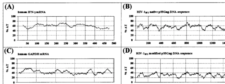
coding sequences with a clear reduction
in overall AT content compared to that of the native
gag
(Fig.
1). In fact, the percentage of A and T nucleotides was reduced
from 56 to 32%, a level more consistent with increased mRNA
stability and translation efficiency (14, 15). The AT content of
the modified
gag
more closely resembled that of the human
glyceraldehyde-3-phosphate dehydrogenase (GAPDH) gene,
which encodes a relatively stable mRNA compared with the
relatively unstable AU-rich human IFN-
␥
mRNA (Fig. 1).
The in vitro expression efficiency of the modified HIV-1
SF2 [image:3.612.78.527.72.241.2]gag
(pCMVKm2.GagMod.SF2) was compared to that of the
native SF2
gag
in a construct (pCMVLink.Gag.SF2.PRE) that
also contained the hepatitis B virus PRE. The pCMVLink.
Gag.SF2.PRE plasmid previously had been found to express
Gag at substantially higher levels than a similar plasmid
con-taining the HIV-1
SF2-derived
gag
gene without the PRE (S. W.
Barnett, unpublished data). The expression levels for these
plasmids were determined in several independent experiments
after transfection of three different cell lines, RD, 293, and
COS-7 (Table 1). Cell supernatants and lysates were tested at
48 and 60 h posttransfection. Gag expression levels were
clearly much higher for the modified
gag
plasmid at all time
points and in all three cell lines tested. The increased
expres-sion was most dramatic in the supernatants of the transfected
human 293 cell line, where expression from the modified
gag
FIG. 1. Comparison of the percentages of A and T nucleotides in genes encoding relatively unstable versus stable mRNA molecules. The human IFN-␥gene and the native HIV-1SF2gagDNA sequences both encode relatively unstable transcripts (A and B) and have an average AT content of 55 to 60%. In contrast, the stable human GAPDH gene and the modified HIV-1SF2gagcoding regions have reduced AT contents of 40 and 30%, respectively (C and D). The calculation of the AT content was done using MacVector software (Oxford Molecular Ltd.); the window size was set at 50.
on November 9, 2019 by guest
http://jvi.asm.org/
was 322- to 966-fold greater than that of the native HIV-1
SF2gag
plasmid tested. The improvement in Gag expression levels
in 293 cell lysates was also apparent, but less so than in the
supernatants, which could be indicative of more-efficient
bud-ding of p55
Gagparticles in cells where expression levels are
elevated. To exclude possible effects on the transfection
effi-ciency depending on the plasmid used, flow cytometry and
direct fluorescence microscope analysis were done in parallel
transfections or by cotransfection using GFP plasmid DNA.
On average, 70% of the cells were transfected using either
method with no differences in transfection efficiency between
the native and modified
gag
plasmids noted (data not shown).
The modified HIV-1
SF2gag
gene encodes p55
GagVLP of the
expected density and morphology.
Supernatants and cell
ly-sates from transfected 293 cells were subjected to immunoblot
and density gradient sedimentation analysis. The results
con-firmed the previous data from the p24 capture assay with
respect to the relative level of p55 expression from the
modi-fied
gag
plasmid. The expected p55
Gagband was detected using
human HIV-1 patient serum (Fig. 2) or an anti-p24 MAb for
the immunostaining (data not shown). Supernatants from 293
cells transfected with the native and modified
gag
genes were
subjected to rate zonal sedimentation to isolate p55
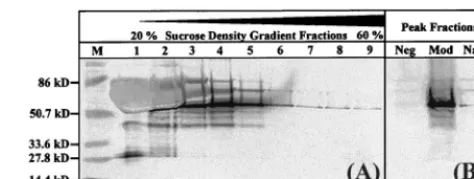
Gagparti-cles of the reported density (32). Gradient fractions were
an-alyzed by p24 capture ELISA (data not shown) and Western
blotting (Fig. 2A) to determine the peak fraction of each
sam-ple. Western blot analysis showed that the p55
Gagband for the
modified Gag expression cassette was stronger than that for
the best native
gag
plasmid (Fig. 2B).
To confirm that VLP were being expressed, COS-7 cells
transfected with pCMVKm2.GagMod.SF2 were harvested at
24 h posttransfection and electron microscopy was performed.
As shown in Fig. 3A, budding and free immature particles
could be observed. These data confirm that the sequence
mod-ifications for the
gag
gene did not adversely affect the p55
Gagparticle assembly or VLP morphology.
Construction and characterization of sequence-modified
gagprotease
gene cassettes.
As a first step in the design of
modified HIV immunogens with increased representation of
Pol-specific epitopes, two different modified
gagprotease
gene
constructs were evaluated for expression and VLP formation.
The protease coding sequences in these constructs were (i)
codon optimized, with subsequent INS inactivation as
de-scribed above for
gag
(GP1), or (ii) modified by INS
inactiva-tion alone (GP2). Like the modified
gag
plasmid, in the
ab-sence of Rev both versions of the modified
gagprotease
exhibited high-level expression of Gag proteins in supernatants
and cell lysates of transiently transfected COS-7 and 293 cell
lines (Table 2). In fact, the expression levels measured in
lysates of 293 cells transfected with the
gagprotease
plasmids
were higher than those seen with the modified
gag
alone. This
result could be partially or wholly attributed to more-efficient
recognition of processed Gag (mostly p24) than of
unproc-essed p55
Gagby the Coulter p24 antigen capture assay, as has
been previously described (29). This apparent increase in p24
expression in cell lysates was not observed in COS-7 cells,
possibly due to lower overall expression of p55
Gagin this cell
line.
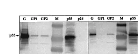
Sucrose density gradient analyses of supernatants from 293
and COS-7 cells transiently transfected with either
gagprotease
or
gag
constructs were performed, and the peak fractions were
subsequently analyzed by Western blotting. The efficiency of
VLP formation varied between the cell lines tested and was
found to be lower for
gagprotease
than for the modified
gag
plasmid (Fig. 4). The levels of VLP formation from the two
gagprotease
constructs in 293 cells were similar (Fig. 4A; GP1
and GP2), but the analysis of the codon-optimized and
INS-inactivated
gagprotease
plasmid, GP1, in COS-7 cells suggested
the production of relatively small amounts of VLP (Fig. 4B).
Polyproteins expressed from both of the modified versions of
gagprotease
were correctly processed by the encoded viral
pro-tease. Bands corresponding to unprocessed p55
Gagand
com-pletely processed p24 were detectable using a MAb specific for
p24 (data not shown) or HIV-1
⫹patient serum (Fig. 4A and B)
(p17 levels were too low to be detected with the HIV
⫹sera
used).
the sucrose density gradient from transfection supernatants of the modifiedgag plasmid. (B) Immunoblot comparing peak fractions collected in the density range expected for HIV-1 VLP after transfection with modified (Mod) or native (Nat) HIV-1SF2gagplasmids. Vector alone (Neg) was used as a negative trans-fection control, and the prestained broad-range molecular weight marker (M) was used as the size standard.
1
Nat
Sup
RD
48
5.6
Mod
Sup
RD
48
66 (12)
Nat
Sup
RD
60
7.8
Mod
Sup
RD
60
70.2 (9)
2
Nat
Lys
RD
60
1.9
Mod
Lys
RD
60
7.8 (4)
1
Nat
Sup
COS-7
48
0.4
Mod
Sup
COS-7
48
33.4 (84)
2
Nat
Sup
COS-7
48
0.4
Mod
Sup
COS-7
48
10 (25)
Nat
Lys
COS-7
48
3
Mod
Lys
COS-7
48
14 (5)
aNat, native (pCMVLink.Gag.SF2.PRE); Mod, modified (pCMVKm2.Gag-Mod.SF2).
bSup, supernatant; Lys, lysate.
on November 9, 2019 by guest
[image:4.612.312.549.72.161.2]Electron microscopic analysis of COS-7 cells transfected
with the two different sequence-modified
gagprotease
con-structs confirmed the results of the sucrose gradient analysis.
COS-7 cells transfected with the codon-optimized and
INS-inactivated version (GP1) showed very little VLP formation
(data not shown) compared to those transfected with the
non-optimized INS-inactivated
gagprotease
(GP2; Fig. 3B). A
pos-sible explanation for this observation is that codon
optimiza-tion of the
protease
coding sequences may have resulted in its
overexpression relative to Gag and the prevention of the
effi-cient budding of particles (19). The GP2 version of
gagprotease
,
in which the INS of the protease-coding region was inactivated
without codon optimization, reduced protease overexpression,
and thus VLP of the mature and immature phenotypes could
be detected.
Increased immunogenicity of the modified
gag
DNA in vivo.
To evaluate and compare the immunogenicities of the
modi-fied and the native
gag
plasmids, mice were immunized
intra-muscularly with plasmid DNA doses titrated from 20 to 0.02
g per mouse. Serum was collected at 4 weeks
postimmuniza-tion and tested in a p24
Gag-specific antibody ELISA. Antibody
responses to Gag could be detected in mice immunized with as
little as 0.2
g of the modified
gag
expression cassette, whereas
the native
gag
cassette was able to induce an antibody response
only at the 20-
g DNA dose (Fig. 5A). This represented the
induction of an antibody response using the modified
gag
at a
single DNA dose 100 fold lower than that necessary for the
native
gag
. In parallel groups of animals, a second dose of
DNA was given at 4 weeks to determine if antibody responses
to the modified
gag
had reached maximal values at the 20-
g
dose and if the lowest DNA dose of 0.02
g could induce an
antibody response after a second immunization. As shown in
Fig. 5B, the Gag-specific antibody titers increased after the
second immunizations for all DNA doses except for the
0.02-
g DNA dose group, which remained negative.
[image:5.612.74.532.74.226.2] [image:5.612.52.292.523.671.2]Measurements of the cellular immune responses following
DNA immunization with the modified
gag
demonstrated a
similar pattern. Gag-specific CTL responses were inducible at
DNA amounts at least 10-fold lower than those necessary with
FIG. 3. Modifiedgagandgagproteaseform VLP in transiently transfected COS-7 cells. Shown are electron micrographs of immature p55GagVLP in COS-7 cells transfected with the modified HIV-1SF2gag(A) and mature (arrows) and immature VLP obtained using the modified HIV-1SF2gagprotease(GP2) (B). Transfected cells were fixed at 24 (gag) or 48 h (gagprotease) posttransfection and subsequently analyzed by electron microscopy as described in Materials and Methods (magnification,⫻100,000). Cells transfected with vector alone (pCMVKm2) served as the negative control (data not shown). [image:5.612.316.546.531.621.2]FIG. 4. Expression and processing of p55Gagpolyproteins in VLP using mod-ified HIV-1gagprotease. Supernatants from transfected cell cultures were col-lected at 60 h posttransfection and centrifuged through 20 to 60% sucrose density gradients. Gradient fractions were collected, and peak fractions were run on an SDS–8 to 16% polyacrylamide gel and analyzed by Western blotting using HIV-1 patient serum as described in Materials and Methods. (A) Peak fractions from 293 cells. Results for the modifiedgag(G) are compared to those for codon-optimized, INS-inactivatedgagprotease(GP1) and for INS-inactivated-onlygagprotease(GP2). (B) Immunoblot comparing peak fractions from trans-fected COS-7 cells using the same plasmids as those described for panel A. Purified HIV-1SF2p24 (Chiron) and baculovirus-derived p55Gagproteins were used as additional controls. Prestained broad-range markers (Bio-Rad) were used as size standards (M).
TABLE 2. In vitro expression from modified
gag
and
gagprotease
plasmids in supernatants and lysates from transiently
transfected cells
Plasmida Material
assayedb Cell line posttransfectionHours ng p24Totalc
Gag
Sup
293
60
760
GagProt (1)
Sup
293
60
380
GagProt (2)
Sup
293
60
320
Gag
Lys
293
60
78
GagProt (1)
Lys
293
60
1,250
GagProt (2)
Lys
293
60
400
Gag
Sup
COS-7
72
40
GagProt (1)
Sup
COS-7
72
150
GagProt (2)
Sup
COS-7
72
290
Gag
Lys
COS-7
72
60
GagProt (1)
Lys
COS-7
72
63
GagProt (2)
Lys
COS-7
72
58
aGag, pCMVKm2.GagMod.SF2; GagProt (1), pCMVKm2.GagProtMod.SF2 (GP1) (gagproteasewith codon optimization and inactivation of INS inprotease); GagProt (2), pCMVKm2.GagProtMod.SF2 (GP2) (gagproteasewith only inacti-vation of INS inprotease).
bSup, supernatant; Lys, lysate.
cRepresentative results from three independent experiments for each cell line tested.
on November 9, 2019 by guest
http://jvi.asm.org/
the native
gag
expression cassette (Fig. 6). Gag-specific CTL
were detectable after a single immunization with a dose of the
modified
gag
plasmid DNA as low as 0.02
g, whereas a dose
of 0.2
g of the native
gag
plasmid was required for the
induc-tion of detectable CTL. In a subsequent study, the modified
gag
plasmid DNA was further diluted (down to 0.2 ng) and used to
immunize additional groups of mice. As shown in Fig. 7,
Gag-specific IFN-
␥
-positive CD8
⫹T cells were scored in mice
re-ceiving as little as 2 ng of the modified
gag
DNA.
Induction of CTL responses in rhesus macaques immunized
with the modified
gag
plasmid.
Based on the increased potency
[image:6.612.131.477.75.206.2]observed in mouse immunizations with the modified
gag
plas-mid DNA, studies with nonhuman primates were initiated.
Four rhesus macaques were given three intramuscular
immu-nizations with 1-mg doses of
gag
plasmid at 4-week intervals.
PBMC were harvested prior to immunization and at 2 weeks
after the second and third immunizations. PBMC were
cul-tured with Gag peptide pools or with rVV
gag-pol
-infected
au-tologous PBMC to stimulate the expansion and differentiation
of CTL and tested against Gag peptide pool-pulsed,
51Cr-labeled, autologous B-LCL targets in 4-h
51Cr release assays.
No Gag-specific cytolysis in PBMC was observed prior to
im-munization (not shown). However, after
gag
DNA
immuniza-tion, all four macaques showed cytolytic activity against
autol-ogous B-LCL pulsed with at least one Gag peptide pool. In
addition, two of the four macaques reacted with two or three
Gag peptide pools (Fig. 8). Percent specific lysis of Gag-pulsed
target cells varied among animals and among pools and
reached as high as 80% at the highest effector cell/target cell
ratio (Fig. 8C). A Gag-specific antibody response (antibody
titer, 164) was detected in one of the four animals 2 weeks after
the second immunization. This animal also had an anamnestic
immune response 2 weeks after the third immunization, with a
fivefold increase of the antibody titer (890). A second animal
in the tibialis anterior muscles with titrated amounts of DNA in 10-fold dilutions from 20g down to 0.002g. Sera were collected at 0 and 4 weeks and tested for HIV-1 p24-specific antibody titers by ELISA as described in Materials and Methods. (A) Comparison of humoral immune responses at different DNA doses using the native and modifiedgagplasmid DNA. Values represent the geometric mean antibody titers and the standard deviations of the midpoint antibody titers for each group. The values in parentheses indicate the percentages of responders (percent seroconversion) in each group. (B) Antibody responses were boosted following a second immunization with the modifiedgagplasmid DNA. Four weeks after the first immunization, additional groups of mice received a second immunization with the same amount of titrated plasmid DNA. Sera collected at weeks 0, 4, and 6 were analyzed by p24 antibody ELISA.FIG. 6. CTL responses in CB6F1 mice after a single immunization with titrated plasmid DNA. Nine weeks after immunization mice were challenged with an intraperitoneal dose of 107PFU of rVVgag-pol. Five days later effector (E) spleen cells were tested for cytolytic activity in a 4-h51Cr release assay using 51Cr-labeled SVBALB tumor target (T) cells (5,000 targets per well) that had been pulsed for 1 h with a 1-g/ml concentration of the H-2Kd-binding HIV-1 Gag peptide p7g (Œ). Target cells pulsed with the negative-control HIV-1 Gag
peptide pgagb(䊐) and major histocompatibility complex-mismatched (H-2b), p7g peptide-pulsed RMA target cells (F) were employed as negative controls.
FIG. 7. Quantification of Gag-specific, IFN-␥-producing CD8⫹T lympho-cytes in mice after a single immunization of titrated modifiedgagplasmid DNA followed by rVVgag-polchallenge. Splenic IFN-␥-positive CD8⫹T lymphocytes specific for the p7g Gag peptide were enumerated by flow cytomerty as described in Materials and Methods. mock, results using spleen cells from naive mice.
on November 9, 2019 by guest
[image:6.612.56.288.423.651.2] [image:6.612.313.549.555.683.2]had a very low titer 2 weeks after the second immunization
(65), which later dropped below the detection level. These
results reflect the induction of robust and relatively broad CTL
responses using the modified
gag
plasmid following DNA
im-munization of nonhuman primates and warrant further study
with these plasmids. This contrasts with previous results in
which weak and transient CTL responses were observed in
only one of four macaques given seven immunizations with
1-mg doses of the pCMVLink.Gag.SF2.PRE plasmid
contain-ing the native HIV-1
SF2gag
(X. Paliard and C. Walker,
un-published data).
DISCUSSION
To increase the potency of HIV-1 DNA vaccines, we
mod-ified the genes coding for HIV-1
SF2Gag and Protease to
over-come Rev dependence and to increase expression levels.
Changes in codon usage to that utilized by highly expressed
human genes in combination with inactivation of INS regions
dramatically increased Gag expression from these constructs in
the absence of Rev. Expression levels from the modified
gag
plasmid pCMVKm2.GagMod.SF2 were increased between
322- and 966-fold in 293 cells compared with those from
pCMVLink.Gag.SF2.PRE, which contained the native
HIV-1
SF2gag
gene. Density gradient and electron microscopy
anal-ysis demonstrated that the modified
gag
genes efficiently
ex-pressed particles with the density and morphology expected for
HIV VLP (Fig. 2 and 3). Similarly modified
gagprotease
plas-mids that also showed high levels of Rev-independent
expres-sion were constructed (Table 2), but the expresexpres-sion cassette in
which the codons for protease were optimized in combination
with INS inactivation showed evidence of protease
overexpres-sion and reduced formation of VLP in transfected COS-7 cells
(GP1; Fig. 4B). In contrast, both immature and mature VLP
were produced from
gagprotease
constructs in which the INS
were inactivated without codon optimization (GP2; Fig. 3 and
4).
In light of the improved expression levels from the modified
gag
, mouse studies were conducted to evaluate immune
re-sponses to this construct when administered as a DNA vaccine.
When the modified
gag
plasmid was employed, Gag
antigen-specific IFN-
␥
-secreting CD8
⫹T cells could be measured
fol-lowing a single immunization with as little as 2 ng of plasmid
DNA (Fig. 7). CTL responses were observed in a lysis assay
after a single immunization with 20 ng of the modified
gag
plasmid. These results combined indicate a 10- to 100-fold
improvement over the native
gag
plasmid, for which at least
200 ng of DNA was required for the induction of a detectable
antigen-specific CTL response (Fig. 6). The improved potency
of the modified
gag
was also reflected in the humoral
re-sponses. A single dose of 200 ng of the modified
gag
was
sufficient to induce measurable anti-Gag antibody responses in
25% of the mice (Fig. 1A), while 100-fold more (20
g) of the
native
gag
plasmid was required for the detection of
Gag-specific antibodies.
The improved potency of the codon-modified
gag
expression
plasmid observed in mouse studies was confirmed with rhesus
macaques. Four of four macaques had detectable Gag-specific
CTL after two or three 1-mg doses of modified
gag
plasmid. In
contrast, in a previous study, only one of four macaques given
1-mg doses of plasmid DNA encoding the wild-type HIV-1
SF2Gag showed strong CTL activity, which was not apparent until
after the seventh immunization (X. Paliard and C. Walker,
unpublished data). Further evidence of the potency of the
modified
gag
plasmid was the observation that CTL from two
of the four rhesus macaques reacted with three
nonoverlap-ping Gag peptide pools, suggesting that as many as three
dif-ferent Gag peptides are recognized and indicating that the
CTL response is polyclonal. Additional quantification and
specificity studies are in progress to further characterize the
T-cell responses to Gag in plasmid-immunized rhesus
ma-caques. DNA immunization of macaques with the modified
gag
plasmid did not result in significant antibody responses, with
only two of four animals seroconverting at low titers. In
con-trast, the majority of macaques in additional groups
immu-nized with p55
Gagprotein seroconverted and had strong
Gag-specific antibody titers (G. Otten, unpublished data). These
preliminary data together with data from other investigators
indicate that a prime-boost strategy, with DNA prime and
protein boost, could be very promising for the induction of
strong CTL and antibody responses.
These results indicate that sequence-modified high-level
ex-pression cassettes for HIV structural genes can improve the
potency of plasmid-vectored HIV vaccines. Sequence-modified
genes may also enhance the potency of virus-vectored vaccines
and increase the production efficiency of HIV structural
pro-teins for use in subunit vaccines.
ACKNOWLEDGMENTS
We thank Diana Atchley, Pedro Benitez, Debbie Swinarski, and
Charles Vitt for their excellent help with the mouse immunization
studies, Kathy Brasky and Robert Geiger from the Southwest
Foun-dation for immunization, handling, and care of the rhesus macaques,
and Benedict Yen and Ivy Hsieh at the San Francisco VA Medical
Center for performing the electron microscopy.
J.M. was supported by a postdoctoral fellowship from the Ernst
Schering Research Foundation (Berlin, Germany).
REFERENCES
1.Barnett, S. W., and M. A. Liu.1999. DNA vaccines coming of age. Annu. Rep. Med. Chem.34:149–158.
[image:7.612.54.292.73.209.2]2.Bertoletti, A., F. Cham, S. McAdam, T. Rostron, S. Rowland-Jones, S.
FIG. 8. Cytolytic T cells from peripheral blood of four individual rhesus macaques immunized with pCMVKm2.GagMod.SF2. PBMC were isolated 2 weeks after the second immunization (A and B) or 2 weeks after the third immunization (C and D). PBMC were cultured for 8 days in the presence of pools of synthetic Gag peptides (A and B) or with rVVgag-pol-infected PBMC (C and D). PBMC cultures were harvested and serially diluted as described in Materials and Methods, and Gag-specific cytolytic activity was assayed using autologous B-LCL target cells that had been pulsed with Gag peptide pools. (A) PBMC from rhesus macaque 63 stimulated with pool 1 and assayed on targets pulsed with pool 1 (F) or pool 5 (E), stimulated with pool 4 and assayed on
targets pulsed with pool 4 (Œ) or pool 8 (‚), and stimulated with pool 5 and
assayed on targets pulsed with pool 5 (■) or pool 1 (䊐); (B) PBMC from rhesus macaque 68 stimulated with pool 4 and assayed on targets pulsed with pool 4 (F)
or pool 8 (E); (C) PBMC from rhesus macaque 77 stimulated with rVVgag-pol
and assayed on targets pulsed with pool 1 (■), pool 5 (Œ), pool 8 (F), or an Env
peptide pool (E); (D) PBMC from rhesus macaque 78 stimulated with rVV
gag-poland assayed on targets pulsed with pool 2 (F) or an Env peptide pool (E).
on November 9, 2019 by guest
http://jvi.asm.org/
19:3979–3986.
7.Clerici, M., and G. Shearer.1996. Correlates of protection in HIV infection and the progression of HIV infection to AIDS. Immunol. Lett.51:69–73. 8.Doe, B., and C. Walker.1996. HIV-1 p24 Gag-specific cytotoxic
T-lympho-cyte responses in mice. AIDS10:793–794. (Letter.)
9.Donello, J. E., A. A. Beeche, G. J. Smith III, G. R. Lucero, and T. J. Hope. 1996. The hepatitis B virus posttranscriptional regulatory element is com-posed of two subelements. J. Virol.70:4345–4351.
10. Donnelly, J. J., J. B. Ulmer, J. W. Shiver, and M. A. Liu.1997. DNA vaccines. Annu. Rev. Immunol.15:617–648.
11. Durali, D., J. Morvan, F. Letourneur, D. Schmitt, N. Guegan, M. Dalod, S. Saragosti, D. Sicard, J. Levy, and E. Gomard.1998. Cross-reactions between the cytotoxic T-lymphocyte responses of human immunodeficiency virus-infected African and European patients. J. Virol.72:3547–3553.
12. Elvin, J., C. Potter, T. Elliott, V. Cerundolo, and A. Townsend.1993. A method to quantify binding of unlabeled peptides to class I MHC molecules and detect their allele specificity. J. Immunol. Methods158:161–171. 13. Falk, L., F. Deinhardt, M. Nonoyama, L. G. Wolfe, and C. Bergholz.1976.
Properties of a baboon lymphotropic herpesvirus related to Epstein-Barr virus. Int. J. Cancer18:798–807.
14. Haas, J., E. Park, and B. Seed.1996. Codon usage limitation in the expres-sion of HIV-1 envelope glycoprotein. Curr. Biol.6:315–324.
15. Hentze, M.1991. Determinants and regulation of cytoplasmic mRNA sta-bility in eukaryotic cells. Biochim. Biophys. Acta1090:281–292.
16. Huang, J., and T. J. Liang.1993. A novel hepatitis B virus (HBV) genetic element with Rev response element-like properties that is essential for ex-pression of HBV gene products. Mol. Cell. Biol.13:7476–7486.
17. Huang, Z. M., and T. S. Yen.1994. Hepatitis B virus RNA element that facilitates accumulation of surface gene transcripts in the cytoplasm. J. Virol. 68:3193–3199.
18. Huang, Z. M., and T. S. Yen.1995. Role of the hepatitis B virus posttran-scriptional regulatory element in export of intronless transcripts. Mol. Cell. Biol.15:3864–3869.
19. Karacostas, V., E. J. Wolffe, K. Nagashima, M. A. Gonda, and B. Moss.1993. Overexpression of the HIV-1 gag-pol polyprotein results in intracellular
grande, A. J. McMichael, and J. I. Bell.1995. Persistent high frequency of human immunodeficiency virus-specific cytotoxic T cells in peripheral blood of infected donors. Proc. Natl. Acad. Sci. USA92:5773–5777.
25. Nasioulas, G., A. S. Zolotukhin, C. Tabernero, L. Solomin, C. P. Cunning-ham, G. N. Pavlakis, and B. K. Felber.1994. Elements distinct from human immunodeficiency virus type 1 splice sites are responsible for the Rev de-pendence ofenvmRNA. J. Virol.68:2986–2993.
26. Paliard, X., B. Doe, and C. Walker.1998. The T cell repertoire primed by antiviral vaccination is influenced by self-tolerance. Cell. Immunol.188:73– 79.
27. Rabin, H., R. H. Neubauer, N. J. Woodside, J. L. Cicmanec, W. C. Wallen, B. A. Lapin, V. A. Agrba, L. A. Yakoleva, and G. N. Chuvirov.1976. Viro-logical studies of baboon (Papio hamadryas) lymphoma: isolation and char-acterization of foamyviruses. J. Med. Primatol.5:13–22.
28. Rowland-Jones, S., J. Sutton, K. Ariyoshi, T. Dong, F. Gotch, S. McAdam, D. Whitby, S. Sabally, A. Gallimore, T. Corrah, et al.1995. HIV-specific cyto-toxic T-cells in HIV-exposed but uninfected Gambian women. Nat. Med. 1:59–64.
29. Schneider, R., M. Campbell, G. Nasioulas, B. K. Felber, and G. N. Pavlakis. 1997. Inactivation of the human immunodeficiency virus type 1 inhibitory elements allows Rev-independent expression of Gag and Gag/protease and particle formation. J. Virol.71:4892–4903.
30. Schwartz, S., M. Campbell, G. Nasioulas, J. Harrison, B. Felber, and G. Pavlakis.1992. Mutational inactivation of an inhibitory sequence in human immunodeficiency virus type 1 results in Rev-independentgagexpression. J. Virol.66:7176–7182.
31. Steimer, K. S., J. P. Puma, M. D. Power, M. A. Powers, C. George-Nasci-mento, J. C. Stephans, J. A. Levy, R. Sanchez-Pescador, P. A. Luciw, and P. J. Barr.1986. Differential antibody responses of individuals infected with AIDS-associated retroviruses surveyed using the viral core antigen p25gag expressed in bacteria. Virology150:283–290.
32. Wagner, R., H. Fliessbach, G. Wanner, M. Motz, M. Niedrig, G. Deby, A. von Brunn, and H. Wolf.1992. Studies on processing, particle formation, and immunogenicity of the HIV-1 gag gene product: a possible component of a HIV vaccine. Arch. Virol.127:117–137.