0022-538X/95/$04.0010
Copyright 1995, American Society for Microbiology
Human Immunodeficiency Virus Type 1 TAR Element Revertant
Viruses Define RNA Structures Required for Efficient
Viral Gene Expression and Replication
DAVID HARRICH, GOPINATH MAVANKAL, ANNE METTE-SNIDER,ANDRICHARD B. GAYNOR*
Division of Molecular Virology, Departments of Internal Medicine and Microbiology, University of Texas Southwestern Medical Center, Dallas, Texas 75235-8594
Received 14 February 1995/Accepted 12 May 1995
The TAR element is a viral regulatory element extending from11 to160 in the human immunodeficiency
virus type 1 (HIV-1) long terminal repeat, which is critical for activation by the transactivator protein Tat. Jurkat cell lines chronically infected with viruses containing HIV-1 TAR element mutations are extremely defective for both gene expression and replication. We previously demonstrated that viruses containing mutations of the TAR RNA stem, bulge, or loop structures have 200- to 5,000-fold-reduced levels of gene expression compared with lymphoid cells harboring wild-type virus. In this study, we characterized several Jurkat cell lines infected with TAR element mutant viruses which spontaneously produced culture superna-tants with wild-type-like levels of reverse transcriptase activity. These viral supernasuperna-tants were used to infect Jurkat cells, and following PCR amplification of the viral long terminal repeats, their DNA sequences were analyzed. This analysis demonstrated that revertant viruses isolated from these cell lines retained the original TAR mutations but also contained additional compensatory mutations within TAR. In gel retardation analysis, recombinant Tat protein bound to higher levels to in vitro-transcribed revertant TAR RNAs than the original TAR RNA mutants. Both the original and revertant TAR elements were inserted into both chloramphenicol acetyltransferase reporter and HIV-1 proviral constructs and assayed following transfection of Jurkat cells. Constructs containing revertant TAR element mutations were capable of strong activation by Tat in contrast to constructs containing the original TAR mutations. Analysis of the secondary structure of TAR RNA sequences suggested that TAR RNA structures which differed from that of wild-type TAR were still capable of strong activation in response to Tat. These results further define critical sequences in TAR RNA that are
required fortatactivation. In addition, since TAR structures with lower free energy that preserve the loop and
bulge structures may be favored over fully formed TAR RNA with higher stable free energy, these results
implicate nascent RNA rather than the fully formed TAR RNA structure as the target fortatactivation.
Human immunodeficiency virus type 1 (HIV-1) gene expres-sion is regulated by a number of different cis-acting elements in the long terminal repeat (LTR) (18). These include several upstream elements including the NF-kB (39), SP1 (21, 26), and TATA (5, 35, 40, 41) regions in addition to a region down-stream of the transcription initiation site known as TAR (44). The HIV-1 transactivator protein Tat is also required for ac-tivation of high levels of gene expression (9, 13). Although upstream regulatory elements are important, the TAR element is essential for Tat activation. For instance, even heterologous promoter constructs which contain a TAR element inserted downstream of the transcription initiation site are strongly activated by Tat (5, 38, 42, 43, 52). Thus, a detailed analysis of TAR is critical for a better understanding of the elements involved in regulating HIV-1 gene expression.
Previous mutagenesis of TAR indicates that it forms a stable RNA stem-loop structure extending from11 to160 and that the maintenance of this RNA structure is critical for tat acti-vation (7, 38). Within TAR, several features, including the preservation of stem base pairing in the upper stem between 119 and144 (7, 12, 16, 23, 25, 46, 48), a 3-nucleotide bulge between122 and124 (2, 6, 8, 11, 45, 46), and a 6-nucleotide loop between130 and135 (2, 4, 12, 16, 45, 46, 54), are critical
for TAR function. The TAR RNA bulge structure binds the transactivator Tat (8, 10, 11, 45, 51) while the TAR RNA loop structure has been demonstrated to bind a 185-kDa cellular factor, known as TRP-185 or TRP-1 (50, 54). In addition, other cellular proteins have been demonstrated to bind to the TAR RNA stem structure (17). A variety of DNA-binding proteins also bind to sequences within TAR (15, 27, 30, 53), and it has been suggested that these proteins may be involved in the synthesis of short or nonprocessive transcripts which terminate in the HIV-1 promoter at160 (43, 49). Although it is clear that RNA synthesized from TAR is critical for tat activation, the mechanism by which TAR functions in this process remains to be determined.
The role of TAR in regulating HIV-1 gene expression has been extensively investigated by using both in vitro transcrip-tion (20, 31, 36, 37), transient expression analysis (2, 12, 16, 23, 25, 44, 46, 48), and nuclear run-on experiments (28, 33, 34, 43). These studies demonstrate that the TAR RNA loop, bulge, and stem structures are each critical for tat activation. How-ever, the role of these elements on viral replication were not analyzed until recently because of the inability to obtain high-titer stocks of TAR mutant viruses because of their marked defects in gene expression. To overcome this problem, a vari-ety of TAR mutations were placed into HIV-1 proviral con-structs which contained the neomycin resistance gene and used to isolate G418-resistant 293 cell lines, which produce high-titer HIV-1 culture supernatants (22). Viruses with mutations in the TAR loop, bulge, or stem exhibited 200- to 5,000-fold
* Corresponding author. Mailing address: Division of Molecular Virology, Department of Internal Medicine, U.T. Southwestern Med-ical Center, 5323 Harry Hines Blvd., Dallas, TX 75235-8594. Phone: (214) 648-7570. Fax: (214) 648-8862.
4906
on November 9, 2019 by guest
http://jvi.asm.org/
decreases in both replication and gene expression following infection of both T-cell lines and peripheral blood lymphocytes (22). In this study, we characterize TAR revertant viruses which arose from Jurkat cell lines infected with a variety of different HIV-1 TAR mutant viruses. The analysis of viral revertants provides a powerful approach which complements standard mutagenesis studies to further characterize sequences in TAR required for efficient HIV-1 gene expression.
MATERIALS AND METHODS
Cell lines and infections.Stable Jurkat cell lines infected with HIV-1 TAR mutant viruses have been previously described (22). Jurkat cell line were main-tained in RPMI 1640 supplemented with 10% fetal bovine serum, 1% of both penicillin and streptomycin, and 2 mg of G418 (Bethesda Research Laborato-ries) per ml. All cultures were split 1:5 twice weekly. Once a week, the cell-free supernatant from each culture was assayed for reverse transcriptase (RT) activity by the ‘‘mini’’ RT procedure (22). When cultures became RT positive, the culture supernatant was filtered through a 0.4-mm-pore-size membrane and assayed for RT activity, and 106cpm of32P-RT activity was used to infect 23106
Jurkat cells. At 2 h postinfection, the Jurkat cells were washed twice in 13
phosphate-buffered saline and placed in complete culture medium. At 2 weeks postinfection, the culture was placed in complete culture medium supplemented with 2 mg of G418 per ml. Drug-resistant cells were subjected to PCR analysis. HeLa cells were grown in Iscove’s medium supplemented with 5% newborn calf serum, 2.5% fetal bovine serum, and 1% of both penicillin and streptomycin.
PCR analysis and mutagenesis.Chromosomal DNAs from G418-selected Jurkat cells were obtained with Qiagen DNA extraction reagents and used in subsequent PCR analysis. PCR mixtures contained 100 ng of chromosomal DNA and 100 ng of each oligonucleotide (59-CCCAAACAAGACAAGAGATTGA-39
[2436 to 2415, sense] and 59-CCTGCGTCGAGAGAGCTCCTCTGG-39
[1242 to1219, antisense]) (47) in 13PCR buffer–1.5 mM MgCl2–2.5 U of Taq
DNA polymerase (Promega). Each reaction was subjected to 35 cycles each at 55, 72, and 958C for 1 min at each temperature. The resulting PCR products were ligated into the pCRII (Invitrogen) and sequenced with Sequenase reagents (U.S. Biochemical Corp.).
The T7 promoter was added to the HIV-1 TAR sequences by PCR mutagen-esis. An oligonucleotide containing the T7 promoter and HIV-1 TAR sequences,
59-GCTAATACGACTCACTATAGGGTCTCTCTGGTTAGAC-39(sense),
and a reverse primer, 59-CACTCAAGGCAAGCTTTATT-39(antisense), were used to amplify HIV-1 wild-type, TAR mutant, and TAR revertant LTRs. The sequence of the T7 promoter is underlined. The PCR products were ligated into pCRII, and EcoRI-HindIII fragments were subcloned into pUC19. All clones were verified by DNA sequencing.
CAT constructs and analysis.For both wild-type and revertant viruses in the TAR structures, a221 (PvuII) to180 (HindIII) DNA fragment was isolated from each virus, cloned into a wild-type HIV-1 chloramphenicol acetyltrans-ferase (CAT) vector extending from2177 to180 and confirmed by sequencing. One day prior to transfection, 60-mm-diameter plates were seeded with 23105
HeLa cells, and 50 to 70% confluent plates were transfected with 2mg of each HIV-1-CAT plasmid, 0.5mg of Rous sarcoma virus (RSV) tat expression plas-mid, and 1mg of an RSV–b-galactosidase expression plasmid. To determine HIV-1 basal gene expression levels, all HIV-1-CAT plasmids were transfected with 0.5mg of an RSV–b-globin expression plasmid and 1mg of an RSV–b -galactosidase expression plasmid. The transfections were harvested after 48 h and the extract was assayed for both CAT (19) andb-galactosidase (24) activities. CAT assays were quantitated on a Molecular Dynamics PhosphorImager.
Proviral constructs and analysis.For each HIV-1 LTR mutant studied, a DNA fragment containing sequences from221 to180 was first subcloned into an LTR shuttle vector extending from 2160 (AvaI) to1988 (SphI), and a
PvuI-HindIII fragment from each shuttle was ligated into the proviral construct
pBRDH1 cut with the same restriction enzymes. The HIV vector pBRDH1 contains a permutation of HIV-1 sequences at a unique MroI located in U3 region of the HIV-1 LTR. This complete molecular clone is not infectious when transfected into permissive cells unless it is first linearized with the restriction enzyme MroI. After the transfection of these constructs into a permissive cell line, the linear fragments concatenate and express HIV-1 gene products and virus (21, 22). For each transfection, 107
Jurkat cells were transfected with 5mg of either the wild-type, original TAR mutant, or TAR revertant proviral con-struct previously digested with the restriction enzyme MroI and 5mg of RSV–
b-galactosidase linearized with ScaI by the DEAE-Dextran method. On day 2 posttransfection, 20% of each culture was harvested, assayed forb-galactosidase, and used to control for transfection efficiency. The supernatant from each trans-fection was assayed for p24 by enzyme-linked immunosorbent assay (ELISA; Abbott) on days 0, 4, and 7, as described by the manufacturer.
Gel retardation assay for Tat binding.The purification of Tat protein has been previously described (54). Briefly, overnight cultures of wild-type Tat in pGEX-2T were grown in Escherichia coli and induced with isopropyl-b-D -thio-galactopyranoside (IPTG). Cells were harvested by centrifugation and resus-pended in 13PBS supplemented with 0.5 mM phenolmethylsulfonyl fluoride
(PMSF) and 1 mM dithiothreitol. Cells were lysed on ice by sonication, spun at 12,000 rpm for 15 min, and purified on a glutathione-Sepharose affinity column. The resulting Tat was loaded for a second passage on a glutathione-Sepharose column and incubated in column washing buffer and thrombin for 40 min at room temperature with mixing. The flow-through fractions that contained throm-bin-released Tat were collected, dialyzed extensively against 20 mM Tris (pH 7.9)–0.2 mM EDTA–100 mM KCl–20% glycerol–1 mM dithiothreitol–0.5 mM PMSF, and stored at2708C.
The gel retardation assay conditions with TAR RNA and Tat have been previously described (54). The probe for the binding assay was prepared by in vitro transcription of a plasmid directing the synthesis of nucleotides11 to180 of the HIV-1 LTR with T7 polymerase and [a-32P]GTP (3,000 Ci/mmol). The
transcribed RNA was gel purified and eluted, and approximately 1.5 ng of TAR RNA probe was mixed with 100 ng of purified Tat protein and 2mg of poly(I)-poly(C) and a final concentration of 10 mM Tris (pH 7.4)–0.1 mM EDTA–50 mM KCl–1 mM 2-mercaptoethanol–10% glycerol in a total volume of 50ml. The binding was allowed to occur at room temperature for 30 min and the samples were loaded onto a 4% polyacrylamide gel containing 13TBE (Tris-borate-EDTA) and 2% glycerol, subjected to electrophoresis at 180 V in 13TBE at room temperature, and then subjected to autoradiography.
RESULTS
Isolation of HIV-1 TAR revertant viruses. Previously, we
described a method to generate high titers of HIV-1 TAR mutant viruses, using G418 to select 293 cell clones (22). Vi-ruses containing mutations in either the upper portion of TAR RNA extending from 118 to 121 or139 to142, the loop sequences from130 to133, a point mutation in the bulge at 122, a deletion of the bulge extending from122 to124, or a disruption of both the upper and lower portions of TAR from 110 to113 and139 to142 were used to infect Jurkat cells (15, 22, 54). These viruses are referred to as 118/121,139/ 142,130/133,122,D(122/124), and (110/113)/(139/142), respectively. Since these TAR mutant viruses contained the neomycin resistance gene in place of nef, G418 selection of Jurkat cells infected with these viruses could be performed. Jurkat cells containing these proviruses exhibited decreases in gene expression from 200- to 5,000-fold compared with Jurkat cells harboring wild-type provirus (22).
These Jurkat cell lines were continuously cultured and as-sayed weekly for RT activity. Initially, cell lines containing the TAR mutants yielded no detectable RT activity and produced less than 1 ng of p24 antigen per ml (22). However, several of these Jurkat cell lines harboring the TAR mutant proviruses 122,118/122,139/142, and D(122/124) produced detect-able RT activity on days 21, 44, 56, and 105, respectively (Fig. 1). In the remaining text, we will refer to the original TAR mutants118/121,139/142,D(122/124), and122 as TAR1, TAR2, TAR3, and TAR4, respectively. The RT activities of the cells containing these TAR mutant viruses were roughly equivalent to that of the wild-type virus within 2 weeks of becoming positive, except for TAR3, which consistently pro-duced lower levels of RT. In contrast, no RT activity was detected after 6 months of culture in Jurkat cells which were mock infected or infected with the TAR mutant viruses130/ 133 and (110/113)/(139/142) though these later viruses con-tinued to express 300 to 800 pg of p24 antigen per ml (data not shown).
Compensatory TAR mutations are found in replication
com-petent HIV-1 TAR mutant viruses.It was important to
deter-mine whether the supernatants from Jurkat cells infected with RT-positive TAR revertant viruses were capable of infecting an uninfected parental Jurkat cell line. Filtered supernatants from the RT-positive Jurkat cell lines containing either wild-type, TAR1, TAR2, TAR3, or TAR4 or the RT-negative Jur-kat cell lines containing either 130/133 or (110/113)/(139/ 142) or that were mock infected were used to infect Jurkat cells. At 2 weeks postinfection, cultures were supplemented with 2 mg of G418 per ml. Drug-resistant Jurkat cells infected
on November 9, 2019 by guest
http://jvi.asm.org/
with supernatants containing either wild-type, TAR1, TAR2, TAR3, or TAR4 were obtained 2 to 3 weeks later, but G418-resistant cell lines were not obtained for the RT-negative su-pernatants which contained either TAR mutant viruses130/ 133 or (110/113)/(139/142) or that were mock infected (data not shown). All infections were repeated at least twice, with the identical results.
Next, we determined whether changes in either TAR or upstream regulatory elements were responsible for the in-creased HIV-1 gene expression noted in the G418-selected Jurkat cell lines. Chromosomal DNAs were isolated from both the original RT-positive Jurkat cell lines containing wild-type, TAR1, TAR2, TAR3, and TAR4 viruses in addition to the reinfected Jurkat cells. PCR amplification was performed from three to five times with oligonucleotides which were capable of generating a 619-bp fragment corresponding to a portion of the 59 HIV LTR extending from 2436 to 1183. DNA se-quence analysis indicated that no sese-quence changes were ob-served in any of the PCR products obtained from Jurkat cells containing the wild-type HIV-1 LTR (Fig. 2A). However, PCR products obtained from amplification of chromosomal DNA isolated from the initial RT-positive Jurkat cell lines contain-ing either TAR1 (Fig. 2B), TAR2 (Fig. 2C), TAR3 (Fig. 2D), or TAR4 (Fig. 2E) consisted of either the original mutation alone or the original mutation plus additional nucleotide al-terations. These latter TAR revertants, designated TAR*, con-tained the following mutations: for TAR1*, a T-to-G transver-sion at139 [(118/121)/T-393 G] (Fig. 2B); for TAR2*, a single nucleotide at121 which was changed from A to C [A-21 3 C/(139/142)] (Fig. 2C); for TAR3*, a dinucleotide CA-to-AT transversion at 118 and 119 [C-18, A-19 3 A,T/ D(122/124)] (Fig. 2D); and for TAR4*, a single A-to-T trans-version at122, recreating the wild-type sequence (A-223T) (Fig. 2E). As expected, all PCR products derived from the chromosomal DNA isolated from Jurkat cells reinfected with the cell-free virus supernatant had the revertant sequences of TAR1*, TAR2*, TAR3*, or TAR4*, respectively.
To determine if additional nucleotide alterations were present in other portions of the HIV-1 LTR, a region
extend-FIG. 1. RT levels in Jurkat cells infected with HIV-1 TAR mutant viruses. Drug-selected Jurkat cells infected with HIV-1 TAR mutant viruses were con-tinuously cultured, and cell-free culture supernatants were monitored weekly for RT activity. Jurkats cells infected with wild-type HIV-1 (Q) remained positive for RT activity, while Jurkat cells infected with HIV-1 TAR mutant viruses were initially negative for RT activity. However, Jurkat cells infected with TAR mu-tant viruses122 (Ç),118/121 (h),139/142 ({), andD(122/124) (µ) tested positive for RT activity on days 21, 42, 56, and 96, respectively. Jurkat cells infected with either130/133 (π) or (110/113)/(139/142) (E) and mock in-fected (É) remained negative for RT activity.
FIG. 2. Schematic of original HIV-1 LTR TAR mutations and compensatory mutations identified in revertant viruses. The positions of the mutations in TAR RNA are shown. (A) wild type; (B) TAR1 (118/121) and TAR1* [(118/121)/U-40)3G]; (C) TAR2 (139/142) and TAR2* [A-223C22/(139/142)]; (D) TAR3 [D(122/124)] and TAR3* [C-19, A-203A,U/D(122/124)]; (E) TAR4 (122) and TAR4* (A-223U). The original TAR mutations are boxed and the compensatory changes are indicated by arrows.
on November 9, 2019 by guest
http://jvi.asm.org/
[image:3.612.61.297.72.217.2]ing from2170 to180 was subject to DNA sequence analysis (Fig. 3). An additional G nucleotide at position244 that was not present in the original HIV-1 LTR was found in all of these LTRs. An A-to-G transition was also noted in TAR1* at nu-cleotide position 2120, although subsequent mutagenesis studies indicated that this change did not affect HIV-1 gene expression (data not shown). No other changes were detected for any of the TAR revertants in any upstream LTR promoter regulatory elements, such as TCF-1a(2149 to2126), NF-kB (2104 to281), Sp1 (278 to246), or the TATA element (227 to223) (Fig. 3). It was also possible that the revertant phe-notypes were due to compensatory mutations in the tat gene resulting in a protein which activated the new TAR RNA structure more efficiently than wild-type tat. Therefore, the tat gene was amplified from a subset of TAR mutant and TAR revertant chromosomes. No nucleotide substitutions that al-tered the amino acid sequence of the tat gene were detected (data not shown).
Tat can bind to revertant TAR RNAs in vitro.While the
mechanism of tat activation remains unclear, high levels of HIV-1 gene expression correlate well with the ability of Tat protein to bind TAR RNA. A 72-amino-acid Tat protein fused to glutathione S-transferase protein was produced in E. coli and purified by multiple passages on glutathione-agarose af-finity columns, and the Tat protein was liberated by cleavage with thrombin at a recognition site engineered into the fusion protein. The Tat was judged to be.95% pure as determined by sodium dodecyl sulfate-polyacrylamide gel electrophoresis and Coomassie staining (54).
To determine if Tat could bind to either the original TAR RNA mutants or the corresponding TAR RNA revertants, we performed gel retardation analysis with in vitro-transcribed TAR RNA and purified Tat protein. The first nucleotide syn-thesized by these constructs corresponded to the G residue at 11 shown in Fig. 2. TAR RNA probes were labeled to the same specific activity and corresponded to either the wild type (Fig. 4A, lanes 1 and 2), the upper stem mutant TAR1 (lanes 3 and 4), the upper stem mutant TAR2 (lanes 5 and 6), the bulge deletion mutant TAR3 (lanes 7 and 8), or the bulge point mutant TAR4 (lanes 9 and 10). These probes were incubated either without Tat (Fig. 4A, lanes 1, 3, 5, 7, and 9) or with 100 ng of purified Tat (lanes 2, 4, 6, 8, and 10). The wild-type TAR RNA probe bound Tat in the gel retardation assays (Fig. 4A, lane 2), whereas a much reduced level of Tat binding was observed with the TAR1 and TAR2 mutants (Fig. 4A, lanes 4 and 6). There was no Tat binding detected with the TAR3 and TAR4 probes (Fig. 4A, lanes 8 and 10). Next, the TAR rever-tants were also assayed by gel retardation analysis. TAR RNA probes corresponding to the wild type (Fig. 4B, lanes 1 and 2), TAR1* (lanes 3 and 4), TAR2* (lanes 5 and 6), TAR3* (lanes 7 and 8), or TAR4* (lanes 9 and 10) were incubated with either 100 ng of purified Tat protein (Fig. 4B, lanes 2, 4, 6, 8, and 10) or without Tat (lanes 1, 3, 5, 7, and 9). Similar Tat binding was observed for the wild-type (Fig. 4B, lane 2) and TAR rever-tants TAR1* (lane 4), TAR2* (lane 6), and TAR4* (lane 10). We detected only weak Tat protein binding to the TAR3* revertant (Fig. 4B, lane 8). Thus, the nucleotides introduced in the revertant TAR sequences increased the ability of the Tat protein to bind to TAR RNA.
TAR revertants restore Tat-mediated activation of HIV-1
FIG. 3. DNA sequence analysis of HIV-1 LTR TAR mutant and revertant viruses. Sequences of the HIV-1 LTR DNA extending from2169 to181 are shown for either wild type (lines 1) or TAR mutants TAR1 (lines 2), TAR2 (lines 4), TAR3 (lines 6), and TAR4 (lines 8) or TAR revertant mutants TAR1* (lines 3), TAR2* (lines 5), TAR3* (lines 7), and TAR4* (lines 9). Boxed regions indicate important upstream DNA regulatory elements, including TCF-1a,
[image:4.612.60.299.73.435.2]NF-kB, Sp1, and TATA, and the positions of origins and compensatory TAR mu-tations. All LTRs have an additional G nucleotide shown in bold at nucleotide position244.
FIG. 4. Gel retardation analysis of Tat using mutant and revertant TAR RNAs. TAR RNA synthesized from the wild type (lanes 1 and 2) or TAR mutants (lanes 3 to 10) were used in gel retardation assays in the absence of Tat (lanes 1, 3, 5, 7, and 9) or in the presence of bacterial produced Tat (lanes 2, 4, 6, 8, and 10). (A) TAR RNAs from wild type (lanes 1 and 2), TAR1 (lanes 3 and 4), TAR2 (lanes 5 and 6), TAR3 (lanes 7 and 8), and TAR4 (lanes 9 and 10); (B) wild type (lanes 1 and 2), TAR1* (lanes 3 and 4), TAR2* (lanes 5 and 6), TAR3* (lanes 7 and 8), and TAR4* (lanes 9 and 10).
on November 9, 2019 by guest
http://jvi.asm.org/
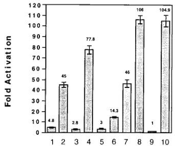
gene expression.We next determined if each construct con-taining the TAR RNA reversions had a response to tat differ-ent from that of each original TAR mutant. DNA fragmdiffer-ents obtained from each of the original or revertant LTRs extend-ing from 221 to 180 were inserted into a wild-type HIV-1 fragment extending from2177 to220. The promoter fusions were cloned into a promoterless CAT expression plasmid to determine the effects of tat on each of these mutant TAR elements. These CAT constructs containing either the wild type (Fig. 5, lanes 9 and 10), the original TAR mutant LTRs (TAR1, TAR2, TAR3, and TAR4) (lanes 1, 3, 5, and 7, re-spectively), or the revertant TAR LTRs (TAR1*, TAR2*, TAR3*, and TAR4*; lanes 2, 4, 6, and 8, respectively) were cotransfected into HeLa cells in both the presence and absence of a tat expression plasmid. All transfections included an RSV– b-galactosidase construct to normalize CAT expression to b-galactosidase activity. No significant differences were ob-served in the basal levels of CAT activity for the wild-type construct compared with either the original TAR mutants or the TAR revertant mutants (data not shown). The tat expres-sion vector induced wild-type HIV-1 gene expresexpres-sion approx-imately 105-fold (lanes 9 and 10). All of the TAR revertant constructs gave much higher levels of tat activation than the original TAR mutants from which they were derived (lanes 2, 4, 6, 8, and 10). The original TAR mutant construct TAR1 gave a 4.8-fold increase (lane 1) in response to tat compared with a 45-fold increase observed for the revertant construct TAR1* (lane 2). The TAR2 construct gave a 2.8-fold increase in gene expression in the presence of tat, while the revertant construct TAR2* gave a 78-fold increase in tat-induced gene expression (lanes 3 and 4). The TAR3 mutant that deleted the TAR RNA bulge element exhibited a 3-fold increase in HIV-1 gene expression (lane 5) while the revertant construct TAR3* exhibited a 14.3-fold increase (lane 6). Point mutants in the TAR RNA bulge have been found to be much more defective for gene expression when assayed in stable cell lines rather than following transient expression assays. For example, the point bulge mutant TAR4 gave a 45-fold increase in response to tat (lane 7), while the revertant TAR4* (lane 8) exhibited wild-type levels of tat-induced gene expression. Thus, each TAR revertant construct was greatly enhanced in its ability to
respond to the Tat protein and increase HIV-1 gene expres-sion.
Revertant TAR RNA structures restore proviral gene
ex-pression.We next determined if proviral constructs containing
TAR reversions resulted in increased viral gene expression compared with those containing the original TAR mutants. TAR elements extending from nucleotides 222 to 180 ob-tained from either the original mutants, TAR1, TAR2, TAR3, and TAR4, or the corresponding revertants, TAR1*, TAR2*, TAR3*, and TAR4*, were cloned into the HIV-1 provirus pBRDH1 in place of the wild-type TAR element. Proviral constructs corresponding to TAR1, TAR2, TAR3, TAR4, and the wild type (Fig. 6, lanes 1, 3, 5, 7, and 9, respectively) or the corresponding revertant constructs TAR1*, TAR2*, TAR3*, TAR4* (lanes 2, 4, 6, and 8, respectively) were transfected into Jurkat cells following restriction digestion with MroI. All trans-fections included an RSV–b-galactosidase construct linearized with the restriction enzyme ScaI to normalize p24 antigen expression to b-galactosidase activity. A cell-free culture su-pernatant from each transfection was assayed for p24 antigen or either day 4 and 7 posttransfection.
The wild-type proviral construct produced 0.85 and 2.1 ng of p24 antigen per ml in the culture supernatant on days 4 and 7, respectively (Fig. 6, lane 10). Nearly equivalent levels of p24 antigen were noted for the revertant TAR4* (lane 8). The p24 antigen levels for the other three TAR revertants were lower than that of the wild-type construct, although there was a marked increase in HIV-1 gene expression compared with the levels of expression in constructs that contained the original TAR mutants. Specifically, TAR1* gave a 112-fold increase on day 4 and a 139-fold increase on day 7 compared with TAR1 (Fig. 6, lanes 1 and 2), the TAR2* revertant gave a 141-fold increase on day 4 and a 145-fold increase on day 7 compared with TAR2 (lanes 3 and 4), and TAR3* gave a 38-fold increase on day 4 and a 20-fold increase on day 7 compared with TAR3 (lanes 5 and 6). The increase for the TAR4* revertant was 6-fold on day 4 and 33-fold on day 7 compared with the original TAR4 construct (lanes 7 and 8). Thus, the nucleotide changes identified in the revertant TAR mutant viruses resulted in TAR elements which were able to substantially increase the level of HIV-1 gene expression in response to tat.
Tat can act through low-energy TAR RNA stem
[image:5.612.91.262.70.212.2]intermedi-ates.The position of nucleotide alterations identified in each
FIG. 5. Tat-induced gene expression from HIV-1 LTR CAT constructs con-taining TAR revertants and original TAR mutants. HeLa cells were transfected with RSV-tat and HIV-1 LTR CAT vectors containing the TAR mutants includ-ing either TAR1 (lane 1), TAR2 (lane 3), TAR3 (lane 5), TAR4 (lane 7) or the TAR reversion mutants TAR1* (lane 2), TAR2* (lane 4), TAR3* (lane 6), or TAR4* (lane 8). The wild-type HIV-1 CAT vector was cotransfected with either RVSb-globin (lane 9) to determine basal gene expression or RSV tat (lane 10) to determine tat activation. All results were normalized by transfection with an RSV–b-galactosidase vector, and the standard deviations for three identical experiments are indicated.
FIG. 6. Analysis of HIV-1 gene expression from proviruses containing either TAR mutants or TAR revertants. Jurkat cells were transfected with HIV-1 proviruses containing either TAR1 (lane 1), TAR2 (lane 3), TAR3 (lane 5), TAR4 (lane 7) or the HIV-1 TAR reversion mutants TAR1* (lane 2), TAR2* (lane 4), TAR3* (lane 6), and TAR4* (lane 8). These mutant constructs were compared with the wild-type construct (lane 9) or pBR322 as a negative control (lane 10). Each transfection was repeated three times with similar results and included an RSV–b-galactosidase vector, which was used to normalize the trans-fection efficiency.u, day 4;■day 7.
on November 9, 2019 by guest
http://jvi.asm.org/
[image:5.612.316.557.72.195.2]TAR revertant virus suggested that restoration of the TAR RNA bulge structure was the primary mechanism which in-creased the level of HIV-1 gene expression. To better under-stand which elements in TAR were critical for tat induction, computer analysis of the TAR RNA secondary structures and calculation of the amount of free energy of both the original TAR mutants and TAR revertants were performed by using the Zuker algorithm (Fig. 7) (14, 55). The wild-type sequence folded into the previously described stem-loop structure with a relative free energy of 224.8 kcal/mol (approximately 2104 kJ/mol) (Fig. 7a). The algorithm predicted two RNA stem-loop structures for the TAR1 mutant. One formed an alternate 7-nucleotide loop, GAGCCTG, separated by 5 bp from a 4-nu-cleotide bulge, UCCU bulge, and had a free energy of218.2 kcal/mol (approximately276.1 kJ/mol) (Fig. 7b). The second predicted structure had a lower free energy of216.3 kcal/mol (approximately 268.2 kJ/mol) and maintained the wild-type loop sequence CUGGGA, which was separated by 4 bp from a double bulge structure containing the sequences CGCUCU and UCU (Fig. 7c). The TAR2 mutant had a free energy of 216.8 kcal/mol (approximately270.2 kJ/mol) and yielded only one significant RNA structure which maintained the wild-type loop sequences separated by 6 bp from a bulge with the se-quence UCGC (Fig. 7f). As expected, the TAR3 mutant lacked a bulge element and formed a highly stable stem-loop structure with a free energy of 230.0 kcal/mol (approximately 2126 kJ/mol) (Fig. 7i). TAR4, which contains the bulge point mu-tation, has a TAR RNA structure and stem energy similar to that of wild-type TAR RNA (Fig. 7k).
TAR1* yielded two potential RNA structures (Fig. 7d and e). The first potential structure had a free energy of 219.1 kcal/mol (approximately 279.9 kJ/mol) and maintained the wild-type loop, upper stem, and bulge elements (Fig. 7d). How-ever, there was a disruption of the stem in which base119 (C) failed to pair with141 (U). The second potential structure for TAR1* had a free energy of218.2 kcal/mol (approximately 276.1 kJ/mol), which was identical to the TAR1 structure except for a U-393G transversion (Fig. 7e). In contrast to the more stable TAR1* structure, this latter structure was unlikely to contain the bulge structure necessary for tat activation. There were two potential stable RNA structures generated for
the revertant TAR2* that differed in energy by 3.2 kcal/mol (approximately 13 kJ/mol) (Fig. 7g and h). The most stable RNA structure has a free energy of220.7 kcal/mol (approxi-mately 286.6 kJ/mol) and maintained the wild-type loop se-quences but not the bulge element (Fig. 7g). A less stable RNA stem-loop that had a free energy of217.5 kcal/mol (approxi-mately 273.2 kJ/mol) disrupted the stem where nucleotides 118 (C) and119 (A) failed to base pair respectively with142 (A) and141 (G) (Fig. 7h). Since this structure maintained the TAR RNA bulge, it is more likely the tat-responsive RNA structure. The TAR3* RNA structure had a relative free en-ergy of222.5 kcal/mol (approximately294.1 kJ/mol) and con-tained a dinucleotide transversion which generated a bulge structure at119 (U), which is located 6 bp below the wild-type loop sequence (Fig. 7j). The defective nature of this revertant in both in vitro binding with Tat and in vivo assays of gene expression is in agreement with previous mutagenesis studies that have demonstrated that the spacing between the bulge and the loop was critical for optimum tat-induced gene expression (3, 4, 10).
DISCUSSION
trans-activation of HIV-1 by the Tat protein is mediated
through a cis-acting RNA element (TAR) transcribed from the HIV-1 LTR (4, 6, 7, 12, 16, 22, 23, 25, 32, 44–46, 48). Although the mechanism by which Tat activates HIV-1 gene expression is unknown, several mutagenesis studies demonstrate that Tat protein acts through a minimal RNA stem structure extending from119 to144, which includes the upper stem and the bulge and loop structures (12, 16, 23, 25, 46, 48). Mutations which alter the loop, bulge, and upper stem decrease tat activation of the HIV-1 LTR (2, 4, 6–8, 11, 12, 16, 22, 23, 25, 32, 45, 46, 48). In addition, experiments have demonstrated that spacing be-tween the bulge and loop sequences is critical for optimal
tat-mediated increases in HIV-1 gene expression. These data
suggest that Tat recognizes a nascent TAR RNA structure, and in conjunction with cellular cofactors which bind to TAR loop sequences, such as TRP-185/TRP-1, forms a complex which is capable of increasing the proportion of productive transcrip-tional elongation complexes.
FIG. 7. Computer generated HIV-1 TAR RNA stem-loop structures. Computer generated stem-loop structures for HIV-1 TAR RNAs are shown for the wild type (a), TAR1 (b and c), TAR1* (d and e), TAR2 (f), TAR2* (g and h), TAR3 (i), TAR3* (j), TAR4 (k), and TAR4* (l). The predicted relative free energy (DG) for each structure is indicated, as are the results for in vitro Tat binding, gene expression in the presence of Tat, and the level of proviral gene expression.
on November 9, 2019 by guest
http://jvi.asm.org/
Recently, we constructed a variety of viruses containing TAR mutations and demonstrated marked defects of these viruses in both gene expression and replication (22). Since these viruses contained the neomycin resistance gene (1), we were able to select G418 Jurkat cell lines which contained each of these TAR mutant viruses. However, we found that pro-longed culture of several of these cell lines results in the gen-eration of viruses with compensatory changes in TAR. The ability to isolate revertant viruses which have increased levels of gene expression from HIV-1 LTR regulatory region mutants has been described previously (6, 29, 32). For instance, con-structs containing extensive mutations of TAR between 11 and110 which disrupt the TAR stem structure give rise to a variety of revertants which restore this stem structure. HIV-1 constructs which contain mutations of the SP1 binding sites give rise to revertants which contain changes in the TATA element and have increased levels of gene expression (29). Thus, second site mutations have previously been found to restore gene expression from HIV-1 LTR regulatory region mutants.
In the current study, we isolated revertant viruses which restored gene expression from TAR mutants viruses. Although we were able to isolate these revertants from viruses containing mutants in the TAR RNA bulge and stem, we never identified revertants which complemented viruses containing loop muta-tions, indicating the critical nature of the loop sequences for tat activation. One of these reversion mutants not surprisingly restored the UCU bulge between122 and 124 by changing the A to U at 122. However, each of the three other TAR revertants reflected interesting features required for TAR function. Two of the upper stem mutants which disrupted stem base pairing were found to contain additional mutations which only partially restored stem base pairing. These additional mu-tations resulted in TAR RNAs that were able to bind Tat protein and restore tat activation. Thus the ability to form a nascent RNA structure capable of Tat binding appears to be critical for subsequent tat activation. Furthermore, complete stem base pairing in the TAR stem is not critical for Tat activation. Another TAR mutant, which deleted the bulge be-tween122 and124, gave rise to a revertant with a dinucle-otide transversion such that a 1-nucledinucle-otide bulge structure was created. Although we could detect only minimal binding of Tat to this mutant TAR RNA by gel retardation analysis and the levels of tat-induced gene expression were markedly lower than the levels with the wild-type virus, it is likely that in vivo nascent RNA from this virus could transiently interact with Tat, although with low affinity. These results indicate that the maintenance of the bulge structure in the nascent RNA is critical for tat activation.
Computer analysis using the Zuker algorithm confirmed the formation of the previously published TAR RNA structure whose presence both in vivo and in vitro has been supported by both physiological and biochemical evidence (14, 55). The po-tential RNA structures of TAR revertants were also analyzed. The most stable TAR RNA structure of the revertant TAR1* would be predicted to be activated by Tat since it contains both bulge and loop structures. However, the most stable RNA structure which is capable of forming the revertant TAR2* is unlikely to be an effective target for tat activation as it is unable to form a bulge structure which is appropriately positioned relative to the loop sequences. This revertant is also capable of forming an alternate TAR RNA structure with a lower free energy which forms a bulge structure in nascent TAR RNA that is appropriately positioned relative to the loop sequences. Since TAR2* is activated by Tat in vivo, these results would suggest that Tat may act through this lower-energy structure
before the formation of a complete TAR RNA structure. Al-though the analyses of these RNA structures are valuable for our understanding of tat activation, it is probable that the binding of cellular factors is also involved in stabilizing RNA structures that more efficiently interact with Tat. Our results suggest that nascent TAR RNA with appropriately positioned loop and bulge sequences is likely a major determinant for subsequent tat activation.
ACKNOWLEDGMENTS
We thank Ty Lawrence and Elizabeth Folta for the preparation of the manuscript.
These studies were supported by the NIH and the Robert Welch Foundation.
REFERENCES
1. Beck, E., G. Ludwig, E. A. Auerswald, B. Reiss, and H. Scholler. 1982. Nucleotide sequence and exact localization of the neomycin phosphotrans-ferase gene from transposon Tn5. Gene 19:327–336.
2. Berkhout, B., A. Gatignol, A. B. Rabson, and K. T. Jeang. 1990. TAR-independent activation of the HIV-1 LTR: evidence that tat requires specific regions of the promoter. Cell 62:757–767.
3. Berkhout, B., and K. T. Jeang. 1989. trans activation of human immunode-ficiency virus type 1 is sequence specific for both the single-stranded bulge and loop of the trans-acting-responsive hairpin: a quantitative analysis. J. Virol. 63:5501–5504.
4. Berkhout, B., and K. T. Jeang. 1991. Detailed mutational analysis of TAR RNA: critical spacing between the bulge and loop recognition domains. Nucleic Acids Res. 19:6169–6176.
5. Berkhout, B., and K. T. Jeang. 1992. Functional roles for the TATA pro-moter and enhancers in basal and Tat-induced expression of the human immunodeficiency virus type 1 long terminal repeat. J. Virol. 66:139–149. 6. Berkhout, B., and B. Klaver. 1993. In vivo selection of randomly mutated
retroviral genomes. Nucleic Acids Res. 21:5020–5024.
7. Berkhout, B., R. H. Silverman, and K. T. Jeang. 1989. Tat trans-activates the human immunodeficiency virus through a nascent RNA target. Cell 59:273– 282.
8. Calnan, B. J., S. Biancalana, D. Hudson, and A. D. Frankel. 1991. Analysis of arginine-rich peptides from the HIV Tat protein reveals unusual features of RNA-protein recognition. Genes Dev. 5:201–210.
9. Dayton, A. I., J. G. Sodroski, C. A. Rosen, W. C. Goh, and W. A. Haseltine. 1986. The trans-activator gene of the human T cell lymphotropic virus type III is required for replication. Cell 44:941–947.
10. Dingwall, C., I. Ernberg, M. J. Gait, S. M. Green, S. Heaphy, J. Karn, A. D.
Lowe, M. Singh, and M. A. Skinner.1990. HIV-1 tat protein stimulates transcription by binding to a U-rich bulge in the stem of the TAR RNA structure. EMBO J. 9:4145–4153.
11. Dingwall, C., I. Ernberg, M. J. Gait, S. M. Green, S. Heaphy, J. Karn, A. D.
Lowe, M. Singh, M. A. Skinner, and R. Valerio.1989. Human immunodefi-ciency virus 1 tat protein binds trans-activation-responsive region (TAR) RNA in vitro. Proc. Natl. Acad. Sci. USA 86:6925–6929.
12. Feng, S., and E. C. Holland. 1988. HIV-1 tat trans-activation requires the loop sequence within TAR. Nature (London) 334:165–167.
13. Fisher, A. G., M. B. Feinberg, S. F. Josephs, M. E. Harper, L. M. Marselle,
G. Reyes, M. A. Gonda, A. Aldovini, C. Debouk, and R. C. Gallo.1986. The trans-activator gene of HTLV-III is essential for virus replication. Nature (London) 320:367–371.
14. Freier, S. M., R. Kierzek, J. A. Jaeger, N. Sugimoto, M. H. Caruthers, T.
Neilson, and D. H. Turner.1986. Improved free-energy parameters for predictions of RNA duplex stability. Proc. Natl. Acad. Sci. USA 83:9373– 9377.
15. Garcia, J. A., D. Harrich, E. Soultanakis, F. Wu, R. Mitsuyasu, and R. B.
Gaynor.1989. Human immunodeficiency virus type 1 LTR TATA and TAR region sequences required for transcriptional regulation. EMBO J. 8:765– 778.
16. Garcia, J. A., F. K. Wu, R. Mitsuyasu, and R. B. Gaynor. 1987. Interactions of cellular proteins involved in the transcriptional regulation of the human immunodeficiency virus. EMBO J. 6:3761–3770.
17. Gatignol, A., W. A. Buckler, B. Berkhout, and K. T. Jeang. 1991. Charac-terization of a human TAR RNA-binding protein that activates the HIV-1 LTR. Science 251:1597–1600.
18. Gaynor, R. 1992. Cellular transcription factors involved in the regulation of HIV-1 gene expression. AIDS 6:347–363.
19. Gorman, C. M., L. F. Moffat, and B. H. Howard. 1982. Recombinant ge-nomes which express chloramphenicol acetyltransferase in mammalian cells. Mol. Cell. Biol. 2:1044–1051.
20. Graeble, M. A., M. J. Churcher, A. D. Lowe, M. J. Gait, and J. Karn. 1993. Human immunodeficiency virus type 1 transactivator protein, tat, stimulates
on November 9, 2019 by guest
http://jvi.asm.org/
transcriptional read-through of distal terminator sequences in vitro. Proc. Natl. Acad. Sci. USA 90:6184–6188.
21. Harrich, D., J. Garcia, R. Mitsuyasu, and R. Gaynor. 1990. TAR indepen-dent activation of the human immunodeficiency virus in phorbol ester stim-ulated T lymphocytes. EMBO J. 9:4417–4423.
22. Harrich, D., C. Yu, E. Race, and R. Gaynor. 1994. Differential growth kinetics of human immunodeficiency virus type 1 TAR mutant viruses. J. Virol. 68:5899–5910.
23. Hauber, J., and B. R. Cullen. 1988. Mutational analysis of the trans-activa-tion-responsive region of the human immunodeficiency virus type 1 long terminal repeat. J. Virol. 62:673–679.
24. Huang, L., A. Joshi, R. Willey, J. Orenstein, and K. T. Jeang. 1994. Human immunodeficiency viruses regulated by alternative trans-activators; genetic evidence for a novel non-transcriptional function of Tat in virion infectivity. EMBO J. 13:2886–2896.
25. Jakobovits, A., D. H. Smith, E. B. Jakobovits, and D. J. Capon. 1988. A discrete element 39of human immunodeficiency virus 1 (HIV-1) and HIV-2 mRNA initiation sites mediates transcriptional activation by an HIV trans activator. Mol. Cell. Biol. 8:2555–2561.
26. Jones, K. A., J. T. Kadonaga, P. A. Luciw, and R. Tjian. 1986. Activation of the AIDS retrovirus promoter by the cellular transcription factor, Sp1. Sci-ence 232:755–759.
27. Jones, K. A., P. A. Luciw, and N. Duchange. 1988. Structural arrangements of transcription control domains within the 59-untranslated leader regions of the HIV-1 and HIV-2 promoters. Genes Dev. 2:1101–1114.
28. Kao, S. Y., A. F. Calman, P. A. Luciw, and B. M. Peterlin. 1987. Anti-termination of transcription within the long terminal repeat of HIV-1 by tat gene product. Nature (London) 330:489–493.
29. Kashanchi, F., R. Shibata, E. K. Ross, J. N. Brandy, and M. A. Martin. 1994. Second site long terminal repeat (LTR) revertants of replication on virus infectivity, LTR-directed expression in vitro RNA synthesis and binding of basal transcription factors TFIID and TFIIA. J. Virol. 68:3298–3307. 30. Kato, H., M. Horikoshi, and R. G. Roeder. 1991. Repression of HIV-1
transcription by a cellular protein. Science 251:1476–1479.
31. Kato, H., H. Sumimoto, P. Pognonec, C. H. Chen, C. A. Rosen, and R. G.
Roeder.1992. HIV-1 Tat acts as a processivity factor in vitro in conjunction with cellular elongation factors. Genes Dev. 6:655–666.
32. Klaver, B., and B. Berkhout. 1994. Evolution of a disrupted TAR RNA hairpin structure in the HIV-1 virus. EMBO J. 13:2650–2659.
33. Laspia, M. F., A. P. Rice, and M. B. Mathews. 1989. HIV-1 Tat protein increases transcriptional initiation and stabilizes elongation. Cell 59:283–292. 34. Laspia, M. F., A. P. Rice, and M. B. Mathews. 1990. Synergy between HIV-1
Tat and adenovirus E1A is principally due to stabilization of transcriptional elongation. Genes Dev. 4:2397–2408.
35. Lu, Y., M. Stenzel, J. G. Sodroski, and W. A. Haseltine. 1989. Effects of long terminal repeat mutations on human immunodeficiency virus type 1 repli-cation. J. Virol. 63:4115–4119.
36. Lu, X., T. M. Welsh, and B. M. Peterlin. 1993. The human immunodeficiency virus type 1 long terminal repeat specifies two different transcription com-plexes, only one of which is regulated by Tat. J. Virol. 67:1752–1760. 37. Marciniak, R. A., and P. A. Sharp. 1991. HIV-1 Tat protein promotes
formation of more-processive elongation complexes. EMBO J. 10:4189–4196. 38. Muesing, M. A., D. H. Smith, and D. J. Capon. 1987. Regulation of mRNA
accumulation by a human immunodeficiency virus trans-activator protein. Cell 48:691–701.
39. Nabel, G., and D. Baltimore. 1987. An inducible transcription factor acti-vates expression of human immunodeficiency virus in T cells. Nature (Lon-don) 326:711–713.
40. Olsen, H. S., and C. A. Rosen. 1992. Contribution of the TATA motif to Tat-mediated transcriptional activation of human immunodeficiency virus gene expression. J. Virol. 66:5594–5597.
41. Ou, S.-H. I., L. G. Garcia-Martinez, E. J. Paulssen, and R. B. Gaynor. 1994. Role of flanking E box motifs in human immunodeficiency virus type 1 TATA element function. J. Virol. 68:7188–7999.
42. Peterlin, B. M., P. A. Luciw, P. J. Barr, and M. D. Walker. 1986. Elevated levels of mRNA can account for the trans-activation of human immunode-ficiency virus. Proc. Natl. Acad. Sci. USA 83:9734–9738.
43. Ratnasabapathy, R., M. Sheldon, L. Johal, and N. Hernandez. 1990. The HIV-1 long terminal repeat contains an unusual element that induces the synthesis of short RNAs from various mRNA and snRNA promoters. Genes Dev. 4:2061–2074.
44. Rosen, C. A., J. G. Sodroski, and W. A. Haseltine. 1985. Location of cis-acting regulatory sequences in the human T-cell leukemia virus type I long terminal repeat. Proc. Natl. Acad. Sci. USA 82:6502–6506.
45. Roy, S., U. Delling, C. H. Chen, C. A. Rosen, and N. Sonenberg. 1990. A bulge structure in HIV-1 TAR RNA is required for Tat binding and Tat-mediated trans-activation. Genes Dev. 4:1365–1373.
46. Roy, S., N. T. Parkin, C. Rosen, J. Itovitch, and N. Sonenberg. 1990. Struc-tural requirements for trans activation of human immunodeficiency virus type 1 long terminal repeat-directed gene expression by tat: importance of base pairing, loop sequence, and bulges in the tat-responsive sequence. J. Virol. 64:1402–1406.
47. Sanchez, P. R., M. D. Power, P. J. Barr, K. S. Steimer, M. M. Stempien, S. S.
Brown, W. W. Gee, A. Renard, A. Randolph, and J. A. Levy.1985. Nucleotide sequence and expression of an AIDS-associated retrovirus (ARV-2). Science
227:484–492.
48. Selby, M. J., E. S. Bain, P. A. Luciw, and B. M. Peterlin. 1989. Structure, sequence, and position of the stem-loop in tar determine transcriptional elongation by tat through the HIV-1 long terminal repeat. Genes Dev.
3:547–558.
49. Sheldon, M., R. Ratnasabapathy, and N. Hernandez. 1993. Characterization of the inducer of short transcripts, a human immunodeficiency virus type 1 transcriptional element that activates the synthesis of short RNAs. Mol. Cell. Biol. 13:1251–1263.
50. Sheline, C. T., L. H. Milocco, and K. A. Jones. 1991. Two distinct nuclear transcription factors recognize loop and bulge residues of the HIV-1 TAR RNA hairpin. Genes Dev. 5:2508–2520.
51. Weeks, K. M., and D. M. Crothers. 1991. RNA recognition by Tat-derived peptides: interaction in the major groove? Cell 66:577–588.
52. Wright, C. M., B. K. Felber, H. Paskalis, and G. N. Pavlakis. 1986. Expres-sion and characterization of the trans-activator of HTLV-III/LAV virus. Science 234:988–992.
53. Wu, F., J. Garcia, D. Sigman, and R. Gaynor. 1991. tat regulates binding of the human immunodeficiency virus trans-activating region RNA loop-bind-ing protein TRP-185. Genes Dev. 5:2128–2140.
54. Wu, F. K., J. A. Garcia, D. Harrich, and R. B. Gaynor. 1988. Purification of the human immunodeficiency virus type 1 enhancer and TAR binding pro-teins EBP-1 and UBP-1. EMBO J. 7:2117–2130.
55. Zuker, M. 1989. On finding all suboptimal foldings of an RNA molecule. Science 244:48–52.