Journal Name
COMMUNICATION
a.WestCHEM/Department of Pure & Applied Chemistry, University of Strathclyde,
99 George Street, Glasgow, G1 1RD, U.K.
b.Departament de Química Inorgànica i Orgànica, Universitat Jaume I, 12071
Castelló, Spain Fax: +34 964728214; Tel: +34 964729155; E-mail: [email protected].
c.Advanced Science Research Center (ASRC) and Hunter College, City University of
New York, 85 St Nicholas Terrace, New York NY10027, United States; E-mail: [email protected]
Received 00th January 20xx, Accepted 00th January 20xx
DOI: 10.1039/x0xx00000x
www.rsc.org/
Metastable hydrogels from aromatic dipeptides
M. P. Conte,a N. Singh,b I.R. Sasselli,a B. Escuder b* and R. V. Ulijn a,c,d,e*
We demonstrate that the well-known self-assembling dipeptide diphenylalanine (FF) and its amidated derivative (FF-NH2) can form
metastable hydrogels upon sonication of the dipeptide solutions. The hydrogels show instantaneous syneresis upon mechanical contact resulting in rapid expulsion of water and collapse into a semi-solid gel.
Supramolecular self-assembly provides an effective bottom-up approach for the fabrication of functional nanomaterials.1, 2
Among the available molecular building blocks, peptides and peptide derivatives have attracted attention due to their chemical versatility and the inherent possibility to provide a functional interface with biological systems.3-5 Very short
peptides 6-8 and their derivatives 9 are of particular interest in
this context due to their chemical simplicity, low cost and remarkable properties. 10
The first example of nanostructure formation by self-assembly of dipeptides dates back to the early 2000s, when Gorbitz et
al. observed the formation of supramolecular structures upon
crystallization of four dipeptides (dileucine (LL), leucine-phenylalanine (LF), phenylalanine-leucine (FL) and diphenylalanine (FF). 11 Among these four dipeptides FF, the
key structural motif of Alzheimer’s β-amyloid polypeptide, is probably the most well-known, thanks to pioneering work by Gazit and co-workers6, 12-15. They observed that this dipeptide
self-assembles into hollow tubular nanostructures in aqueous solution, through a combination of hydrogen-bonding and π-π interactions.6 Following this observation, unexpected
properties not typically associated with biological matter have been reported for FF nanotubes. This includes demonstration that these nanostructures are stable in boiling water and organic solvent,13 and that they are remarkably stiff, having a
Young’s modulus of ~ 20 GPa, which places them among the stiffest biological materials known.16 Moreover, by varying
assembly conditions FF-based building blocks have been shown to self-assemble into a variety of different structures, such as vesicles, ribbons and fibres. 17-19 Li et al. showed that
FF may also act as an organo-gelator, forming organogels in chloroform or aromatic solvents. 20 So far, the only example of
an (unmodified) dipeptide that is able to self-assemble to form a hydrogel is the isoleucine-phenylalanine (IF) reported by Ventura et al. 21 Other examples of dipeptide nanostructures
include WF and FW nanoparticles 22 and VA and AV which
self-assemble into different structural morphologies in various solvent media. 23 It is increasingly appreciated that in addition
to the molecular structure of the building blocks, the self-assembly pathway is critical to properties of the supramolecular assemblies. 24
In this work, we show the formation of metastable FF (free acid and amide) hydrogels obtained upon sequential solvent switching and sonication of dipeptide solutions. Once formed, the gels show rapid syneresis upon mechanical stimulation (Fig. 1 and S1). Syneresis is the contraction of a gel accompanied by sudden release of the moisture initially contained within the gel network. This phenomenon has been mostly studied in polymer-based gels 25-27 and only a few
reports exist about the syneresis of supramolecular hydrogels.
28-30 Adams and co-workers reported a few Fmoc-based
peptide gels that show syneresis and recently they provided further insights into the origin of the process for a peptide-based gelator with an oligophenylene vinylene core. 31
To form the hydrogels, 5 mg of FF or FF-NH2 dipeptide is first
dissolved in 80 µl of 1,1,1,3,3,3-hexafluoro-2-propanol (HFIP) and then diluted up to 1 mL in 100 mM sodium phosphate buffer pH 8 giving rise to a final molar concentration of 16 mM.6 Upon sonication (5 to 10 ultrasound pulses of 10 s each)
the colourless, transparent solution turns into a white opaque gel (Fig. S1). The critical gelation concentration (CGC) was determined by consecutive dilution with buffer and found to be ~ 8 mM for FF (~ 0.25 wt%) and ~ 4 mM for FF-NH2 (~ 0.12
Gels were formed exclusively when the combination of the two stimuli (ultrasound and solvent switch) is applied. Amorphous aggregates were observed when the dipeptides are dissolved in HFIP and buffer without sonication.
Sonication is commonly used in supramolecular chemistry to modulate self-assembly and gelation processes. 32-37
Ultrasound can assist the gelation process allowing conformational rearrangements and reorganisation to maximize non-covalent interactions. 32, 35 Both gels formed are
stable at room temperature for several weeks when left undisturbed, but they exhibit rapid syneresis when they undergo mechanical stress (Fig. 1). This rapid expulsion of water is likely related to the extremely hydrophobic nature of the peptide fibres formed. To demonstrate the hydrophobicity of the fibres we employed a fluorimetric assay based on 8-Anilino-1-naphthalenesulfonic acid (ANS). ANS is a fluorescent dye frequently employed in various fields of protein analysis as it binds to hydrophobic regions of proteins. Interactions of ANS with hydrophobic binding sites is accompanied by an increase in fluorescence and a blue shift of the peak maximum. 38 5 mg
of FF or FF-NH2 dipeptide are first dissolved in 80 µl of HFIP
and then diluted up to 1 mL in 100 μM ANS buffered solution (see section 6 and 16 in ESI). The ANS buffered solution fluoresces around 540 nm (Figure S7). When the dye is mixed with the dipeptides, a significant increase in the fluorescence intensity emission is observed, accompanied by a blue shift of the maximum peak. This effect is even more evident when the peptide solutions are sonicated and the gel is formed. Upon mechanical disruption, the collapsed gel and the exuded liquid was separated and collected. The fluorescence emission confirms the hydrophobic nature of the collapsed gel, whereas the spectrum of the exuded liquid does not show any significant peak (Figure S7). Since the rapid expulsion of the moisture is observed upon mechanical disruption, we wanted to verify whether hydrophobic compounds would show release or remained bound to the network of fibres. We employed the hydrophobic dye thioflavin T (ThT), which is
commonly used to stain β-sheet like fibres. 39 When ThT is
added to the peptide solutions, a bright yellow gel is obtained. Upon syneresis, a transparent liquid is exuded, in contrast to the collapsed gel that remains yellow, suggesting that the hydrophobic dye is retained into the supramolecular aggregates. A UV-Vis assay confirmed that more than 99% of the initial concentration of the dye is retained into the collapsed gel (See Fig. 1c and section 7 in the ESI).
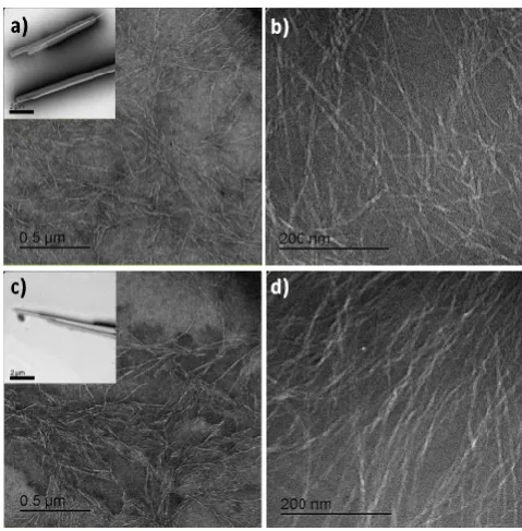
The collapsed structures obtained upon syneresis remain intact and can still be handled and characterised. Due to the immediate collapse upon handling, the following characterisation was carried out on the collapsed structures. Transmission and scanning electron microscopy (TEM and SEM) were used to gain insight into the nanoscale morphology of the dipeptide assemblies. Before sonication, the TEM images of both FF-NH2 and FF (Fig. 2a and 2c, insets) show the
presence of tubular structures of several micrometres of length and hundreds of nanometres of diameter, as previously reported.6, 17
Upon sonication, in addition to remaining nanotubes, much smaller fibres were observed, with a length of hundreds of nanometres and a diameter of approximately 10 nm (Fig. 2).
Diphenylalanine has been previously reported to self-assemble into nanofibrils in organic solvents, 19, 20 but to our knowledge
this is the first report of diphenylalanine self-assembly into nanofibers in predominantly aqueous environment. The SEM pictures of both FF-NH2 and FF confirmed the presence of an
extended network of fibres and tubes (Fig. S4). As a control, the dipeptide phenylalanine-tyrosine (FY) did not form any observable aggregate or gel upon sonication demonstrating that the FF sequence is critical to the observed behaviour. TEM images of the reference sample FY showed that this dipeptide
[image:2.595.41.287.43.229.2]Figure 1. (a) Chemical structures of the dipeptides. (b) Schematic of the gelation process for FF and FF-NH2. The opaque gel forms upon vortexing and sonication of the dipeptide solution. A fluorescent dye (ThT) was added to help visualize the process and demonstrate that the nanofiber network effectively sequesters hydrophobic compounds, releasing uncoloured liquid upon syneresis.Mechanical touching with a spatula causes the rapid syneresis (seconds) of the gel which collapses to approximately 40% of its original volume. (c) Screenshots to illustrate the process. The full video is available in the ESI.
[image:2.595.309.549.335.579.2]forms spherical aggregates and that the structure of the aggregates is not altered upon sonication (Fig. S3).
Gazit et al. hypothesized that FF molecules dissolved in HFIP arrange in a 2D extended β-sheet which with time should close along an axis to give a stable nanotubular structure.40
However, upon sonication, the transition of dissolved dipeptide molecules from HFIP to buffer may induce rapid change in solubility assisting the formation of several nucleation sites. We propose that this favours the supramolecular organization of the FF molecules in kinetically trapped nanofibers, possibly preventing the extended 2D β-sheets (and subsequent larger tubes) to form. To investigate this, the gels formed upon sonication of the dipeptide solutions were taken through several heat/cool cycles in order to unlock kinetic aggregates. In the TEM images of the heated/cooled samples (Fig. S5) the only morphology observed is the nanotubes, confirming that this is the most stable form of aggregation and that the nanofibrous gels represent a metastable, kinetic trapped state.
Dynamic frequency sweep experiments were carried out to assess the mechanical properties of the collapsed material. For both FF and FF-NH2 the rheology revealed a gel-like behaviour,
providing an elastic modulus G’ of 1.78 kPa for FF and 30.1 kPa for FF-NH2 (Fig. S6) which is in the range of values previously
reported for other peptide-based hydrogels. 41
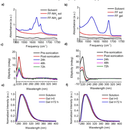
Fourier transform infrared (FTIR) spectroscopy is a valuable
tool to analyse the secondary structure content of proteins, specifically in the amide I region (1600-1700 cm-1). The
enhancement of the signal in this region of the spectra is due to coupling of hydrogen bonds between amide groups. The
absorption associated with the amide I band for protein solution is typically observed around 1650-1655 cm-1. 7 In
β-sheet-based supramolecular structures the presence of hydrogen bonds between the amide groups causes this band to shift towards lower frequencies. In the FTIR spectrum of FF-NH2 gel (Fig. 3) the presence of a dominant absorption peak at
1625 cm-1 confirms a β-sheet-like arrangement of the amide
groups. 7 The absorption around 1650 cm-1 is normally
associated with a “random coil” conformation in proteins. For short peptides, this peak can be assigned to imperfect stacking of amide groups. 7, 42 The peak around 1585 cm-1 can be
assigned to the free amine group at the N-terminus. A shoulder is visible around 1610 cm-1, which could correspond
to the peak visible in the solvent spectrum or to a different amide group environment in the stack (Fig. 3a and 3b, black line). The FTIR spectrum of FF gel is quite different and shows a peak around 1570 cm-1 due to the free carboxylic acid group
and a peak around 1670 cm-1 which confirms the presence of
H-bonding between the dipeptide molecules suggesting a well ordered structure.7 For the reference sample FY, no peaks
were observed in the amide region and no difference was observed between the spectra measured before and after sonication, indicating a lack of organized amide bonds (Fig. S2). The observed CD spectrum for both FF-NH2 and FF shows a
positive signal around 218 nm (Fig. 3c and 3d). This signal is also visible for the dipeptides in solution, consistent with previously reported results 13 and the intensity of the peak
decreases after sonication. For FF, upon sonication an additional (negative) peak around 232 nm (Fig. 3d) indicates an additional chiral component for the stacking of the phenylalanine residues. 20, 43, 44 Fluorescence spectroscopy
was used to measure the emission spectra of the dipeptides in solution and of the gels formed upon sonication. For the dipeptide solutions the emission peak of the phenyl groups is observed around 285 nm. For both FF and FF-NH2 the
fluorescence emission is red-shifted upon sonication and gel formation (~ 6 nm for FF and ~ 5 nm for FF-NH2, Fig. 3e and 3f).
This suggests a more efficient π-π stacking of the phenyl groups upon sonication as previously reported for proteins 45
and self-assembled amyloid fibres. 20, 46 Based on the above
observations, we suggest that sonication reorganizes the molecules, giving rise to enhanced π-π stacking between the aromatic groups and the intermolecular hydrogen bonding providing the driving force to form extended supramolecular aggregates.
In summary, we have observed the formation of metastable hydrogels by the diphenylalanine dipeptide and its amidated derivative. By sonication, we were able to achieve unstable kinetically trapped hydrogels. The gels exhibit extreme syneresis upon mechanical stimulation but the collapsed structures still behave as a gel-like material. Thanks to their sensitivity to external mechanical stimuli and the rapid supramolecular collapse and the highly hydrophobic nature of the fibres formed, materials of this type might find applications as pressure sensors or as selective scavengers for small hydrophobic molecules. Spectroscopic analysis of the dipeptide solutions and of the gels suggests that sonication
c) d)
220 240 260 280 300 320 340 -5 0 5 10 15 20 E lli p tic ity ( m de g) Wavelength (nm) Pre-sonication Post-sonication 24h 48h 72h
220 240 260 280 300 320 340 -10 0 10 20 30 40 50 E lli pt ic ity ( m de g) Wavelength (nm) Pre-sonication Post-sonication 24h 48h 72h a) b)
1500 1550 1600 1650 1700 17500
1 2 3 A bs or ba nc e ( a. u. )
Frequency (cm-1)
Solvent FF sol FF gel
1500 1550 1600 1650 1700 17500
1 2 A bs or ba n ce ( a. u. )
Frequency (cm-1)
Solvent FF-NH2 sol
FF-NH2 gel
e) f)
280 300 320 340 360 380 400 0.0 0.2 0.4 0.6 0.8 1.0 N or m al iz e d In te ns ity ( a .u .) Wavelength (nm) Solution Gel t=0 Gel t=72 h
[image:3.595.45.285.377.624.2]280 300 320 340 360 380 400 0.0 0.2 0.4 0.6 0.8 1.0 N or m al iz e d In te ns ity ( a .u .) Wavelength (nm) Solution Gel t=0 Gel t=72 h
induces a reorganization of hydrogen bonding and an enhancement of π-π stacking, allowing the formation of an extended network of supramolecular aggregates.
The authors gratefully acknowledge the financial support by the EC 7th Framework Programme Marie Curie Actions via the European ITN SMARTNET No. 316656. They also acknowledge Margaret Mullin from University of Glasgow for help in TEM and SEM imaging, Dr. Sharon Kelly from University of Glasgow for help in CD, Dr. Neil Hunt from University of Strathclyde for using the FTIR equipment and Dr. Yousef Abul-Haija for the help with the preparation of free standing gels.
Notes and references
1. M. J. Webber, E. A. Appel, E. W. Meijer and R. Langer, Nat
Mater, 2016, 15, 13-26.
2. T. Aida, E. W. Meijer and S. I. Stupp, Science, 2012, 335,
813-817.
3. M. Zelzer and R. V. Ulijn, Chemical Society Reviews, 2010,
39, 3351-3357.
4. R. deLaRica, K. I. Fabijanic, A. Baldi and H. Matsui,
Angewandte Chemie International Edition, 2010, 49,
1447-1450.
5. S. Zhang, Nature Biotechnology, 2004, 22.
6. M. Reches and E. Gazit, Science, 2003, 300, 625-627.
7. P. W. J. M. Frederix, G. G. Scott, Y. M. Abul-Haija, D.
Kalafatovic, C. G. Pappas, N. Javid, N. T. Hunt, R. V. Ulijn and T. Tuttle, Nature chemistry, 2015, 7, 30-37.
8. S. Maity, S. Nir, T. Zada and M. Reches, Chemical
communications, 2014, 50, 11154-11157.
9. S. Fleming and R. V. Ulijn, Chem Soc Rev, 2014, DOI:
10.1039/c4cs00247d.
10. L. Adler-Abramovich and E. Gazit, Chemical Society
Reviews, 2014, 43, 6881-6893.
11. C. H. Görbitz, Chemistry – A European Journal, 2001, 7,
5153-5159.
12. M. Reches and E. Gazit, Nat Nano, 2006, 1, 195-200.
13. L. Adler-Abramovich, M. Reches, V. L. Sedman, S. Allen, S.
J. B. Tendler and E. Gazit, Langmuir : the ACS journal of
surfaces and colloids, 2006, 22, 1313-1320.
14. P. Tamamis, L. Adler-Abramovich, M. Reches, K. Marshall,
P. Sikorski, L. Serpell, E. Gazit and G. Archontis,
Biophysical Journal, 2009, 96, 5020-5029.
15. I. Azuri, L. Adler-Abramovich, E. Gazit, O. Hod and L.
Kronik, Journal of the American Chemical Society, 2014, 136, 963-969.
16. N. Kol, L. Adler-Abramovich, D. Barlam, R. Z. Shneck, E.
Gazit and I. Rousso, Nano Letters, 2005, 5, 1343-1346.
17. X. Yan, P. Zhu and J. Li, Chemical Society Reviews, 2010,
39, 1877-1890.
18. I. W. Hamley, Angewandte Chemie International Edition,
2014, 53, 6866-6881.
19. J. Wang, K. Liu, L. Yan, A. Wang, S. Bai and X. Yan, ACS
Nano, 2016, DOI: 10.1021/acsnano.5b06567.
20. X. Yan, Y. Cui, Q. He, K. Wang and J. Li, Chemistry of
Materials, 2008, 20, 1522-1526.
21. N. S. de Groot, T. Parella, F. X. Aviles, J. Vendrell and S.
Ventura, Biophysical Journal, 2007, 92, 1732-1741.
22. Z. Fan, L. Sun, Y. Huang, Y. Wang and M. Zhang, Nat Nano,
2016, 11, 388-394.
23. H. Erdogan, E. Babur, M. Yilmaz, E. Candas, M. Gordesel,
Y. Dede, E. E. Oren, G. B. Demirel, M. K. Ozturk, M. S. Yavuz and G. Demirel, Langmuir : the ACS journal of
surfaces and colloids, 2015, 31, 7337-7345.
24. J. Raeburn, A. Zamith Cardoso and D. J. Adams, Chemical
Society Reviews, 2013, 42, 5143-5156.
25. H.-J. Schneider and R. M. Strongin, Accounts of Chemical
Research, 2009, 42, 1489-1500.
26. C. P. McCoy, C. Rooney, C. R. Edwards, D. S. Jones and S.
P. Gorman, Journal of the American Chemical Society, 2007, 129, 9572-9573.
27. J. A. Lucey, T. van Vliet, K. Grolle, T. Geurts and P. Walstra,
International Dairy Journal, 1997, 7, 389-397.
28. F. Xie, L. Qin and M. Liu, Chemical communications, 2016,
52, 930-933.
29. S.-L. Zhou, S. Matsumoto, H.-D. Tian, H. Yamane, A. Ojida,
S. Kiyonaka and I. Hamachi, Chemistry – A European
Journal, 2005, 11, 1130-1136.
30. D. J. Adams, L. M. Mullen, M. Berta, L. Chen and W. J.
Frith, Soft Matter, 2010, 6, 1971-1980.
31. A. M. Castilla, M. Wallace, L. L. E. Mears, E. R. Draper, J.
Doutch, S. Rogers and D. J. Adams, Soft Matter, 2016, DOI: 10.1039/C6SM01194B.
32. X. Yu, L. Chen, M. Zhang and T. Yi, Chemical Society
Reviews, 2014, 43, 5346-5371.
33. G. Cravotto and P. Cintas, Chemical Society Reviews, 2009,
38, 2684-2697.
34. C. G. Pappas, P. W. J. M. Frederix, T. Mutasa, S. Fleming, Y.
M. Abul-Haija, S. M. Kelly, A. Gachagan, D. Kalafatovic, J. Trevino, R. V. Ulijn and S. Bai, Chemical communications, 2015, 51, 8465-8468.
35. C. G. Pappas, T. Mutasa, P. W. J. M. Frederix, S. Fleming, S.
Bai, S. Debnath, S. M. Kelly, A. Gachagan and R. V. Ulijn,
Materials Horizons, 2015, 2, 198-202.
36. T. Naota and H. Koori, Journal of the American Chemical
Society, 2005, 127, 9324-9325.
37. S. Maity, P. Das and M. Reches, Scientific Reports, 2015, 5,
16365.
38. A. Hawe, M. Sutter and W. Jiskoot, Pharmaceutical
Research, 2008, 25, 1487-1499.
39. M. Biancalana and S. Koide, Biochimica et Biophysica Acta
(BBA) - Proteins and Proteomics, 2010, 1804, 1405-1412.
40. M. Reches and E. Gazit, Nano Letters, 2004, 4, 581-585.
41. V. Jayawarna, S. M. Richardson, A. R. Hirst, N. W. Hodson,
A. Saiani, J. E. Gough and R. V. Ulijn, Acta Biomaterialia, 2009, 5, 934-943.
42. S. Fleming, P. W. J. M. Frederix, I. Ramos Sasselli, N. T.
Hunt, R. V. Ulijn and T. Tuttle, Langmuir : the ACS journal
of surfaces and colloids, 2013, 29, 9510-9515.
43. K. N. N. Berova, R. W. Woody, Circular Dichroism:
Principles and Applications, Wiley, 2nd Edition edn., 2000.
44. Q. Li, Y. Jia, L. Dai, Y. Yang and J. Li, ACS Nano, 2015, 9,
2689-2695.
45. G. B. McGaughey, M. Gagné and A. K. Rappé, Journal of
Biological Chemistry, 1998, 273, 15458-15463.
46. E. Gazit, The FASEB Journal, 2002, 16, 77-83.
1. M. J. Webber, E. A. Appel, E. W. Meijer and R. Langer, Nat
2. T. Aida, E. W. Meijer and S. I. Stupp, Science, 2012, 335, 813-817.
3. M. Zelzer and R. V. Ulijn, Chemical Society Reviews, 2010,
39, 3351-3357.
4. R. deLaRica, K. I. Fabijanic, A. Baldi and H. Matsui,
Angewandte Chemie International Edition, 2010, 49,
1447-1450.
5. S. Zhang, Nature Biotechnology, 2004, 22.
6. M. Reches and E. Gazit, Science, 2003, 300, 625-627.
7. P. W. J. M. Frederix, G. G. Scott, Y. M. Abul-Haija, D.
Kalafatovic, C. G. Pappas, N. Javid, N. T. Hunt, R. V. Ulijn and T. Tuttle, Nature chemistry, 2015, 7, 30-37.
8. S. Maity, S. Nir, T. Zada and M. Reches, Chemical
communications, 2014, 50, 11154-11157.
9. S. Fleming and R. V. Ulijn, Chem Soc Rev, 2014, DOI:
10.1039/c4cs00247d.
10. L. Adler-Abramovich and E. Gazit, Chemical Society
Reviews, 2014, 43, 6881-6893.
11. C. H. Görbitz, Chemistry – A European Journal, 2001, 7,
5153-5159.
12. M. Reches and E. Gazit, Nat Nano, 2006, 1, 195-200.
13. L. Adler-Abramovich, M. Reches, V. L. Sedman, S. Allen, S.
J. B. Tendler and E. Gazit, Langmuir : the ACS journal of
surfaces and colloids, 2006, 22, 1313-1320.
14. P. Tamamis, L. Adler-Abramovich, M. Reches, K. Marshall,
P. Sikorski, L. Serpell, E. Gazit and G. Archontis,
Biophysical Journal, 2009, 96, 5020-5029.
15. I. Azuri, L. Adler-Abramovich, E. Gazit, O. Hod and L.
Kronik, Journal of the American Chemical Society, 2014, 136, 963-969.
16. N. Kol, L. Adler-Abramovich, D. Barlam, R. Z. Shneck, E.
Gazit and I. Rousso, Nano Letters, 2005, 5, 1343-1346.
17. X. Yan, P. Zhu and J. Li, Chemical Society Reviews, 2010,
39, 1877-1890.
18. I. W. Hamley, Angewandte Chemie International Edition,
2014, 53, 6866-6881.
19. J. Wang, K. Liu, L. Yan, A. Wang, S. Bai and X. Yan, ACS
Nano, 2016, DOI: 10.1021/acsnano.5b06567.
20. X. Yan, Y. Cui, Q. He, K. Wang and J. Li, Chemistry of
Materials, 2008, 20, 1522-1526.
21. N. S. de Groot, T. Parella, F. X. Aviles, J. Vendrell and S.
Ventura, Biophysical Journal, 2007, 92, 1732-1741.
22. Z. Fan, L. Sun, Y. Huang, Y. Wang and M. Zhang, Nat Nano,
2016, 11, 388-394.
23. H. Erdogan, E. Babur, M. Yilmaz, E. Candas, M. Gordesel,
Y. Dede, E. E. Oren, G. B. Demirel, M. K. Ozturk, M. S. Yavuz and G. Demirel, Langmuir : the ACS journal of
surfaces and colloids, 2015, 31, 7337-7345.
24. J. Raeburn, A. Zamith Cardoso and D. J. Adams, Chemical
Society Reviews, 2013, 42, 5143-5156.
25. H.-J. Schneider and R. M. Strongin, Accounts of Chemical
Research, 2009, 42, 1489-1500.
26. C. P. McCoy, C. Rooney, C. R. Edwards, D. S. Jones and S.
P. Gorman, Journal of the American Chemical Society, 2007, 129, 9572-9573.
27. J. A. Lucey, T. van Vliet, K. Grolle, T. Geurts and P. Walstra,
International Dairy Journal, 1997, 7, 389-397.
28. F. Xie, L. Qin and M. Liu, Chemical communications, 2016,
52, 930-933.
29. S.-L. Zhou, S. Matsumoto, H.-D. Tian, H. Yamane, A. Ojida,
S. Kiyonaka and I. Hamachi, Chemistry – A European
Journal, 2005, 11, 1130-1136.
30. D. J. Adams, L. M. Mullen, M. Berta, L. Chen and W. J.
Frith, Soft Matter, 2010, 6, 1971-1980.
31. A. M. Castilla, M. Wallace, L. L. E. Mears, E. R. Draper, J.
Doutch, S. Rogers and D. J. Adams, Soft Matter, 2016, DOI: 10.1039/C6SM01194B.
32. X. Yu, L. Chen, M. Zhang and T. Yi, Chemical Society
Reviews, 2014, 43, 5346-5371.
33. G. Cravotto and P. Cintas, Chemical Society Reviews, 2009,
38, 2684-2697.
34. C. G. Pappas, P. W. J. M. Frederix, T. Mutasa, S. Fleming, Y.
M. Abul-Haija, S. M. Kelly, A. Gachagan, D. Kalafatovic, J. Trevino, R. V. Ulijn and S. Bai, Chemical communications, 2015, 51, 8465-8468.
35. C. G. Pappas, T. Mutasa, P. W. J. M. Frederix, S. Fleming, S.
Bai, S. Debnath, S. M. Kelly, A. Gachagan and R. V. Ulijn,
Materials Horizons, 2015, 2, 198-202.
36. T. Naota and H. Koori, Journal of the American Chemical
Society, 2005, 127, 9324-9325.
37. S. Maity, P. Das and M. Reches, Scientific Reports, 2015, 5,
16365.
38. A. Hawe, M. Sutter and W. Jiskoot, Pharmaceutical
Research, 2008, 25, 1487-1499.
39. M. Biancalana and S. Koide, Biochimica et Biophysica Acta
(BBA) - Proteins and Proteomics, 2010, 1804, 1405-1412.
40. M. Reches and E. Gazit, Nano Letters, 2004, 4, 581-585.
41. V. Jayawarna, S. M. Richardson, A. R. Hirst, N. W. Hodson,
A. Saiani, J. E. Gough and R. V. Ulijn, Acta Biomaterialia, 2009, 5, 934-943.
42. S. Fleming, P. W. J. M. Frederix, I. Ramos Sasselli, N. T.
Hunt, R. V. Ulijn and T. Tuttle, Langmuir : the ACS journal
of surfaces and colloids, 2013, 29, 9510-9515.
43. K. N. N. Berova, R. W. Woody, Circular Dichroism:
Principles and Applications, Wiley, 2nd Edition edn., 2000.
44. Q. Li, Y. Jia, L. Dai, Y. Yang and J. Li, ACS Nano, 2015, 9,
2689-2695.
45. G. B. McGaughey, M. Gagné and A. K. Rappé, Journal of
Biological Chemistry, 1998, 273, 15458-15463.