
Vol. 42,No. 1 JOURNALOFVIROLOGY,Apr.1982,p.208-219
0022-538X/82/040208-12$02.00/0
Structural
Analysis of Virion
Proteins of the Avian
Coronavirus Infectious Bronchitis Virus
DAVID F.STERN,',2*LOYDBURGESS,1 ANDBARTHOLOMEWM. SEFTON2
Departmentof Biology, University of California, San Diego, La Jolla, California 92093,1 and Tumor Virology
Laboratory, The SalkInstitute, SanDiego, California921382
Received13August 1981/Accepted 25 November 1981
We have found six major polypeptides in virions of the avian coronavirus
infectious bronchitis virus grown in tissue culture: four glycoproteins, GP84,
GP36, GP31, and GP28, and two non-glycosylated proteins, P51 and P23. In
addition, wedetected three minor species: two glycoproteins, GP90 and GP59,
and onenon-glycosylated protein, P14. Two-dimensionaltryptic peptidemapping showed that GP36, GP31, GP28, and P23 comprise a group of closely related proteins which we have designated the "P23 family," but that the other proteins
aredistinct. Analysis by partial proteolytic digestion of the P23 family, labeled
biosynthetically with [3
S]methionine,
andP23, labeled with[35S]formyl-methio-nine by in vitro translation of RNA from infected cells, revealed that the proteins
ofthe P23family differin theiramino-terminaldomains. Similaranalysisof GP31 andGP36labeled with[3H]mannoseshowed that thepartialproteolytic fragments unique to these proteins were glycosylated. This suggests that differences in
glycosylationin the amino-terminaldomains contributes to the marked
polymor-phismof the P23family.The results arediscussed with respect to possible models
forsynthesis ofthe virionproteins.
The coronaviruses are large enveloped viruses with positive-stranded RNA genomes. The genomeof the coronaviruses is the largest of any RNA virus characterized, with estimates of its
sizeranging from 6x
106
to 9x 10⁶daltons (20). These viruses are of interest because of thediverse diseases caused by members of this
familyand theunique mode of gene expression.
Thepositive-stranded animal viruses charac-terized to date employ two different strategies
forthe expression oftheirgenetic information.
The picornavirus proteins are synthesized by
translation of a single mRNA identical in
se-quence tothe viral genome (11). In contrast, the
alphaviruses employ two mRNAs, thegenomic
RNA which is translated to produce the
non-structural proteins, and a single subgenomic
mRNA, corresponding to a 3'-terminal portion of the genome, which directs the synthesis of virionstructural proteins. The translated regions of these mRNAs do notoverlap; translation of thegenome terminates at apoint upstream from
thatcorresponding to the 5' end of the subgeno-mic mRNA, and the smaller mRNA is translated from aninitiation site latent in the genome (3, 27). The production of two mRNAs permits independent transcriptional regulation of the synthesis of the nonstructural and structural
polyproteins.
Coronavirus infection results in thesynthesis
ofatleast fivesubgenomicRNAspecies(13,28,
30). These RNAs are likely to be functional mRNAs because they are polyadenylated, are
present inpolysomes, and can be translated in vitro to yield viralproteins (21, 25; see below). Ribonuclease T1 oligonucleotide fingerprint
analysis ofthese RNAsdemonstrates thatthey
form a nested set (13, 30). Comparison of the
oligonucleotide composition of the subgenomic
mRNAs of the avian coronavirus infectious
bronchitis virus(IBV) witha
T,
oligonucleotideorder obtainedforthe IBV genomerevealed that each of thefivemajorRNAsiscolinearwith the 3' end of the genome (31). In this sense, the
transcriptional pattern of the coronaviruses
re-semblesthatof thealphaviruses.
We wish to understand eventually how the
genetic information in these overlapping
mRNAs is expressed. As a first step we have characterized the IBV virion proteins because
they are likely to be thepolypeptides encoded
by these mRNAs. There has been some dis-agreementas tothepolypeptide contentof
cor-onavirusparticles. Virions ofthemurine
corona-viruses contain a large glycoprotein with an
estimated molecular weight of approximately
90,000,asmallerglycoproteinorgroup of
glyco-proteins with molecular weights of 20,000 to
30,000, and aphosphoprotein with amolecular weight of 50,000 to 60,000 (24, 32, 33, 35).
Analysis of the structure of viral particles has
shown that most of the large glycoprotein is
208
on November 10, 2019 by guest
http://jvi.asm.org/
CORONAVIRUS PROTEINS 209
external to thelipid bilayer and forms the char-acteristic coronaviruspeplomers,that the small-erglycoprotein is a membrane protein which is
largely but not completely protected from
prote-ase digestion in the intact virion, and that the
phosphoprotein and associated genomic RNA comprise the helical nucleocapsid of the virus (32-35).
Whereas a rather similar polypeptide compo-sition of IBV has been reported (6, 9, 16), others have identified as many as sixteen proteins in
IBV virions (2, 8). The variation among these studies may be attributed to the differentstrains
of virus employed, the different cellular and
embryonic hosts used to grow the virus,the lack
ofuniformityamongprocedures for virus
purifi-cationandelectrophoretic analysis, and the
dif-ficulty in distinguishing minor viral proteins
fromcontaminating host-encoded proteins. Wedemonstrate here that virions of the
Beau-dette strain of IBV contain six majorproteins, GP84, P51, GP36, GP31, GP28, and P23, and three minor proteins, GP90, GP59, and P14, when grown in tissue culture. GP36, GP31, GP28, and P23 appear to be closely related to one another. Further analysis suggests that theseproteins differ in their amino-terminal do-mains and that it is theamino-terminal portions
whichare glycosylated in GP36 and GP31. The
remaining proteins which were mapped
ap-peared unrelated to one another. We discuss
these resultswithrespect tothoseobtained with other coronaviruses and possible models for
translation of the coronavirus mRNAs.
MATERIALSANDMETHODS
Virus andcells. TheBeaudette strain(strain42) of IBV was propagated inprimary chicken embryo
kid-ney (CEK)cells asdescribedpreviously(30), except
that the cells were seeded in Dulbeccomodified Eagle medium (DMEM) containing10ohorse serum. Cells wereincubated at 37 or 38.5°C.
Virusgrowthandpurification.CEK cells in 160-mm culture dishes (Falcon Plastics) were washed once with Tris-buffered saline and infected with IBV at a
multiplicityofapproximately10PFU/cell.After
incu-bationfor 90 min, the inoculum was replaced with 12 mlof DMEMcontaining2%calf serum(fornonlabeled
virions) orwith 11 ml of one of the labeling media
described below. At 4 hpostinfection (defined relative to the endof the adsorption period), radioactive label was added in 1.0 ml oflabeling medium, giving a final volume of 12 ml. Cultures were labeled with
[35S]methionine(1,200Ci/mmol;AmershamCorp.) at
a concentration of 0.08 mCi/ml in methionine-free DMEM. Cells were labeled with [35S]cysteine (1,150
Ci/mmol; Amersham)at aconcentration of 0.08mCi/
ml in cysteine-free DMEM. Cells were labeled with
3H-mixedamino acids(Amersham) at a concentration
of 0.08mCi/mlinamino acid-free DMEM
supplement-ed with glutamine. Cultures were labeled with
[2-3H]mannose (14.5 Ci/mmol; New England Nuclear
Corp.)at aconcentrationof0.13mCi/mlor
[6_3H]glu-cosamine (NewEngland Nuclear) at a concentration of 0.13mCi/ml in minimal essential medium supple-mented with 5 mM pyruvate and with nonessential amino acids. All labeling media contained 2% calf serum dialyzed against phosphate-buffered saline. At 15 h postinfection the medium from the infected cultures was harvested and clarified.Clarification and all subsequent steps were performedat4°C. Virions were sedimented through 6.5 ml of20%o(wt/vol) su-crosein TNE(50 mM Tris [pH7.4], 100 mMNaCl,1 mM EDTA) onto a cushion of 5.0 ml of55% (wt/wt) sucrose-TNE by centrifugation in a SW27 rotor at 75,000 x g for 3 h. The virus was located visually, aspirated, diluted with TNE, and centrifuged to equi-librium in 16-ml linear 20%-55% sucrose-TNE gradients at 75,000 xgfor 18 h in an SW27 rotor. The virus was then diluted and layered on linear10 to50%o
(vol/vol) Renografin (Renografin-76;E. R.Squibb and
Sons,Inc.)-TNEgradientswhich werecentrifuged to
equilibrium under the same conditions. Finally the virus wasdiluted with TNE, and virions were pelleted by sedimentation at 65,000 x g for 3 h in a 30 rotor. Pellets were stored at -70°C until used.
SDS-polyacrylamide gel electrophoresis. Virions were suspended in sample buffer (5 mM phosphate buffer [pH 7.0], 2% sodium dodecyl sulfate [SDS], 10%o mercaptoethanol, 100 mM dithiothreitol, 10%o glycerol, bromophenol blue), boiled for 30 s, and analyzed by electrophoresis on discontinuous 15% acrylamide-0.090o bisacrylamide gels as described be-fore (23). Analytical gels were 14 cm longby 1 mm thick. For peptide mapping GP90 was isolated from a gel 40 cm long by 1.5 mmthick. All otherproteins were isolated from gels 14 cm long by 2 cmthick.
Molecular weight determination. The molecular weights of virion proteins were estimated by compari-sonof theirelectrophoretic mobilities to thoseof the
following standards (and molecular weights): horse
heart cytochrome c (12,400); soybeantrypsininhibitor (21,500); chicken ovalbumin (45,000); bovine serum albumin (68,000); human transferrin (82,000); rabbit musclephosphorylase A (90,000); and Escherichia coli
RNApolymerase(155,000 and 165,000).
Molar ratiosand relativeglycosylationofvirion pro-teins. The fluorograph of virions labeled with 3H-mixed amino acids(Fig.1, laneb) was scanned with a Zeineh soft laser scanning densitometer (LKB Instru-ments, Inc.), and peak areas were computed with a Hewlett-Packard digitizer. The relative molarities of theproteins were calculated and normalized to a value of 1.0 for the abundance of GP31 as follows
Relativeabundance. =
areaofpeak,, area of
peako3l
molecularweight,, molecular
weightGp1
The fluorographs of virion proteins labeled with[3H]mannoseand[3H]glucosamine(Fig. 1,lanes a and
c) were scanned and quantified as above. The man-nose and glucosamine contents of theproteins were calculatedper polypeptide chain bycorrecting for the relative abundance of each of the proteins and the values were normalized to a figure of 1.0 for the mannose andglucosamine contents of GP31 asfollows VOL. 42,1982
on November 10, 2019 by guest
http://jvi.asm.org/
210 STERN, BURGESS, AND SEFTON Relative
content.
=areaofpeaks. areaofpeak3p
relativeabundance, relativeabundance0n1 Tryptic peptide mapping. Virions were labeled
bio-syntheticallywith[3"Simethionineor[35S]cysteineand
purifiedas described above. For maps of
methionine-labeled peptides, virions were purified and pooled from three to five radiolabeled, infected cultures. Virions purified from two labeled and six unlabeled
cultureswerepooledformapsof[35S]cysteine-labeled
proteins. The virion proteins were resolved on 2-mm-thick 15% acrylamide-0.090o bisacrylamide gels as described above and eluted by electrophoresis as previously described (36) up to thefirstlyophilization step.Bovine immunoglobulin Gcarrier(10jig)was then
added, and the proteins were precipitated with
tri-chloroacetic acid, oxidized with performic acid, and digested with tolylsulfonyl phenylalanyl chloromethyl ketone-trypsin (WorthingtonDiagnostics) as described before (1). GP84 waspurifiedby adifferentprocedure. Virion proteins were fractionated on a preparative gel 14cm long by 2.0 mm thick, and GP84 waseluted by homogenization of the excised gel slice(1). The pro-tein was then dissolved in electrophoresis sample buffer and applied to a 14-cm by 1.0-mmgel. Electro-phoresis of this gel was allowed to continue for 4 h after the bromophenol blue dye had reached the bot-tom reservoir. GP84 was eluted from the gel by
homogenizationandpreparedforpeptide mappingas
described above. Two-dimensional peptide mapping
on 0.1-mmthin-layercellulose plates(E. M.Reagents) byelectrophoresis at pH 4.7 in the first dimension, and ascending chromatography in the second dimension was performed asdescribed previously (1). The plates were prepared for fluorography (5) and exposed to preflashedfilm(Kodax X-Omat R) at -70°C.
Partialproteolytic analysis ofvirion proteins. Virion
proteinswerereduced andcarboxymethylatedas
fol-lows. Thepurifiedvirions wereresuspendedin 1.0 M Tris (pH 8.8)-2% 2-mercaptoethanol-2% SDS and
boiledfor 1 min.lodoacetamide(1 M, BDH
Biochemi-cals)was added toyieldafinal concentrationof 0.33
M, and the suspension wasincubated at20°Cfor 30
min.Theproteinswereprecipitatedwith
trichloroace-tic acid, and the pellets were washed twice with
ethanol and once with ethanol-ether(1:1,vol/vol)and
suspended in sample buffer. The proteins were
re-solved in a 1-mm 15% preparative acrylamide gel which was dried without fixation. Polypeptides were detected byautoradiography (3H-labeled bandswere
locatedaccording to the positions of15S-labeled pro-teins in adjacent lanes) andmapped asdescribedby Cleveland et al. (7), except that EDTAwasomitted from the gel solutions, and the buffers for enzyme dilution andrehydrationof thegelslicescontained1
mM2-mercaptoethanol. The gel slices were inserted
intowellsof a20% acrylamide-0.08% bisacrylamide
discontinuous SDS-polyacrylamide gel prepared as
described previously (23). Proteolytic digestion was
for 30 min at room temperature.
Preparation ofcytoplasmicRNAfor in vitro
transla-tion.Six CEK cultures in160-mmdisheswerewashed with Tris-buffered saline and infected with IBV at a
multiplicity ofapproximately 10PFU/cell. After the
90-min adsorption period, the inoculum of one plate was replaced with 20 ml of phosphate-free DMEM
containing1,ugof actinomycin D
(Calbiochem-Boehr-ing Corp.) per ml and 2% heat-inactivatedcalf serum dialyzed against 0.9% NaCl. After 1 h 32p (ICN Pharmaceuticals; 285 Ci/mg) was added tothis culture to afinal concentration of 0.05mCi/ml.The remaining cultures were incubated in DMEM containing 2% heat-inactivated calf serum. At 8 hpostinfection the cells were washed three times in cold phosphate-buffered saline and once with cold TNE, scraped into cold TNE, andpelleted at 4°C (labeled andunlabeled cultures were worked up separately and inparallel). After resuspension of thepellets inTNE, three vol-umes of TNE containing 2% Triton N-101 (Sigma ChemicalCo.)wasadded, and the cells weredissolved
by repeatedinversion for 1 min at room temperature
andcentrifuged at 4°C to remove remainingcellsand nuclei. The supernatants were transferred to tubes containing SDS, the pellets were reextracted with2% Triton N-101-TNE, and the resulting supernatants were pooled with thesupernatantsobtained from the first extraction. The final concentration of SDS was
2%. The supernatants were extracted with an equal
volume ofphenol-chloroform (1:1, vol/vol; saturated with TNE), the organic phases werereextracted with TNE, and the combined aqueous phases were then extracted once more withphenol-chloroformand
pre-cipitatedwith ethanol.
Nucleic acid recovered from the labeled and unla-beled cultures waspooled, and polyadenylated RNA was selected by two cycles of chromatography on
oligodeoxythymidylic acid-cellulose (Collaborative
Research, Inc., type 3) as previously described (7), except that the RNA was not boiled before
chromatog-raphy. The RNA was then precipitated twice with
ethanol, suspended,lyophilized,anddissolved in
wa-ter to afinal concentration of 1mg/ml.
Aportion of the RNA was fractionated by
prepara-tivepolyacrylamidegelelectrophoresis in a 2%
acryla-mide-0.1%bisacrylamide gel as described previously
(30), except that the chamberbuffer contained0.2% SDS. The IBV intracellular RNAs were located by
autoradiography ofthe wetgelandexcised. RNA was
eluted from the gel slices by homogenization and
preparedfor in vitro translation as described
previous-ly (10).
In vitrotranslation. Samples (0.5 ,ul)of RNA were translated in amessenger-dependentreticulocyte ly-sate(19)asdescribed before(1).[3"S]formylmethionyl
tRNAP"et
(4.5 x 10'cpm/,l)waspreparedessentially asdescribedbyStanley (29),except that itwastreatedwithCUSO4to hydrolyzemethionyl tRNAfPet (22).It
was agift from B. Adkins and J. Cooper (The Salk Institute). In vitro translation wasperformedasabove, except that 1 pAof[3"S]formylmethionyltRNAfetwas added instead of [35S]methionine, and the reaction mixture was supplemented with 0.4 mM methionine and0.04 mMdithiothreitol.
RESULTS
Identification of IBV structural proteins. All
experiments reportedhereemployedIBVgrown
in CEK cells in tissue culture. Virions were
purified bycentrifugation onto asucrose
cush-J.VIROL.
on November 10, 2019 by guest
http://jvi.asm.org/
CORONAVIRUS PROTEINS 211
ion, sedimentation to equilibrium in sucrose,
andsedimentationtoequilibriuminRenografin.
Virions pelleted fromtheRenografin band were
boiled in the presence of SDS, dithiothreitol,
and 2-mercaptoethanol, and the proteins were
analyzed by SDS-polyacrylamide gel electro-phoresis on 15% low-bisacrylamide gels. Six major and three minorpolypeptides were found in virions labeled with
3H-mixed
amino acids (Fig. 1, laneb).Themolecularweights ofthese proteins were estimated by comparisonof theirelectrophoretic mobilities withthose of several
marker proteins, and the bands in Fig. 1 are
labeled accordingly. P51, P23, and P14 were
apparently notglycosylated because
they
were notlabeled witheither[3H]mannoseor[VH]glu-cosamine (Fig. 1, lanes a and c, respectively).
The remaining proteins, GP90, GP84, GP59,
GP36, GP31, and GP28, were labeled with one
orboth
3H-sugars
and havetherefore beendes-ignated GP.
The molarratios of virionproteinswere deter-mined from a densitometric scan of the fluoro-graph in Fig. 1, lane b, which shows virion proteins labeled with an amino acid mixture
(Table 1). There is some variabilityin the
pro-TABLE 1. Molar ratios and relative glycosylation of IBVvirionproteins
Relativeamt Relativecontentofmannose
Protein of andglucosamineb
polypeptide4 Mannose Glucosamine
GP90 0.001 2.2 1.5
GP84 0.01 0.8 0.5
GP59 0.006 NDC ND
P51 0.5
GP36 0.07 1.0 5.0
GP31 1.0 1.0 1.0
GP28 0.04 1.0 0
P23 0.06
P14 0.01
aThefluorographofvirionproteins labeled with
3H-mixed amino acids (Fig. 1, lane b)wasscanned, and therelative molarity of each protein was calculated fromthe peak areasasdescribed in thetext.Thedata havebeenpormalized such that the relative abundance of GP31 is 1.0.
bThe fluorograph of virion proteins labeled with [3H]mannose and[3H]glucosamine (Fig. 1, lanesaand c)wasscanned, and peak areas werecomputed. The mannose and glucosamine contents per polypeptide chain were calculated and normalized as described above, except that the valueswerecorrected for the relativeabundance of each polypeptide.
cND, Notdetermined.
abc
II"
GP90-" GP84 GP59
' P51
GP36 GP31 GP28
P23
[image:4.492.257.453.82.222.2]P14
FIG. 1. VirionproteinsofIBVgrowninCEKcells. Virions labeled biosynthetically with [3H]mannose, 3H-mixedaminoacids,or[3H]glucosaminewere puri-fiedandanalyzedby SDS-polyacrylamide gel
electro-phoresis as described in the text. Approximately 50,000cpmofradioactive viruswasanalyzedineach
lane, and proteins were visualized by fluorography
withpreflashedfilm (KodakX-Omat R)for8daysat
-70°C. Lanes:a,[3H]mannose-labeledIBVvirions;b,
3H-mixed aminoacid-labeled virions; lanec,
[3H]glu-cosamine-labeledvirions.
portion of the less prominent proteins among
different preparations, so these numbers are
necessarily approximate. The major structural
proteins were GP31 and P51. Relatively small amountsofGP90, GP59,and P14 were present. The extent of labeling by [3H]mannose and
[3H]glucosamine was also calculated (Table 1).
The mannose content of the five glycoproteins wasapproximately equal. In contrast, there was
significant variation in glucosamine content.
Particularly striking was the lack oflabelingof
GP28by[3H]glucosamine(Fig. 1, lane c).Again,
becauseof thevariability in proportions ofthese
proteins, these numbers must be considered
approximate.
Tryptic peptide analysis of IBV proteins. To determine whether the proteins represented
unique gene products or were related to one
another,wepreparedtryptic peptidemapsofthe
structuralproteins labeled with methionine and cysteine (Fig. 2, 3, and 4). It was difficult to obtain preparations of GP90 and GP84 free of cross-contamination when theproteins were iso-lated from 14-cm preparative gels. To circum-vent this problem we either used two consecu-tive gel purification steps, with the second gel
runforalonger time than usual(GP84, Fig. 2),
orfractionated the virion proteins onasingle
40-cmgel(GP90, Fig. 2).
Itwas evidentfrom maps of
methionine-con-tainingpeptides thatGP90 and GP84were
struc-VOL.42,1982
on November 10, 2019 by guest
http://jvi.asm.org/
[image:4.492.120.178.346.581.2]CY) (N
0
9
r
co CN
I
0
(5
CY)
p
0a
4
I
U-* ~ ~~~CL
o~~~~
i.0E
!~~~~~~~~~~~~~~~~~~~~~~~N
< C:~~~~~~~~~~~(
LUv
S
0.
a
$
0
U
S co
}a.
0:
U,
gn3
0
$
0
0
4
#i
on November 10, 2019 by guest
http://jvi.asm.org/
CORONAVIRUS PROTEINS 213
turallydistinct (Fig. 2A and B). GP90 contained
twomajor tryptic peptides (designated2 and3) and a streak (designated 1); GP84 yielded five major tryptic peptides, designated A, B, C, D,
and E. Mixture of peptides from the two
pro-teins confirmed that none of the peptides de-rived from themcomigrated (Fig. 3A).
Prelimi-nary data suggest that the minor glycoprotein
GP59maybe related toGP84.
Themapof P51, labeled with [35S]methionine (Fig. 2C), contained eight peptides, none of which was present in the maps of the other proteins. Thus P51 appeared unrelated to the othervirion proteins.
Themapsofmethionine-labeledGP36, GP31,
GP28, and P23 (Fig. 2E, F, G, and H) each contained one major peptide, designated a.
Analysis of mixtures showed that these peptides had the same mobility (Fig. 3 E and F). The ovoid shape of peptide ain thesemaps wasdue
to overloading of the thin-layer plate. When smaller amounts of material were analyzed it
was possible to resolve this peptide as a small round spot(Fig. 3D).
Because of the simplicity of the maps of methionine-containing peptides obtained from theseproteins, weprepared maps ofthe
cyste-ine-containing tryptic peptides as well. These
maps contained four majorpeptides each (Fig. 4A, B, C, and D) and werealmost identical. A mixture ofpeptidesfrom GP31 and P23(Fig.4E) showed that all of these peptides comigrated. Weconcluded that GP36, GP31, GP28, and P23 constitute afamily of closely related proteins, whichwewill referto asthe "P23family."
The map of methionine-labeled P14(Fig. 2D) containedtwomajortrypticpeptides (I and II).
PeptideIhadamobility similartothatofpeptide
a, butamixture ofpeptidesfrom P14 and GP31 (Fig. 3C) showed that peptides I and a do not
have identical mobilities. P14therefore appears tobe unrelatedtoothervirionproteins.
OneminorpeptideinGP90 (seearrowinFig.
2A) had the same mobility as peptide a of the
P23 family (compare Fig. 2A, Fig. 3D, and the mixture in Fig. 3C). Thispeptide was also de-tected insomepreparations of GP84.It was not,
however, visible in the preparation of GP84 mapped in Fig. 2. The amount of this peptide
washighly variable in preparations ofGP90and
GP84, which suggestedthat thepeptidewas not
derivedfrom either ofthe twoproteins.Amore
likely explanation is that some preparations of
GP90 and GP84 are contaminated with
aggre-gates of one of the P23 family proteins (see
below).
Sequence relationships within the P23 family. Wewishedtolocalizethe structuraldifferences betweenproteins ofthe P23family. Partial
pro-teolytic digestion products oftheproteins were
therefore analyzed by the method ofCleveland
etal. (7).Thisanalysiswasinitially complicated by the tendencyof proteinsof the P23familyto
aggregate, aphenomenon first reported by
Stur-man (32). The mapping procedure involves gel
purification ofaprotein followedbyproteolytic
digestion and electrophoresis in a second gel.
The paucity ofreducing agent in the digestion buffer apparently promotes the formation of multimers. Tominimize aggregation itwas
nec-essary to reduce andalkylate theproteins prior
togelfractionation. Thistreatmentlessened, but did not completely eliminate, aggregation;
dis-cretemultimers ofgel-purifiedGP31 arevisible
inFig. 5 and Fig. 6, section 1, lane 31. Carboxy-methylation did not significantly alter the gel profile obtained for the structuralproteins (data
notshown).
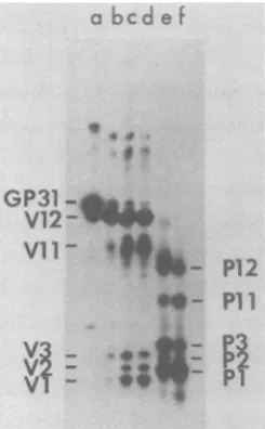
Figure 5 shows a partial proteolytic map of GP31. Theundigested protein is in lanea, and lanesb, c, and d show the results ofdigestion with increasing amounts of the protease from Staphylococcus aureus, strain V8. With10ng of
protease(Fig.5,lane b)onemajorfragmentwas
observed. This fragment was slightly smaller than theundigested protein and has been desig-nated fragment V12. With higher amounts of
protease (lanescandd) smallerfragments,
des-ignated Vll, V3, V2, and
Vi,
appeared. Diges-tion with 10and 50ng of papain produced five fragments, whichwehavedesignated P12, P11, P3, P2, and Pl (Fig. 5, lanes e and f). Thepresence of the V12fragment in laneawasdue
tothe leakage of theproteasefrom the adjacent lane.Thisfragmentwasnotvisible inundigested GP31 in similarmappingexperiments where the proteinwaswell separated from lanes containing
protease(e.g., Fig. 6, sectionI, lane 31).
InFigure 6the proteolysis products of GP31
are compared to those derived from GP36, GP28, and P23. Thefigure is divided into three
sections,eachcontaining all fourproteinsof the
P23family. Section I showsthe untreated
pro-teins,sectionIIshows the resultsof proteolysis
with50 ngofS. aureus protease,andsectionIII
shows the results ofproteolysis with 10 ng of papain. The first four lanes in each section show theprofiles obtained for the GP36, GP31, GP28, and P23 from virions. Bands smaller than the
undigested GP31 and P23 may have resulted
from spontaneous proteolysis. P23 did not
mi-grate as a doublet in other experiments. The
smaller S. aureusproteaseandpapainfragments
(Vi,
V2,V3,P1,P2,and P3)wereofequalsize inall four proteins. This demonstrated the ho-mology ofthefour proteins whichwasapparentfrom the peptide maps. The large S. aureus
protease fragments
Vii
and V12 and the largepapain fragments
Pit
and P12 varied in sizeamong theproteinsinmuchthe same way as the
VOL.42, 1982
on November 10, 2019 by guest
http://jvi.asm.org/
90 +84
D
31+14
II
I,x
3
'm
36 +31
FIG. 3. Mapsof mixtures ofmethionine-containingtryptic peptides of IBV virion proteins.Mapsofmostof
theproteins analyzed in this figurearealsoshownin Fig. 2. However, GP31 inpanels B, C, and Dwasfroma
dif-ferentpreparationthantheothermapsofthisprotein. Mixturesof peptidesobtained from the virionproteins
wereanalyzedintwodimensions. Exposureswerefor 21or28days. "O" indicates theorigin in panel A. The remainingsymbolsarediscussed in thetext.Panels: A, GP90plus GP84;B,GP90 plusGP31; C, GP31plusP14; D, GP31;E, GP36plus GP31;andF,GP28plus GP23.
A
B
90+
31
E
4f c
A
0.
-.3 t2 0C;
C
D
31
I.
*0(
E
F
28+23
a 4b
on November 10, 2019 by guest
http://jvi.asm.org/
[image:7.492.68.418.31.650.2]VOL.42,1982~~~~~~CORONAVIRUSPROTEINS 215
E 31+23
0
FIG. 4. Two-dimensionaltrypticpeptidemapsof IBV virionproteinslabeled with[35S]cysteine.Virionswere
labeled with [35S]cysteine, andtryptic digestswerepreparedfortwo-dimensionalmappingasdescribed in the text;10,000to40,000cpmwasappliedtoeachplate,andfluorographywasfor 5days."0"designatestheorigin
inpanelA. Panels: A,GP36;B, GP31, C,GP28;D, P23;andE,GP31 plusP23.
A
36
B
31
9
0
4*
'S
S
0
C
28
D
23
4p 0
$
L
VOL. 42,1982on November 10, 2019 by guest
http://jvi.asm.org/
[image:8.492.106.404.98.616.2]216 STERN, BURGESS, AND SEFTON
intact proteinsvary. For example,theV12
frag-mentfromGP36 was slightlylargerthan theV12
fragmentofGP31,which in turnwaslargerthan
the V12 of GP28, whichwassimilarly larger than the V12 of P23. Thus, cleavage of all four proteins with S. aureus protease orpapain
re-sulted in a similar spectrum offragments. The smallerfragmentswerethesamesize for all four proteins and must have been derived from
re-gions of the molecules which are homologous. The largest of these common fragments was
papainfragment P3.Wehaveestimated the size of this fragment to be approximately 9,000 to
10,000daltons, so atleast half the length ofP23,
and perhaps more, is homologous tothe other proteins.
Aminoterminusof P23.Synthesis ofP23by in vitro translation enabledus todeterminewhich proteolyticfragmentsincluded the amino
termi-nus of that protein. A 23K protein synthesized
by translation of RNA extracted from
IBV-infectedcells wasanalyzed by partial
proteoly-sis(Fig.6,lane A). The undigested protein (Fig. 6, section I, lane A) comigrated with virion protein P23 and the proteolytic cleavage pat-terns of thetwo proteinswerealmost identical (Fig. 6, sections II and III, lanes A and P23). Thus, this in vitro translation product is
indistin-a
bcdef
4.*
GP31 *;
Vll-
0P12
"-
P1l
.. a; ....
v-
is
4-
Pi2
FIG. 5. PartialproteolyticmapofGP31.
Carboxy-methvlatedGP31containing approximately 3,000cpm wasmappedby partial proteolysisasdescribed in the
text.Theproteolyticcleavage fragmentsare
designat-ed as discussed in the text. The fluorograph was
exposedfor 60days. Lanes: a, noprotease;b,GP31 digestedwith10ngofS.aureus V8protease (Miles Laboratories);c,50ngofS.aureusprotease;d,100ng
ofS.aureusprotease;e,10ngofpapain(Sigma);and f,50ngofpapain.
guishable from P23. The 23K in vitro translation
products in lanes B and C were prepared by
translation of the same RNA as the protein in
laneA, but were labeled with
[35]formyl
methi-onine by synthesis in the presence of[35S]formyl
methionyltRNAmfet. Under these conditions the label should be incorporated only attheamino terminus of the protein. Digestion of the formyl methionine-labeled 23K protein with S. aureus
protease andpapain (Fig. 6,sections II and III,
lanesB andC) revealed that only the unique S.
aureusproteasefragmentsVll and V12 and the
unique papain fragments P11 and P12 contained
label. These fragments therefore must include
the aminoterminus of P23. The lack of labeling
ofthe shared fragments Vi, V2, V3, Pl, P2, and
P3 demonstrated that the formyl methionine labelwasnotincorporated internally.
Glycosylated fragmentsofGP31.GP31 labeled with [3H]mannoseor[3H]glucosamine was
ana-lyzedby partial proteolysistodetermine which fragments containoligosaccharides (Fig.7).The variable S. aureus protease fragments Vii and
V12werelabeled with mannose(Fig. 7, lane 1)
andglucosamine (Fig. 7, lane 3). The variable papain fragments Pit and P12werealso labeled with the 3H-monosaccharides (Fig. 7, lanes 4 and 6). However, the common fragments Vi, V2, V3, P1, P2, and P3 were not labeled with either sugar. Therefore, the variable fragments of GP31 beartheoligosaccharide moieties. Simi-lar results were obtained for GP36 (data not shown).
DISCUSSION
Wehave found nineproteinsinvirions of IBV
grownintissue culture andsubjected to
exten-sive purification. The major polypeptides are
P51, GP84, and afamily ofstructurally related
proteins,GP36, GP31,GP28, and P23. We also
found three minor proteins in IBV virions,
GP90,GP59, andP14.Thisprotein composition
is in virtually complete agreement with the
re-sults of Cavanagh (6), who characterized the structuralproteinsof M41 (Massachusetts)IBV produced in ovo and with the results of
Lom-niczi andMorser(14). Our results also resemble
to some extentthose obtainedby several other
groupsfor avian infectious bronchitis virus (12,
16, 17, 34a) and mammalian coronaviruses (4,
15, 20, 32, 35).Itislikelythat GP84 isthelarge
glycoproteincomprisingthepeplomers.Proteins in the P23 family, especially the predominant
GP31, probablycorrespondtothesmallermajor
glycoprotein(s) describedbyothergroups.
Several observations suggest thatGP84, P51,
GP36, GP31,GP28,andP23arevirus-inducedor
virus-encoded proteins: (i) infection of CEK
cells with IBVresults in de novo synthesis of
proteinscomigrating with GP84, P51, G31, and J.VIROL.
on November 10, 2019 by guest
http://jvi.asm.org/
[image:9.492.80.203.376.574.2]CORONAVIRUS PROTEINS 217
36 31 28 23A B C
11
36 312823A B C
III
3631 2823 A
0
S. lb
*1
* 04
'I,
_
A*
4>_
0.40 ~~4
P.l4 _04
ot_
-V3
-VV
_
w*0
P3 lw*
P2,4
P1
;
FIG. 6. Partialproteolyticmapsof the P23familyandthe 23K in vitro translationproduct.A23Kproteinwas synthesizedbyin vitro translation of RNA isolated fromIBV-infectedcells asdescribed in thetext.Gel-purified
RNACwastranslated forlanes A andB,andunfractionated RNAwastranslated for lane C. The 23Kprotein waslabeledby translationinthepresenceof[ISlmethionine(lanes A)or[35S]formyl methionyltRNAfCt(lanes
B andC).[I5S]methionine-labeledvirionproteinsand the invitro translation productswerecarboxymethylated
andmappedby partial proteolysisasdescribedin thetext.Fluorographywasfor 90days.Sections:I,undigested
proteins; II, productsofdigestionwith50ngofS.aureusprotease;andIII, productsofdigestion with 10ngof
papain.Lanes36, 31, 28,and 23 contain virionproteinsGP36,GP31, GP28,and P23.LanesAcontainthe 23K in vitrotranslation product synthesized by translation of RNA C and labeled with[35S]methionine; lanes B contain the 23K productsynthesized from RNA C and labeled with[35S]formylmethionine; and lanes C contain the23K product synthesized from unfractionated RNA and labeled with [35S]formyl methionine. The unique S.aureus
proteaseandpapainfragmentsaremarkedasfollows: *, V12;0,Vll;>, Pi2; ,Pl1.
P23; (ii) proteins comigrating with GP84, P51, GP31, GP28,and P23canbe
immunoprecipitat-ed from infected, but not mock-infected, cell
lysateswithrabbitantiserumraised against
puri-fied IBV virions; and (iii) P51 and P23 can be
translated in vitro from cytoplasmic RNA
ex-tractedfromIBV-infected, butnot
mock-infect-ed,cells.
We do not yetknow whetherthe minor
pro-teins are virus-specific or host cell
contami-nants. It is clearfromourtryptic peptidemaps
thatGP90 and P14areunrelatedtotheremaining
structuralproteins.Cavanagh (6)concluded that
GP94,whichmaybeanalogoustoourGP90,isa
virus-specific protein, but that P14 was host
specific. Interestingly, Siddell et al. (26) have
detected a14K protein synthesized incells
in-fected with the murine coronavirus JHM virus
whichwasnotfound in JHM virus virions.
Two-dimensionaltryptic peptide mapping and
one-dimensional mapping bylimitedproteolysis
demonstrated that GP36, GP31, GP28, and P23
constitute agroupof relatedproteinswhichwe
have designated the P23 family. It is probable
thatsuchafamilyof proteins isafeatureof other
coronaviruses,sinceothergroupshavereported aheterogeneous series ofproteins in this size range(6, 21,26). Forexample,Siddelletal. (26)
B C
n a
9
C::W
0
VOL. 42,1982
lw
on November 10, 2019 by guest
http://jvi.asm.org/
[image:10.492.49.450.68.413.2]218 STERN, BURGESS, AND SEFTON
A
1 2 3
V 12 W..
V12
V3 V2
VI
*
B
4 5 6
.._
_*
FIG. 7. Partial proteolytic maps ofG with[3H]mannose, [355]methionine, andI
mine. GP31 obtained from[35S]methionini
rions andportionsof themannose-andg labeled virion preparations shown in
mapped by partial proteolysisasdescribec
Fluorographywasfor 42days. Proteolysis
ngof S.aureusprotease(panel A) and 10 (panel B). The cleavagefragments ared4
described in the text. Lanes: 1, GP31 1
[3H]mannose; 2,GP31labeledwith[35S]m4
GP31 labeled with [3H]glucosamine; 4, G
with [3H]mannose; 5,GP31 labeled with
nine; and6, GP31labeledwith[3H]glucos identifieda23Knon-glycosylatedprc
murine coronavirus JHM as wellas
slightly larger glycoproteins, and C Anderson(6a) have shown thattwos
hepatitis virus proteinsare structura]
Our results implythatasfewas th
many as nine viral genes mayencod structuralproteins. Since thereare a
subgenomic mRNAs it is possible
virionproteinisencodedby adiffere The IBV mRNAs comprise a 3'
nested set. Each mRNA contains quences of the smaller RNA species
tional"unique"sequencesatits5'ei SincethemRNAsoverlap,itisconce
the IBVproteinswould besynthesize lation of the same region in differen
However,wehaveshown that theIB
exceptingthe P23family,arelikely t
turally distinct,soanyoverlappingtn
the same reading frame must only 4
short regions. The simplest hypotht each mRNA istranslated only over
portion at its 5' end, as occurs wil
alphavirus mRNAs. A prediction o:
overlappingtranslational model isth;
ofprimary translation products would reflect the
length of the unique sequences at the 5' end of
each mRNA. Results which we have obtained
by translation of fractionated IBV mRNAs in
vitro are consistent with this model since the
sizeofthe translationproductis not
proportion-alto the size of the mRNA. IBV RNA A, which
is 0.8 x 10⁶ daltons in size,apparentlyencodes
P12
P51,
and RNAC,
which is 1.3 x 10⁶ daltons insize, can be translated to synthesize P23 (our
unpublished results). Similar results have been
P11 reported by others (21, 25). This model for
translation of coronavirus mRNAs implies the
existenceof multiplecryptic sites for initiation
P3 of translation on the genome, each of which
P2 wouldonly be activated when locatedproximal
P1 tothe 5' endofanmRNA.
The mechanismby which proteins of theP23
family arise is unclear. We obtained indirect
,P31
labeled evidencethatproteins
of theP23family
differin[3H]glucosa-
their amino-terminal domains. Limitedproteo-e-labeled vi- lytic digestion of each of the proteins witheither
lucosamine- S. aureus protease or papain produced five
Fig. 1 was fragments. Some fragments from all four
pro-1 in the text. teins were the same size, but the largest
frag-waswith 50 ments were unique to each protein. Thelargest
ngof
papain
uniquefragment produced by cleavage of eachesignated
as protein with S. aureus V8 protease wasdesig-abeled with nated V12. TheV12fragments apparently
differ
ethionine; 3,
rP31 labeled in size in the same way as the intact
proteins
[35S]methio-
differ
(Fig. 6). The simplest explanation for thissamine. is that the V12 fragments were generated by
cleavage of the fourproteinsathomologous sites which results in equal reductions in size of the )tein in the proteins. We showed that the V12 fragments
aseries of derived from P23 labeled with
[35S]formyl-me-Mheley and thionine retain the label. Thus, the P23 V12
,uchmouse fragmentcontains the amino terminus of P23. If
Ily related. all of the V12fragmentsarein factgenerated by
iree andas cleavageatthesamesite, then eachshouldalso
le the IBV contain the amino terminus of theproteinsfrom
It least five which it is derived. Thus, it is likely that the
that each proteins of the P23 familydiffer in their amino
ntmRNA. terminaldomains.
-coterminal The P23 familycould arise from synthesis of
all the se- fourprimarytranslationproducts, bydifferential
s and addi- processing of a single translation product, or
nd(30,31). through some combination of both schemes.
livablethat Some ofthe differences among the P23 family
dbytrans- proteinsmustbe ascribedtoglycosylation since
it mRNAs. P23 is not glycosylated and theremaining
pro-Vproteins, teinsare.Itispossiblethatthefourproteinsare
tobestruc- variants ofP23whichareglycosylatedto
differ-anslationin entextentsandcontainidenticalprimaryamino
occur over acid sequences. This model is consistent with
esis is that the fact thatP23canbesynthesizedinvitroand the unique the finding that the unique partial proteolytic
th the two fragments of GP36 andGP31areglycosylated.
If this non- We arenowattemptingtodistinguishbetween
atthe sizes thesepossibilitieswithpulse-chase experiments
J.VIROL.
on November 10, 2019 by guest
http://jvi.asm.org/
[image:11.492.50.241.73.271.2]CORONAVIRUS PROTEINS 219
and bythe generation of non-glycosylated forms
of GP36, GP31, and GP23 by using tunicamycin
andendoglycosidase H. We are also translating
the fractionatedviral RNAs in vitro. This should
revealthe unprocessed form of each of the viral
polypeptides. Inaddition, itwill permit
determi-nation of the coding assignments for the viral
mRNAs, which,since they have been mapped to
the genome, will reveal the genetic map of the virus.
ACKNOWLEDGMENTS
We thank Tony Hunter for discussions and Jon Cooper for advice about the manuscript.
D.F.S. and L.B. weresupported by Public Health Service training grants GM 07313 and CA 09345 from the National Institutes of Health. This work was supported by Public Health Service grants CA 14195 and CA 17289 from the NationalInstitutes of Health.
LITERATURECITED
1. Beemon, K., and T. Hunter. 1978. Characterization of Rous sarcoma virus src gene products synthesized in vitro. J. Virol.28:551-566.
2. Biagham,R. W. 1975. The polypeptide composition of avianinfectious bronchitis virus. Arch. Virol. 49:207-216. 3. Bonattl,S., N.Sonenberg, A. J.Shatkln, andR. Cancedda. 1980. Restricted initiation of protein synthesis on the
potentiallypolycistronic Sindbis virus42S RNA. J. Biol. Chem.255:11473-11477.
4. Bond, C. W., J. L. Leibowltz, and J. A. Robb. 1979. Pathogenic murinecoronaviruses.II. Characterization of
virus-specific proteins of murine coronaviruses, JHMV andA59V. Virology94:371-384.
5. Bonner, W. M., and J. D. Stedman. 1978. Efficient fluo-rography of3Hand14C on thin layers. Anal. Biochem.
89:247-256.
6. Cavanagh, D. 1981. Structural polypeptides of corona-virusIBV. J. Gen. Virol.53:93-103.
6a.Cheley,S., and R.Anderson. 1981. Cellular synthesis and modification of murine hepatitis virus polypeptides. J.
Gen.Virol.54:301-311.
7.Cleveland,D. W., S. G.Fischer, M. W.Kirchner,and U. K.Laemmli.1977. Peptide mapping by limited proteolysis in sodiumdodecyl sulfate and analysis by gel electropho-resis. J. Biol. Chem.252:1102-1106.
8. Coffln,M. S.,and D. J.Alexander.1980. Avian infectious
bronchitisvirusstructuralpolypeptides:effect of different
conditions of disruption and comparison of different
strainsand isolates. Arch. Virol. 63:239-251.
9. Davies, H. A., R. R. Dourmashkln, and M. R.
Mac-Naughton. 1981. Ribonucleoprotein of avian infectious bronchitis virus. J. Gen. Virol. 53:67-74.
10. Hunter, T., and J. I.Garrels. 1977. Characterization of themRNAs fora-,P-,and-y-actin. Cell12:767-781.
11. Kltamura,N., B. L.Semler, P. G. Rothberg, G. R.Larsen,
C. J. Adler, A. J. Doruer, E. A. Emini, R. Hanecak, J. J. Lee, S. vanderWerf, C. W.Anderson, and E.Whmmer. 1981. Primarystructure, geneorganizationand polypep-tide expression of poliovirus RNA. Nature (London)
291:547-553.
12.Lanser, J. A., and C. R.Howard.1980. The polypeptides ofinfectious bronchitis virus (IBV-41 strain). J. Gen. Virol.46:49-361.
13. Lelbowltz,J. L., K. C.Wilhehusen,and C. W. Bond. 1981. Thevirus-specific intracellularRNAspecies of two mu-rinecoronaviruses:MHV-A59 and MHV-JHM. Virology
114:39-51.
14. Lomnzi, B., and J.Morser. 1981. Polypeptides of infec-tious bronchitis virus. I. Polypeptides ofthe virion. J. Gen. Virol.55:155-164.
15. MacNaughton, M. R. 1980. The polypeptides of human and mouse coronaviruses. Arch. Virol. 63:75-80. 16. MacNaughton, M. R., and J. H.Madge. 1977. The
poly-peptide composition of avian infectious bronchitis virus particles. Arch. Virol.55:47-54.
17. Nagy,E., and B.Lomnnzl.1979. Polypeptidepatternsof infectious bronchitis virus serotypes fall into two catego-ries. Arch. Virol. 61:341-345.
18. Papkoff, J., T. Hunter, and K. Beemon. 1980. In vitro translation of virion RNA from Moloney murine sarcoma virus. Virology 101:91-103.
19. Pelham, H. R. B., and R. J. Jackson. 1976. An efficient mRNA-dependent translation system from reticulocyte
lysates.Eur. J. Biochem. 67:247-256.
20. Robb, J. A., and C. W. Bond. 1979. Coronaviridae, p. 193-247. In H. Fraenkel-Conrat and R. R.Wagner(ed.), Comprehensive virology, vol. 14. Plenum Publishing Corp., New York.
21. Rottier, P. J. M., W. J.Spaan,M. C.Horzlnek,and B. A. M. van der ZeIst. 1981. Translation of three mouse hepatitis virus strain A59 subgenomic RNAs in xenopus laevis oocytes. J. Virol.38:20-26.
22. Schofield, P., and P. C. Zamecnlk. 1968. Cupric ion catalysis in hydrolysis of amino acyl-tRNA. Biochim. Biophys. Acta155:410-416.
23. Sefton, B. M., K. Beemon, and T. Hunter. 1978. Compari-son of the expression of the src gene in vitro and in vivo. J. Virol. 28:957-971.
24. SIddell, S. G., A. Barthel, and V. Ter Meulen. 1981. Coronavirus JHM: a virion-associated protein kinase. J. Gen. Virol. 52:235-243.
25. Siddell,S., H. Wege, A.Barthel,and V. TerMeulen.1980. Coronavirus JHM: cell-free synthesis ofstructuralprotein p60. J. Virol.33:10-17.
26. Skddeil,S., H. Wege, A. Barthel, and V. Ter Meulen. 1981.
Coronavirus JHM:Intracellularprotein synthesis. J. Gen. Virol. 53:145-155.
27. Sinmons, D. T., and J. H.Strauss. 1974. Translation of Sindbis virus 25S RNA and 49S RNA inlysatesofrabbit
reticulocytes. J. Mol. Biol.86:397-409.
28. Spaan,W. J. M., P. J. M.Rottier,M. C.Horzinek, and B. A. M.vanderZeUst.1981. Isolation and identification of virus-specific mRNAs in cells infected with mouse hepati-tisvirus(MHV-A59).Virology 108:424-434.
29. Stanley, W. M., Jr. 1972. Preparation and analysis of
L-[35S]-methioninelabeled transfer ribonucleic acids from
rabbit liver. Anal. Biochem. 48:202-216.
30. Stern, D. F.,andS. I. T.Kennedy. 1980. The coronavirus multiplicationstrategy. I. Identification and characteriza-tion of virus-specific RNA. J. Virol. 34:665-674. 31. Stern, D. F., and S. I. T.Kennedy. 1980. Coronavirus
multiplication strategy. II. Mapping the avian infectious bronchitis virus intracellular RNA species to the genome. J. Virol.36:440-449.
32. Sturman,L.S.1977. Characterization of a coronavirus.I.
Structural proteins: effects of preparative conditions on
themigration of protein in polyacrylamide gels. Virology 77:637-649.
33. Sturman, L. S., and K. V. Holmes. 1977. Characterization of acoronavirus. II. Glycoproteins of the viral envelope: tryptic peptide analysis. Virology77:650-660.
34. Sturman, L. S., K. V. Holmes, and J. Behnke. 1980. Isolation of coronavirus envelope glycoproteins and inter-action with the viral nucleocapsid. J. Virol. 33:449-462.
34a.Wadey, C. N., and E. G. Westaway. 1981. Structural proteins and glycoproteins of infectious bronchitis virus particles labeled during growth in chick embryo cells. Intervirology 15:19-27.
35. Wege, H., H. Wege, K.Nagashima,and V. Ter Meulen. 1979. Structural polypeptides of the murine coronavirus JHM. J. Gen. Virol. 42:37-47.
36. Welch, W. J., B. M.Sefton,and F. S.Esch.1981. Amino
terminalsequence analysis of alphavirus polypeptides. J. Virol.38:968-972.
VOL.42, 1982
on November 10, 2019 by guest
http://jvi.asm.org/


![FIG.4.labeledtext;in panelTwo-dimensional tryptic peptide maps of IBV virion proteins labeled with [35S]cysteine](https://thumb-us.123doks.com/thumbv2/123dok_us/1462974.98966/8.492.106.404.98.616/labeledtext-paneltwo-dimensional-tryptic-peptide-proteins-labeled-cysteine.webp)

![FIG. 7.,P31with Partial proteolytic maps of G [3H]mannose, [355]methionine, andI[3H]glucosa-](https://thumb-us.123doks.com/thumbv2/123dok_us/1462974.98966/11.492.50.241.73.271/fig-with-partial-proteolytic-maps-mannose-methionine-glucosa.webp)
