Arm-cranking exercise assisted by Functional Electrical Stimulation in
C6 tetraplegia: a pilot study
S. Coupaud1,2,∗, H. Gollee1, K.J. Hunt1,2, M.H. Fraser2, D.B. Allan2 and A.N. McLean2
1Centre for Rehabilitation Engineering, Department of Mechanical Engineering, University of Glasgow, Glasgow G12 8QQ. 2Queen Elizabeth National Spinal Injuries Unit, Southern General Hospital, 1345 Govan Road, Glasgow G51 4TF.
∗Corresponding Author: Dr Sylvie Coupaud, Centre for Rehabilitation Engineering, Department of Mechanical Engineering, University
Abstract
Tetraplegic volunteers undertook progressive exercise training, using novel systems for arm-cranking ex-ercise assisted by Functional Electrical Stimulation (FES). The main aim was to determine potential training effects of FES-assisted arm-crank ergometry (FES-ACE) on upper limb strength and cardiopul-monary fitness in tetraplegia. Surface FES was applied to the biceps and triceps during exercise on an instrumented ergometer. Two tetraplegic volunteers with C6 Spinal Cord Injury (SCI) went through mus-cle strengthening, baseline exercise testing and three months of progressive FES-ACE training. Repeat exercise tests were carried out every four weeks during training, and post-training, to monitor upper-limb strength and cardiopulmonary fitness. At each test point, an incremental test was carried out to determine peak work rate, peak oxygen uptake, gas exchange threshold and oxygen uptake-work rate relationship during FES-ACE. Peak oxygen uptake for Subject A increased from 0.7 l/min to 1.1 l/min, and peak power output increased from 7 W to 38 W after FES-ACE training. For Subject B, peak oxygen uptake was unchanged, but peak power output increased from 3 W to 8 W. These case studies illustrate potential benefits of FES-ACE in tetraplegia, but also the differences in exercise responses between individuals.
1
Introduction
People with spinal cord injury (SCI) can expect a near-normal lifespan, provided acute and long-term care are adequately managed, but are susceptible to recurrent health problems that affect their quality of life. These include spasticity, skin degradation with susceptibility to pressure sores, and recurrent infections in the chest and urinary tract [27]. Appropriate management can reduce their frequency or severity. Some individuals with chronic SCI are living to an old age, and the long-term health consequences have only recently emerged. These are associated with greatly reduced physical activity and extensive muscle disuse [12], which include osteoporosis in the paralysed limbs (with increased risk of fracture) [5], and much reduced cardiopulmonary fitness (with increased risk of cardiovascular disease) [9]. These secondary health complications tend to be more frequent or more severe in tetraplegia than in paraplegia. This is related to the more extensive paralysis as well as extensive dysfunction of the autonomic nervous system that results from higher levels of SCI. This autonomic dysfunction has implications for the control of heart rate, breathing and blood pressure in response to exercise [18]. A simple example is the blunted heart rate response to exercise observed in tetraplegia and high paraplegia.
In the able-bodied population, exercise may be prescribed to tackle issues such as reduced cardiopul-monary fitness, osteoporosis and reduced muscle strength. In tetraplegia, the options for exercise are limited. Only a small proportion of tetraplegics are able to perform forms of voluntary upper limb exercise that can stress the cardiopulmonary system adequately. Over the past thirty years, exercising using Functional Elec-trical Stimulation (FES) for paralysed muscles has been explored for people with SCI [26]. This technique uses small pulses of electrical current to the peripheral nerves innervating paralysed muscle to induce con-traction in the muscle. Where there is no significant lower motor-neurone damage to the nerve supplying a paralysed muscle, FES can be applied to activate motor units using surface electrodes placed on the skin or implanted electrodes placed on, around or attached to the motor nerves. A number of lower-limb FES exer-cise modalities (most of which use surface FES) have been developed to date, including FES cycling [11, 17], FES rowing [34] and FES ambulation [14]. However, most of these are designed for people with paraplegia. FES cycling has been shown to be beneficial in both tetraplegia [16] and paraplegia [19], but we pro-pose that there is a need for an exercise modality aimed specifically at tetraplegics. We hypothesised that FES-assisted arm-cranking exercise (FES-ACE), using electrical stimulation applied to the biceps and tri-ceps of both arms, could benefit an individual with tetraplegia in terms of both upper limb strength and cardiopulmonary fitness. Needham-Shropshireet al. (1997) [24] demonstrated an increase in manual muscle test scores in people with tetraplegia training with arm-cranking assisted by FES of the triceps (for elbow extension). This set-up restricted the exercise modality to individuals with voluntary control of elbow flex-ors: those with an injury at C5 and below. In this study, no cardiopulmonary measurements were made over the training period to identify possible fitness benefits in the tetraplegic subjects of regular use of the exercise modality.
The system developed in our study for FES-ACE [13, 6] allows stimulation to be set independently for each of four target muscles, which are the biceps and triceps muscles of both arms. This means that those with voluntary control of some of the muscles in the upper limbs can benefit from the additional muscle force for the exercise achieved by applying FES to paralysed or weakened muscle groups. This would potentially result in more effective and strenuous exercise. The muscles can be built up progressively and the training sessions can be made gradually more intensive, with associated peripheral adaptations within the exercising muscles. We speculated that this could result in increased cardiopulmonary fitness through peripheral and, possibly, central adaptations in tetraplegics who typically have peak oxygen uptakes significantly below those of neurologically intact sedentary people in the general population.
in the response of tetraplegic patients to FES-ACE suggests that this novel exercise intervention would be suitable for some of the tetraplegic population.
2
Methods
2.1 Subjects
Inclusion criteria were: (i) traumatic cervical SCI at C4–C6 (ii) no history of recurring autonomic dysreflexia, (iii) neurological stability, (iv) psychological stability, (v) no extensive denervation of the biceps or triceps muscles, and (vi) no excessive spasticity. The study was approved by the South Glasgow University Hospitals Research Ethics Committee, and written informed consent was obtained from the subjects.
The details for the subjects who completed the FES-ACE training and testing are given in Table 1. The grade of completeness of SCI is given according to the American Spinal Injury Association (ASIA) [21] scale.
Subject SCI level ASIA Grade TSI Age Sex
A C6 B 18 years 38 M
B C6 A 8 months 52 F
Table 1: Subject details. ‘Age’ in years and ‘Time since injury’ (TSI) are as at the start of participation.
2.2 Methods for FES-assisted Arm Crank Ergometry
The hardware consisted of a commercially available instrumented arm-crank ergometer (TheraVital, Medica Medizintechnik, Germany), an eight-channel stimulator (Motive8, Stanmore, UK) and a PC interface. Four channels were used for FES-ACE, to apply the electrical stimulation through pairs of gel surface electrodes (PALS Ultraflex, Massachussets, US) placed over the bulk of the biceps brachii and the triceps brachii of each arm. A pattern generator was used to co-ordinate the stimulation to the four muscles with the position of the cranks. This was implemented in software, running in Matlab/Simulink (The Mathworks, Massachussets, US). The stimulation pulses used were constant-current, monophasic and charge-balanced. The stimulation current amplitude was variable in 10 mA increments up to 140 mA (but the maximum used here was 30 mA), the stimulation frequency was fixed at 20 Hz, and the stimulation pulsewidth was variable from 0 to 500µs. The operator could change the stimulation pulsewidth using a potentiometer.
The FES-ACE instrumentation allowed us to control the work rate through the PC interface. The range of work rates achievable with this FES-ACE set-up included a “negative power output” range and a positive power output range, with transition through unloaded arm-cranking. During the negative range, the motor provided assistance to the subject’s arm-cranking motion. Unloaded arm-cranking refers to the point at which the subject’s arms were producing just enough power to maintain the desired cadence, but with no resistance to that motion. For positive power outputs (i.e. above unloaded), the subject had to work against resistance provided by the motor.
Full technical details of the systems for FES-assisted Arm Crank Ergometry (FES-ACE) are described elsewhere [6, 13].
During FES-ACE sessions, the subject was sitting in his/her own wheelchair. The height of the ergometer was such that the center of the cranks was horizontally aligned with the subject’s shoulders. The subject was positioned with the elbows in slight flexion when the crank was at the furthest distance from the body, and the wrists were stabilised in the armrests using straps and bandages.
2.3 Schedule of training and testing
negative workrates to zero-load arm-cranking over the muscle-strengthening period. Only those subjects who at the end of this phase were able to maintain zero-load arm-cranking for at least ten minutes were invited to progress to the next phase of the programme.
After the muscle-strengthening phase, a baseline incremental exercise test was performed in Week 0. This was Test Point (TP) 1. At the test points, the exercise testing sessions replaced the training sessions. During incremental tests, the work rate started from a zero-load baseline and was incremented in equal steps every minute until the subject could no longer continue. Incremental tests were used to determine peak power output, peak oxygen uptake, and the gas exchange threshold.
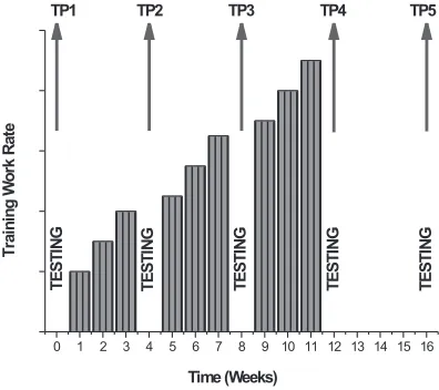
Formal FES-ACE training began in Week 1. A progressive training schedule was used, with both session duration and work rates being gradually increased over the training period. Each subject trained three times per week (for 15 to 30 minutes per session), and the FES-ACE exercise intervention period spanned 12 weeks. Repeat exercise tests were carried out during weeks 4 (TP2), 8 (TP3), and 12 (TP4). One month after the end of the FES-ACE exercise intervention period, an additional test was carried out (Week 16, TP5). The general time-line that was used for training and testing is depicted in Figure 1.
0 1 2 3 4 5 6 7 8 9 10 11 12 13 14 15 16
T
E
S
T
IN
G
T
E
S
T
IN
G
T
E
S
T
IN
G
T
E
S
T
IN
G
TP1 TP2 TP3 TP4 TP5
T
ra
in
in
g
W
o
rk
R
a
te
Time (Weeks)
T
E
S
T
IN
[image:5.612.200.398.246.422.2]G
Figure 1: Timeline representing training and testing time points, and illustrating relative work rates during training sessions. At test points, testing sessions replaced training sessions. TP5 represents the follow-up test; no training took place between TP4 and TP5.
Subjects performed no arm-cranking exercise between TP4 and TP5. Tests at TP5 allowed us to evaluate positive carry-over effects of the training intervention, or identify any negative de-training effects. For Subject A, a final test (TP6) was performed 8 weeks after TP5. During the 8 weeks between TP5 and TP6, voluntary (without FES) arm-cranking exercise training was carried out by this subject at home, using a commercially available arm-crank ergometer.
2.4 Methods for exercise testing
Each subject was familiarised with the physiological measurement equipment over a minimum of two sessions prior to baseline testing. The system used to communicate with the subject during exercise testing involved eye-blinking (one eye-blink for a “yes” and two eye-blinks for a “no” answer to the investigator’s question). Prior to each test, the breath-by-breath cardiopulmonary measuring system’s volume transducer was calibrated using a 3-litre syringe, and the O2and CO2gas analysers were calibrated using two references gases of known concentrations.
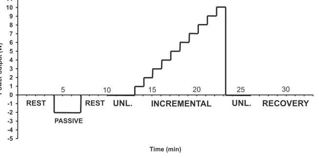
The investigator determined the step-size of the increments for the incremental test and programmed the sequence of work rate changes in the Matlab/Simulink FES-ACE exercise testing software. A schematic diagram illustrating the programmed time course of the incremental test is shown in Figure 2.
-5 -4 -3 -2 -1 0 1 2 3 4 5 6 7 8 9 10 11
Time (min)
P
o
w
e
r
o
u
tp
u
t
(W
)
PASSIVE
UNL. INCREMENTAL
REST UNL. RECOVERY
REST
[image:6.612.141.468.177.339.2]5 10 15 20 25 30
Figure 2: Example of timeline for an incremental exercise test. ‘UNL.’ refers to unloaded exercise.
Subjects were asked to refrain from eating, smoking, and drinking alcohol or coffee for at least two hours prior to the test. They were required to empty their urine bag before starting the test. Ideally, the subjects had not performed any exercise for 24 hours prior to the test.
After electrode positioning and stimulation testing, the subject was set-up at the arm-crank ergometer. When the subject had settled down, the mask was placed over the nose and mouth and tightened to ensure a good seal. At least two minutes of resting data were recorded.
Following recorded rest, the exercise began with three minutes of passive arm-cranking in order to loosen the muscles. A second resting phase was then implemented. When cardiopulmonary data stabilised during this second rest period, the subject performed three minutes of unloaded arm-cranking, at a target cadence of 50 rpm. Visual feedback of actual and target cadence was provided in real time on the PC screen. If necessary, electrical stimulation was applied to the target muscles to achieve or maintain the desired cadence. After three minutes of unloaded arm-cranking, the resistance was automatically stepped up every minute to increase the work rate in pre-programmed equal steps. The size of these steps varied between tests and between subjects. The incremental phase generally lasted between the desired optimal 8 to 12 minutes [2]. In order to maintain the cadence at increasing work rates, the stimulation intensity was increased manually by the investigator when necessary.
When the desired cadence of 50 rpm could no longer be maintained (dropping consistently to below 35 rpm), the load was reduced back down to zero. Unloaded arm-cranking during this active recovery phase lasted for 2 to 4 minutes, depending on how the subject was feeling.
2.5 Data analysis
We used commercially available statistical software (Origin 7.5, OriginLab, Massachussets, U.S.) to edit the raw cardiopulmonary data (to remove outliers) and to analyse the edited data.
The data from incremental tests were used to determine the following:
• Peak power output(in W) — calculated as the mean power output over 60 s at the highest completed work rate.
• Peak oxygen uptake (in l/min) — the breath-by-breath data were edited to remove outliers, and time aligned with the power output data. The peak oxygen uptake was calculated as the mean oxygen uptake (after editing) over the last 30 s at the highest completed work rate.
• Gas exchange threshold (in l/min) — a modified V-slope method was used to estimate the gas exchange threshold (GET) using the incremental portion of the edited data only. ˙VCO2 was
plot-ted against ˙VO2, and the ˙VO2 at the point of inflexion in the relationship was taken to represent
the GET [1, 23]. To increase the confidence that the threshold represented metabolic acidosis, the information obtained from the V-slope method was correlated with characteristic changes in the respi-ratory exchange ratio, and in the end-tidal tensions and ventilatory equivalents for oxygen and carbon dioxide.
• Oxygen uptake-work rate relationship— a linear regression was fitted to the ˙VO2-WR
relation-ship for each incremental test, and the slope of the regression was calculated.
3
Results
The data for the two case studies are presented together for comparison.
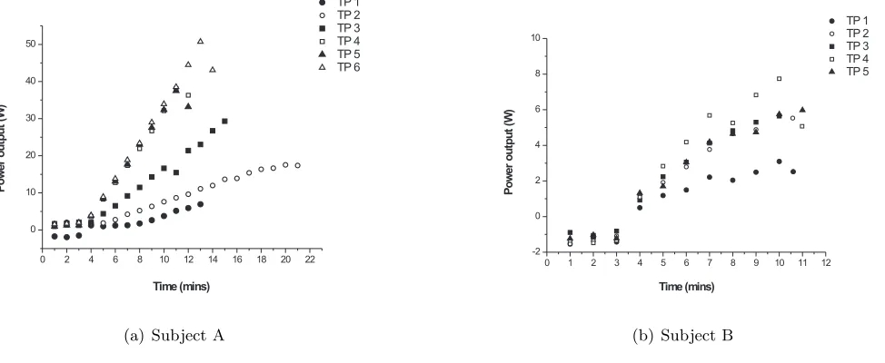
The profiles of increasing work rate during each incremental test performed by Subjects A and B are depicted in Figure 3. The graphs illustrate that for most tests the increment step-size was appropriate to limit the incremental phase to between 8 and 12 minutes, with the exception of TP2 for Subject A.
0 2 4 6 8 10 12 14 16 18 20 22 0
10 20 30 40 50
TP 1 TP 2 TP 3 TP 4 TP 5 TP 6
Time (mins)
P
o
w
e
r
o
u
tp
u
t
(W
)
(a) Subject A
0 1 2 3 4 5 6 7 8 9 10 11 12 -2
0 2 4 6 8 10
TP 1 TP 2 TP 3 TP 4 TP 5
Time (mins)
P
o
w
e
r
o
u
tp
u
t
(W
)
[image:7.612.63.550.446.646.2](b) Subject B
Figure 3: Power output profile during each incremental test.
were more frequent with Subject B, as seen in Figure 3(b), with the effect being apparent at a number of test points: at the 8th minute at TPs 1 and 4, and at the 9th minute at TPs 2 and 5. The tests were not
terminated at these points as the decreases in power output were only transient in nature, and the subject indicated that he/she was able to continue.
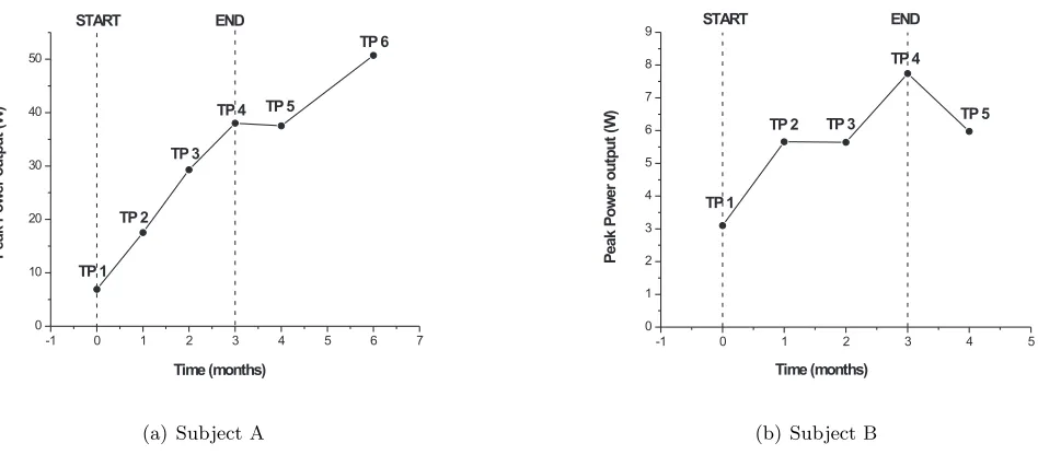
Peak values for power output (or work rate) are shown for each test point in Figure 4. Both subjects showed a gradual increase in peak power output from the start of FES-ACE exercise (TP1) to the end of training (TP4). For Subject A, the baseline peak was 7 W and the end-of-training peak was 38 W, equivalent to a rate of increase of around 10 W per month. The increase over the training period was much lower for Subject B, from 3 W to 8 W. After one month without exercise, Subject A maintained a similar peak power output (37 W) as at the end of FES-ACE training, whilst Subject B appeared to lose some upper limb strength, down to a peak power output of 6 W (TP5). Further improvements were achieved by Subject A following 8 further weeks of arm-cranking training at home (without FES), reaching a new peak power output of 51 W (TP6).
-1 0 1 2 3 4 5 6 7
0 10 20 30 40 50 TP 1 TP 2 TP 3
TP 4 TP 5
TP 6 START END Time (months) P e a k P o w e r o u tp u t (W )
(a) Subject A
-1 0 1 2 3 4 5
0 1 2 3 4 5 6 7 8 9 TP 5 TP 4 TP 3 TP 2 TP 1 START END P e a k P o w e r o u tp u t (W ) Time (months)
[image:8.612.66.540.232.440.2](b) Subject B
Figure 4: Peak power output at each test point. The start and end of the FES-ACE training period are highlighted.
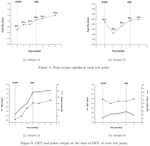
Peak values for oxygen uptake are shown for each test point in Figure 5. For Subject A, peak oxygen uptake at baseline was 0.71 l/min and increased by 50% over the FES-ACE training period to 1.05 l/min, and then to 1.21 l/min with two additional months of training at home. Subject B showed no clear improvement in peak oxygen uptake overall from the start (0.53 l/min) to the end of training (0.55 l/min).
For both subjects, the GET followed a similar pattern to the peak oxygen uptake, as did the power outputs at the GET, as shown in Figure 6.
-1 0 1 2 3 4 5 6 7 0.2 0.4 0.6 0.8 1.0 1.2 1.4 1.6 TP 6 TP 5 TP 4 TP 3 TP 2 TP 1 Time (months) P e a k V O2 (l /m in ) START END
(a) Subject A
-1 0 1 2 3 4 5
0.30 0.35 0.40 0.45 0.50 0.55 0.60 0.65 0.70 TP 1 TP 2 TP 5 TP 3 START END Time (months) P e a k V O2 (l /m in
) TP 4
(b) Subject B
Figure 5: Peak oxygen uptake at each test point.
-1 0 1 2 3 4 5 6 7 0.6 0.7 0.8 0.9 1.0 1.1 1.2 1.3 1.4 -10 0 10 20 30 40 50 START END
Time (months) G E T ( l / m i n ) P o w e r o u t p u t ( W )
(a) Subject A
-1 0 1 2 3 4 5
0.0 0.1 0.2 0.3 0.4 0.5 0.6 0.7 0.8 0 2 4 6 8 10 12 14 16 18 20 START END
Time (months) G E T ( l / m i n ) P o w e r o u t p u t ( W )
[image:9.612.56.547.48.526.2](b) Subject B
Figure 6: GET and power output at the time of GET, at each test point.
Test Point Gradient of linear regression of ˙VO2 vs Work Rate
Subject A Subject B
1 0.026 (R2=0.40) 0.029 (R2= 0.63)
2 0.023 (R2=0.75) 0.019 (R2= 0.61) 3 0.024 (R2=0.76) 0.026 (R2= 0.78)
4 0.015 (R2=0.87) 0.019 (R2= 0.51)
5 0.017 (R2=0.87) 0.024 (R2= 0.70)
6 0.017 (R2=0.87) no data
Table 2: Gradient of the linear regression of ˙VO2-WR relationship, for each incremental test. The correlation
4
Discussion
In this pilot study, two case studies are presented to illustrate the feasibility of FES-ACE in C6 tetraplegia and the potential for positive training effects following an intensive programme of FES-ACE exercise.
4.1 Upper limb strength
Regular recording of peak power output during FES-ACE exercise every four weeks allowed us to monitor upper limb strength over the training phase, and for a period of time after cessation of exercise intervention. In both case studies, overall increases in peak power production following FES-ACE exercise intervention were demonstrated.
With Subject A, the effectiveness of a progressive FES-ACE training programme was evident from the steady increase in peak power output at a rate of approximately 10 W per month, to an end-of-training value of 38 W. With such rates of improvement, individuals with similar neurological deficits following their SCI potentially may able to benefit from regular use of FES-ACE in order to enhance the strength of upper limb muscles, including those still under voluntary control. With a greater degree of initial impairment than Subject A, Subject B gained upper body strength over the period of FES-ACE training but at a slower rate. The difference between subjects was too large to have been explained by other factors alone, such as sex, age, and arm girth prior to training. Starting with a baseline peak power output of 3 W, and with an average increase of only around 1.5 W per month, it would have taken Subject B longer and possibly a more intensive training schedule to achieve the end-of-training peak power output of 38 W that Subject A had reached.
This pilot study therefore provides preliminary evidence that FES-ACE systems, when used for regular and progressive training, can build up upper body strength in some individuals with C6 SCI. The complete-ness of the injury, degree of disuse and denervation atrophy of the upper limb muscles prior to training, and the level of motivation of the individual, are all likely to influence the potential for training effects in this group. It should be noted that care would need to be taken after a long period of training that adverse degenerative effects at the shoulder did not develop.
The contribution of FES muscle recruitment to the arm-cranking exercise, in comparison to that of voluntary muscle use, cannot be discerned from the results of this pilot study. It is likely that a training schedule similar to that described here would benefit patients with sufficient initial voluntary upper body strength, without using FES. However, we can speculate that without using FES in C6 tetraplegia, an important percentage of the force-producing muscle fibres (mostly triceps) would not be recruited during arm-cranking based solely on the use of voluntarily-controlled muscles (mostly elbow flexors). FES is also likely to have contributed to the build-up of voluntarily-controlled muscle, in particular in the initial stages of the training programme. Without the simultaneous use of the biceps of one arm and the triceps in the contralateral arm, fatigue of the biceps may occur earlier on in both training and testing than with FES-assisted ACE.
4.2 Cardiopulmonary Fitness
succeeded in elevating his peak oxygen uptake to 1.05 l/min. This is above the average for tetraplegics and into the lower range of what paraplegics would achieve during voluntary arm-cranking exercise [31, 15, 7]. Subject B did not show such an improvement. With a pre-training value of 0.53 l/min, her baseline peak oxygen uptake was already below the tetraplegic average provided by Van Loan et al. [31]. Subject B’s peak oxygen uptake did not increase discernibly over the training period, remaining low (0.55 l/min) even after three months of FES-ACE training three times per week.
Another indicator of cardiopulmonary fitness, the gas exchange threshold (GET), was also estimated. The GET is taken to be representative of the lactate threshold (LT), defined as the “level of work or O2 consumption just below that at which metabolic acidosis and the associated changes in gas exchange occur” [33]. In general, the GET coincides with the LT, as it represents the onset of excess carbon dioxide production in response to lactate accumulation [1].
The validity and usefulness of the lactate threshold in FES-exercise need to be explored further. The physiological characteristics of most muscles no longer under voluntary control appear to change following SCI. The muscle composition shifts from predominantly type I prior to injury to predominantly type II muscle fibres post-injury, due to preferential atrophy of the former [3, 20]. With the latter mostly being the high-glycolytic fibres, there is little remaining potential for the muscle to perform the work required during FES-induced exercise without a significant anaerobic component.
The pattern of nerve-fibre recruitment in the muscles stimulated by external electrical pulses seems to be the inverse of the natural pattern seen in voluntary muscular contraction [25]. High glycolytic (type IIb) fibres are preferentially recruited during FES-exercise due to the lower activation threshold of their motor neurones which are typically of large diameter. Therefore, one would expect a strong anaerobic component to the metabolic activity of the working muscles early on during FES-exercise. High oxidative (type I) fibres with their smaller-diameter motor neurones may then be recruited to a greater extent later on in the exercise, after the fast fibres have fatigued. If this were the case, would FES-exercise be expected to result in a reverse LT?
This may be so for exercise relying solely on electrical stimulation for contraction of the muscles involved in the exercise, although the authors are unaware of any study showing empirical data to support this theory. In our application of FES-ACE exercise in C6 SCI the exercise was performed through a combination of the remaining volitional component and the artificially-controlled FES component. This makes the interpretation of the LT even less straightforward. Although purely speculative, we suggest that the GET determined for FES-ACE may be more a feature of the artificial control of stimulation and activation of the fibres through FES, rather than a true representation of the aerobic capacity of the muscles being stimulated. Hence, the LT may not be a reliable indicator of cardiopulmonary fitness in individuals performing FES-ACE exercise, or any other form of FES-exercise.
Training effects were also investigated using the ˙VO2-WR relationship. In the able-bodied population,
one would expect most healthy people regardless of age, sex or training status to follow a rather robust ˙
VO2-WR relationship for any specific exercise modality [32]. The position of the slope should not alter
dramatically from person to person, except in the case of obesity due to the increased oxygen cost of maintaining the higher mass of the limbs in motion at any particular work rate. Positive training effects would normally allow the trained individual to work to a higher level up that slope (approximately 0.01 l/min of ˙VO2 per W for leg-cycling), but with no obvious change in its gradient. This is because the gradient is
representative of the efficiency of the exercise, which should reflect the underlying metabolic requirements for the muscles to perform that work.
We observed an apparent change in the slope of the ˙VO2-WR relationship over the training period, which
4.3 Physiological limitations in tetraplegia
One of the major factors limiting cardiovascular responses to exercise in tetraplegia is the extent of dysfunc-tion of the sympathetic nervous system (SNS) resulting from the SCI, the effects of which tend to become more pronounced, the higher the lesion [30]. It mostly affects those with a lesion above T6 and results in reduced resting systolic and diastolic blood pressure. During exercise there is also an impaired cardiovas-cular response to the requirements for increased blood flow and oxygen delivery to the exercising muscles as evidenced from the blunted heart rate response [29]. McLeanet al.[22] highlighted the variability in the blunted heart rate response and SNS dysfunction between individuals with tetraplegia. In most cases of clinically-complete SCI leading to tetraplegia (as determined through ASIA grade classification) a significant amount of SNS dysfunction is expected, but there may be some preserved sympathetic pathways [4]. In incomplete injuries, this likelihood is increased even further. It has been suggested that with incomplete SCI lesions “the clinical examination can predict the severity of impairment of the spinal sympathetic system in only about 50% of patients” [8]. The need for diagnostic tests relating to SNS dysfunction is being addressed by a team led by Ellaway [10]. Should such tests be incorporated into routine clinical investigations, an indication of SNS dysfunction and its possible effect on exercise capacity could be obtained on a case-by-case basis prior to exercise testing and exercise prescription.
The very different cardiopulmonary responses to training with FES-ACE in two different people with the same level of cervical SCI illustrate some important points: (i) the variability of exercise responses in tetraplegia, even between individuals with the same lesion level, which may be related to the completeness of the injury; (ii) the likelihood of severe central limitations to exercise, which may be more important in complete SCI, and reduced in some cases of incomplete SCI; (iii) the need for diagnostic tests prior to exercise prescription in tetraplegia. Such tests would enable tailor-made exercise programmes to be developed to suit the individual’s capabilities and limitations.
If a larger-scale evaluation of FES-assisted arm-cranking exercise can identify its benefits and limitations in the wider tetraplegic population, FES-ACE may be considered as one of the possible exercise options in this group.
5
Conclusions
We propose that FES-ACE training could have a useful role to play in tackling some of the secondary complications of tetraplegia resulting from SCI, by providing cardiopulmonary and upper-limb conditioning. High-precision work rate control of FES-ACE exercise enabled the investigators to evaluate upper limb strength and cardiopulmonary fitness indicators at regular test points throughout a three-month FES-ACE training programme with two tetraplegic volunteers. Both subjects showed improvements in peak power output as a result of training, but only the subject with incomplete C6 SCI showed clear increases in cardiopulmonary fitness indicators. The data from our pilot study cannot be used to determine to what extent the improvements in upper limb strength and cardiopulmonary fitness were attributable to the use of FES, compared to recruitment of muscles under voluntary control. The extrapolation of potential benefits of FES-assisted arm-cranking exercise observed with these subjects to other individuals with tetraplegia would not be justified without further evaluation of the system and proposed methods.
Acknowledgements
References
[1] W.L. Beaver, K. Wasserman, ja B.J. Whipp. A new method for detecting anaerobic threshold by gas exchange. J. Appl. Physiol., 60(6):2020–2027, 1986.
[2] M.J. Buchfuhrer, J.E. Hansen, T.E. Robinson, D.Y. Sue, K. Wasserman, ja B.J. Whipp. Optimizing the exercise protocols for cardiopulmonary assessment. J. Appl. Physiol., 55(5):1558–1564, 1983.
[3] R. Burnham, T. Martin, R. Stein, G. Bell, I. MacLean, ja R. Steadward. Skeletal muscle fibre type transformation following spinal cord injury. Spinal Cord, 35:86–91, 1997.
[4] P. Cariga, M. Catley, C.J. Mathias, G. Savic, H.L. Frankel, ja P.H. Ellaway. Organisation of the sympathetic skin response in spinal cord injury. J. Neurol. Neurosurg. Psych., 72(3):356–360, 2002.
[5] B. Chen ja A. Stein. Osteoporosis in spinal cord injury. Top. Spinal Cord Inj. Rehabil., 9(1):26–35, 2003.
[6] S. Coupaud, H. Gollee, K.J. Hunt, D.B. Allan, M.H. Fraser, ja A.N. McLean. Methods & protocols for for incremental exercise testing in tetraplegia, using arm-cranking exercise assisted by Functional Electrical Stimulation. Biomed. Signal Process. Control, 4:67–78, 2009.
[7] S. Coupaud, A.N. McLean, ja D.B. Allan. Characterisation of a clinically-feasible exercise test for use in paraplegic annual reviews — a tool for cardiopulmonary fitness assessment. Physical Therapy, 2008 (under review).
[8] A. Curt, C. Weinhardt, ja V. Dietz. Significance of sympathetic skin response in the assessment of autonomic failure in patients with spinal cord injury. J. Autonom. Nerv. Syst., 61(2):175–180, 1996.
[9] A.J. Dallmeijer, L.H.V. Van der Woude, G.J. Van Kamp, ja A.P. Hollander. Changes in lipid, lipoprotein and apolipoprotein profiles in persons with spinal cord injuries during the first 2 years post-injury.Spinal Cord, 37(2):96–102, 1999.
[10] P.H. Ellaway, P. Anand, E.M.K. Bergstrom, M. Catley, N.J. Davey, H.L. Frankel, A. Jamous, C. Math-ias, A. Nicotra, G. Savic, ja D. Short. Towards improved clinical and physiological assessments of recovery in spinal cord injury: a clinical initiative. Spinal Cord, 42(6):325–337, 2004.
[11] P.D. Faghri, R.M. Glaser, ja S.F. Figoni. Functional Electrical Stimulation leg cycle ergometer exer-cise: training effects on cardiorespiratory responses of spinal cord injured subjects at rest and during submaximal exercise. Arch. Phys. Med. Rehab., 73:1085–1093, 1992.
[12] B. Fernhall, K. Heffernan, S.Y. Jae, ja B. Hedrick. Health implications of physical activity in individuals with spinal cord injury: a literature review. J. Health Hum. Serv. Adm., 30(4):468–502, 2008.
[13] H. Gollee, K.J. Hunt, S. Coupaud, A.N. McLean, ja M.H. Fraser. An apparatus for FES-assisted arm-cranking exercise in tetraplegia. Kirjassa Proc. 7th Ann. Conf. Int. FES Soc., ss. 298–300, Ljubljana, Slovenia, July 2002.
[14] D. Graupe ja K.H. Kohn. Functional Electrical Stimulation for ambulation by paraplegics. Krieger publishing company, ensimm¨ainen laitos, 1994.
[16] S.P. Hooker, S.F. Figoni, M.M. Rodgers, R.M. Glaser, T. Mathews, A.G. Suryaprasad, ja S.C. Gupta. Metabolic and hemodynamic responses to concurrent voluntary arm crank and electrical-stimulation leg cycle exercise in quadriplegics. J. Rehabil. Res. Dev., 29(3):1–11, 1992.
[17] K.J. Hunt, B. Stone, N.O. Neg˚ard, T. Schauer, M.H. Fraser, A.J. Cathcart, C. Ferrario, S.A. Ward, ja S. Grant. Control strategies for integration of electric motor assist and functional electrical stimulation in paraplegic cycling: Utility for exercise testing and mobile cycling. IEEE Trans. Neural Syst. Rehabil. Eng., 12(1):89–101, 2004.
[18] P.L. Jacobs ja M.S. Nash. Exercise recommendations for individuals with spinal cord injury. Sports Med., 34(11):727–751, 2004.
[19] T.W.J. Janssen, R.M. Glaser, ja D.B. Shuster. Clinical efficacy of electrical stimulation exercise training: Effects on health, fitness and function. Top. Spinal Cord Inj. Rehabil., 3(3):33–49, 1998.
[20] T.P. Martin, R.B. Stein, P.H. Hoeppner, ja D.C. Reid. Influence of electrical stimulation on the morphological and metabolic properties of paralyzed muscle. J. Appl. Physiol., 72(4):1401–1406, 1992.
[21] F.M. Maynard, M.B. Bracken, G. Creasey, J.F. Ditunno (Jr), W.H. Donovan, T.B. Ducker, S.L. Garber, R.J. Marino, S.L. Stover, C.H. Tator, R.L. Waters, J.E. Wilberger, ja W. Young. International Stan-dards for Neurological and Functional Classification of Spinal Cord Injury. Spinal Cord, 35:266–274, 1997.
[22] K.P. McLean, P.P. Jones, ja J.S. Skinner. Exercise prescription for sitting and supine exercise in subjects with quadriplegia. Med. Sci. Sports Exerc., 27:15–21, 1995.
[23] T. Meyer, A. Lucia, C.P. Earnest, ja W. Kinderman. A conceptual framework for performance diagnosis and training prescription from submaximal gas exchange parameters - theory and application. Int. J. Sports Med., 26:S38–S48, 2005.
[24] B.M. Needham-Shropshire, J.G. Broton, T.L. Cameron, ja K.J. Klose. Improved motor function in tetraplegics following neuromuscular stimulation-assisted arm ergometry. J. Spinal Cord Med., 20:49– 55, 1997.
[25] D.B. Popovic ja T. Sinkjaer.Control of movement for the physically disabled. Center for Sensory-Motor Interaction, Department of Health Science and Technology, Aalborg University, Denmark, toinen laitos, 2003.
[26] K.T. Ragnarsson. Functional electrical stimulation after spinal cord injury: current use, therapeutic effects and future directions. Spinal Cord, 46(4):255–274, 2008.
[27] K.T. Ragnarsson, A.B. Stein, A.M. Spungen, ja W.A. Bauman. Medical complications after spinal cord injury: spasticity, pain, and endocrine/metabolic changes. Top. Spinal Cord Inj. Rehabil., 10(2):86–106, 2004.
[28] J. Roca, B.J. Whipp, A.G.N. Agusti, S.D. Anderson, R. Casaburi, J.E. Cotes, C.F. Donner, M. Estenne, H. Folgering, T.W. Higgenbottam, K.J. Killian, P. Palange, A. Patessio, C. Prefaut, R. Sergysels, P.D. Wagner, ja I. Weisman. Clinical exercise testing with reference to lung diseases: indications, standardization and interpretation strategies. Eur. Respir. J., 10:2662–2689, 1997.
[30] R.W. Teasell, J.M.O. Arnold, A. Krassioukov, ja G.A. Delaney. Cardiovascular consequences of loss of supraspinal control of the sympathetic nervous system after spinal cord injury. Arch. Phys. Med. Rehab., 81:506–516, 2000.
[31] M. VanLoan, S. McCluer, J.M. Loftin, ja R.A. Boileau. Comparison of maximal physiological responses to arm exercise among able-bodied, paraplegics and quadriplegics. Paraplegia, 25:397–405, 1987.
[32] K. Wasserman, J. Hansen, D. Sue, B. Whipp, ja R. Casaburi. Principles of exercise testing and interpretation. Lippincott, Williams and Wilkins, kolmas laitos, 1999.
[33] K. Wasserman, B.J. Whipp, S.N. Koyal, ja W.L. Beaver. Anaerobic threshold and respiratory gas exchange during exercise. J. Appl. Physiol., 35(2):236–243, 1973.