R E S E A R C H
Open Access
Abdominal functional electrical stimulation
to assist ventilator weaning in critical
illness: a double-blinded, randomised,
sham-controlled pilot study
Euan J. McCaughey
1,2*, Annemijn H. Jonkman
3, Claire L. Boswell-Ruys
1,2,4, Rachel A. McBain
1,2,4,
Elizabeth A. Bye
1,2,4, Anna L. Hudson
1,2, David W. Collins
4, Leo M. A. Heunks
3, Angus J. McLachlan
5,
Simon C. Gandevia
1,2,4and Jane E. Butler
1,2Abstract
Background:For every day a person is dependent on mechanical ventilation, respiratory and cardiac complications increase, quality of life decreases and costs increase by > $USD 1500. Interventions that improve respiratory muscle function during mechanical ventilation can reduce ventilation duration. The aim of this pilot study was to assess the feasibility of employing an abdominal functional electrical stimulation (abdominal FES) training program with critically ill mechanically ventilated patients. We also investigated the effect of abdominal FES on respiratory muscle atrophy, mechanical ventilation duration and intensive care unit (ICU) length of stay.
Methods:Twenty critically ill mechanically ventilated participants were recruited over a 6-month period from one metropolitan teaching hospital. They were randomly assigned to receive active or sham (control) abdominal FES for 30 min, twice per day, 5 days per week, until ICU discharge. Feasibility was assessed through participant compliance to stimulation sessions. Abdominal and diaphragm muscle thickness were measured using ultrasound 3 times in the first week, and weekly thereafter by a blinded assessor. Respiratory function was recorded when the participant could first breathe independently and at ICU discharge, with ventilation duration and ICU length of stay also recorded at ICU discharge by a blinded assessor.
Results:Fourteen of 20 participants survived to ICU discharge (8, intervention; 6, control). One control was
transferred before extubation, while one withdrew consent and one was withdrawn for staff safety after extubation. Median compliance to stimulation sessions was 92.1% (IQR 5.77%) in the intervention group, and 97.2% (IQR 7.40%) in the control group (p= 0.384). While this pilot study is not adequately powered to make an accurate statistical conclusion, there appeared to be no between-group thickness changes of the rectus abdominis (p= 0.099 at day 3), diaphragm (p= 0.652 at day 3) or combined lateral abdominal muscles (p= 0.074 at day 3). However, ICU length of stay (p= 0.011) and ventilation duration (p= 0.039) appeared to be shorter in the intervention compared to the control group.
(Continued on next page)
© The Author(s). 2019Open AccessThis article is distributed under the terms of the Creative Commons Attribution 4.0 International License (http://creativecommons.org/licenses/by/4.0/), which permits unrestricted use, distribution, and reproduction in any medium, provided you give appropriate credit to the original author(s) and the source, provide a link to the Creative Commons license, and indicate if changes were made. The Creative Commons Public Domain Dedication waiver (http://creativecommons.org/publicdomain/zero/1.0/) applies to the data made available in this article, unless otherwise stated.
* Correspondence:[email protected]
1
Neuroscience Research Australia, 139 Barker Street, Randwick, NSW 2031, Australia
2School of Medical Sciences, University of New South Wales, Kensington,
NSW 2052, Australia
(Continued from previous page)
Conclusions:Our compliance rates demonstrate the feasibility of using abdominal FES with critically ill mechanically ventilated patients. While abdominal FES did not lead to differences in abdominal muscle or diaphragm thickness, it may be an effective method to reduce ventilation duration and ICU length of stay in this patient group. A fully powered study into this effect is warranted.
Trial registration:The Australian New Zealand Clinical Trials Registry,ACTRN12617001180303. Registered 9 August 2017.
Keywords:Critical illness, Electrical stimulation, Mechanical ventilation, Respiratory function, Respiratory muscles
Background
Approximately 33% of critically ill patients treated in in-tensive care units (ICUs) require mechanical ventilation to support respiration, some for a few hours, and others for months [1]. During this time, disuse atrophy of the major respiratory muscles, namely the diaphragm, abdominal and intercostal muscles, may occur [2]. This reduces respiratory function and leads to a range of complications including difficulty weaning from mech-anical ventilation [3], increased mortality, respiratory and cardiac complications, readmissions to hospital and intensive care [2–6], and decreased quality of life [7,8]. While a lifesaving intervention, need for mechanical ventilation is also associated with additional health care costs [9]. Interventions that reduce respiratory muscle atrophy or increase respiratory muscle strength are likely to reduce mechanical ventilation duration, with a direct impact on morbidity and mortality, quality of life and costs to the health care provider.
Functional electrical stimulation (FES) is the applica-tion of a train of electrical pulses to a motor nerve, caus-ing the associated muscle to contract. Transcutaneous FES of the abdominal muscles, termed abdominal FES, can improve respiratory function [10–13] and assist ven-tilator weaning in spinal cord injury [12,14]. Unlike in-spiratory muscle training, which has been shown to improve weaning outcomes for difficult to wean patients [15,16], abdominal FES does not require patient partici-pation or cooperation [16]. A pilot study of 25 ventilated critically ill participants showed that FES of the rectus abdominis and pectoral muscles maintained respiratory muscle thickness to a greater degree than sham stimula-tion and shortened ICU length of stay [17]. This is des-pite the rectus abdominis muscles making minimal contribution to expiratory pressures [18, 19], and that stimulation was not applied in synchrony with respir-ation (increasing the risk of patient-ventilator asyn-chrony and increasing the load of breathing). FES of muscles in the upper legs of ventilated critically ill patients has also been shown to reduce ventilation duration [20]. Although widely advocated as an effective technique to maintain muscle mass and reduce critical illness polyneur-omyopathy for critically ill patients [21, 22], it does not directly target the respiratory muscles. As the abdominal
muscles play an active role in cough generation and res-piration during respiratory distress [23] and we have pre-viously shown that abdominal FES is an effective way to improve cough function [13], abdominal FES may provide a more direct, practical and efficacious way to reduce mechanical ventilation duration in critical illness. This hy-pothesis is further supported by the fact that respiratory muscle strength, as measured by maximum expiratory pressure (MEP) and cough peak flow (CPF), has been shown to be an independent predictor of delayed extuba-tion, weaning success, morbidity and mortality [24, 25]. The primary aim of this pilot study was to assess the feasi-bility of employing an abdominal FES training program with critically ill mechanically ventilated patients. Second-ary objectives were to investigate the effect of abdominal FES on muscle atrophy, mechanical ventilation duration and ICU length of stay. The data collected from this study will be used to assess feasibility and estimate sample size, for a fully powered study to ascertain whether abdominal FES can reduce mechanical ventilation in critical illness.
Methods Study design
A double-blinded, randomised, sham-controlled pilot study was conducted in the 12 bed ICU of a metropol-itan teaching hospital. The study was approved by the local research ethics board.
Participants
All consecutive admissions (n = 273) between 1 Novem-ber 2017 and the 12 April 2018 were screened against the eligibility criteria (Fig. 1). Patients were eligible if they were ≥18 years of age and dependent on mechan-ical ventilation due to critmechan-ical illness. Patients were ex-cluded if they were expected to be ventilated for < 24 h or already ventilated for > 72 h, were pregnant, had non-pharmacological paralysis (e.g. spinal cord injury), had physical obstacles that prevent abdominal FES (e.g. ab-dominal trauma, pacemaker), had a diagnosed terminal illness, had no response to abdominal FES (e.g. lower motor neuron impairment or obese) or had abdominal surgery within 4 weeks prior to potential inclusion.
Similar criteria were used in a previous trial of the ef-fectiveness of FES of the quadriceps to reduce critical ill-ness polyneuromyopathy in critically ill mechanically ventilated patients [22].
Stimulation
Twenty participants were randomised to receive active (intervention) or sham (control) abdominal FES (Table 1). Participants received the first session of their allocated intervention ~ 48 h post initiation of mechan-ical ventilation (enabling washout of neuromuscular blocking agents). The set up for both groups was identi-cal, with the only difference being the stimulation pa-rameters. Stimulation was applied for 30 min, twice per
[image:3.595.61.540.263.716.2]day, 5 days per week (including first 5 days consecu-tively), until discharge from the ICU, via surface elec-trodes (5 cm × 10 cm rectangular, UF2040, Axelgaard, USA). Electrodes were placed posteriorlaterally over the abdominal wall designed to activate the transversus ab-dominis and internal and external oblique muscles as pre-viously described [18]. Stimulation was applied during exhalation using a commercially available abdominal FES device (Empi Continuum, Empi Inc., USA) with automatic synchronisation with the participant’s breathing achieved using an investigational device (VentFree VF03-K, Liber-ate Medical LLC, USA, note not approved for therapeutic use) connected between the y-piece of the mechanical ventilator and the endotracheal tube. The active group re-ceived abdominal FES at an intensity that caused a strong visible muscle contraction (median 60 mA [range 50–65 mA]), with a frequency of 30 Hz and a pulsewidth of 350μs. The stimulation current in the control group was set at 10 mA (possible sensation but no muscle contrac-tion), with a frequency of 10 Hz and a pulsewidth of 350μs. Similar training protocols have been used in other studies performed by the research team [11,12,14].
To achieve blinding, the researcher administering ab-dominal FES drew each participant’s bedside curtain
while preparing the device. This person did not perform any outcome measurements. The machine was covered with a towel or sheet, and the participants’ abdomen covered with a bed sheet so that participants, family members and caregivers could not see the machine or whether stimulation resulted in muscle contractions. Outcome assessors were never in the room when stimu-lation was delivered. Although participants were not in-formed of their randomisation allocation, they could notice the contractions caused by abdominal FES (com-pared to the control) and therefore could become aware of the allocation. Participants were instructed not to dis-cuss their perception of allocation with outcome asses-sors, other participants or clinical staff.
Data collection
[image:4.595.57.541.118.435.2]Ultrasound was performed at the end of exhalation (without stimulation) to measure the thickness of the rectus abdominis, internal and external oblique and transversus abdominis muscles and diaphragm before the first abdominal FES session, twice more in the first week of participation, and then weekly until ICU dis-charge. All measurements were taken from muscles on the right-hand side of the participant by the same Table 1Participant information. All ventilator settings refer to first day of study. APACHE III score was calculated in the first 24 h of ICU admission as described by Knaus et al. [27].PEEPpositive end-expiratory pressure,FiO2fraction of inspired oxygen,IQR interquartile range
Active (n= 10)
Control (n= 10)
Age (years) [median (IQR)]
56.5 (18.50) 61.0 (17.25)
Gender
[M/F] 7/3 5/5
Severity of illness at ICU admission
APACHE III score [median (IQR)] 81.5 (37.75) 82.0 (14.00)
Diagnostic category at admission [n(%)]
Brain injury 6 (60%) 2 (20%)
Sepsis/septic shock 0 3 (30%)
Respiratory failure 0 2 (20%)
Trauma 0 0
Post-surgical 1 (10%) 0
Meningitis 1 (10%) 1 (10%)
Other 2 (20%) 2 (20%)
Baseline ventilation characteristics
Mode of ventilation [n(%)]
Synchronized intermittent-mandatory ventilation 10 (100%) 9 (90%)
Adaptive pressure ventilation 0 1 (10%)
PEEP (cmH2O) [median (IQR)] 10.0 (3.50) 10.0 (2.25)
FiO2(%) [median (IQR)] 25.0 (6.75) 30.0 (10.00)
Exposure to intervention (min) [median (IQR)]
assessor at all assessment sessions. To measure the rec-tus abdominis, the probe was firstly placed on the mid-line of the abdomen, 2 cm above the umbilicus to identify the linea alba. The probe was then moved lat-erally until the right rectus abdominis muscle became visible, with the probe then moved in the cranial and caudal directions until the maximum thickness of the muscle was identified. From the position of the rectus abdominis, the probe was moved to the right until the lateral abdominal muscles became visible, and moved laterally until the upper and lower limits of each muscle were parallel to each other. This was approximately at the anterior axillary line. Minimal pressure to the skin was applied during these measurements to limit muscle deformation. For the diaphragm, the probe was placed parallel to the anterior axillary line in the intercostal space between the 9th and 10th rib and moved in the cranial and caudal directions until the pleural line was identified. From this point, the probe was moved approximately 1 or 2 intercostal spaces lower to identify the costal diaphragm in the zone of apposition. In all measurements the, probe was placed perpendicular to the skin.
Respiratory function was measured via forced vital capacity (FVC), forced expiratory volume in 1 s (FEV1), peak expiratory flow (PEF), maximum inspiratory pres-sure (MIP) and maximum expiratory prespres-sure (MEP), as soon as possible after the participant was able to breathe independently. FVC, FEV1and PEF were measured with a handheld spirometer (One Flow FVC Memo, Clement Clarke International, UK) by asking the participant to exhale as fully and as forcefully as possible (verbal en-couragement provided) from total lung capacity. MIP and MEP were measured using a hand-held pressure meter (MicroRPM, Vyaire Medical, USA), with partici-pants inhaling and exhaling as fully and as forcefully as possible (verbal encouragement provided) against an oc-cluded airway from residual volume and total lung cap-acity, respectively. The size of the filter approved for use with our mouth pressure device was not compatible with the tracheostomies being used at the study site. As such, MIP and MEP were not recorded from patients with tracheostomies. All measurements were recorded with the participant supine. When possible, each measure-ment was repeated until three reproducible results within 5% were registered, and the greatest value used for analysis [28].
Ventilation duration (defined as the total number of days from the onset of ventilation until the first success-ful extubation of more than 48 h during ICU stay [22]) and ICU length of stay (the number of days from ICU admission to ICU discharge) were obtained via chart re-view by a blinded assessor at ICU discharge, while mor-tality was obtained from the participants’medical record by the same blinded assessor 6 weeks post ICU
discharge. Participants without a tracheostomy were extubated by progressively reducing ventilator support. Here, ventilator rate, pressure support, and positive end-expiratory pressure (PEEP) were decreased while respira-tory rate, respirarespira-tory effort, tidal volume and blood gases were monitored. When support reached low levels, typ-ically PEEP and pressure support of 8 and 7 cmH2O, re-spectively, participants were extubated based on clinical judgement. Participants with a tracheostomy were weaned from ventilatory support via a similar scheme and then progressive ventilator-free breathing.
Analysis
Due to the small sample size, and the risk of a normality test being underpowered, we followed the statistical methods in Dall’Acqua et al. [17], where the distribution of muscle thickness was assumed normal.
There are no respiratory function measures for the participants who died during the study. The Mann-Whitney U test, as a distribution-free non-parametric test, was used to analyse respiratory function data. Ven-tilation duration and ICU length of stay were analysed using Gray’s test in the survival analysis [30], with the competing risks of death or withdrawal of treatment (e.g. ventilator support) with the intention of subsequent death. Gray’s test compares cause-specific cumulative in-cidence curves. In the case where less than 50% of par-ticipants achieved the outcome, due to either competing events or censoring, the median time to the outcome was not estimable. The sample size required for a larger study was also calculated based on survival test and cause-specific hazard approach accounting for compet-ing events. All analyses were performed uscompet-ing SPSS (Version 22, IBM Corp, NY, USA).
Results
Study population and compliance
Twelve males and eight females, with a median age of 56.5 years in the active group and 61.0 years in the con-trol group, were recruited for this study (Table1). Four-teen patients survived to ICU discharge (8 active, 6 control). One control participant was transferred to an-other hospital before extubation. After extubation, one further control participant withdrew consent and one was withdrawn due to violent behaviour. The median time on mechanical ventilation before starting the inter-vention was 1.5 days (IQR 1 day). The median time on the study for participants who died was 11.5 days (IQR 13.25 days). Median compliance for the training sessions was 92.1% (IQR 5.77%) in the active group and 97.2% (IQR 7.40%) in the control group (p= 0.384), with active participants having a median exposure to the interven-tion of 366 min (IQR 293.8 min) and control participants 555 min (IQR 492.5 min) (Table1).
Adverse events
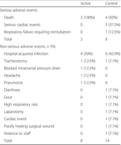
There were eight non-serious adverse events in the ac-tive group and 14 in the control group and two serious adverse events (death) in the active group and eight in the control group (four death, three cardiac events, one re-intubation) (Table 2). Only one participant, who was a control, suffered multiple serious adverse events (2 cardiac events and death). For adverse events, two active participants experienced three events (2 hospital-ac-quired infections and pneumonia; 2 hospital-achospital-ac-quired in-fections and tracheostomy), and one had one adverse event and a serious adverse event (blocked pressure
drain and death). In the control group, one participant experienced four adverse events (2 hospital-acquired in-fections, pneumonia and tracheostomy), two had three adverse events and a serious adverse event (high respira-tory rate, hospital-acquired infection, diarrhoea and ser-ious cardiac event; 2 hospital-acquired infections, laparotomy and reintubation) and one experienced an adverse event and serious adverse event (poor wound healing and death). All other adverse or serious adverse events were experienced by individual participants. An independent safety data monitoring committee judged that none of the serious adverse events were related to the intervention.
Muscle atrophy
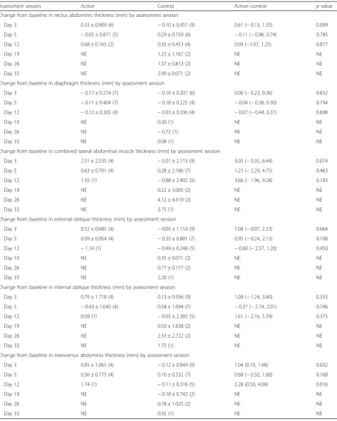
[image:6.595.305.540.452.732.2]There was no difference in the longitudinal changes from baseline in the thickness of rectus abdominis, dia-phragm and combined lateral abdominal muscles be-tween groups at any assessment sessions (Table 3). Further analyses were also conducted on each individual muscle within the combined lateral abdominal muscles, where there did appear to be a change in the thickness
Table 2Adverse events stratified by group. Serious adverse events and non-serious adverse events are reported separately. All data are reported as absolute number of events, as well as the proportion of all serious adverse or non-serious adverse events per group. While some participants experienced multiple adverse events (see the“Results”section:“Adverse events”), none of the adverse events reported here were deemed to be related to the intervention
Active Control
Serious adverse events
Death 2 (100%) 4 (50%)
Serious cardiac events 0 3 (37.5%)
Respiratory failure requiring reintubation 0 1 (12.5%)
Total 2 8
Non-serious adverse events,n(%)
Hospital-acquired infection 4 (50%) 6 (42.9%)
Tracheostomy 1 (12.5%) 1 (7.1%)
Blocked intracranial pressure drain 1 (12.5%) 0
Headache 1 (12.5%) 0
Pneumonia 1 (12.5%) 0
Diarrhoea 0 1 (7.1%)
Gout 0 1 (7.1%)
High respiratory rate 0 1 (7.1%)
Laparotomy 0 1 (7.1%)
Cardiac event 0 1 (7.1%)
Poorly healing surgical wound 0 1 (7.1%)
Violence to staff 0 1 (7.1%)
Table 3Group comparison of change from baseline in thicknesses of rectus abdominis, diaphragm, combined lateral abdominal muscle and each individual muscle by assessment session. Data are summarised as mean ± SD (n). Active versus control analysed using a least square mean difference based on a mixed effects model for repeated measures.pvalues based on a mixed effects model for repeated measures.NEnot estimable
Assessment session Active Control Active–control pvalue
Change from baseline in rectus abdominis thickness (mm) by assessment session
Day 3 0.33 ± 0.909 (6) −0.10 ± 0.451 (9) 0.61 (−0.13, 1.35) 0.099
Day 5 −0.03 ± 0.871 (5) 0.29 ± 0.759 (6) −0.11 (−0.96, 0.74) 0.785
Day 12 0.68 ± 0.165 (2) 0.35 ± 0.453 (4) 0.09 (−1.07, 1.25) 0.877
Day 19 NE 1.22 ± 1.167 (2) NE NE
Day 26 NE 1.37 ± 0.813 (2) NE NE
Day 33 NE 2.90 ± 0.071 (2) NE NE
Change from baseline in diaphragm thickness (mm) by assessment session
Day 3 −0.17 ± 0.274 (7) −0.18 ± 0.207 (6) 0.06 (−0.23, 0.36) 0.652
Day 5 −0.11 ± 0.404 (7) −0.18 ± 0.225 (4) −0.04 (−0.38, 0.30) 0.794
Day 12 −0.13 ± 0.305 (4) −0.03 ± 0.336 (4) −0.07 (−0.44, 0.31) 0.698
Day 19 NE 0.30 (1) NE NE
Day 26 NE −0.72 (1) NE NE
Day 33 NE 0.08 (1) NE NE
Change from baseline in combined lateral abdominal muscle thickness (mm) by assessment session
Day 3 2.51 ± 2.535 (4) −0.01 ± 2.113 (9) 3.05 (−0.35, 6.44) 0.074
Day 5 0.63 ± 0.701 (4) 0.28 ± 2.186 (7) 1.23 (−2.29, 4.75) 0.463
Day 12 1.55 (1) −0.88 ± 2.902 (5) 3.66 (−1.96, 9.28) 0.183
Day 19 NE 0.22 ± 3.005 (2) NE NE
Day 26 NE 4.12 ± 4.419 (2) NE NE
Day 33 NE 3.75 (1) NE NE
Change from baseline in external oblique thickness (mm) by assessment session
Day 3 0.52 ± 0.685 (4) −0.05 ± 1.153 (9) 1.08 (−0.07, 2.23) 0.064
Day 5 0.09 ± 0.954 (4) −0.35 ± 0.881 (7) 0.95 (−0.24, 2.13) 0.108
Day 12 −1.14 (1) −0.49 ± 0.248 (5) −0.68 (−2.57, 1.20) 0.450
Day 19 NE 0.35 ± 0.071 (2) NE NE
Day 26 NE 0.77 ± 0.177 (2) NE NE
Day 33 NE 2.20 (1) NE NE
Change from baseline in internal oblique thickness (mm) by assessment session
Day 3 0.79 ± 1.718 (4) 0.13 ± 0.936 (9) 1.08 (−1.24, 3.40) 0.335
Day 5 −0.43 ± 1.040 (4) 0.58 ± 1.694 (7) −0.37 (−2.74, 2.01) 0.746
Day 12 0.09 (1) −0.05 ± 2.385 (5) 1.61 (−2.16, 5.39) 0.375
Day 19 NE 0.50 ± 1.838 (2) NE NE
Day 26 NE 2.33 ± 2.722 (2) NE NE
Day 33 NE 1.75 (1) NE NE
Change from baseline in transversus abdominis thickness (mm) by assessment session
Day 3 0.85 ± 1.065 (4) −0.12 ± 0.849 (9) 1.04 (0.10, 1.98) 0.032
Day 5 0.56 ± 0.775 (4) 0.10 ± 0.532 (7) 0.68 (−0.32, 1.68) 0.168
Day 12 1.74 (1) −0.11 ± 0.318 (5) 2.28 (0.50, 4.06) 0.016
Day 19 NE −0.18 ± 0.742 (2) NE NE
Day 26 NE 0.78 ± 1.025 (2) NE NE
of the transversus abdominis at day 3 (0.85 vs. −0.12,
p= 0.032). It should be noted that the results for assess-ment sessions beyond day 5 were not stable or not es-timable due to the small or no sample sizes.
Respiratory function
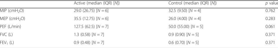
Respiratory function was assessed at a median of 6 days (IQR 3 days) from randomisation for the active group and 15 days (IQR 15 days) from randomisation for the control group (p= 0.084). In the active group, one par-ticipant was unable to perform all respiratory function measures due to delirium and one was unable to ad-equately perform MIP and MEP measurements due to tracheostomy. In the control group, one participant was unable to perform all respiratory measures due to trans-fer to another hospital and one was unable to perform MIP and MEP due to tracheostomy. There was no dif-ference in FVC (p= 0.371), FEV1(p= 0.371), MIP (p= 0.762), MEP (p = 0.283) or PEF (p = 0.061) between groups (Table4).
Clinical outcomes
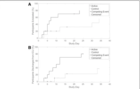
Ventilation duration (median 6.5 versus 34 days, Gray’s testp= 0.039) and ICU length of stay (median 11 versus not estimable days, Gray’s testp= 0.011) were shorter in the active compared to the control group (Fig.2). Of the 13 participants liberated from mechanical ventilation, nine (69.2%, 6 active, 3 control) were liberated by redu-cing ventilator support. The four remaining participants (two active, two control) were extubated via progressive ventilator-free breathing. The median time from initi-ation of progressive ventilator-free breathing to extuba-tion was 9.5 days (IQR 7.75 days).
There was no difference in mortality between groups (p= 0.629).
Discussion
The aim of this pilot study was to assess the feasibility of employing an abdominal FES training program with crit-ically ill mechancrit-ically ventilated patients. We also
investigated the effect of abdominal FES on respiratory muscle atrophy, mechanical ventilation duration and ICU length of stay. Our compliance to the stimulation sessions of > 90% in both groups demonstrates the feasi-bility of applying this intervention in the critically ill mechanically ventilated population. While this pilot study is not adequately powered to make an accurate statistical conclusion, we did not find a longitudinal dif-ference in respiratory muscle thickness between groups. However, ICU length of stay and duration of mechanical ventilation duration were shorter in the abdominal FES than the control group. This provides justification for a fully powered study to determine whether abdominal FES can reduce mechanical ventilation in critical illness. Such a study would require 254 participants (based on a cause-specific hazard approach accounting for compet-ing events, assumcompet-ing 60% of intervention and 45% of controls being liberated from the ventilator by day 9, logrank test (2 sided),α= 0.05 (two sided),β= 0.1, mor-tality at day 9 = 20%, 10% loss to follow-up).
[image:8.595.57.537.647.731.2]Routsi et al. [22] demonstrated that participants who received FES of the quadriceps had a ventilation dur-ation of 7 days compared to 10 days for controls (p = 0.07). Abu-Khaber et al. [20] found the same technique reduced ventilation duration from 12 to 9 days in a simi-lar group of patients (p = 0.048). However, while advo-cated clinically as a way to reduce ventilation duration [21, 22], this technique does not directly target the re-spiratory muscles. Here, we found abdominal FES ap-peared to reduce ventilation duration and ICU length of stay. In agreement, Dall’Acqua et al. [17] found that FES of the rectus abdominis and intercostal muscles reduced ICU length of stay (p= 0.045). In contrast, Routsi et al. [22] found that FES of the quadriceps did not change ICU length of stay (p= 0.11). With each ICU bed day in Australian public hospitals estimated at $A6141 (com-pared to $A2351 for a general ward bed) [31], a reduc-tion in ICU length of stay would result in a significant cost saving for local health care providers. As such, abdominal FES may offer a useful clinical addition or
Table 4Respiratory function. Respiratory function is analysed as soon as possible after the participant is able to breathe independently. There are no respiratory function measures for the six participants who died during the study. See the section
“Analysis”for further information relating to who participated in respiratory function measurements. Analysis was performed using the Mann-WhitneyUtest. All data are shown as: Median (interquartile range (IQR)) [number of participants providing data (N)].MIP maximum inspiratory pressure,MEPmaximum expiratory pressure,PEFpeak expiratory flow,FVCforced vital capacity,FEV1forced exhaled volume in 1 s
Active (median (IQR) [N]) Control (median (IQR) [N]) pvalue
MIP (cmH2O) 29.0 (26.75) [N= 6] 32.5 (9.50) [N= 4] 0.762
MEP (cmH2O) 35.5 (12.75) [N= 6] 26.0 (4.00) [N= 4] 0.283
PEF (L/min) 127.5 (62.5) [N= 7] 50.0 (55.00) [N= 5] 0.061
FVC (L) 1.3 (0.58) [N= 7] 0.9 (0.90) [N= 5] 0.371
alternative to FES of the quadriceps and is worthy of further exploration.
Our finding that there appeared to be no longitudinal change in the thickness of the rectus abdominis muscles in either the intervention or control group is in contrast to Dall’Acqua et al. [17], who found no change in rectus abdominis thickness in patients who received FES of the rectus abdominis and intercostal muscles, but a significant 16.3% decrease in the control group. This, coupled with the fact that we did not observe a difference in diaphragm thickness between the groups, may indicate that the mechanisms of abdominal FES to reduce ventilation dur-ation are not solely based on muscle thickness. Further-more, MIP and MEP are good indicators of respiratory muscle strength. [32] While these outcomes and those of lung function were not different between the two groups, a previous systematic review has shown that abdominal FES can improve respiratory function in spinal cord injury [13]. Further study of the effect of abdominal FES on respiratory function in this population is warranted.
Our average recruitment rate of 4 participants per month was higher than expected and shows the feasibility
of a larger study, particularly if it were multi-institutional. However, 12.6% of all interventions in this study were double sessions (stimulation applied for 1 consecutive hour, as opposed to two 30-min sessions), largely due to staffing issues and difficulty accessing participants (e.g. they were away for a procedure). This suggests that one training session per day may be more practical for a fol-low-up study. The mortality rate in this study (30%) was slightly lower than that in a larger study by Routsi et al. (35%) that employed the same inclusion and exclusion cri-teria [22], but is in line with large epidemiological studies of ICU patients [33].
Limitations
[image:9.595.59.539.87.393.2]led to changes in muscle architecture unrelated to atro-phy or abdominal FES. However, it should be noted that fluid imbalance alone has been shown not to affect dia-phragm thickness [34,35]. Difficulty with the ultrasound measurements led to a number of sessions having to be excluded from the analysis. As a result, more robust methods are needed to measure respiratory muscle thickness in a large clinical trial.
The majority of the analysis in this pilot study was af-fected by post-randomisation events and effects, particu-larly death. This was only accounted for in the analysis of ventilation duration and ICU length of stay, which employed Gray’s test with death and withdrawal of treat-ment treated as competing events or censoring. As such, there may be some bias in the other outcome measures due to the larger number of control participants not completing the study. Analysis of a larger study will need to account for these post-randomisation events in all outcome measures.
Conclusion
This pilot study demonstrates the feasibility of employ-ing an abdominal FES trainemploy-ing program with critically ill mechanically ventilated patients. While there were no longitudinal changes in respiratory muscle thickness be-tween groups, participants who received abdominal FES had a shorter mechanical ventilation duration and ICU length of stay. A fully powered study into this effect is now warranted, with a positive outcome likely to lead to the rapid clinical translation of this technique. This should lead to reduced morbidity and mortality, improved quality of life and a significant cost saving for the health care provider.
Abbreviations
FES:Functional electrical stimulation; FEV1: Forced exhaled volume in 1 s;
FiO2: Fraction of inspired oxygen; FVC: Forced vital capacity; ICU: Intensive
care unit; IQR: Interquartile range; MEP: Maximum expiratory pressure; MIP: Maximum inspiratory pressure; PEEP: Positive end-expiratory pressure; PEF: Peak expiratory flow
Acknowledgements
The authors would like to thank John Shen and Jason Cai from OcTech Medical for their statistical contribution to the project and review of the final manuscript. The authors would also like to thank Mr. Kamal Aryal, ICU Research Manager, for his assistance in coordinating outcome reporting. Finally, we would also like to thank all of the clinicians and staff in the ICU for their assistance in screening patients and the Safety and Data Monitoring Committee for their assistance in monitoring this trial.
Authors’contributions
EJM, SCG, AJM and JEB came up with the study concept. All authors developed the study design and protocol. EJM, CBR, RAM and ELB collected the study data. All authors were involved in the analysis and interpretation of data. EJM prepared the first draft of the manuscript. All co-authors pro-vided input and critical review of the manuscript leading to the final version. All authors read and approved the final manuscript.
Funding
This study was supported by grants from Liberate Medical, National Science Foundation, Kentucky Cabinet for Economic Development, the National
Health and Medical Research Council (NHMRC), The Prince of Wales Hospital Foundation (POWHF) and The Australian Academy of Technological Sciences and Engineering (ATSE). AJM from Liberate Medical assisted in the design of the trial and in reviewing the manuscript. Liberate Medical had no role in the collection of data. The NHMRC, POWHF and ATSE had no role in the design of the study; collection, analysis and interpretation of data; and writing the manuscript.
Availability of data and materials
The datasets used and analysed during the current study are available from the corresponding author on reasonable request.
Ethics approval and consent to participate
This study was approved by the South Eastern Sydney Local Health District Human Research Ethics Committee (Local Code: 17/050). As most
participants were lacking the capacity to give consent, approval was granted by the New South Wales Civil and Administrative Tribunal to take consent from the‘Person Responsible’for each patient (Case Number: 2017/ 00237843).
Consent for publication
Not applicable.
Competing interests
AJM is employed by Liberate Medical LLC, a medical device company that is developing an abdominal muscle stimulator. EJM has previously received financial support from Liberate Medical LLC to conduct a systematic review [13].
Author details
1
Neuroscience Research Australia, 139 Barker Street, Randwick, NSW 2031, Australia.2School of Medical Sciences, University of New South Wales,
Kensington, NSW 2052, Australia.3Department of Intensive Care Medicine, Amsterdam UMC, Vrije Universiteit Amsterdam, De Boelelaan, 1117 Amsterdam, The Netherlands.4Prince of Wales Hospital, Randwick, NSW 2031, Australia.5Liberate Medical LLC, 6400 Westwind Way, Suite A,
Crestwood, KY 40014, USA.
Received: 1 November 2018 Accepted: 16 July 2019
References
1. Esteban A, Anzueto A, Frutos F, Alia I, Brochard L, Stewart TE, et al. Characteristics and outcomes in adult patients receiving mechanical ventilation: a 28-day international study. JAMA. 2002;287(3):345–55. 2. Schellekens WJM, van Hees HWH, Doorduin J, Roesthuis LH, Scheffer GJ, van
der Hoeven JG, et al. Strategies to optimize respiratory muscle function in ICU patients. Crit Care. 2016;20(1):103.
3. Larsson L, Friedrich O. Critical illness myopathy (CIM) and ventilator-induced diaphragm muscle dysfunction (VIDD): acquired myopathies affecting contractile proteins. Compr Physiol. 2016;7(1):105–12.
4. Cook DJ, Walter SD, Cook RJ, Griffith LE, Guyatt GH, Leasa D, et al. Incidence of and risk factors for ventilator-associated pneumonia in critically ill patients. Ann Intern Med. 1998;129(6):433–40.
5. Klompas M, Khan Y, Kleinman K, Evans RS, Lloyd JF, Stevenson K, et al. Multicenter evaluation of a novel surveillance paradigm for complications of mechanical ventilation. PLoS One. 2011;6(3):e18062.
6. Mutlu GM, Mutlu EA, Factor P. GI complications in patients receiving mechanical ventilation. Chest. 2001;119(4):1222–41.
7. Chelluri L, Im KA, Belle SH, Schulz R, Rotondi AJ, Donahoe MP, et al. Long-term mortality and quality of life after prolonged mechanical ventilation. Crit Care Med. 2004;32(1):61–9.
8. Combes A, Costa MA, Trouillet JL, Baudot J, Mokhtari M, Gibert C, et al. Morbidity, mortality, and quality-of-life outcomes of patients requiring >or= 14 days of mechanical ventilation. Crit Care Med. 2003;31(5):1373–81. 9. Dasta JF, McLaughlin TP, Mody SH, Piech CT. Daily cost of an intensive care
unit day: the contribution of mechanical ventilation. Crit Care Med. 2005; 33(6):1266–71.
11. McBain RA, Boswell-Ruys CL, Lee BB, Gandevia SC, Butler JE. Abdominal muscle training can enhance cough after spinal cord injury. Neurorehabil Neural Repair. 2013;27(9):834–43.
12. McCaughey EJ, Berry HR, McLean AN, Allan DB, Gollee H. Abdominal functional electrical stimulation to assist ventilator weaning in acute tetraplegia: a cohort study. PLoS One. 2015;10(6):e0128589.
13. McCaughey EJ, Borotkanics RJ, Gollee H, Folz RJ, McLachlan AJ. Abdominal functional electrical stimulation to improve respiratory function after spinal cord injury: a systematic review and meta-analysis. Spinal Cord. 2016;54(9): 628–39.
14. Lee BB, Boswell-Ruys C, Butler JE, Gandevia SC. Surface functional electrical stimulation of the abdominal muscles to enhance cough and assist tracheostomy decannulation after high-level spinal cord injury. J Spinal Cord Med. 2008;31(1):78–82.
15. Vorona S, Sabatini U, Al-Maqbali S, Bertoni M, Dres M, Bissett B, et al. Inspiratory muscle rehabilitation in critically ill adults a systematic review and meta-analysis. Ann Am Thorac Soc. 2018;15(6):735–44.
16. Martin AD, Smith BK, Davenport PD, Harman E, Gonzalez-Rothi RJ, Baz M, et al. Inspiratory muscle strength training improves weaning outcome in failure to wean patients: a randomized trial. Crit Care. 2011;15(2):R84. 17. Dall'Acqua AM, Sachetti A, Santos LJ, Lemos FA, Bianchi T, Naue WS, et al.
Use of neuromuscular electrical stimulation to preserve the thickness of the abdominal and chest muscles of critically ill patients: a randomized clinical trial. J Rehabil Med. 2017;49:40–8.
18. McCaughey EJ, Boswell-Ruys C, Hudson AL, Gandevia SC, Butler JE. Optimal electrode position for abdominal functional electrical stimulation. J Appl Physiol. 1985;125(4):1062–1068.
19. DiMarco AF, Romaniuk JR, Kowalski KE, Supinski G. Mechanical contribution of expiratory muscles to pressure generation during spinal cord stimulation. J Appl Physiol (1985). 1999;87(4):1433–9.
20. Abu-Khaber HA, Abouelela AMZ, Abdelkarim EM. Effect of electrical muscle stimulation on prevention of ICU acquired muscle weakness and facilitating weaning from mechanical ventilation. Alexandria J Med. 2013;49:309–15. 21. Medrinal C, Combret Y, Prieur G, Robledo Quesada A, Bonnevie T, Gravier
FE, et al. Comparison of exercise intensity during four early rehabilitation techniques in sedated and ventilated patients in ICU: a randomised cross-over trial. Crit Care. 2018;22(1):110.
22. Routsi C, Gerovasili V, Vasileiadis I, Karatzanos E, Pitsolis T, Tripodaki E, et al. Electrical muscle stimulation prevents critical illness polyneuromyopathy: a randomized parallel intervention trial. Crit Care. 2010;14(2):R74.
23. Doorduin J, Roesthuis LH, Jansen D, van der Hoeven JG, van Hees HWH, Heunks LMA. Respiratory muscle effort during expiration in successful and failed weaning from mechanical ventilation. Anesthesiology. 2018;129(3): 490–501.
24. De Jonghe B, Bastuji-Garin S, Durand MC, Malissin I, Rodrigues P, Cerf C, et al. Respiratory weakness is associated with limb weakness and delayed weaning in critical illness. Crit Care Med. 2007;35(9):2007–15.
25. Jiang C, Esquinas A, Mina B. Evaluation of cough peak expiratory flow as a predictor of successful mechanical ventilation discontinuation: a narrative review of the literature. J Intensive Care. 2017;5:33.
26. Whitehead AL, Julious SA, Cooper CL, Campbell MJ. Estimating the sample size for a pilot randomised trial to minimise the overall trial sample size for the external pilot and main trial for a continuous outcome variable. Stat Methods Med Res. 2016;25:1057–73.
27. Knaus WA, Wagner DP, Draper EA, Zimmerman JE, Bergner M, Bastos PG, et al. The APACHE III prognostic system. Risk prediction of hospital mortality for critically ill hospitalized adults. Chest. 1991;100(6):1619–36.
28. Miller MR, Hankinson J, Brusasco V, Burgos F, Casaburi R, Coates A, et al. Standardisation of spirometry. Eur Respir J. 2005;26(2):319–38. 29. Mallinckrod CH, Lane PW, Schnell D, Peng Y, Mancuso JP.
Recommendations for the primary analysis of continuous endpoints in longitudinal clinical trials. Ther. Innov. Regul. Sci. 2008;42:303–19. 30. Gray RJ. A class of K-sample tests for comparing the cumulative incidence
of a competing risk. Ann Stat. 1988;16(3):1141–54.
31. Carter HE, Winch S, Barnett AG, Parker M, Gallois C, Willmott L, et al. Incidence, duration and cost of futile treatment in end-of-life hospital admissions to three Australian public-sector tertiary hospitals: a retrospective multicentre cohort study. BMJ Open. 2017;7(10):e017661. 32. Caruso P, Albuquerque AL, Santana PV, Cardenas LZ, Ferreira JG, Prina E, et
al. Diagnostic methods to assess inspiratory and expiratory muscle strength. J Bras Pneumol. 2015;41(2):110–23.
33. Beduneau G, Pham T, Schortgen F, Piquilloud L, Zogheib E, Jonas M, et al. Epidemiology of weaning outcome according to a new definition. The WIND Study. Am J Respir Crit Care Med. 2017;195(6):772–83. 34. Goligher EC, Laghi F, Detsky ME, Farias P, Murray A, Brace D, et al.
Measuring diaphragm thickness with ultrasound in mechanically ventilated patients: feasibility, reproducibility and validity. Intensive Care Med. 2015; 41(4):642–9.
35. Goligher EC, Dres M, Fan E, Rubenfeld GD, Scales DC, Herridge MS, et al. Mechanical ventilation-induced diaphragm atrophy strongly impacts clinical outcomes. Am J Respir Crit Care Med. 2018;197(2):204–13.
Publisher’s Note