Development of Analytical Methods and
Standard Reference Materials for Analysis of
Trace Elements and Isotopic Ratios in Sulphides
by
Sarah E. Gilbert
(BSc. Honours, University of Tasmania)
A thesis submitted in fulfilment of the requirements for the
degree of Doctor of Philosophy.
This thesis does not contain any material which has been accepted for the award of any other degree or diploma in any tertiary institution, and to the best of my knowledge and belief, contains no material previously published or written by another person, except where due reference is made in the text of the thesis, nor does the thesis contain any material that infringes copyright.
Signed: Date: 13/05/2015
Authority of Access
The publishers of the papers comprising Chapters 2, 3 and 4 hold the copyright for those contents. Access to those materials should be sought from and Journal of Analytical Atomic Spectrometry and Geostandards and Geoanalytical Research. The remaining non-published content of the thesis may be made available for loan and limited copying in accordance with the Copyright Act of 1968.
The following people and institutions contributed to the publication of work undertaken as part of this thesis:
Candidate Sarah Gilbert, Candidate (CODES, University of Tasmania)
Author 1 Leonid Danyushevsky (CODES, University of Tasmania)
Author 2 Ross Large (CODES, University of Tasmania)
Author 3 Sebastien Meffre (CODES, University of Tasmania)
Author 4 Helen Thomas (CODES, University of Tasmania)
Author 5 Phil Robinson (CODES, University of Tasmania)
Author 6 Karsten Goemann (CSL, University of Tasmania)
Author 7 Thomas Rodemann (CSL, University of Tasmania)
Author 8 David Death (Agilent Technologies Inc, Australia)
Author 9 Cora Wohlgemuth-Ueberwasser (University of Johannesburg, South Africa)
Author 10 Norman Pearson (GEMOC, Macquarie University, Australia)
Author 11 Dany Savard (Université du Québec à Chicoutimi, Canada)
Author 12 Marc Norman (Australian National University, Australia)
Author 13 Jacob Hanley (St. Mary’s University, Canada)
Author 14 Nobu Shimizu (Woods Hole Oceanographic Institution, USA)
Author 15 Andrey Gurenko (Centre de Recherches Pétrographiques et Géochimiques, Université de Lorraine, France)
Author Details and Their Roles:
Paper 1. Fractionation of sulphur relative to iron during Laser Ablation-ICP-MS analyses of sulphide minerals: implications for quantification. Located in Chapter 2.
The Candidate (80%) was the primary author.
Author 1 (10%) contributed to the idea, its formalisation and development.
Author 6 (5%) provided access, assistance and advice in the laboratory at the CSL, University of Tasmania.
determination of the platinum-group elements and gold by LA-ICP-MS. Located in Chapter 3.
The Candidate (70%) was the primary author.
Author 1 (10%) contributed to the idea, its formalisation and development.
Author 5 (5%) provided assistance in the production of the Ni-sulphide reference material. Author 10 (5%) provided a specimen of reference material to analyse and compare, and provided access, assistance and advice in the laboratory at GEMOC, Macquarie University.
Author 9 (2.5%), Author 11 (2.5%), Author 12 (2.5%)and Author 13 (2.5%) provided specimens of reference materials to analyse and compare.
Paper 3. Optimisation of laser parameters for the analysis of sulphur isotopes in sulphide minerals by Laser Ablation ICP-MS. Located in Chapter 4.
The Candidate (70%) was the primary author.
Author 1 (10%), Author 2 (5%) and Author 3 (5%) contributed to the idea, its formalisation and development.
Author 7 (2%), Author 14 (2%) and Author 15 (2%) provided independent analyses for the characterisation of the reference material developed in the study.
Author 4 (2%) provided samples and SHRIMP analytical data for the case study comparison by LA-ICP-MS.
Author 8 (2%) provided project funding and support.
Signed:
Date: 13/05/2015
Leonid Danyushevsky Supervisor
Discipline of Earth Sciences School of Physical Sciences University of Tasmania
Signed:
Date:
John Dickey Head of School
Laser Ablation Inductively Coupled Plasma Mass Spectrometry (LA-ICP-MS) is a micro-sampling
technique for the analysis of isotopic ratios and elemental concentrations in solid materials. It has become a widely used analytical technique for geological research; however its application has been limited in the field of ore deposit research by the small range of mineral specific reference materials available and an incomplete understanding of the fundamental processes occurring
during the laser-sample interaction for sulphide minerals. In this study the influence of laser parameters such as fluence, spot size and repetition rate on the ablation of sulphide minerals were investigated for a range of laser ablation systems: a solid state 213 nm wavelength laser, ~4 ns pulse width (New Wave UP213); a solid state 193 nm wavelength laser, ~4 ns pulse width (New
Wave UP193ss); and an ArF excimer 193 nm wavelength laser, ~20 ns pulse width (Resonetics Resolution S155). These laser systems were coupled with Agilent quadrupole ICP-MS instruments for analysis.
The ablation characteristics of a range of sulphide minerals were investigated: for bornite, chalcopyrite, pentlandite, pyrite, pyrrhotite, sphalerite and tetrahedrite. The ablation craters in
each mineral were imaged using a Field Emission Scanning Electron Microscope (FE-SEM) and significant melting was found to occur around the rim and on the crater base for some minerals. The amount of melting was partially dependent on the type of laser with the 213 nm laser producing slightly more melting than the 193 nm excimer laser. However, the relative order of minerals for the amount of melting observed was the same for both lasers, with tetrahedrite >
bornite, chalcopyrite > pentlandite, pyrrhotite > pyrite, sphalerite. There is also a strong correlation between the amount of melting and the bond strength as estimated from the Gibbs free energy of formation. This suggests that the extent of melting occurring during ablation is primarily dependent on the physical properties of the mineral, rather than the type of laser used. Three types of particles produced during ablation were identified: large droplets of solidified melt
(0.1-1.0 µm), rounded condensate particles (20-100 nm) and loose agglomerations of very fine condensates. For some minerals the proportions and size of each particle type were different between laser systems, indicating that the particle formation processes are influenced by the laser wavelength and pulse width.
Similarly using pyrite as the RM for calibration of S concentrations using Fe as the internal
standard can cause an over estimation of S by up to 50% for tetrahedrite and 30-40% for bornite and chalcopyrite. These differences can be reduced however, to <1 ‰ for 34S/32S ratios and 10-15% for chalcopyrite by using high fluence (4.6 J cm-2) with a 193 nm excimer laser. Despite this improvement in mineral dependent fractionation at high fluence, the amount of down-hole fractionation is more significant for spot ablations under these conditions and therefore is not
recommended for routine analyses. There is a positive correlation between the elemental fractionation between S and Fe and the amount of melting around the ablation crater. This is attributed to preferential volatilisation of S from molten material due to its lower boiling point compared to Fe.
The need for matrix match reference materials for accurate LA-ICP-MS has been demonstrated for
the ns pulse width lasers used, however there are few widely available RM’s for sulphide minerals. Two new RM’s have been developed in this study: NiS-3 for PGE and Au trace element analysis in the range 20-24 µg g-1 and PPP-1 pyrite for S isotopic analysis with δ34SV-CDT 5.3 ± 0.2 ‰.
Two additional secondary isotope RMs were also developed: Po-10 pyrrhotite (δ34SV-CDT 6.0 ± 0.3
‰) and N-11 bornite (δ34SV-CDT -4.4 ± 0.6 ‰). The NiS-3 RM was used to quantify the PGE and Au
concentrations in four RMs used and developed in other LA-ICP-MS facilities: 8b, PGE-A, Po727-T1, Po724-T and the Lombard meteorite. These concentrations were compared against their published values and the consistency for calibration was investigated between these RMs. Po727-T1 and 8b were consistent for all elements to within 5%, however all other RMs showed some
inconsistencies. Po724-T, Po727-T1 and the Lombard meteorite were found to be homogeneous for all elements with the analytical uncertainties, whereas the other RMs were heterogeneous for some elements especially Os in 8b and Pd in NiS-3.
The interface tubing through which the ablated material is transported from the laser ablation sample chamber to the ICP-MS can have an influence on both the precision and the length of time
for the signal to fall to background levels after analysis (washout time). This study investigated the effects of interface tubing configuration on the precision and washout of LA-ICP-MS analyses of sulphides. The washout time for the S can be longer than the signals for other elements: when using a single straight interface tube the S signal from ablating pyrite takes >120 s to washout,
the precision (from 0.66 to 0.28 ‰ uncertainty on a single 34S/32S analysis) and the washout time for S after pyrite ablation (to ~20 s).
By gaining a better understanding of the ablation processes in sulphide minerals, improvements in the washout time for S, and the characterisation of new mineral specific RMs, new applications for quadrupole ICP-MS have been developed: 1) Characterising the 34S/32S isotopic ratios in pyrite and pyrrhotite to within 1 ‰ precision and accuracy; and 2) being able to accurately measure S in
I would like to acknowledge Agilent Technologies for their financial support through their
University Relations Program; and the ARC Industry Transformation Research Hub: Transforming Mining Value Change for its support during the final year of this project. This research was also conducted under the ARC Centre of Excellence funding to CODES.
My biggest thanks are to my supervisor Leonid Danyushevsky for his patience and guidance
throughout and for persuading me to start this PhD in the first place. I have learnt a lot from him over the years about science, work and life in general. He has been very influential in helping me to become the person that I am today and for that I am very grateful.
Thank you to Ross Large, Sebastien Meffre, Marcel Guillong and Chris Hollitt for their
contributions and ideas in the preliminary stages of this research. Also thank you to all my co-authors for their contributions of samples, ideas and suggestions for improvements to the papers. Thank you to the editors and reviewers of each of the publications for their valuable editing and suggestions for improving the manuscripts. Also thank you to Paul Olin for his thorough proof reading and feedback. Thank you to Karsten Goemann, Sandrin Feig, Thomas Rodemann, Christine
Cook and Keith Harris at the CSL for their analytical expertise.
This PhD has been the culmination of learning and experimentation with the LA-ICP-MS technique while working in the CODES Analytical Facility over the past twelve years. Thank you to everyone I’ve worked with at CODES during this time for making it an enjoyable and stimulating place to work, especially to office mates and fellow lab staff past and present: Katie McGoldrick, Chris
Hollit, Terrie Sawyer, Marcel Guillong, Ian Little, Jay Thompson, Elena Lounejeva, Paul Olin, Sebastien Meffre, Ivan Belousov and Sasha Stepanov.
Declaration of Originality ... ii
Authority of Access ... ii
Statement of Co-authorship ... iii
Abstract …….… ... v
Acknowledgments ... viii
Table of Contents ... ix
Chapter 1 Introduction ... 1
LA-ICP-MS Analytical Technique ... 1
1.1 1.1.1 Instrumentation ... 1
1.1.2 Quantification ... 4
1.1.3 LA-ICP-MS in the Earth Sciences ... 6
Laser Ablation Processes ... 7
1.2 1.2.1 Ablation Mechanisms... 7
1.2.2 Particle Formation Processes ... 8
1.2.3 Element Fractionation... 9
Trace Element and Isotopic Reference Materials ... 11
1.3 Aims of This Study... 12
1.4 Chapter 2 Sulphide Ablation Processes and Element Fractionation ... 14
Abstract ... 14
Introduction ... 15
2.1 Methodology ... 16
2.2 Results and Discussion ... 18
2.3 2.3.1 Effects of Laser Energy ... 18
2.3.2 Ablation Characteristics of Sulphides ... 19
2.3.3 Ablation Rate... 24
2.3.4 Fe and S yields ... 25
2.3.7 Down Hole Fractionation ... 31
Conclusions ... 33
2.4 Acknowledgements ... 34
2.5 Chapter 3 Reference Materials for PGE Analysis ... 35
Abstract ... 35
Introduction ... 35
3.1 Experimental Procedure ... 37
3.2 3.2.1 NiS-3 Reference Material ... 37
3.2.2 Reference Materials Compared by LA-ICP-MS ... 38
3.2.3 Laser Ablation ICP-MS ... 40
Results ... 45
3.3 3.3.1 Characterisation of NiS-3 ... 45
3.3.2 PGE Concentrations by LA-ICP-MS ... 47
Discussion ... 50
3.4 3.4.1 Homogeneity of NiS-3 ... 50
3.4.2 Homogeneity of Reference Materials ... 51
3.4.3 Comparability of the PGE Content of Reference Materials ... 53
3.4.4 Matrix Effects ... 55
Conclusions ... 55
3.5 Acknowledgements ... 56
3.6 Chapter 4 Technique Development for Sulphur Isotope Analysis ... 57
Abstract ... 57
Introduction ... 57
4.1 Experimental ... 60
4.2 4.2.1 Instrumentation and analytical procedures ... 60
4.2.4 Mineral samples ... 65
4.3 Results and Discussion ... 66
4.3.1 Characterisation of mineral reference materials ... 66
4.3.2 Effect of laser ablation parameters ... 68
4.3.3 Effect of interface tubing configuration... 70
4.3.4 Tubing material and sulphur background ... 71
4.3.5 Isotopic drift ... 72
4.3.6 Matrix effects between minerals ... 74
4.3.7 Geological case study ... 76
Conclusions ... 77
4.4 Acknowledgements ... 78
4.5 Chapter 5 Conclusions and Outlook ... 79
LA-ICP-MS Analysis of Sulphur in Sulphides ... 79
5.1 Reference Materials ... 81
5.2 Application to the Earth Sciences ... 82
A.1 Other Publications ... 97
A.1.1 Co-authored Publications ... 97
A.1.2 Conference Presentations and Posters ... 98
A.1.3 Co-authored Conference Abstracts... 98
A.2 Supplementary Material for
Chapter 2 ... 100A.2.1 Analytical conditions for electron microbrobe analyses ... 100
A.2.2 Ablation crater profiles ... 101
A.2.3 Determination of the amount of melting around the ablation craters .... 103
A.2.4 Major Element Concentration of Sulphides ... 106
A.3 Supplementary Material for
Chapter 4 ... 107A.3.1 Reduction of O2 interference on masses 32 and 34 ... 107
Electronic Appendices
A.4 Publications
A.4.1 Chapter 2: Gilbert et al., 2014, JAAS, 29, 1024 A.4.2 Chapter 3: Gilbert et al., 2013, GGR, 37, 51 A.4.3 Chapter 4: Gilbert et al., 2014, JAAS, 29, 1042 A.4.4 Other Publications
A.5 Data Presented in Chapter 2
A.5.1 EDS data for ablated material in pyrite and pyrrhotite
A.5.2 Measurements of ablation rate in sulphides
A.5.3 Chemical composition of sulphides
A.5.4 Relative S concentrations measured by LA-ICP-MS
A.5.5 Effect of changing laser fluence on S fractionation
A.5.6 Down-hole fractionation in pyrite, pyrrhotite and chalcopyrite
A.6 Data Presented in Chapter 3
A.7.1 Characterisation of reference materials
A.7.2 Matrix effects for S isotopes
A.7.3 Interface configuration and S washout
A.7.4 Long term drift of Pyrite-2 analyses
Figure
1.1: Schematic of LA-ICP-MS instrumentation ... 1Figure
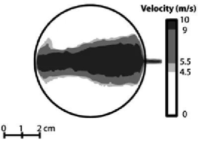
1.2: Calculated flow velocities from a barrel shaped sample chamber with internal volume33 cm3 and 0.5 L min-1 He gas flow. ... 3
Figure
1.3: Schematic cross section of the Laurin two-volume laser ablation cell ... 4Figure
2.1: Fe and S yields (cps/µg g-1) for pyrite, pyrrhotite and chalcopyrite with changing laser fluence with the 213 nm Nd:YAG and 193 nm excimer lasers.. ... 19Figure
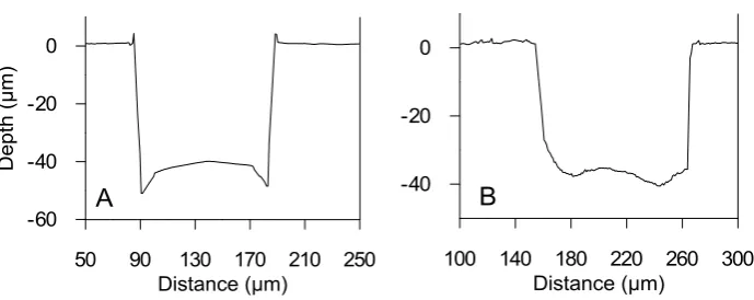
2.2: Examples of representative cross-sectional profiles of the ablation craters: A) insphalerite with the 193 nm excimer laser and B) in pyrite with the 213 nm Nd:YAG laser. ... 20
Figure
2.3: Secondary electron images of pyrite craters from: A) the 193 nm excimer laser and B) the 213 nm Nd:YAG laser. ... 20Figure
2.4: Relationship of Gibbs Free Energy with the amount of melting in and around the ablation crater.. ... 21Figure
2.5: Secondary electron images of material surrounding the ablation craters. ... 22Figure
2.6: Ablation rate (µm/pulse) for the 213 nm Nd:YAG and 193 nm excimer lasers at 4.2and 2.7 J cm-2, respectively. ... 24
Figure
2.7: Relationship between ablation rate (µm/pulse) and amount of melting with the 213nm Nd:YAG and 193 nm excimer lasers ... 25
Figure
2.8: Signal washout for A) 32S and B) 57Fe, after ablation of pyrite and pyrrhotite with either a straight tube or with the squid and coiled tube mixing devices between the laserablation sample cell and the ICP-MS. ... 27
Figure
2.9: Concentrations of S measured by LA-ICP-MS relative to the expected concentration. . 29excimer lasers. ... 31
Figure
2.12: Dependence of DHF with laser spot size, 213 nm Nd:YAG laser. ... 32Figure
2.13: Relationship of DHF and S fractionation, relative to pyrite reference standard, for spot analyses with the 213 nm Nd:YAG and the 193 nm excimer lasers. ... 33Figure
3.1: Backscattered electron image of NiS-3 showing fine lamellae of Ni3S2 and Ni7S6. ... 45Figure
3.2: LA-ICP-MS time-resolved signal for an analysis of NiS-3, with 30 s of gas background recorded when the laser was not firing, preceding 60 s of ablation with the laser on. ... 51Figure
3.3: Homogeneity of reference materials.. ... 52Figure
3.4: LA-ICP-MS time-resolved signal for the analyses of NiS-3 (Ni-sulphide) and Po724-T(Fe-sulphide) with instrument setup 2, and the Lombard meteorite analysed with instrument setup 1. ... 54
Figure
4.1: A) Isotopic fractionation in pyrite with changing fluence with the 193 nm excimer and Nd:YAG lasers. B) The effect of fluence on the measurement uncertainty with the 193 nm excimer and Nd:YAG lasers. ... 68Figure
4.2. Laser ablation analyses of pyrite (excimer laser, 50 μm beam size, 2.7 J cm-2) with the32
S signal overlain from three separate analyses using different interface configurations. .... 71
Figure
4.3. Rate of drift on multiple analyses of PPP-1 starting 15 min after the ICP-MS plasmawas switched on. ... 73
Figure
4.4. Long term drift for analyses of Pyrite-2 over seven analytical sessions, ... 73Figure
4.5. Matrix effects measured with the 193 nm Nd:YAG and excimer lasers, calibrated against PPP-1. . ... 75Figure
4.6. The degree of matrix effects with changing fluence for pyrite, pyrrhotite and bornite using the excimer laser ... 75Figure
4.7. Comparison of SHRIMP II and LA-ICP-MS analyses on the same pyrite grains from theList of Tables
Table 1.1: Laser systems and parameters commonly used for LA-ICP-MS. ... 2
Table 2.1: Laser Ablation systems used in this study. ... 18
Table 2.2: Concentrations of S and Fe in solidified melt and ablated particulates surrounding the ablation craters in pyrite and pyrrhotite. ... 24
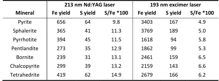
Table 2.3: Fe and S yields (cps per µg g-1) for the Nd:YAG 213 nm and 193 nm excimer lasers. ... 26
Table 2.4: Fe and S concentrations of sulphide minerals analysed by electron microprobe. ... 28
Table 3.1: Optimised instrument conditions for solution ICP-MS analyses. ... 38
Table 3.2: Optimised instrument conditions for LA-ICP-MS analyses at CODES (University of Tasmania) and GEMOC (Macquarie University) ... 41
Table 3.3: Concentrations (µg g-1) of PGE and Au in NiS-3 derived by solution ICP-MS after aqua regia digestion. ... 46
Table 3.4: Measured and published concentrations (µg g-1) and LA-ICP-MS heterogeneity (% RSD) for five LA-ICP-MS reference materials and the Lombard meteorite. ... 48
Table 3.5: Consistency between reference materials. ... 53
Table 4.1: Laser Ablation and ICP-MS instrument parameters. ... 63
Table 4.2: Average isotopic ratios for reference minerals. ... 67
Table 4.3: Isotopic measurements of PPP-1, relative to a 45 and 50 μm spot analysis with the excimer and Nd:YAG lasers respectively. ... 69
Chapter 1
Introduction
LA-ICP-MS Analytical Technique
1.1
1.1.1 Instrumentation
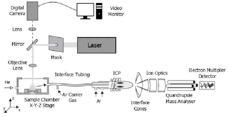
Laser Ablation Inductively Couple Plasma Mass Spectrometry (LA-ICP-MS) enables rapid, in-situ
analysis of solid materials by focusing a pulsed laser beam onto a sample surface with a spatial resolution of ~5-200 µm. The linear dynamic range of modern ICP-MS instruments allows for the simultaneous detection of both major and trace elements with low detection limits (~1 ng g-1) in a wide range of materials. The samples are sealed in a sample chamber which is mounted on a
movable x-y-z stage positioned under a stationary laser beam. A continuous flow of gas (typically either Ar or He; Gunther and Heinrich, 1999) is used as the carrier gas to transport the ablated material from the chamber, through the interface tubing to the ICP-MS. The ablated aerosol is atomised and ionised in the ICP, and for the quadrupole ICP-MS system used in this study, focused through a series of ion lenses, filtered by mass via a quadrupole and the signal counts are
[image:17.595.126.526.440.646.2]detected by an electron multiplier (Figure
1.1).Figure 1.1: Schematic of LA-ICP-MS instrumentation (modified after: www.analchem.ugent.be/ams_
onderzoek _solid)
pulse width lasers (Table
1.1). Nd:YAG crystals produce a laser at the fundamental wavelength of 1064 nm and the 5th harmonics of 213 nm is most commonly used for analysis. The photon coupling with many minerals is improved at shorter wavelengths (Gonzalez et al., 2002; Guillong and Gunther, 2002; Russo et al., 2002b) resulting in more efficient ablation despite the total beamenergy being sequentially reduced for each harmonic. ArF excimer lasers generate photons at the fundamental wavelength of 193 nm which allows for analysis at higher fluence if required. These high energy, short wavelength lasers are advantageous in providing more controlled ablation for materials transparent in the visible and near-UV spectrum (Gonzalez et al., 2002; Guillong and Gunther, 2002; Gunther et al., 1997).
Table 1.1: Laser systems and parameters commonly used for LA-ICP-MS.
Laser Source Laser Type
Wavelength (nm)
Energy Range (mJ)
Nd:YAG Solid state 1064 100-2000
CO2 Gas 1060 100-2000
Ti:sapphire Femtosecond pulse width 750-800 < 5
Ruby Solid state 694 100-1000
XeCl Excimer 308 20-200
Nd:YAG Solid state (4th harmonic) 266 1-100
KrF Excimer 248 20-200
Nd:YAG Solid state (5th harmonic) 213 0.1-20
Ti:sapphire Femtosecond pulse width 198 < 1
Nd:YAG Solid state (OPO) 193 0.1-5
ArF Excimer 193 20-200
LA-ICP-MS analyses are recorded in time resolved mode where variations in the signal over time reflect changes in the material being ablated, such as trace element zonation in minerals (Claeson et al., 2007; Maslennekov et al., 2009), depth profiling in layered samples (Farinas et al., 2010;
Pisonero and Gunther, 2008; Steely et al., 2014), and characterising boundaries between mineral grains and small inclusions (Large et al., 2009; Steadman et al., 2013). Analyses can be performed in two modes: 1) spot ablations where the laser ablates a crater into a sample deepening with time; or 2) as a line ablation where the sample is moved under the stationary laser beam creating
a channel across the surface of the material.
via a single outlet (Figure
1.2). However, it has been shown that the gas flow can vary in different regions of these chambers dependent on the gas flow path though the large volume chamber and the amount of turbulence created, resulting in variable entrainment of ablated aerosol (Bleiner and Bogaerts, 2007; Bleiner and Gunther, 2001; Loewen and Kent, 2012). Loewen & Kent (2012)showed that there can be up to 20 % variation in signal response for highly volatile elements (e.g. Zn, Se, Pb, Cd) between regions of high and low He gas flow. In addition, the time for the sample aerosol to washout from the ablation chamber can be >5 s for some barrel-shaped chamber designs due to their large internal volume (Bleiner and Gunther, 2001; Hu et al., 2008). Several small-volume barrel-type cells have been developed to improve this washout time (Gurevich and
[image:19.595.213.413.306.453.2]Hergenroder, 2007; Hu et al., 2008; Monticelli et al., 2009).
Figure 1.2: Calculated flow velocities from a barrel shaped sample chamber with internal volume 33 cm3
and 0.5 L min-1 He gas flow (from Loewen and Kent, 2012).
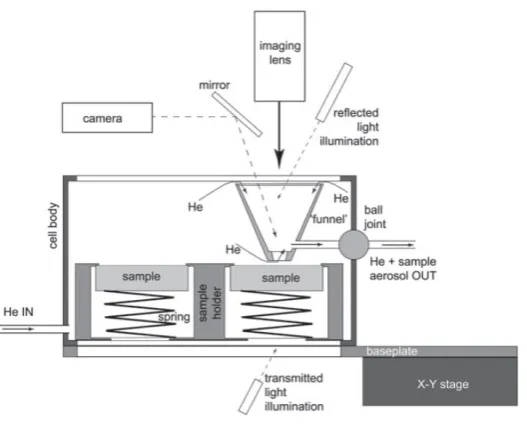
A two volume chamber (also referred to as a constant-geometry chamber) was conceptualised and developed at the Australian National University, and several in-house variations have been developed (Autrique et al., 2008; Linder et al., 2010) and commercialised by Laurin Technic Inc. (Muller et al., 2009) and Photon-Machines Inc. These sample chambers have a small volume
funnel positioned over the ablation site and the samples move in a large chamber below the
funnel (Figure
1.3). This funnel enables a constant gas flow configuration at the ablation site regardless of the sample position within the chamber, providing both fast washout (<1 s per orderFigure 1.3: Schematic cross section of the Laurin Technic two-volume laser ablation cell (from Muller et al.,
2009).
1.1.2 Quantification
Trace elements measured by LA-ICP-MS can be calibrated against an external reference material (RM) providing the ablation characteristics are similar between the RM and sample materials. The yield (the sensitivity for a given concentration in a sample; cps per µg g-1) of an element is dependent on a number of factors, including: 1) the chemical and physical properties of the
material and how well the laser couples with the material during ablation; 2) the laser wavelength, fluence, repetition rate, beam size and ablation geometry (spot or line ablations); 3) the carrier gas composition and flow rate, and the transport efficiency of the ablated aerosol through the interface tubing; and 4) ICP-MS plasma and lens settings. These numerous factors prevent direct and accurate comparison of an individual element’s yield between different
materials. However, the relative yield between two elements was found to be consistent between materials with similar ablation properties (Longerich et al., 1996). This principle can be used for quantification as described below.
The concentration (C) of an element (el) in a sample (Sa) is calculated by comparing the relative yields between elements via the following relationship (Longerich et al., 1996):
Where S is the cps sensitivity of the element and N is the normalised sensitivity as defined by:
(
)
Equation 1.2
This method requires the concentration of one element to be known in a sample which is referred
to as the IS element (IS i.e. the term in Equation
1.2). For LA-ICP-MS the internal standardmust not only be present in the sample but also occur at sufficiently high concentrations in both the sample and RM. For mineral analyses a major element is typically used and the concentration
is determined via an independent analytical technique (e.g. electron microprobe) or calculated from mineral stoichiometry (e.g. Fe in pyrite or Ca in clinopyroxene).
For all ICP-MS analyses the sensitivity of an element is prone to drift with time, and is generally greater for light atomic masses (Longerich et al., 1996). This can cause relative changes in
sensitivity between heavy mass trace elements and the typically light mass internal standard element. However, the drift can be corrected by interpolation between frequent analyses of the external RM.
The detection limit (DL) is the lowest concentration that can be confidently measured above the uncertainty of the background signal. Longerich et al. (1996) defines this uncertainty (UBG) as the
standard error of the background calculated over both the signal and background time intervals:
√
Equation 1.3
Where n is the total number of sweeps in the analyses, and σBG is the standard deviation of the
background signal. The DL is calculated as the concentration of three times the background uncertainty (UBG):
Equation 1.4
laser beam size, energy and repetition rate). Therefore, it is best practice to calculate a separate
DL for each element in each analysis.
1.1.3 LA-ICP-MS in the Earth Sciences
In the past decade LA-ICP-MS has become a widely used technique for geological research, especially in the field of economic geology (Heinrich et al., 2003; Kosler, 2001; Shaheen et al.,
2012). It has been used to characterise trace element zonation and isotopic composition of sulphide minerals which can aid in the understanding of ore forming processes (Large et al., 2007; Samson et al., 2008; Scott et al., 2009; Su et al., 2009; Thomas et al., 2011) and for improving ore extraction practices (Leichliter et al., 2011; Parbhakar-Fox et al., 2013). Spot analyses by
LA-ICP-MS have been used to determine the trace element concentrations in a range of sulphides (Cabri et al., 2003; Ciobanu et al., 2013; Cook et al., 2011; Cook et al., 2009; Maslennekov et al., 2009; Pina et al., 2013; Ulrich et al., 2011), silicate minerals (Beurlen et al., 2011; Flem et al., 2002; Humayun et al., 2010; Jeffries et al., 1995), fluid inclusions (Guillong et al., 2008; Guillong and Pettke, 2012; Kouzmanov et al., 2010; Leisen et al., 2012) and other minerals (Jochum et al., 2012;
Weiss et al., 2008).
Trace element imaging using LA-ICP-MS has become a widely used technique in both biological (Austin et al., 2010; Konz et al., 2014) and geological sciences (Gregory et al., 2013; Large et al., 2009; Large et al., 2013; Steadman et al., 2013) to provide detailed spatial relationships between elements which are not possible by spot analyses alone. For this technique the laser is rastered in
a series of parallel lines across a mineral grain and the time resolved signals stacked to produce a two-dimensional image of trace element distribution. With recent advances in processing software, three-dimensional images of multiple, stacked ablations are also possible (Chirinos et al., 2014; Hare et al., 2010). These images can reveal trace element zonation at < 10 µm resolution
within an individual mineral grain, which can aid in the understanding of mineral growth and inclusion chemistry.
LA-ICP-MS is also used for determining isotopic ratios, for example: U-Pb geochronology in zircons, apatites, monazites and other minerals (Burrett et al., 2014; Halpin et al., 2014; Jackson et
of isotope dwell times and acquisition parameters; the use of signal smoothing devices in the
interface tubing and the characterisation of several mineral specific reference materials (Aramendia et al., 2010).
Laser Ablation Processes
1.2
1.2.1 Ablation Mechanisms
The physical processes involved in the laser-sample interaction and the mechanisms of laser ablation are complex and not fully understood for many geological materials. The laser-sample interactions can be unique for different laser type and material combinations (Guillong et al., 2003a; Kosler et al., 2005; Shaheen et al., 2013a). Therefore, characterising and understanding
the ablation processes for a range of lasers and minerals is essential for accurate analyses.
A range of models have been described in the literature for the thermal processes involved for ns pulse width laser ablation: rapid, localised heating of the sample causing vaporisation, atomisation and ionisation of the material; the development of a plume of material ejected from
the ablation site; and the formation of a laser-induced plasma (Bogaerts et al., 2003; Gusarov and Smurov, 2005; Hergenroder, 2006c; Hirata and Miyazaki, 2007). These processes are governed partly by the laser parameters (i.e. wavelength, fluence and pulse width) and also on the physical properties of the material (e.g. bond strength, opacity and heat capacity; Guillong and Gunther, 2002; Kelley and Fallick, 1990; Wagner et al., 2002). Inefficient ablation can occur from loss of
energy via heating and melting of the surrounding material or transmission of photons through the sample (Gonzalez et al., 2002; Hergenroder, 2006c; Hirata and Miyazaki, 2007). The latter is most apparent for minerals such as calcite, quartz or fluorite which are transparent to visible and long wavelength UV light (Jeffries et al., 1998). For opaque minerals such as sulphides and metallic samples, loss of energy via transmission is minor but significant melting can occur around the
ablation crater especially with ns pulse width lasers (Bogaerts et al., 2003; Hergenroder, 2006a, c). A resultant melt layer can potentially be detrimental for accurate LA-ICP-MS analyses as it can
cause element and isotopic fractionation (as detailed in Section
1.2.3 below) and a loss of spatialresolution. Femtosecond lasers can reduce the amount of melting due to the pulse length being shorter than the rate of thermal diffusion for metallic samples (<1 ps for Cu metal; Hirata and Miyazaki, 2007; Shaheen et al., 2013a).
material and also on the wavelength of the laser. For fs to ps pulse width lasers the ablation
threshold for Cu metal has been shown to increase with increasing pulse width (Gamaly et al., 2002; Hashida et al., 2002). At very low fluence heating and evaporation processes can dominate rather than ablation (i.e. atomisation and ionisation; Zhigilei et al., 2003). Similarly at very high fluence, excessive temperatures can occur at the ablation site resulting in the development of a thicker melt layer at the base of the crater. The optimal fluence for ablation is dependent on the
relationship between fluence and sensitivity, ablation rate and element fractionation which varies for different minerals. To date these relationships have been described for some metals, glasses and monazites (Bogaerts et al., 2003; D'Abzac et al., 2010; Diwakar et al., 2013; Gunther et al., 1997). However, there has been little discussion in the literature of strategies to determine the
optimal conditions for other minerals. An approach for this has been developed for sulphide
ablation in this study, as discussed in
Chapter 2.1.2.2 Particle Formation Processes
After the initial ablation process, a plume of particulates is generated via several mechanisms. Particles can be formed within this cooling plume via condensation and coalescence of small condensed particles via collision (D'Abzac et al., 2012; Hergenroder, 2006b). Hergenroder (2006b) describes the hydrodynamic expansion of the plume in two stages: firstly free-flight expansion
when the internal energy of the plume is high, in which all nucleation and condensation of particulates occurs; this is followed by shockwave expansion into the ambient gas causing coalescence of previously formed particles. These processes occurring within the cooling ablation plume are thought to be common to all wavelengths and pulse widths, including fspulse width
lasers, if near complete vaporisation of a sample occurs (D'Abzac et al., 2012; Jackson and Gunther, 2003; Koch et al., 2002; Outridge et al., 1997). The rate and angle of expansion of the ablation plume has been shown to be dependent on the back pressure and type of carrier gas in the sample chamber, where finer particles are generated in a He atmosphere, compared to Ar, due to the more rapid expansion of the ablation plume (Hirata and Miyazaki, 2007; Horn and
Gunther, 2003; Jackson and Gunther, 2003).
free droplets forming in regions of liquid instability created from successive laser pulses; or 2) be
displaced from around the crater rim by the shock wave caused by the expanding laser plume.
The proportions of each particulate type formed are both sample and laser dependent. For example, hydrodynamic sputtering was found to cause bi-modal particle size distribution for the ablation of brass (Koch et al., 2004), whereas condensation-coalescence and fragmentation processes are predominant in the ablation of silicates with femtosecond lasers (D'Abzac et al.,
2012). The size distribution of the particles formed is partially dependent on the laser parameters: wavelength (Guillong et al., 2003a; Horn et al., 2001; Horn and Gunther, 2003; Jeffries et al., 1998), pulse width (Koch et al., 2004; Russo et al., 2002a), fluence (Cromwell and Arrowsmith, 1995; Horn et al., 2001) and the type of carrier gas in the sample chamber (Gunther and Heinrich,
1999; Horn and Gunther, 2003; Jackson and Gunther, 2003; Kosler et al., 2014; Kosler et al., 2002). Shorter wavelengths (193 nm) have been demonstrated to produce smaller and more uniformly distributed aerosols (Guillong et al., 2003a; Kuhn et al., 2004), whereas bimodal distribution of particles can occur with the ablation of glasses and zircons with a 266 nm laser (Koch et al., 2004; Kosler et al., 2005). The transparency of the material ablated has also be shown
to effect the particle size distribution, with finer particles forming with the ablation of the dark blue SRM NIST-610 compared to the translucent NIST-614 (Guillong and Gunther, 2002).
1.2.3 Element Fractionation
For LA-ICP-MS analyses, the term element fractionation is commonly used to describe the
non-stoichiometric sampling of material due to various ablation, particle formation, transport and ionisation effects (Heinrich et al., 2003).
Element fractionation can occur during the ablation and particle formation processes described above and has been shown to be predominantly dependent on the volatility of the element or
oxide species (Koch et al., 2002; Mason et al., 2006; Outridge et al., 1997). Heating of the material around the ablation crater can cause preferential evaporation of volatile elements (Chenery et al., 1992; Cromwell and Arrowsmith, 1995; Hergenroder, 2006a, c; Koch et al., 2002; Kuhn and Gunther, 2003) and the migration of elements within the molten material can lead to
over a longer condensing time being enriched in the less volatile element and the smaller particles
enriched in the more volatile elements (D'Abzac et al., 2012; Kosler et al., 2005).
Different sized particles can have different transport efficiencies through the interface tubing from the laser sample chamber to the ICP-MS (Cromwell and Arrowsmith, 1995; D'Abzac et al., 2012; Hergenroder, 2006a, b; Kuhn and Gunther, 2003, 2004; Loewen and Kent, 2012; Outridge et al., 1997). Larger particles formed by fracturing and hydrodynamic sputtering are not easily
entrained in the carrier gas and can be deposited on the sample surface around the ablation crater or can settle within the transport tubing due to gravity. The latter can also occur for the larger condensate particles. In addition, highly volatile elements which may remain in the gaseous state, can become depleted by condensation on the inner surface of the sample chamber
and interface tubing between the laser and ICP-MS (Koch et al., 2002; Kosler et al., 2005; Kovacs and Gunther, 2008; Kuhn and Gunther, 2004; Outridge et al., 1997). Several studies have demonstrated that significant fractionation can occur in the ICP itself if larger particles are incompletely ionised (Guillong and Gunther, 2002; Kuhn et al., 2004; Kuhn and Gunther, 2004). Therefore, optimisation of the laser parameters and ablation conditions is important to control
the particle size distribution and minimise fractionation.
Several studies have investigated the effect of laser wavelength on element fractionation for ns lasers using the NIST 610-614 range of glass RMs (Guillong et al., 2003a; Jeffries et al., 1998). They found that the shorter the wavelength, the finer the particle size distribution and hence reduced
element fractionation due to near complete ionisation of all particles in the ICP. This has more recently been extended to include fs lasers, where reduced levels of element fractionation have been found for glasses and some silicate minerals, compared to ns lasers (D'Abzac et al., 2010; Diwakar et al., 2013; Koch et al., 2004; Shaheen et al., 2008). However, Ohata et al. (2014) demonstrated that under certain conditions (795 nm wavelength and high fluence) the levels of
fractionation are similar between fs and 193 nm excimer lasers.
Therefore, to fully understand the processes which cause element fractionation and for further method development of new applications, it is important to characterise mineral specific ablation characteristics using a range of laser wavelengths, pulse widths and fluence conditions. For
2004; Kosler et al., 2005; Kuhn and Gunther, 2003, 2004; Outridge et al., 1997). To date there has
been limited research undertaken into the ablation mechanisms for pyrite and other sulphide minerals (Chenery et al., 1992; D'Abzac et al., 2013; Kosler et al., 2002).
Trace Element and Isotopic Reference Materials
1.3
One of the major limitations for LA-ICP-MS analyses is the small range of SRMs available for primary calibration. This remains one of the biggest setbacks for the wider acceptance of the
technique for geological applications. The physical properties of a mineral can influence the laser-sample interaction and the amount of element fractionation (Guillong and Gunther, 2002; Kelley and Fallick, 1990; Wagner et al., 2002), therefore for accurate analyses it is important to have the same matrix, and hence the same ablation processes occurring in both the sample and RM. This
has been demonstrated to be of most importance for ns pulse width lasers (Danyushevsky et al., 2011; Gaboardi and Humayun, 2009; Kroslakova and Gunther, 2007; Sylvester, 2008b), as used in this study.
There are two sets of synthetic glass SRMs available which are suitable as primary RMs for the analysis of silicate minerals: 1) the NIST SRM 610–614 silicate reference glasses contain many
commonly analysed trace elements at 4-400 µg g-1. However, they are heterogeneous for many volatile and siderophile elements (e.g. Ni, Se, Pd and Pt; Jochum et al., 2011; Sylvester and Eggins, 1997) and their major element composition (Si, Al, Na and Ca rich) is different from many geological minerals leading to potential matrix effects (Yu et al., 2003); and 2) the USGS basaltic glasses GSC-1G, GSD-1G and GSE-1G which contain trace elements at 5-500 µg g-1. These have a more suitable matrix composition but to date remain poorly characterised for some elements (Guillong et al., 2005; Jochum and Enzweiler, 2014). There are also several other glass RMs which cover a range of silicate matrices: the MPI-DING glasses (Jochum et al., 2000) and the USGS basaltic glasses BCR-2G, BIR-1G and BHVO-1G (Jochum et al., 2005; USGS, 1996, 2004). These RMs
do not contain all trace elements of interest at sufficiently high concentrations or homogeneity for calibration, due to the natural abundances of elements in these glasses (Jochum et al., 2005). They are suitable however, as secondary RMs for many elements (Jochum and Enzweiler, 2014).
There is currently a lack of well characterised, mineral specific RMs for sulphide analyses by LA-ICP-MS (Jochum and Enzweiler, 2014). The USGS developed a precipitated synthetic sulphide
down hole fractionation and aerosol particle size distribution (Perkins et al., 1997). In addition, it
is heterogeneous for some elements (e.g. Au and Pt; Danyushevsky et al., 2011; Sylvester et al., 2005; Wilson et al., 2002)
Many analytical facilities have developed in-house reference materials for trace element analyses in sulphide minerals, for example: the STDGL2b2 glass characterised for a wide range of trace elements, produced at CODES, University of Tasmania (Danyushevsky et al., 2011); the Laflamme
Po700 series synthetic FeS RMs (prototype known as Po41), developed at Memorial University, specifically for the analysis of platinum group elements (PGEs; Cabri et al., 2010; Sylvester, 2006; Sylvester et al., 2005) and a range of fused Fe, Cu and Ni mono-sulphides doped with PGEs at University of Bonn (Wohlgemuth-Ueberwasser et al., 2007). Most naturally occurring sulphide
minerals are heterogeneous for many trace elements, but can be isotopically homogeneous. Crowe and Vaughan (1996) characterised the S isotope ratios in a series of sulphides (e.g. pyrite, pyrrhotite, chalcopyrite and sphalerite).
Very little cross calibration has been performed to date between individual RMs or between LA-ICP-MS instrumentation. In addition, most RMs have generally been produced in limited
quantities and due to the destructive nature of LA-ICP-MS analyses, many are no longer available.
Aims of This Study
1.4
This thesis presents a series of three publications aimed at addressing some of the current limitations of the LA-ICP-MS technique as highlighted above. This study has focused on both the fundamental aspects of ablation, in terms of characterising ablation processes and determining
which laser parameters influence this; and also practical geological applications with the development of new reference materials and isotopic techniques.
The main aims of this research are to:
Understand how sulphide minerals ablate during LA-ICP-MS analysis and whether there is
a mineral or laser dependency on the efficiency of ablation.
Investigate which laser parameters influence element fractionation and to develop
methodology to minimise its effects.
Develop new sulphide-specific reference materials for trace element and isotopic analyses
Design new analytical methods for quadrupole LA-ICP-MS to enhance the technique for
geological applications.
Chapter 2, published in Journal of Analytical Atomic Spectrometry (vol. 29, 2014) describes the ablation processes for a range of sulphide minerals with contrasting chemical and physical
properties, and investigates transport mechanisms and the amount of fractionation between Fe and S using three different laser ablation systems.
Chapter 3, published in Geostandards and Geoanalytical Research (vol. 37, 2013) details the development of a new Ni-sulphide RM for the analysis of the platinum group elements and Au, and details the consistency of calibration between this and five other independently characterised RMs.
Chapter 2
Sulphide Ablation Processes and Element
Fractionation
Fractionation of sulphur relative to iron during Laser Ablation ICP-MS analyses of sulphide
minerals: implications for quantification.
As published in full: Gilbert, S., Danyushevsky, L., Goemann, K., Death, D., (2014), Journal of Analytical Atomic Spectrometry, 29, 1024-1033.
Abstract
In this study we investigate the effect that the mineral composition has on the quantification of sulphur by Laser Ablation ICP-MS (LA-ICP-MS) between a range of sulphide minerals: pyrite,
pyrrhotite, bornite, chalcopyrite, sphalerite, pentlandite and tetrahedrite. The amount of S fractionation relative to Fe was compared between three different nano-second pulse width laser ablation systems: a 213 nm Nd:YAG, a 193 nm Nd:YAG and a 193 nm ArF excimer. Significant matrix effects were seen for some minerals. With the 213 nm Nd:YAG laser, the yield (sensitivity
per µg g-1) of S relative to Fe is up to 50% higher for tetrahedrite and approximately 30% higher for bornite and chalcopyrite when compared to the yields of pyrite, whereas no fractionation was seen between Cu and Fe. For analyses on a fixed position on the sample, significant down-hole fractionation (DHF) occurred where S/Fe ratios increased during an analysis. The rate of DHF is also mineral specific, emphasising the need for matrix matched standards for accurate S analysis.
The ablation properties of the minerals were also investigated by characterising the shape of the ablation craters and the composition and morphology of the deposited aerosol material around the ablation site using a field emission scanning electron microscope (FE-SEM). At fluences below 3.5 J cm-2, pyrite is ablated efficiently by all laser systems with minimal melting around the ablation site, producing steep sided ablation craters. However, some melting occurs in and around the craters for most other sulphide minerals. The amount of melting is mineral specific and primarily dependent on its physical properties (e.g., bond strength and melting point). The greater the extent of melting the more S fractionation occurs, consistent with the higher volatility
Introduction
2.1
LA-ICP-MS has become a widely used analytical technique for trace element and isotopic analysis for a wide range of geological applications (Becker, 2002; Large et al., 2009; Shaheen et al., 2012; Sylvester, 2001a, 2008a; Vanhaecke et al., 2009; Woodhead et al., 2009). It is important to characterise the physical and chemical processes occurring during ablation for accurate analyses and to better understand the limitations of the technique. For geological applications, the
ablation mechanics and particle formation processes for zircons and other silicate minerals, glasses and metal alloys have been studied extensively with a range of ns and fs pulse width lasers (Cromwell and Arrowsmith, 1995; D'Abzac et al., 2012; Gonzalez et al., 2007; Gusarov and Smurov, 2005; Hirata and Miyazaki, 2007; Koch et al., 2002; Koch et al., 2004; Kosler et al., 2005;
Kuhn and Gunther, 2003; Outridge et al., 1997). However, the ablation mechanisms for pyrite (Chenery et al., 1992) and other sulphide minerals have not been fully characterised to date.
The physical mechanisms for ionisation, atomisation and aerosol formation due to the sample-laser interaction are complex (D'Abzac et al., 2012; Hergenroder, 2006b; Hirata and Miyazaki, 2007). In some materials the energy from the laser pulse can be dissipated into the sample
causing localised heating and melting in and around the laser crater. Following the ablation process, particles can be formed by several mechanisms (D'Abzac et al., 2012; Gonzalez et al., 2007; Gusarov and Smurov, 2005; Hergenroder, 2006c; Hirata and Miyazaki, 2007; Koch et al., 2004): 1) removal of melt within the ablation crater by the shock wave caused by the expanding laser induced plasma (hydrodynamic sputtering); 2) fracturing and fragmentation of the solid
mineral; 3) condensation from the cooling plume of vaporised material; and 4) coalescence of small condensed particles via collision. Elements can be partitioned into different particles dependent on the element volatility, where particles formed later in the ablation process (via late condensation and coalescence) tend to be smaller and enriched in the more volatile elements
(Cromwell and Arrowsmith, 1995; D'Abzac et al., 2012; Hergenroder, 2006a, b; Koch et al., 2004; Kosler et al., 2005; Kuhn and Gunther, 2003, 2004; Loewen and Kent, 2012; Outridge et al., 1997). The differences in transport efficiencies of these particles, from the laser sample chamber through the interface tubing to the ICP-MS, and their size-dependent ionisation efficiency in the plasma, can produce element fractionation if the composition of all or some particles are not
et al., 1997), whereas for fs lasers fragmentation and vaporisation-condensation mechanisms
dominate (D'Abzac et al., 2012). However, for some materials the fractionation effects between volatile and refractory elements may be reduced by increasing the laser fluence (using a 266 nm Nd:YAG laser; Cromwell and Arrowsmith, 1995).Particle formation processes with all laser types are dominated by the element interactions in the cooling ablation plume, and are common to all wavelengths and pulse widths, including fs pulse width lasers, if near complete vaporisation of a
sample occurs (D'Abzac et al., 2012; Jackson and Gunther, 2003; Koch et al., 2002; Outridge et al., 1997). For short pulse width lasers, the ablation plume will be smaller and shorter lived and hence less fractionation would be expected to occur. The laser pulse will interact differently with each material ablated depending on the wavelength and pulse width of the laser and also on the
physical properties of the mineral (e.g., bond strength, opacity and heat capacity; Guillong and Gunther, 2002; Kelley and Fallick, 1990; Wagner et al., 2002). These differences in the laser-solid interaction in turn influence the particle forming processes and the amount of element fractionation (matrix effects). With ns pulse width lasers, accurate analyses require matrix matching of reference standards and samples (Danyushevsky et al., 2003; Gaboardi and
Humayun, 2009; Kroslakova and Gunther, 2007; Sylvester, 2008b). To date there are few studies (Gusarov and Smurov, 2005) of ablation mechanics conducted with shorter wavelength (213 nm and 193 nm) ns pulse width lasers, as used in this study.
Here we present data for the fractionation of S relative to Fe (a volatile versus refractory element)
in a range of sulphide minerals: pyrite FeS2; pyrrhotite Fe1-xS; pentlandite (Fe,Ni)9S8; sphalerite
(Zn,Fe)S; chalcopyrite CuFeS2; bornite Cu5FeS4; and tetrahedrite (Cu,Fe,Zn,Ag)12Sb4S13. The amount
of melting around the craters and the morphology and composition of the particulates surrounding the craters were also investigated. Understanding the fractionation of S and characterising the need for matrix matched standards is essential for accurate S analyses and is of
particular importance if S is to be used as an internal standard element (Holwell, 2011; Yuan et al., 2012) or for quantification by summing major elements to 100% totals (Gagnon et al., 2008; Humayun et al., 2010; Liu et al., 2008).
Methodology
2.2
A range of Fe-bearing sulphide minerals were analysed in this study. Each mineral was set in a 25
Laboratory, University of Tasmania) with a 2 µm beam diameter at 20 kV accelerating voltage and
15 nA beam current (see Appendix A.2.1 for further details).
Three nano-second pulse width UV lasers were used: a NewWave UP193ss Nd:YAG (UP193), a NewWave UP213 Nd:YAG (UP213) and a Resonetics RESOlution 193 nm excimer laser (193-Ex; see
Table
2.1). Each laser system was coupled to an Agilent 7700s ICP-MS. Both Nd:YAG lasers wereequipped with in-house constant geometry sample chambers, and the excimer laser with a S-155 large volume constant geometry chamber (Laurin Technic, Australia). Samples were ablated in a He atmosphere and the aerosol mixed with Ar carrier gas before being transported to the ICP-MS. The ICP-MS was tuned for low oxide production (ThO/Th < 0.2 %). The laser energy at the sample
was independently verified for each laser system before analysis, using a hand held energy meter (FieldMax-TOP meter and J25LP-MUV sensor, Coherent, USA).
For each mineral, the S concentration was measured by LA-ICP-MS using a pyrite as the reference material and 57Fe as the internal standard element (calculations after Longerich et al, 1996). The analytical conditions used for the reference pyrite for each laser system were kept constant
throughout all tests to allow for direct comparison between laser sessions (details are listed in
Table
2.1). The pyrite was measured in triplicate before and after each set of samples (up to 18analyses, ~1 hr) and used to correct for instrument drift. Total acquisition time for each analysis was 90 s, consisting of 30 s of gas background, acquired with the laser switched off, and 60 s of ablation signal. Potential O2 interferences on 32S and 34S isotopes were minimised by flushing the
interface tubing with Ar when the ICP-MS was idle (i.e., overnight) to prevent the ingress of atmospheric O2 and reduce the instrument stabilisation time after plasma ignition (Kovacs and
Gunther, 2008). Both 32S and 34S isotopes were measured and reported concentrations are an average of the two isotopes as they gave consistent concentrations. Other isotopes measured were: 57Fe, 59Co, 60Ni, 65Cu, 66Zn, 109Ag, 111Cd, 121Sb. The dwell times for 32S and 34S were 10 ms, 10-20 ms for other masses, and the total sweep time for all masses was 0.17 s, giving approx. 350 data sweeps during the 60 s analysis.
The depths and cross-section profiles of the ablation craters were measured with a Wyko NT 9100 optical profiler (Veeco, NY, USA). Secondary electron images of the craters and the morphology of the aerosol deposited around each crater were taken with a Hitachi SU-70 field emission scanning electron microscope (FE-SEM; Central Science Laboratory, University of Tasmania) using 1.5 kV
pyrrhotite analyses, pyrite was used as the standard for Fe (Lα) and S (Kα) using 15 kV
accelerating voltage. For chalcopyrite analyses, a chalcopyrite was used as the standard for Fe (Kα), Cu (Kα) and S (Kα) at 7 kV accelerating voltage. The analysis totals were generally low due to the surface roughness of the aerosol particles, and only analyses with 90-100% totals were used.
Table 2.1: Laser Ablation systems used in this study.
NewWave UP193ss NewWave UP213 Resonetics RESOlution
Wave Length 193 nm 213 nm 193 nm
Pulse Width < 4 ns < 4 ns 20 ns
Laser Type Nd:YAG solid state Nd:YAG solid state ArF excimer
Referred to in text UP193 UP213 193-Ex
He gas flow rate 0.85 L/min 0.89 L/min 0.35 L/min
Ar gas flow rate 0.91 L/min 0.89 L/min 1.05 L/min
Analytical conditions for the reference pyrite
Beam Size 50 µm 47 µm 67 µm
Fluence 2.7 J cm-2 4.2 J cm-2 2.7 J cm-2
Repetition rate 10 Hz 10 Hz 10 Hz
Results and Discussion
2.3
2.3.1 Effects of Laser Energy
The Fe and S yields (cps per µg g-1) were measured over a range of fluences from 0.2 to 10 J cm-2 with the UP213 laser and 0.6 to 4.7 J cm-2 with the 193-Ex laser for pyrite, pyrrhotite and chalcopyrite (Figure
2.1). For all minerals there was an increase in the yields with increasing fluence with the most significant rate of change at low fluence. For each mineral there is an inflection point at 2.5-3.0 J cm-2 with the UP213 laser and at 1.5-2.0 J cm-2 with the 193-Ex. The ablation conditions for the reference pyrite for each laser were selected at ~1 J cm-2 above this inflection point where any small fluctuations in fluence during an analysis had little effect on the [image:34.595.124.513.203.362.2]Figure 2.1: Fe and S yields (cps/µg g-1) for pyrite (Py, black), pyrrhotite (Po, grey) and chalcopyrite (Cpy
white) with changing laser fluence with the 213 nm Nd:YAG and 193 nm excimer lasers. The arrow indicates
the inflection point for pyrite. Note: for this experiment chalcopyrite was analysed in a different analytical
session to the pyrite and pyrrhotite with the excimer laser, and differences in instrument tuning account for
the apparent change in relative order of the minerals compared to the 213 nm laser.
2.3.2 Ablation Characteristics of Sulphides
2.3.2.1 Ablation Crater Morphology
The ablation craters and the morphology of the particulates deposited around these craters were imaged using a FE-SEM. The craters from the UP213 and 193-Ex lasers were ablated with ~100 µm
beam size for 300 laser pulses at 4.2 J cm-2 and 2.7 J cm-2 respectively. Overall, the 193-Ex laser produces craters with a uniformly flat base, most likely due to homogeneous energy distribution across the laser beam (Gunther and Hattendorf, 2001). For most minerals there was enhanced
ablation at the edges of the craters with this laser (e.g. Figure
2.2.A). In contrast the UP213 laserproduces a more undulating crater base, most likely caused by irregularities in the laser energy density across the beam where regions of higher fluence would ablate more rapidly (e.g.
Figure
2.2.B; see appendix A.2.2 for additional crater profiles). For both lasers, the pyrite cratersthe base and rim of the craters. The craters in each mineral were ranked based on the amount of
melting using the following criteria: roundness of the crater, slope of the walls, evidence of solidified melt on and around the rim and cooling cracks on the base (see Appendix A.2.3 for additional information). With both laser systems the relative amount of melting between minerals was consistent: pyrite, sphalerite < pyrrhotite < pentlandite < chalcopyrite < bornite < tetrahedrite.
[image:36.595.147.492.220.357.2] [image:36.595.137.502.427.560.2]
Figure 2.2: Examples of representative cross-sectional profiles of the ablation craters: A) in sphalerite with
the 193 nm excimer laser and B) in pyrite with the 213 nm Nd:YAG laser.
Figure 2.3: Secondary electron images of pyrite craters from: A) the 193 nm excimer laser and B) the 213
nm Nd:YAG laser.
A mineral’s bond strength can be estimated by the Gibbs Free Energy of formation at standard pressure and temperature, where the higher the Gibbs Free Energy, the weaker the bond
strengths (Kelley and Fallick, 1990; Wagner et al., 2002). Figure
2.4 shows that higher amounts ofmelting occurred for minerals with weaker bond strengths. We propose that the amount of melting is primarily dependent on the physical properties of that mineral (e.g., bond strengths, melting point and thermal conductivity) rather than the wavelength or pulse width of the laser,
fluence would be expected to create larger amounts of melting overall, however, the relative
[image:37.595.186.455.153.308.2]order of the minerals is likely to remain the same for a given fluence, as the physical properties of the minerals are constant.
Figure 2.4: Relationship of Gibbs Free Energy with the amount of melting in and around the ablation crater.
Bornite (bn); chalcopyrite (cpy); pyrite (py); pyrrhotite (po); sphalerite (sph); tetrahedrite (tet). Values for
Gibbs Free Energy from Wagner et al (2002) and Kelley and Fallick (1990).
2.3.2.2 Ablated Aerosol Morphology
The morphology of the ablated material surrounding the craters can differ between the UP213 (at
4.2 J cm-2) and the 193-Ex (at 2.7 J cm-2) despite similar amounts of melting occurring for each mineral. Three main particle morphologies were observed for the sulphide ablations (Figure
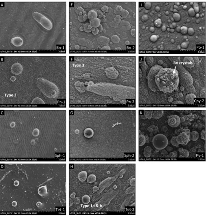
2.5):Type 1) large (approx. 0.1-1.0 µm) solidified droplets of molten material, either spherical or spattered depending on the distance from the ablation crater and the melting point of the mineral; Type 2) small (approx. 20-100 nm) round particles formed by condensation of the ablation plume; and Type 3) diffuse clusters of very fine condensate. Large particles are most abundant close to the ablation craters, and it is unlikely that they are transported to the ICP-MS
due to their size. If the compositions of the smaller particles (Types 2 and 3) vary, with the clusters of finer particles being enriched in volatile elements (Cromwell and Arrowsmith, 1995; D'Abzac et al., 2012; Hergenroder, 2006b; Kuhn and Gunther, 2003), this would result in fractionation of S relative to Fe due to the differences in their transport efficiencies through the interface tubing and ionisation in the plasma (Jackson and Gunther, 2003; Koch et al., 2004; Kuhn and Gunther,
Figure 2.5: Secondary electron images of material surrounding the ablation craters. Mineral-1: 213 nm
Nd:YAG laser at 4.2 J cm-2 (A-D, I, K); Mineral-2: 193 nm excimer laser at 2.7 J cm-2 (E-H, J). Particle Type 1a
& 1b: rounded and spattered droplets; Type 2: small spherical condensate particles coating larger droplets;
Type 3: coating of diffuse condensate clusters. Bornite (Bn); chalcopyrite (Cpy); pentlandite (Pn); pyrite (Py);
pyrrhotite (Po); sphalerite (Sph); tetrahedrite (Tet).
In general the 193-Ex laser produces finer particles than the UP213 laser, due to the shorter wavelength and more efficient ablation (Kuhn and Gunther, 2004). However, the morphology of the ablated material can differ for some minerals between the two laser wavelengths. At the laser fluences tested, pyrite, chalcopyrite and pyrrhotite produce similar particle types with both laser
systems (Figure
2.5.I-K), whereas bornite, pentlandite, sphalerite and tetrahedrite produceBn-1 Bn-2
Pn-1 Pn-2
Sph-1 Sph-2
Tet-1 Tet-2
Po-1
Cpy-2
Py-1
Type 1a & b Type 2
Type 3
A E
B F
C G
D H
I
J
K