Review Article
D
yspepsia is a constellation of symptoms referable to thegas-troduodenal region of the upper gastrointestinal tract. Functional dyspep-sia, a relapsing and remitting disorder, is the most common cause of these symptoms. The current standard for the diagnosis of functional dyspepsia is the Rome III criteria, developed by the Rome III Committees, a multinational group of experts in the field, first convened in 1990, that meets regularly to review and revise the diagnostic criteria for all functional gastrointestinal disorders.
The Rome III criteria for functional dyspepsia consist of a sensation of pain or burning in the epigastrium, early satiety (inability to finish a normal-sized meal), fullness during or after a meal, or a combination of these symptoms (Table S1 in the Supplementary Appendix, available with the full text of this article at NEJM.org). Symptoms must be chronic, occurring at least weekly and over a period of at least 6 months, in the absence of an organic explanation.1 The global prevalence of functional dyspepsia in the community according to this definition is between 5% and 11%.2
Up to 40% of persons who have functional dyspepsia consult a physician,3 and the condition negatively affects attendance and productivity in the workplace.4 Functional dyspepsia has substantial financial implications for patients, health care organizations, and society as a whole; costs associated with the condition in the United States in 2009 were in excess of $18 billion.5 It is therefore important that physicians be able to recognize functional dyspepsia, use investigations and diagnostic tests judiciously, and recommend effective treatments, in order to minimize the potential adverse social and economic effects of the condition.
Diagnosis of Functional Dyspepsia
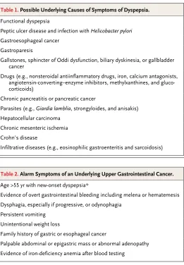
Symptoms do not reliably distinguish between organic and functional forms of the disease,6,7 so the challenge for the physician evaluating a patient with dyspepsia lies in discriminating between functional dyspepsia and organic conditions of the stomach or duodenum that may provoke similar symptoms (Table 1). In most cases, the cause can be clarified by means of upper gastrointestinal endoscopy, a test that generally shows that less than 10% of patients with dyspepsia have a peptic ulcer, less than 1% have gastroesophageal cancer, and more than 70% have functional dyspepsia.8 Celiac disease is the great mimic of many gastrointestinal disorders, but its frequency is not significantly increased among persons who re-port dyspepsia.9 The patient’s medication history should be reviewed, but medica-tion is not usually implicated in causing the dyspepsia.10
Given that upper gastrointestinal endoscopy is associated with a relatively low rate of identification of organic disease, it is neither desirable nor realistic to per-form this test in all patients with dyspepsia. A primary care–based study showed that the cost of detecting each case of upper gastrointestinal cancer among patients
From the Faculty of Health and Medicine, University of Newcastle, Newcastle, NSW, Australia (N.J.T.); and Leeds Gastroenterol-ogy Institute, St. James’s University Hos-pital, and Leeds Institute of Biomedical and Clinical Sciences, University of Leeds — both in Leeds, United Kingdom (A.C.F.). Address reprint requests to Dr. Talley at the Hunter Medical Research Institute, Rm. 3419, University of Newcastle, Kooka-burra Circuit, New Lambton, NSW 2295, Australia, or at nicholas . talley@ newcastle . edu . au.
N Engl J Med 2015;373:1853-63. DOI: 10.1056/NEJMra1501505 Copyright © 2015 Massachusetts Medical Society.
Dan L. Longo, M.D., Editor
with dyspepsia was more than $80,000,11 which provides support for a selective approach. Guide-lines recommend that patients with dyspepsia who report so-called alarm symptoms (Table 2), which may indicate an underlying gastroesopha-geal cancer, be referred urgently for upper gastro-intestinal endoscopy12; however, only a small per-centage of patients who undergo this test have such a cancer, which indicates that alarm symp-toms have only modest predictive capability.13
For patients with simple dyspepsia who do not have alarm symptoms, in whom the likeliest diagnosis is functional dyspepsia, the require-ment for any further diagnostic testing depends on the background prevalence of Helicobacter pylori infection. In populations in which the prevalence of infection is at least 10%, noninvasive testing for H. pylori, with either carbon-13–labeled urea
breath testing or stool antigen testing, is recom-mended.12 In practice, however, because it is unlikely that the physician will be aware of the local prevalence of H. pylori, it is reasonable to use one of these tests as a first-line strategy, given that the testing is neither invasive nor pro-hibitively expensive.
Functional dyspepsia may be confused with other gastrointestinal conditions outside the gas-troduodenal region, including other functional disorders.14 In the past 20 years, there has been a concerted effort to standardize the definitions of functional dyspepsia, in part to minimize the likelihood of overlap with other functional gas-trointestinal disorders. For the most part, this goal has been achieved by excluding from the definition of functional dyspepsia persons with symptoms suggestive of gastroesophageal reflux disease (GERD), such as retrosternal burning pain, regurgitation of acid into the mouth, or the irritable bowel syndrome, which is characterized by lower abdominal pain or discomfort associ-ated with a change in stool form or frequency.1 Despite this effort, in one study, more than 50% of the patients who met the criteria for func-tional dyspepsia and who had normal 48-hour pH studies reported heartburn and regurgita-tion, which were the predominant symptoms in
30% of these patients.15 Common underlying
mechanisms, such as failure of the gastric fundus to relax properly, may account for such symp-toms in patients with overlapping functional dyspepsia and heartburn.16 In a factor-analysis study, the presence of lower gastrointestinal symptoms, such as diarrhea and constipation, increased the ability of physicians to discrimi-nate between people with functional dyspepsia and those without it.17
There is also overlap between symptoms of functional dyspepsia and those of gastroparesis. More than one in four patients with functional dyspepsia have evidence of delayed gastric empty-ing,18 and in one study 86% of the patients with gastroparesis met the criteria for functional dyspepsia,19 which suggests that these condi-tions share similar pathophysiological features; the degree of overlap of symptoms also means that the capacity of diagnostic tests such as gas-tric scintigraphy to discriminate between func-tional dyspepsia and gastroparesis is limited.20 The usefulness of ultrasonography in detecting relevant organic pancreatobiliary disease in pa-Functional dyspepsia
Peptic ulcer disease and infection with Helicobacter pylori Gastroesophageal cancer
Gastroparesis
Gallstones, sphincter of Oddi dysfunction, biliary dyskinesia, or gallbladder cancer
Drugs (e.g., nonsteroidal antiinflammatory drugs, iron, calcium antagonists, angiotensin-converting–enzyme inhibitors, methylxanthines, and gluco-corticoids)
Chronic pancreatitis or pancreatic cancer
Parasites (e.g., Giardia lamblia, strongyloides, and anisakis) Hepatocellular carcinoma
Chronic mesenteric ischemia Crohn’s disease
[image:2.567.37.298.51.427.2]Infiltrative diseases (e.g., eosinophilic gastroenteritis and sarcoidosis)
Table 1. Possible Underlying Causes of Symptoms of Dyspepsia.
Age >55 yr with new-onset dyspepsia*
Evidence of overt gastrointestinal bleeding including melena or hematemesis Dysphagia, especially if progressive, or odynophagia
Persistent vomiting Unintentional weight loss
Family history of gastric or esophageal cancer
Palpable abdominal or epigastric mass or abnormal adenopathy Evidence of iron-deficiency anemia after blood testing
* In regions with a high background prevalence rate of gastric cancer, such as Southeast Asia, a lower age threshold should be considered.
tients with dyspepsia who have normal results on upper gastrointestinal endoscopy was less than 5% in one primary care–based study.21
Cl assification of Functional Dyspepsia
In the past 10 years, the terminology used to describe functional dyspepsia has changed, moving away from grouping patients according to the predominant reported symptom as having ulcer-like, reflux-like, or dysmotility-like func-tional dyspepsia and instead describing them as having one of two newly defined syndromes, the epigastric pain syndrome and the postprandial distress syndrome. The epigastric pain syndrome consists of intermittent pain or burning in the epigastrium, occurring at least once per week, and the postprandial distress syndrome is marked by the occurrence at least several times per week of bothersome postprandial fullness occurring after normal-sized meals or by early satiety that prevents the person from finishing a regular meal (Table S1 in the Supplementary Appendix).1
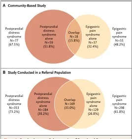
These two syndromes were proposed because as many as 80% of persons with dyspepsia re-port that their symptoms are aggravated by the ingestion of a meal.22 The definitions were also based on factor analysis that showed the group-ing of dyspeptic symptoms into three or four clusters,23,24 with the epigastric pain syndrome and the postprandial distress syndrome appear-ing consistently in several different studies. Sub-sequent community-based cross-sectional sur-veys, which showed good separation between these two subgroups, support this approach.25 Studies in referral populations are less convinc-ing, however, with a greater degree of overlap evident between the epigastric pain syndrome and the postprandial distress syndrome (Fig. 1).7,26 The rationale for assigning patients into these two syndrome subtypes in the clinic is that the classification may help guide therapy.
Pathophysiological Fe atur es of Functional Dyspepsia
Psychological distress, particularly anxiety, is associated with functional dyspepsia and may precede the onset of the disorder in some per-sons.27 In others, the gut symptoms occur before
the onset of anxiety, which suggests that a gut-driven brain disorder may explain some cases.28 Central pain processing may be abnormal in persons with functional dyspepsia,29 although whether it is caused by gut disturbances or is a primary symptom is uncertain.30 Genetic factors have also been implicated in functional dyspep-sia, but the associations remain weak.31
[image:3.567.268.536.55.336.2]Functional dyspepsia has conventionally been attributed to a disturbance of gastric physiologic factors, such as slow gastric emptying, failure of the gastric fundus to relax after a meal (fundic disaccommodation, which is a vagal reflex), or gastric hypersensitivity with distention of the stomach.32 Some patients with functional dys-pepsia have none of these abnormalities, and any link with specific symptoms is unconvinc-ing, except possibly for the inability to finish a normal meal and fundic failure.18,22 Gastric ac-commodation failure is also linked to transient
Figure 1. Overlap between Subcategories of Functional Dyspepsia in Community-Based and Referral Populations.
The Venn diagrams show the degree of overlap between patients with functional dyspepsia who present with the postprandial distress syndrome and those who present with the epigastric pain syndrome. A total of 114 patients with functional dyspepsia were included in a community-based study25 (Panel A), and 482 were included in a study conducted in a referral population7 (Panel B).
B Study Conducted in a Referral Population A Community-Based Study
Postprandial distress syndrome
alone N=59 (51.8%)
Overlap N=18 (15.8%)
Epigastric pain syndrome
alone N=37 (32.4%) Postprandial
distress syndrome
N=77 (67.5%)
Epigastric pain syndrome
N=55 (48.2%)
Postprandial distress syndrome
alone N=184 (38.2%)
Overlap N=169 (35.0%)
Epigastric pain syndrome
alone N=129 (26.8%) Postprandial
distress syndrome
N=353 (73.2%)
Epigastric pain syndrome
relaxations of the lower esophageal sphincter that occur in GERD and may, in part, explain the overlap of GERD with functional dyspepsia.16 Duodenal hypersensitivity to acid or distention has also been reported in patients with func-tional dyspepsia.33
Infections may cause functional dyspepsia, but Koch’s postulates have not yet been fulfilled for any microbe. The occurrence of a postinfec-tious irritable bowel syndrome is well estab-lished; however, gastroenteritis can also lead to functional dyspepsia or to a persistent combina-tion of funccombina-tional dyspepsia and symptoms of the irritable bowel syndrome. Salmonella, Esche-richia coli O157, Campylobacter jejuni, Giardia lamblia, and norovirus may induce functional dyspepsia; risk factors include genetic factors and smok-ing.34 It is conceivable that functional dyspepsia arises when the proximal small intestine or stomach becomes inflamed after an enteric in-fection, whereas the irritable bowel syndrome may arise from involvement of the distal small intestine or colon; if both the proximal and dis-tal small intestine are inflamed, an overlap syndrome (the irritable bowel syndrome and functional dyspepsia) may be likely,35 although this hypothesis needs formal testing.
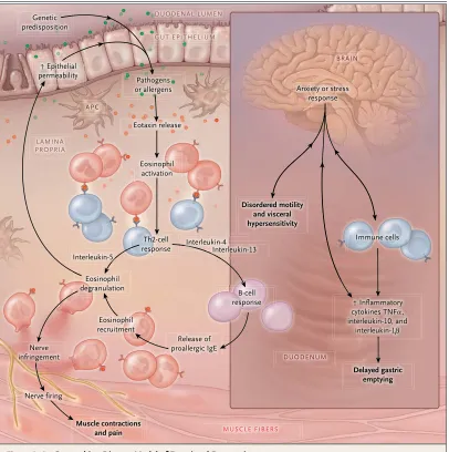
Duodenal inflammation has been observed in up to 40% of patients with functional dyspepsia, particularly subtle duodenal eosinophilia with, in some cases, excess clusters of eosinophils and eosinophil degranulation adjacent to nerves (Fig. S1 in the Supplementary Appendix).36-38 Duodenal eosinophilia has been linked to smok-ing and to symptoms of early satiety and pain; barrier disruption and increased duodenal perme-ability have been documented.36,39 In some cases, mast cells that can recruit eosinophils have also been observed in functional dyspepsia, but the patient population that was studied included patients with both functional dyspepsia and the irritable bowel syndrome.39 Further evidence link-ing intestinal inflammation to functional dys-pepsia is provided by the finding of enhanced small-bowel homing T lymphocytes that are positive for both α4β7 integrin and chemokine receptor 9 in patients with functional dyspepsia — a finding that has been significantly associ-ated with the release of cytokines (including tumor necrosis factor α), a greater severity of symptoms, and delayed gastric emptying,40 thus implicating the duodenum in gastric disorders.41
Together, these findings suggest that some
patients with functional dyspepsia may have an organic mechanism for their symptoms. An-other likely infectious cause is H. pylori. Although H. pylori infection is usually asymptomatic, in a small subgroup of patients with functional dys-pepsia the eradication of infection leads to the long-term resolution of symptoms.42 The role of other components of the microbiome in func-tional dyspepsia is unknown.43
Functional dyspepsia is most often a meal-induced syndrome.22,44 A high-fat meal, for exam-ple, can alter gastroduodenal physiology by means of altered gut-hormone responses,45 including by raising cholecystokinin levels.46 Food intolerance or allergy may play a direct role in functional dyspepsia, but this possibility has been poorly studied.47
An overarching disease model postulates that, in genetically predisposed persons, an allergen or infection leads to antigen presentation, bar-rier disruption, immune activation, and a type 2 helper T-cell response in functional dyspepsia, in which eosinophils are recruited that degranu-late in some patients (Fig. 2).48 In some patients, this process can lead to tissue injury and symp-toms, whereas in others eosinophils may be protective and promote healing. An inflamed duodenum may be sensitive to acid and induce reflex responses and cytokine release that alter gastroduodenal function and result in meal- related symptoms. If this hypothesis is correct, then some patients with functional dyspepsia may have a response to therapy targeted at im-mune activation, but this remains to be estab-lished; however, preliminary data in children suggest that montelukast, a leukotriene-receptor antagonist, reduces symptoms.49
Tr e atment of Functional Dyspepsia
Placebo or Reassurance
reassurance as a treatment strategy in patients with functional dyspepsia, although this ap-proach is often used by physicians. It should be pointed out that any reassurance derived from investigations that have ruled out organic dis-ease is minimal.52
H. pylori Eradication Therapy
[image:5.567.63.469.54.462.2]Although 5% of the cases of dyspepsia in the community are attributable to infection with H. pylori,53 the effect of eradication therapy on the symptoms of functional dyspepsia is mod-est. In a meta-analysis of 17 randomized trials,
Figure 2. An Overarching Disease Model of Functional Dyspepsia.
In the presence of a background genetic disposition, a type 2 helper T (Th2)–cell response may be activated in the duodenum, possibly by allergens or pathogens, which cross through the gut epithelium. Resident and recruited eosin-ophils may be activated by eotaxin, which is expressed constitutively in the lamina propria, and act as antigen-present-ing cells to Th2 lymphocytes, which in turn express interleukin-5. This process can lead to eosinophil degranulation that impinges on nerve fibers, which may then fire, inducing muscle contraction or pain. Duodenal feedback to the stomach by means of interleukin-4 and interleukin-13, also expressed by Th2 cells, may promote immunoglobulin class switching to proallergic IgE antibody expression by B cells, further recruiting eosinophils and leading to de-granulation with increased epithelial permeability. The cytokines tumor necrosis factor α (TNF-α), interleukin-10, and interleukin-1β can then be released into the blood and invoke an anxiety or stress response, which in turn may lead to disordered motility and visceral hypersensitivity in the stomach and duodenum. Gut-homing T cells may also increase in number and produce excess inflammatory cytokines that could then delay gastric emptying. APC denotes antigen-presenting cell.
Pathogens or allergens Genetic
predisposition
Eotaxin release ↑ Epithelial
permeability
Eosinophil activation
Eosinophil degranulation
Nerve infringement
Muscle contractions and pain
Nerve firing
Eosinophil recruitment
Release of proallergic IgE
Anxiety or stress response
Immune cells
↑ Inflammatory cytokines TNFα, interleukin-10, and
interleukin-1β
Delayed gastric emptying Disordered motility
and visceral hypersensitivity
B-cell response Th2-cell
response Interleukin-5
Interleukin-4 nterleukin-4 nterleukin-4 Interleukin-13 DUODENAL LUMEN
GUT EPITHELIUM
LAMINA PROPRIA
APC
BRAIN
MUSCLE FIBERS
involving 3566 patients, the relative risk of per-sistent symptoms was 0.90 (95% confidence in-terval [CI], 0.86 to 0.94), with a number needed to treat of 15.42 Nevertheless, economic model-ing that was based on these data suggests that eradication therapy is a cost-effective strategy for managing functional dyspepsia.54 Evidence continues to accumulate that such therapy is beneficial.55 A trial assessing the effect of eradi-cation therapy according to individual symptoms reported by the patient56 showed a significant effect on epigastric pain and burning but not on early satiety or postprandial fullness. These data suggest that the benefit of eradication therapy may be more pronounced in patients with the epigastric pain syndrome than in others.
Acid-Suppression Therapy
Despite evidence of impaired duodenal clearance of gastric acid and duodenal hypersensitivity to infused gastric acid in persons with functional dyspepsia,33 the efficacy of acid-suppressive drugs such as proton-pump inhibitors (PPIs) or histamine H2-receptor antagonists is modest. A Cochrane meta-analysis of 10 randomized trials of PPIs, involving 3347 patients, reported a relative risk of persistent symptoms of 0.87 (95% CI, 0.80 to 0.96) and a number needed to treat of 10.50 For histamine H2-receptor antagonists, the effect was more pronounced than with PPIs (relative risk, 0.77; 95% CI, 0.65 to 0.92; number needed to treat, 7), but the quality of the trials was lower. The majority of these trials were com-pleted before the Rome III classification of func-tional dyspepsia, and subgroup analyses were therefore conducted according to the predomi-nant symptoms reported by the patients rather than according to whether the patients had the epigastric pain syndrome or the postprandial distress syndrome.
The meta-analysis showed that PPIs were ef-fective in patients reporting reflux-like or ulcer-like functional dyspepsia but not in patients with dysmotility-like functional dyspepsia.12 However, a recent trial conducted in Japan that confirmed the efficacy of the PPI rabeprazole in patients with functional dyspepsia did not show any dif-ference in the effect of treatment according to whether patients met the criteria for the epigas-tric pain syndrome or the postprandial distress syndrome.57 A trial of acid suppression seems to be a worthwhile strategy in most patients with functional dyspepsia, particularly in those who
have negative results on H. pylori testing or in those with positive results on H. pylori testing in whom eradication therapy has not improved symptoms. Antacids, bismuth, and sucralfate are not efficacious in functional dyspepsia.12
Prokinetic Agents
A substantial proportion of patients with func-tional dyspepsia have abnormalities in gastric motility and fundal accommodation.58 Existing prokinetic agents, including cisapride, domperi-done, and itopride, have all been tested in func-tional dyspepsia and have been shown to be more effective than placebo in a meta-analysis of 24 randomized trials.50 Cisapride was withdrawn because of its increased risk of adverse cardiac events, including sudden death due to a pro-longed QT interval, and itopride was no more effective than placebo in two large trials pub-lished after this meta-analysis.59 Metoclopramide is not recommended routinely because of its un-certain efficacy and side effects (including irre-versible tardive dyskinesia), and the prescription of domperidone in the United States requires an Investigational New Drug application to the Food and Drug Administration.60
Partly as a result of the lack of efficacy of these drugs, new agents have been developed and tested in recent years. Acotiamide is an acetyl-cholinesterase inhibitor that accelerates gastric emptying and enhances gastric accommodation.61 In a double-blind, placebo-controlled trial in-volving 897 patients with functional dyspepsia in Japan, symptoms improved in 52% of those assigned to active therapy, as compared with 35% of those assigned to placebo (P<0.001).62 When the effect of acotiamide on individual dyspeptic symptoms was studied, significant improvements were identified in postprandial fullness, upper abdominal bloating, and early satiety but not in upper abdominal pain or dis-comfort. The drug has now been approved for the treatment of the postprandial distress syn-drome in Japan, and phase 3 trials are ongoing in Western populations.
fullness.63 In a double-blind, placebo-controlled study involving 144 patients, the response rate after 4 weeks of treatment with tandospirone was 31.5%, as compared with 12.7% with placebo (P = 0.002).64
Antidepressants
Because of the potential role of the brain–gut axis and abnormal central pain processing in functional dyspepsia,29,30 antidepressants have been suggested as a second-line or third-line therapy for many years, but it is only in the past decade that their efficacy has been tested in large, rigorously designed clinical trials. A trial of venlafaxine in 160 patients with functional dyspepsia showed no benefit after 8 weeks of treatment (37% of the patients were symptom free with venlafaxine, as compared with 39% with placebo).65 In a placebo-controlled trial of sertraline in 193 patients in China, 28% of the patients randomly assigned to the active drug had complete relief of their symptoms, as com-pared with 28% of those assigned to placebo.66
Mirtazapine has also been assessed in 34 patients with functional dyspepsia and weight loss67; significant improvements were seen in early satiety and quality of life at 8 weeks in the patients assigned to mirtazapine, as compared with those assigned to placebo. More recently, in a large North American multicenter trial, 292 patients with functional dyspepsia were assigned to amitriptyline, escitalopram, or placebo.68 The rate of response after 10 weeks was 53% with amitriptyline, 38% with escitalopram, and 40% with placebo (P = 0.05 for the three-way com-parison). Taken together, these data suggest that tricyclic antidepressants, such as amitriptyline, should be preferred over selective serotonin-reuptake inhibitors or serotonin–norepinephrine reuptake inhibitors for the treatment of func-tional dyspepsia.
Psychological Therapy
The use of psychological therapy in functional dyspepsia remains an under-studied area. A Co-chrane review published 10 years ago identified only four studies and highlighted the need for more research addressing this issue.69 Few studies have been published since this review, although a recent trial involving 158 patients with func-tional dyspepsia in Spain randomly assigned participants to receive conventional medical treat-ment or conventional medical treattreat-ment plus
psychotherapy.70 There were significant improve-ments in dyspepsia-related quality-of-life and symptom scores at 10 weeks with psychother-apy, and these effects persisted for as long as 6 months after the end of treatment. However, these data should be regarded as preliminary.
More studies will be required before the place of psychological therapy in the treatment of functional dyspepsia is known. That said, such treatment should probably be considered for patients who have not had any improvement in their symptoms with conventional medical ther-apies, particularly in those with coexisting im-pairment of mood.
Complementary and Alternative Therapy
Given the limited efficacy of the majority of conventional medical therapies, it is not surpris-ing that up to 50% of patients with functional dyspepsia seek out other forms of treatment71; in one study, nearly 50% of the patients were will-ing to accept a 12.7% risk of sudden death with a drug that offered a 99% chance of cure,72 in the hope of improving their symptoms and qual-ity of life. However, consistent evidence of the efficacy of acupuncture, homeopathy, or probiot-ics in functional dyspepsia is lacking.
Some patients may find herbal supplements, such as the nine-herb combination product iberogast (also known as STW5),73 beneficial, and STW5 has been observed to relax the gastric fundus.74 Capsaicin, a component of red pepper, was superior to placebo in terms of the reduc-tion in symptom scores in one small trial,75 but more studies are needed before any definitive conclusions can be drawn. Disordered sleep is more common in patients with functional dys-pepsia than in healthy controls without func-tional dyspepsia, and it appears to be correlated with the severity of symptoms,76 but no interven-tion studies have been performed.
Management of Functional Dyspepsia
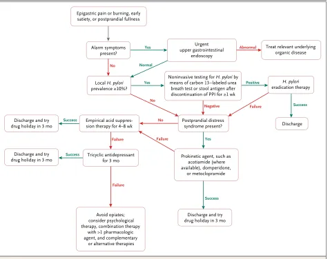
to support the screening of all patients with functional dyspepsia for anxiety or for treating those in whom it is present with an anxiolytic. A treatment algorithm is presented in Figure 3.
Management of Refractory Functional Dyspepsia
In patients whose symptoms do not respond to standard medical therapy, treatment is empiri-cal. In our experience, histamine H2-receptor antagonists may help even if PPIs have failed.
The combination of acid suppression with a pro-kinetic agent appears to benefit some other pa-tients. The combination of peripheral drug ther-apy with psychological treatment is promising.70 If pain is the predominant symptom despite these strategies, the physician should consider other options, although these are empirical and not evidence-based.78 Approaches that may be helpful include adjusting the dose of a tricyclic antidepressant to the full antidepressant level, prescribing an antipsychotic drug such as
levo-Figure 3. Recommended Treatment Algorithm for Patients with a Provisional Diagnosis of Functional Dyspepsia.
This treatment algorithm can be applied in patients who present with epigastric pain or burning, early satiation, or postprandial fullness. In the case of treatment failure, the clinician should reevaluate and reconsider the diagnosis at each step by means of further investiga-tions, such as upper gastrointestinal endoscopy if the procedure has not been performed within the past 5 years; ultrasonography of the abdomen, particularly if the patient has severe, intermittent episodes of pain; serologic testing for celiac disease; and gastric scintigra-phy or carbon-13–labeled octanoic or spirulina (Arthrospira platensis) breath test to assess gastric emptying if the symptoms are severe or resistant to treatment or if the patient has vomiting and prominent weight loss. There are no data from randomized trials in support of using metoclopramide to treat patients with the postprandial distress syndrome; we suggest starting the drug at a low dose owing to the potential for cardiac and neurologic toxic effects. PPI denotes proton-pump inhibitor.
Epigastric pain or burning, early satiety, or postprandial fullness
Alarm symptoms present?
Urgent upper gastrointestinal
endoscopy
Local H. pylori prevalence ≥10%?
H. pylori
eradication therapy
Empirical acid
suppres-sion therapy for 4–8 wk Postprandial distress syndrome present?
Discharge and try drug holiday in 3 mo
Discharge Abnormal
Failure
Failure Negative
Failure Failure No
No
No Discharge and try
drug holiday in 3 mo
Treat relevant underlying organic disease
Discharge and try
drug holiday in 3 mo Prokinetic agent, such as acotiamide (where available), domperidone, or metoclopramide Tricyclic antidepressant
for 3 mo
Avoid opiates; consider psychological therapy, combination therapy
with >1 pharmacologic agent, and complementary
or alternative therapies
Noninvasive testing for H. pylori by means of carbon 13–labeled urea
breath test or stool antigen after discontinuation of PPI for ≥1 wk
Success
Success
Success
Success
Positive Normal
Yes
[image:8.567.32.510.55.431.2]sulpiride,79 or adding an anxiolytic (e.g., buspi-rone) to a tricyclic antidepressant. The combina-tion of an antidepressant with pregabalin or gabapentin is yet another option that appears to relieve pain. Opioids have no therapeutic role in the management of functional dyspepsia and should be avoided because of the risk of depen-dence, the frequent failure of analgesia, and possibility of the narcotic bowel syndrome.80
Prognosis in Functional Dyspepsia
In most patients with functional dyspepsia, the natural history is chronic and fluctuating, with periods of time when the patient is asymptom-atic followed by episodes of symptom relapse. Data from population-based studies suggest that, during extended follow-up, approximately 15 to 20% of people with functional dyspepsia have persistent symptoms and 50% have resolution of symptoms; in the remaining 30 to 35% of pa-tients symptoms will fluctuate and meet the criteria for another functional gastrointestinal
disorder.81 Despite the chronic nature of func-tional dyspepsia, there is no evidence to suggest that it is associated with decreased survival.77
Dr. Talley reports receiving lecture fees from the Rome Foun-dation and Takeda Pharmaceutical, receiving consulting fees from Yuhan, Adelphi Values, Prometheus Medical, Abbott Labora-tories, Forest Laboratories (now Actavis), Furiex, Synergy Pharma-ceuticals, Focus Communications, and Zeria Pharmaceutical, serving as an unpaid consultant to GI Therapies, receiving hono-raria from Janssen, Danone, and GI Care, receiving study medi-cation from Forest Laboratories, receiving grant support from the Rome Foundation, Ironwood Pharmaceuticals, Prometheus Medical, Janssen-Cilag, Takeda Pharmaceutical, Abbott Labora-tories, Datapharm, Pfizer, and Salix Pharmaceuticals, licensing the Bowel Disease Questionnaire and Mayo Dysphagia Ques-tionnaire to the Mayo Clinic, and holding a patent (U.S. 12735358.9-1405/2710383) related to the performance of a bio-marker panel for the irritable bowel syndrome. No other poten-tial conflict of interest relevant to this article was reported.
Disclosure forms provided by the authors are available with the full text of this article at NEJM.org.
We thank Dr. Paul G. Shekelle, director of the Southern Cali-fornia Evidence-Based Practice Center, RAND, for an informal review of an earlier version of the manuscript; Dr. Marjorie M. Walker, University of Newcastle, Australia, for assistance with the preparation of figures in an earlier version of the manuscript and in the Supplementary Appendix; and Dr. Gerald Holtmann, University of Queensland, Australia, Drs. Marjorie M. Walker and Simon Keely, University of Newcastle, and Dr. Nick Powell, King’s College London, for critical review of an earlier version of Figure 2.
References
1. Tack J, Talley NJ, Camilleri M, et al. Functional gastroduodenal disorders. Gas-troenterology 2006; 130: 1466-79.
2. Ford AC, Marwaha A, Sood R, Moay-yedi P. Global prevalence of, and risk fac-tors for, uninvestigated dyspepsia: a meta-analysis. Gut 2015; 64: 1049-57.
3. Ford AC, Forman D, Bailey AG, Cook MB, Axon ATR, Moayyedi P. Who consults with dyspepsia? Results from a longitudi-nal 10-yr follow-up study. Am J Gastroen-terol 2007; 102: 957-65.
4. Sander GB, Mazzoleni LE, Francesconi CF, et al. Influence of organic and func-tional dyspepsia on work productivity: the HEROES-DIP study. Value Health 2011; 14: Suppl 1: S126-S129.
5. Lacy BE, Weiser KT, Kennedy AT, Crowell MD, Talley NJ. Functional dys-pepsia: the economic impact to patients. Aliment Pharmacol Ther 2013; 38: 170-7.
6. Ford AC, Bercik P, Morgan DG, Bolino C, Pintos-Sanchez MI, Moayyedi P. The Rome III criteria for the diagnosis of functional dyspepsia in secondary care are not superior to previous definitions. Gastroenterology 2014; 146: 932-40.
7. Fang YJ, Liou JM, Chen CC, et al. Dis-tinct aetiopathogenesis in subgroups of functional dyspepsia according to the Rome III criteria. Gut 2015; 64: 1517-28.
8. Ford AC, Marwaha A, Lim A, Moayye-di P. What is the prevalence of clinically significant endoscopic findings in
sub-jects with dyspepsia? Systematic review and meta-analysis. Clin Gastroenterol Hepatol 2010; 8: 830-7.
9. Ford AC, Ching E, Moayyedi P. Meta-analysis: yield of diagnostic tests for coe-liac disease in dyspepsia. Aliment Phar-macol Ther 2009; 30: 28-36.
10. Hallas J, Bytzer P. Screening for drug related dyspepsia: an analysis of prescrip-tion symmetry. Eur J Gastroenterol Hepa-tol 1998; 10: 27-32.
11. Vakil N, Talley NJ, van Zanten SV, et al. Cost of detecting malignant lesions by endoscopy in 2741 primary care dyspeptic patients without alarm symptoms. Clin Gastroenterol Hepatol 2009; 7: 756-61.
12. Talley NJ, Vakil NB, Moayyedi P. Amer-ican Gastroenterological Association tech-nical review on the evaluation of dyspep-sia. Gastroenterology 2005; 129: 1756-80.
13. Vakil N, Moayyedi P, Fennerty MB, Talley NJ. Limited value of alarm features in the diagnosis of upper gastrointestinal malignancy: systematic review and meta-analysis. Gastroenterology 2006; 131: 390-401.
14. Pleyer C, Bittner H, Locke GR III, et al. Overdiagnosis of gastro-esophageal reflux disease and underdiagnosis of functional dyspepsia in a USA community. Neuro-gastroenterol Motil 2014; 26: 1163-71.
15. Vakil N, Halling K, Ohlsson L, Werners-son B. Symptom overlap between post-prandial distress and epigastric pain
syn-dromes of the Rome III dyspepsia classification. Am J Gastroenterol 2013; 108: 767-74.
16. Pauwels A, Altan E, Tack J. The gastric accommodation response to meal intake determines the occurrence of transient lower esophageal sphincter relaxations and reflux events in patients with gastro-esophageal reflux disease. Neurogastro-enterol Motil 2014; 26: 581-8.
17. Matsuzaki J, Suzuki H, Asakura K, et al. Classification of functional dyspepsia based on concomitant bowel symptoms. Neurogastroenterol Motil 2012; 24(4): 325-e164.
18. Sarnelli G, Caenepeel P, Geypens B, Janssens J, Tack J. Symptoms associated with impaired gastric emptying of solids and liquids in functional dyspepsia. Am J Gastroenterol 2003; 98: 783-8.
19. Parkman HP, Yates K, Hasler WL, et al. Clinical features of idiopathic gastropare-sis vary with sex, body mass, symptom onset, delay in gastric emptying, and gas-troparesis severity. Gastroenterology 2011; 140: 101-15.
20. Stanghellini V, Tack J. Gastroparesis: separate entity or just a part of dyspepsia? Gut 2014; 63: 1972-8.
21. Heikkinen M, Räsänen H, Färkkilä M. Clinical value of ultrasound in the evalua-tion of dyspepsia in primary health care. Scand J Gastroenterol 2005; 40: 980-4.
et al. Relationship between symptoms and ingestion of a meal in functional dys-pepsia. Gut 2008; 57: 1495-503.
23. Piessevaux H, De Winter B, Louis E, et al. Dyspeptic symptoms in the general population: a factor and cluster analysis of symptom groupings. Neurogastroen-terol Motil 2009; 21: 378-88.
24. Camilleri M, Dubois D, Coulie B, et al. Prevalence and socioeconomic impact of upper gastrointestinal disorders in the United States: results of the US Upper Gastrointestinal Study. Clin Gastroenter-ol HepatGastroenter-ol 2005; 3: 543-52.
25. Zagari RM, Law GR, Fuccio L, et al. Epidemiology of functional dyspepsia and subgroups in the Italian general popula-tion: an endoscopic study. Gastroenterol-ogy 2010; 138: 1302-11.
26. van Kerkhoven LA, Laheij RJ, Meineche-Schmidt V, Veldhuyzen-van Zanten SJ, de Wit NJ, Jansen JB. Functional dyspepsia: not all roads seem to lead to Rome. J Clin Gastroenterol 2009; 43: 118-22.
27. Aro P, Talley NJ, Johansson SE, Agréus L, Ronkainen J. Anxiety is linked to new-onset dyspepsia in the Swedish population: a 10-year follow-up study. Gastroenterology 2015; 148: 928-37.
28. Koloski NA, Jones M, Kalantar J, Weltman M, Zaguirre J, Talley NJ. The brain–gut pathway in functional gastro-intestinal disorders is bidirectional: a 12-year prospective population-based study. Gut 2012; 61: 1284-90.
29. Vandenberghe J, Dupont P, Van Oudenhove L, et al. Regional cerebral blood flow during gastric balloon disten-tion in funcdisten-tional dyspepsia. Gastroenter-ology 2007; 132: 1684-93.
30. Wilder-Smith CH, Li X, Shen L, Cao Y, Ho KY, Wong RK. Dysfunctional endoge-nous pain modulation in patients with functional dyspepsia. Neurogastroenterol Motil 2014; 26: 489-98.
31. Oshima T, Toyoshima F, Nakajima S, Fukui H, Watari J, Miwa H. Genetic fac-tors for functional dyspepsia. J Gastroen-terol Hepatol 2011; 26: Suppl 3: 83-7.
32. Carbone F, Tack J. Gastroduodenal mechanisms underlying functional gas-tric disorders. Dig Dis 2014; 32: 222-9.
33. Samsom M, Verhagen MA, vanBerge Henegouwen GP, Smout AJ. Abnormal clearance of exogenous acid and increased acid sensitivity of the proximal duodenum in dyspeptic patients. Gastroenterology 1999; 116: 515-20.
34. Futagami S, Itoh T, Sakamoto C. Sys-tematic review with meta-analysis: post-infectious functional dyspepsia. Aliment Pharmacol Ther 2015; 41: 177-88.
35. Spiller R. Postinfectious functional dyspepsia and postinfectious irritable bowel syndrome: different symptoms but similar risk factors. Gastroenterology 2010; 138: 1660-3.
36. Walker MM, Aggarwal KR, Shim LS, et al. Duodenal eosinophilia and early
sa-tiety in functional dyspepsia: confirma-tion of a positive associaconfirma-tion in an Austra-lian cohort. J Gastroenterol Hepatol 2014; 29: 474-9.
37. Futagami S, Shindo T, Kawagoe T, et al. Migration of eosinophils and CCR2-/ CD68-double positive cells into the duo-denal mucosa of patients with postinfec-tious functional dyspepsia. Am J Gastro-enterol 2010; 105: 1835-42.
38. Talley NJ, Walker MM, Aro P, et al. Non-ulcer dyspepsia and duodenal eosin-ophilia: an adult endoscopic population-based case-control study. Clin Gastroen-terol Hepatol 2007; 5: 1175-83.
39. Vanheel H, Vicario M, Vanuytsel T, et al. Impaired duodenal mucosal integrity and low-grade inflammation in functional dyspepsia. Gut 2014; 63: 262-71.
40. Liebregts T, Adam B, Bredack C, et al. Small bowel homing T cells are associat-ed with symptoms and delayassociat-ed gastric emptying in functional dyspepsia. Am J Gastroenterol 2011; 106: 1089-98.
41. Azpiroz F, Feinle-Bisset C, Grundy D, Tack J. Gastric sensitivity and reflexes: ba-sic mechanisms underlying clinical prob-lems. J Gastroenterol 2014; 49: 206-18.
42. Moayyedi P, Soo S, Deeks J, et al. Eradication of Helicobacter pylori for non-ulcer dyspepsia. Cochrane Database Syst Rev 2006; 2: CD002096 [Withdrawn: Co-chrane Database Syst Rev 2011 February 16].
43. Walker MM, Talley NJ. Review article: bacteria and pathogenesis of disease in the upper gastrointestinal tract — beyond the era of Helicobacter pylori. Aliment Pharmacol Ther 2014; 39: 767-79.
44. Farré R, Vanheel H, Vanuytsel T, et al. In functional dyspepsia, hypersensitivity to postprandial distention correlates with meal-related symptom severity. Gastroen-terology 2013; 145: 566-73.
45. Bharucha AE, Camilleri M, Burton DD, et al. Increased nutrient sensitivity and plasma concentrations of enteral hor-mones during duodenal nutrient infusion in functional dyspepsia. Am J Gastroen-terol 2014; 109: 1910-20.
46. Feinle-Bisset C, Azpiroz F. Dietary lip-ids and functional gastrointestinal disor-ders. Am J Gastroenterol 2013; 108: 737-47.
47. Zuo XL, Li YQ, Li WJ, et al. Alterations of food antigen-specific serum immuno-globulins G and E antibodies in patients with irritable bowel syndrome and func-tional dyspepsia. Clin Exp Allergy 2007; 37: 823-30.
48. Walker MM, Powell N, Talley NJ. Ato-py and the gastrointestinal tract — a re-view of a common association in unex-plained gastrointestinal disease. Expert Rev Gastroenterol Hepatol 2014; 8: 289-99.
49. Friesen CA, Schurman JV, Colombo JM, Abdel-Rahman SM. Eosinophils and mast cells as therapeutic targets in pedi-atric functional dyspepsia. World J Gas-trointest Pharmacol Ther 2013; 4: 86-96.
50. Moayyedi P, Soo S, Deeks J, Delaney B, Innes M, Forman D. Pharmacological inter-ventions for non-ulcer dyspepsia. Cochrane Database Syst Rev 2006; 4: CD001960.
51. Kaptchuk TJ, Friedlander E, Kelley JM, et al. Placebos without deception: a ran-domized controlled trial in irritable bowel syndrome. PLoS One 2010; 5(12): e15591.
52. van Kerkhoven LA, van Rossum LG, van Oijen MG, Tan AC, Laheij RJ, Jansen JB. Upper gastrointestinal endoscopy does not reassure patients with functional dys-pepsia. Endoscopy 2006; 38: 879-85.
53. Moayyedi P, Forman D, Braunholtz D, et al. The proportion of upper gastrointes-tinal symptoms in the community associ-ated with Helicobacter pylori, lifestyle factors, and nonsteroidal anti-inflamma-tory drugs. Am J Gastroenterol 2000; 95: 1448-55.
54. Moayyedi P, Soo S, Deeks JJ, et al. Sys-tematic review and economic evaluation of Helicobacter pylori eradication treat-ment for non-ulcer dyspepsia. BMJ 2000; 321: 659-64.
55. Mazzoleni LE, Sander GB, Francesconi CF, et al. Helicobacter pylori eradication in functional dyspepsia: HEROES trial. Arch Intern Med 2011; 171: 1929-36.
56. Lan L, Yu J, Chen YL, et al. Symptom-based tendencies of Helicobacter pylori eradication in patients with functional dyspepsia. World J Gastroenterol 2011; 17: 3242-7.
57. Iwakiri R, Tominaga K, Furuta K, et al. Randomised clinical trial: rabeprazole improves symptoms in patients with functional dyspepsia in Japan. Aliment Pharmacol Ther 2013; 38: 729-40.
58. Thumshirn M, Camilleri M, Saslow SB, Williams DE, Burton DD, Hanson RB. Gastric accommodation in non-ulcer dys-pepsia and the roles of Helicobacter pylori infection and vagal function. Gut 1999; 44: 55-64.
59. Talley NJ, Tack J, Ptak T, Gupta R, Giguère M. Itopride in functional dyspep-sia: results of two phase III multicentre, randomised, double-blind, placebo-con-trolled trials. Gut 2008; 57: 740-6.
60. Camilleri M, Parkman HP, Shafi MA, Abell TL, Gerson L. Clinical guideline: management of gastroparesis. Am J Gas-troenterol 2013; 108: 18-37.
61. Kusunoki H, Haruma K, Manabe N, et al. Therapeutic efficacy of acotiamide in patients with functional dyspepsia based on enhanced postprandial gastric accom-modation and emptying: randomized con-trolled study evaluation by real-time ultra-sonography. Neurogastroenterol Motil 2012; 24: 540-5.
62. Matsueda K, Hongo M, Tack J, Saito Y, Kato H. A placebo-controlled trial of aco-tiamide for meal-related symptoms of functional dyspepsia. Gut 2012; 61: 821-8.
functional dyspepsia. Clin Gastroenterol Hepatol 2012; 10: 1239-45.
64. Miwa H, Nagahara A, Tominaga K, et al. Efficacy of the 5-HT1A agonist tando-spirone citrate in improving symptoms of patients with functional dyspepsia: a ran-domized controlled trial. Am J Gastroen-terol 2009; 104: 2779-87.
65. van Kerkhoven LA, Laheij RJ, Aparicio N, et al. Effect of the antidepressant ven-lafaxine in functional dyspepsia: a ran-domized, double-blind, placebo-controlled trial. Clin Gastroenterol Hepatol 2008; 6: 746-52.
66. Tan VP, Cheung TK, Wong WM, Pang R, Wong BC. Treatment of functional dys-pepsia with sertraline: a double-blind ran-domized placebo-controlled pilot study. World J Gastroenterol 2012; 18: 6127-33.
67. Ly HG, Carbone F, Holvoet L, et al. Mirtazapine improves early satiation, nutri-ent intake, weight recovery and quality of life in functional dyspepsia with weight loss: a double-blind, randomized, placebo-controlled pilot study. Gastroenterology 2013; 144: S37.
68. Talley NJ, Locke GR, Saito YA, et al. Effect of amitriptyline and escitalopram on functional dyspepsia: a multi-center, randomized, controlled study. Gastroen-terology 2015; 149: 340-9.
69. Soo S, Moayyedi P, Deeks JJ, Delaney B, Lewis M, Forman D. Psychological inter-ventions for non-ulcer dyspepsia. Cochrane Database Syst Rev 2005; 2: CD002301.
70. Orive M, Barrio I, Orive VM, et al. A ran-domized controlled trial of a 10-week group psychotherapeutic treatment added to stan-dard medical treatment in patients with functional dyspepsia. J Psychosom Res 2015; 78: 563-8.
71. Lahner E, Bellentani S, Bastiani RD, et al. A survey of pharmacological and nonpharmacological treatment of func-tional gastrointestinal disorders. United European Gastroenterol J 2013; 1: 385-93.
72. Lacy BE, Yu J, Crowell MD. Medication risk-taking behavior in functional dys-pepsia patients. Clin Transl Gastroenterol 2015; 6: e69.
73. von Arnim U, Peitz U, Vinson B, Gun-dermann KJ, Malfertheiner P. STW 5, a phytopharmacon for patients with func-tional dyspepsia: results of a multicenter, placebo-controlled double-blind study. Am J Gastroenterol 2007; 102: 1268-75.
74. Pilichiewicz AN, Horowitz M, Russo A, et al. Effects of iberogast on proximal gastric volume, antropyloroduodenal mo-tility and gastric emptying in healthy men. Am J Gastroenterol 2007; 102: 1276-83.
75. Bortolotti M, Coccia G, Grossi G,
Mi-glioli M. The treatment of functional dys-pepsia with red pepper. Aliment Pharma-col Ther 2002; 16: 1075-82.
76. Lacy BE, Everhart K, Crowell MD. Functional dyspepsia is associated with sleep disorders. Clin Gastroenterol Hepa-tol 2011; 9: 410-4.
77. Ford AC, Forman D, Bailey AG, Axon ATR, Moayyedi P. Effect of dyspepsia on survival: a longitudinal 10-year follow-up study. Am J Gastroenterol 2012; 107: 912-21.
78. Törnblom H, Drossman DA. Centrally targeted pharmacotherapy for chronic ab-dominal pain. Neurogastroenterol Motil 2015; 27: 455-67.
79. Arienti V, Corazza GR, Sorge M, et al. The effects of levosulpiride on gastric and gall-bladder emptying in functional dys-pepsia. Aliment Pharmacol Ther 1994; 8: 631-8.
80. Drossman D, Szigethy E. The narcotic bowel syndrome: a recent update. Am J Gastroenterol 2014; 2: 22-30.
81. Olafsdottir LB, Gudjonsson H, Jons-dottir HH, Bjornsson E, Thjodleifsson B. Natural history of functional gastrointes-tinal disorders: comparison of two longi-tudinal population-based studies. Dig Liver Dis 2012; 44: 211-7.