White Rose Research Online [email protected]
Universities of Leeds, Sheffield and York
http://eprints.whiterose.ac.uk/
This is an author produced version of a paper published inPlant Cell
White Rose Research Online URL for this paper:
http://eprints.whiterose.ac.uk/id/eprint/77543
Paper:
Hu, J, Baker, A, Bartel, B, Linka, N, Mullen, RT, Reumann, S and Zolman, BK (2012) Plant peroxisomes: biogenesis and function.Plant Cell, 24 (6). 2279 -2303. ISSN 1040-4651
Plant Peroxisomes: Biogenesis and Function
Jianping Hua,b,1, Alison Bakerc,, Bonnie Barteld, Nicole Linkae, Robert T. Mullenf, Sigrun
Reumanng, and Bethany K. Zolmanh
a
MSU-DOE Plant Research Laboratory and bDepartment of Plant Biology, Michigan State
University, East Lansing, MI 48824.
c
Centre for Plant Sciences, Faculty of Biological Sciences, University of Leeds, Leeds, LS2 9JT,
UK.
d
Department of Biochemistry and Cell Biology, Rice University, Houston, TX 77005.
e
Department of Plant Biochemistry, Heinrich Heine University, Düsseldorf, Germany.
f
Department of Molecular and Cellular Biology, University of Guelph, Guelph, Ontario, Canada
N1G 2W1.
g
Centre for Organelle Research, Faculty of Science and Technology, University of Stavanger,
N-4036 Stavanger, Norway.
h
Department of Biology, University of Missouri – St. Louis, St. Louis, MO 63121
1
To whom correspondence should be addressed: Jianping Hu, MSU-DOE Plant Research
Laboratory, Michigan State University, East Lansing, MI 48824. Email: [email protected]. Phone:
517-432-4620. Fax: 517-353-9168.
Running title: Plant Peroxisomes
Estimated length of the published paper: 24.5 pages
Synopsis: Peroxisomes are highly dynamic organelles that influence a wide variety of plant
processes including primary and secondary metabolism, development, and stress responses. This
review summarizes recent findings on peroxisome biogenesis, multiplication, and functions in
model systems and articulates the challenges and opportunities for translating this knowledge
ABSTRACT
Peroxisomes are eukaryotic organelles highly dynamic both in morphology and metabolism.
Plant peroxisomes are involved in numerous processes, including primary and secondary
metabolism, development, and responses to abiotic and biotic stresses. Considerable progress has
been made in the identification of factors involved in peroxisomal biogenesis, revealing
mechanisms that are both shared with and diverged from non-plant systems. Further, recent
advances have begun to reveal an unexpectedly large plant peroxisomal proteome and increased
our understanding of metabolic pathways in peroxisomes. Coordination of the biosynthesis,
import, biochemical activity, and degradation of peroxisomal proteins allows dynamic responses
of peroxisomal metabolism to meet the needs of a plant. Knowledge gained from plant
peroxisomal research will be instrumental to fully understanding the organelle’s dynamic
behavior and defining peroxisomal metabolic networks, thus allowing the development of
molecular strategies for rational engineering of plant metabolism, biomass production, stress
INTRODUCTION
Peroxisomes were one of the last major cellular organelles to be discovered (De Duve and
Baudhuin, 1966) and their importance in plant metabolism, particularly with respect to fatty acid
β-oxidation, the glyoxylate cycle and photorespiration, was soon realized (reviewed in Beevers,
1979; Huang et al., 1983). In recent years, it has become clear that peroxisomes are highly
dynamic organelles, both morphologically and metabolically, and are involved in a wide range of
plant processes, including primary carbon metabolism, secondary metabolism, development,
abiotic stress response, and pathogen defense. With this understanding, the names of microbody,
glyoxysome, peroxisome, and gerontosome, which were used to define some specialized
peroxisome activities, are now subsumed within the general name of peroxisome
(Pracharoenwattana and Smith, 2008). Here, we review the recent advances in plant peroxisome
research, focusing on de novo biogenesis, multiplication, matrix protein import, protein
degradation, several major metabolic pathways such as β–oxidation of fatty acids, production of
jasmonic acid (JA), conversion of indole 3-butyric acid (IBA) to indole 3-acetic acid (IAA), and
photorespiration, metabolite transport, and strategies to unravel the full peroxisomal proteome.
We also provide perspectives on the future research needed to fully understand the dynamics and
functions of these organelles.
PEROXISOME BIOGENESIS
The Role of the ER in Peroxisome Biogenesis
A Historical Perspective
The biogenetic relationship between the ER and peroxisomes has been highly contentious
(reviewed in Mullen and Trelease, 2006; Schluter et al., 2006; Tabak et al., 2006; Schrader and
Fahimi, 2008). Peroxisomes were initially thought to form exclusively by vesiculation of
specialized ER regions. Nascent soluble and membrane-bound protein constituents were thought
to be synthesized co-translationally on the ER before sequestration, along with membrane lipids,
produce a new functional peroxisome (Figure 1A). This ‘ER-vesiculation’ model (Beevers,
1979) was supported by microscopic observations of peroxisomes commonly associated with the
ER in plants (Huang et al., 1983) and by pulse-chase studies indicating that both peroxisomal
proteins and phospholipids in the peroxisomal membrane first passed through the ER (Moore,
1982; Lord and Roberts, 1983).
However, new techniques and re-evaluation of older data resulted in the ER vesiculation model
losing favor to the ‘growth and division’ model (Trelease, 1984; Lazarow and Fujiki, 1985).
Herein, peroxisomes, like chloroplasts and mitochondria, were considered fully autonomous,
increasing in size by post-translational import of protein constituents from the cytosol and
forming only from the division of pre-existing organelles (Figure 1B). The ER was thought to
serve only as a source of membrane lipids for the enlargement of pre-existing peroxisomes (e.g.,
via phospholipid transfer proteins and/or ER-peroxisome contact sites).
Studies in yeasts and Chinese hamster ovary cells (Kunau, 1998) identified a set of ‘peroxins’
encoded by PEX genes that are required for peroxisome biogenesis. The growth and division
paradigm was challenged by demonstrations that mutant yeast and mammalian cells lacking
certain PEX genes, such as PEX3 and PEX19, were devoid of any obvious peroxisomal
structures, yet the organelles appeared after reintroduction of the wild-type gene (South and
Gould, 1999; Hettema et al., 2000). Also conflicting with the idea that peroxisomes are strictly
autonomous were observations from in vivo trafficking studies of peroxisome membrane proteins
(PMPs) in yeasts, mammals and plants, which demonstrated that at least some PMPs sorted
indirectly to peroxisomes by way of the ER (reviewed in Titorenko and Rachubinski, 2009).
The current working model for peroxisome biogenesis incorporates aspects of both earlier
models plus latest data and considers peroxisomes to be semi-autonomous, arising by two
distinct pathways: de novo biogenesis from specific regions of the ER and by growth and fission
of pre-existing peroxisomes (Figure 1C). This ‘ER semi-autonomous’ model for peroxisome
biogenesis includes at least one important new feature: the involvement of ER-derived
pre-peroxisomes (i.e., vesicles or membrane fragments/lamellae) that deliver phospholipids and
form a new peroxisome (Trelease and Lingard, 2006; Schrader and Fahimi, 2008; Titorenko and
Rachubinski, 2009).
There is a growing appreciation that the processes underlying the de novo synthesis and growth
and fission of peroxisomes may not be controlled completely independently (Koch and Brocard,
2011) and that these processes may vary considerably depending on the species, cell-type, or
physiological status of the organism. Hence, a unified model of peroxisome biogenesis may not
be easy to attain. For instance, in mammals and yeast both de novo synthesis from the ER and
fission contribute to the formation of new (“daughter”) peroxisomes, albeit to different extents
(Nagotu et al., 2010), whereas in plants there is no direct evidence for the de novo synthesis of
peroxisomes from the ER. Instead, the ER appears to serve as a platform from which selected
membrane components are derived and trafficked by an unknown carrier to pre-existing
peroxisomes, which undergo growth and division to produce new peroxisomes.
Membrane Protein Trafficking from the ER to Peroxisomes
The understanding of the ER-to-peroxisome pathway in plants is based primarily on studies of
two types of PMPs: i) cottonseed and pumpkin ascorbate peroxidase (APX), a carboxy
tail-anchored (TA) integral membrane protein that plays a key role in protecting plant cells by
scavenging toxic reactive oxygen species (Yamaguchi et al., 1995; Bunkelmann and Trelease,
1996); and ii) Arabidopsis PEX16, an integral membrane peroxin (Karnik and Trelease, 2005;
Nito et al., 2007). Like most other PMPs that traffic to peroxisomes via the ER (referred to as
Group I PMPs), APX3 and PEX16 contain ER targeting elements that are distinct from typical
signal peptide or signal anchor sequences and overlap with or are adjacent to the elements
responsible for their subsequent targeting from the ER to peroxisomes (Mullen and Trelease,
2000; Karnik and Trelease, 2007). While the precise nature of these ER targeting signals is not
known, APX relies on a post-translational targeting process that involves ATP and various
chaperones (Mullen et al., 1999; Shen et al., 2010). Whether PEX16 and any other plant PMPs
that traffic to peroxisomes via the ER use the same or a different post-translational pathway
Another important, but poorly characterized, aspect of the ER-peroxisome relationship in plants
is the nature of the peroxisomal ER (pER) subdomain, a region of the ER at which
pre-peroxisomes are proposed to be formed (Mullen et al., 1999; Lisenbee et al., 2003). The PMPs
APX3 and Arabidopsis PEX10 localize to subdomains of the rough ER (Lisenbee et al., 2003;
Flynn et al., 2005; Karnik and Trelease, 2005, 2007). However, whether these regions are
identical and how the intra-ER sorting and segregation of APX and PEX10 (or any other PMP in
the ER) is accomplished has not been elucidated. By contrast, Arabidopsis PEX16 localizes
throughout the ‘general’ ER and not to a specific subdomain, as does PEX16 in mammals (Kim
et al., 2006) and certain yeasts (Titorenko and Rachubinski, 1998). Arabidopsis PEX16 also
exists in peroxisomes under steady-state conditions (Karnik and Trelease, 2005) and a pex16
knockdown mutant possesses fewer and enlarged peroxisomes (Nito et al., 2007), suggesting
that, as in mammals, plant PEX16 performs multiple roles depending on its subcellular location.
For instance, PEX16 may modulate peroxisome morphology (Nito et al., 2007) via its potential
role as peroxisomal membrane receptor (Matsuzaki and Fujiki, 2008). At the ER, however,
PEX16 might regulate the early steps of peroxisome biogenesis, including acting as a receptor
for other PMPs and orchestrating their subsequent sorting into the pER (Karnik and Trelease,
2005; Nito et al., 2007); although, to date, no experimental evidence for such a role in plants has
been presented. In addition, PEX16 appears to participate in the biogenesis of other
plant-specific subcellular compartments, such as protein and oil bodies, which also are derived from
the ER (Lin et al., 1999).
Arabidopsis PEX10, which is reported to sort either indirectly to peroxisomes via the ER in
suspension cells (Flynn et al., 2005) or directly to peroxisomes from the cytosol in leaves
(Sparkes et al., 2005), also appears to perform multiple functions, including the biogenesis of
ER-derived protein and oil bodies (Schumann et al., 2003), the maintenance of ER morphology,
the formation of cuticular wax (Kamijaki et al., 2009), and peroxisome and chloroplast
connections (Schumann et al., 2007), and, as discussed further below, the import of matrix
proteins (Nito et al., 2007; Prestele et al., 2010). The relative distribution of PEX10 in the ER
and peroxisomes might exemplify how plant peroxisome biogenesis varies depending on the
species and/or cell-type. Likewise, Arabidopsis PEX3 is reported to target directly to
mammals sort to peroxisomes via the ER (Hoepfner et al., 2005; Toro et al., 2009) and
participate in PMP import and the formation of pre-peroxisomal membrane carriers (e.g.,
vesicles) (van der Zand et al., 2010). Whether plant PEX3 functions independently of the ER is
still an open question, particularly if the protein sorts rapidly through the ER as it does in other
organisms (Hoepfner et al., 2005; Agrawal et al., 2011), making transient intermediates difficult
to detect.
No solid data exist on the pre-peroxisomal membrane carriers that would originate from the pER
and ultimately sort to pre-existing or nascent (“daughter”) peroxisomes in plants, but factors
necessary for forming pre-peroxisomes are beginning to be identified in other organisms, e.g.,
Sec20p, Sec39p and Dsl1p (Perry et al., 2009) and Sec16B (Yonekawa et al., 2011). In plants,
small pre-peroxisomal membrane vesicles may bud from the ER and perhaps, prior to their
fusion with pre-existing peroxisomes, coalesce in a so-called ER-peroxisome intermediate
compartment (ERPIC) (Mullen and Trelease, 2006; Trelease and Lingard, 2006), consistent with
the proposed ER-to-peroxisome vesicular transport pathways in certain yeasts and mammalian
cells (reviewed in Titorenko and Rachubinski, 2009). Alternatively or in addition, plant
pre-peroxisomes may exist as large pleomorphic structures of clustered peroxisomal tubules,
reminiscent of the lamellar extension that detaches en block from the ER in mouse dendritic cells
(Geuze et al., 2003). Independent of their structural features, one important functional attribute
of the pre-peroxisomal membrane vesicles in plants (and in other organisms) is that they are
competent in importing matrix proteins (Mullen et al., 1999) and Group II PMPs that bypass the
ER, i.e., PMPs that sort directly to peroxisomes from the cytosol, such as the Arabidopsis 22 kDa
PMP (PMP22) (Murphy et al., 2003).
Another intriguing possibility is that plant peroxisomes might remain physically attached to the
ER, analogous to recent models for oil body-ER connectivity (Chapman et al., 2012). Some
support for this premise comes from live-cell imaging of peroxisome tubular extensions
(‘peroxules’) in Arabidopsis (Sinclair et al., 2009). The growth and retraction of peroxules
appears to occur along tracks defined by ER tubules (and perhaps driven by cytoskeleton
interactions) and at speeds (i.e., seconds) that argue against the idea that these morphological
pre-peroxisomal carriers (Mathur, 2009). However, because no ultrastructural studies have
revealed any direct connections between ER and peroxisome membranes in any organism,
peroxisome-ER connectivity has to be considered carefully. For instance, the reported dynamic
behavior of peroxules in plants could be due to peroxisome-ER contact sites, akin to that
proposed in yeast (Raychaudhuri and Prinz, 2008).
Peroxisome Multiplication by Growth and Division
In addition to de novo formation from the ER, peroxisomes also multiply through division, which
occurs constitutively (i.e., in association with the cell cycle) or inducibly (i.e., peroxisome
proliferation). Peroxisome division begins with organelle elongation/tubulation and ends in
fission, resulting in the formation of two or more peroxisomes (reviewed in Koch and Brocard,
2011; Schrader et al., 2011). Arabidopsis proteins that operate in peroxisome division have been
identified through sequence similarity-based searches using yeast proteins, forward genetic
screens, and in silico analysis followed by cell biological validations (reviewed in Hu, 2009;
Kaur and Hu, 2009; Aung et al., 2010). As discussed below, plant peroxisome division
machineries consist of evolutionarily conserved and plant-specific factors. Moreover, several
plant peroxisomal division proteins are shared with mitochondria and chloroplasts, a strategy that
might enable plants to coordinate divisions of these metabolically-linked organelles.
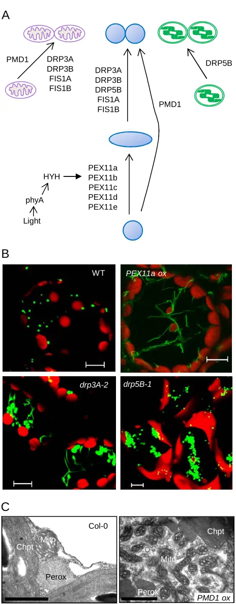
Peroxisome Elongation/Tubulation: PEROXIN11 (PEX11) Proteins Serve as Key Factors
Saccharomyces cerevisiae Pex11p was the first peroxisome division protein identified. Ectopic
expression of ScPEX11 leads to the elongation/tubulation and/or increased numbers of
peroxisomes, whereas the yeast pex11 null mutants contain one or two giant peroxisomes per cell
(Erdmann and Blobel, 1995; Marshall et al., 1995). PEX11 homologs have been identified as
multigene families in various lineages (Hu, 2009; Schrader et al., 2011). Arabidopsis has five
PEX11 homologs categorized into three subfamilies based on sequence, i.e., PEX11a, PEX11b,
and PEX11c to e (Figure 2A). These five isoforms are integral PMPs capable of inducing
peroxisome elongation and/or number increase (Figure 2B) (Lingard and Trelease, 2006; Nito et
mammalian PEX11 homologs complements the yeast mutant phenotype to various degrees,
demonstrating the conserved role of PEX11 across kingdoms (Orth et al., 2007; Koch et al.,
2010).
A recent study in Penicillium chrysogenum showed a role for Pex11p (and possibly other PEX11
homologs) in membrane remodeling. The conserved N-terminal amphipathic helix of PcPex11p
binds to liposomes that have membrane lipid content resembling that of the peroxisome
membrane, and induces liposome tubulation and membrane curvature, possibly through insertion
into the cytosolic leaflet of the phospholipid bilayer (Koch and Brocard, 2011; Opalinski et al.,
2011). Despite sequence and structural similarities, individual PEX11 family members may have
distinct functions (Koch and Brocard, 2011; Huber et al., 2012). The differential roles played by
Arabidopsis PEX11 proteins is indicated by the findings that i) PEX11a has a distinct membrane
topology from the other isoforms (Lingard and Trelease, 2006), and ii) only members of the
PEX11c-e subfamily complement the yeast pex11 mutants (Orth et al., 2007; Koch et al., 2010).
Being a highly abundant component of the peroxisome membrane and rate-limiting factor in
early peroxisome division, PEX11 is regulated at both transcriptional and post-translational
levels in yeast and mammals (Gurvitz and Rottensteiner, 2006; Michalik et al., 2006; Knoblach
and Rachubinski, 2010). In Arabidopsis synchronized cell cultures, the expression of PEX11 and
genes encoding other key division proteins is regulated by the cell cycle, which correlates with
peroxisome duplication (Lingard et al., 2008). A phytochrome A-mediated light signaling
pathway induces PEX11b expression during dark-to-light transitions, in which the bZIP
transcription factor HY5 HOMOLOG (HYH) binds to the PEX11b promoter (Figure 1A) (Desai
and Hu, 2008; Hu and Desai, 2008). Salt stress, abscisic acid (ABA), and jasmonic acid (JA) also
regulate the expression of Arabidopsis and/or rice PEX11 genes (Nayidu et al., 2008; Mitsuya et
al., 2010). Whether plant PEX11 proteins are subjected to post-translational modifications is
unclear.
Following elongation/tubulation, peroxisome division proceeds with membrane constriction and
fission, a process mediated by a protein complex consisting of the integral membrane-anchored
protein FIS1, a dynamin-related protein (DRP), and some lineage-specific cytosolic adaptor
proteins (Benard and Karbowski, 2009; Kaur and Hu, 2009).
Dynamins and DRPs are mechano-chemical enzymes or signaling GTPases that form oligomeric
rings around membranes, enforcing membrane fission or fusion through GTP hydrolysis
(Praefcke and McMahon, 2004; Faelber et al., 2011; Ford et al., 2011). At least three of the 16
Arabidopsis DRPs are involved in peroxisome fission. The closely related DRP3A and DRP3B
proteins are dual-localized and shared by peroxisomal and mitochondrial divisions, with DRP3A
playing a major role in peroxisome fission (Arimura and Tsutsumi, 2002; Arimura et al., 2004;
Logan et al., 2004; Mano et al., 2004; Lingard et al., 2008; Aung and Hu, 2009; Fujimoto et al.,
2009; Zhang and Hu, 2009) (Figure 2). Interestingly, DRP5B (ARC5), a DRP distantly related to
DRP3, targets to chloroplasts and peroxisomes and facilitates the division of both organelles
(Gao et al., 2003; Zhang and Hu, 2010) (Figure 2). Besides having enlarged, dumbbell-shaped
chloroplasts, drp5B mutants also contain aggregated peroxisomes that are impaired in fission
(Figure 2B) and are partially compromised in peroxisomal functions (Zhang and Hu, 2010).
Whereas DRP3A and DRP3B are members of an ancient family of DRPs involved in peroxisome
and mitochondrial division, DRP5B evolved more recently in the plant/algal lineage
(Miyagishima et al., 2008) to mediate chloroplast and peroxisome division.
Most eukaryotic DRPs lack a lipid-binding or transmembrane domain (TMD) and are only
recruited to the division sites by interacting directly or indirectly with a membrane-bound
receptor (Praefcke and McMahon, 2004). A yeast DRP, Dnm1p, is recruited to peroxisomes and
mitochondria by Fis1p, which is tethered to the organelles by its C terminus and extends its
N-terminal tetratricopeptide repeat (TPR) domain into the cytosol (Motley and Hettema, 2007).
Both Arabidopsis FIS1 homologs, FIS1A (BIGYIN) and FIS1B, are dual-targeted to
peroxisomes and mitochondria and play rate-limiting roles in initiating organelle fission (Scott et
al., 2006; Lingard et al., 2008; Zhang and Hu, 2008, 2009) (Figure 2A). Whether AtFIS1 is
required for targeting DRP3A/3B to the organelles has not been verified. Given that DRP5B has
2004), it may not need a receptor for peroxisome association. Physical interactions between FIS1
and PEX11 have been detected in mammals and plants (Kobayashi et al., 2007; Lingard et al.,
2008; Zhang and Hu, 2010), indicating a possible, direct functional link between the peroxisome
elongation and fission machineries.
Possible kingdom-specific factors also exist in the FIS1-DRP complex. Yeast Mdv1p and Caf4p
are two homologous and partially redundant proteins, each possessing a WD40 repeat and a
coiled-coil domain and acting as cytosolic adaptors for DRP recruitment (Tieu et al., 2002;
Griffin et al., 2005; Motley et al., 2008). Although homologs of Mdv1p and Caf4p have not been
identified in mammals, an analysis of the Arabidopsis genome revealed eight proteins with
similar domain structures (Pan and Hu, 2011); whether these proteins are involved in organelle
division remains to be demonstrated.
Peroxisome Division Factors that Act Independently from PEX11, FIS1, and/or DRPs
Mff (Mitochondrial fission factor) is a mammalian-specific coiled-coil protein, which is tethered
to mitochondrial and peroxisome membranes and recruits Drp1 to the organelles in a
Fis1-independent manner (Gandre-Babbe and van der Bliek, 2008; Otera et al., 2010). In the yeast
Yarrowia lipolytica, peroxisome division can be triggered when the β-oxidation enzyme
acyl-CoA oxidase binds to the PMP Pex16p, which subsequently induces lipid biosynthesis in the
membrane and the formation of a division complex containing the DRP Vps1p (Guo et al., 2003;
Guo et al., 2007). Some Arabidopsis mutants defective in β-oxidation or NAD+ transport contain
larger but fewer peroxisomes (Graham et al., 2002; Baker et al., 2006; Mano et al., 2011),
suggesting that accumulation of acyl-CoA or other molecules within the peroxisome may
regulate division.
Arabidopsis PEROXISOMAL and MITOCHONDRIAL DIVISION FACTOR 1 (PMD1) is a
plant-specific organelle division factor that acts independently from PEX11 and the FIS1-DRP3
complex (Aung and Hu, 2011) (Figure 2A). PMD1 is dual-targeted to the membranes of
peroxisomes and mitochondria. Loss-of-function pmd1 mutants contain enlarged peroxisomes
the organelles, which are often aggregated (Figure 2C). Surprisingly, PMD1 fails to show
physical or genetic interaction with any of the known organelle division proteins, indicating that
it is not an Mff counterpart. Furthermore, the PMD1 homolog, PMD2, which can form
complexes with PMD1, is localized only to mitochondria and exclusively involved in
mitochondrial morphogenesis (Aung and Hu, 2011). The mechanism by which PMD1 and
PMD2 impact peroxisome and mitochondrial division and morphogenesis remains to be
elucidated.
Protein Import
Identification of Genes Required for Matrix Protein Import
With the exception of some PMPs that traffic to peroxisomes via the ER (see above), nascent
peroxisomal proteins are imported from the cytosol. The plant peroxins that recognize and
transport peroxisomal proteins (Figure 3) have been identified by a combination of forward and
reverse genetic approaches. First, forward genetic strategies have taken advantage of the role of
peroxisomes in bioactivation of auxin precursors. Indole-3-butyric acid (IBA) and
2,4-dichlorophenoxybutryic acid (2,4-DB) undergo β-oxidation to form indole-3-acetic acid (IAA)
and 2,4-dichlorophenoxyacetic acid (2,4-D), respectively, resulting in the inhibition of root and
hypocotyl elongation. Therefore, IBA or 2,4-DB resistant mutants that display an elongated
phenotype but remain sensitive to the product (IAA or 2,4-D) are readily identified (Hayashi et
al., 1998; Zolman et al., 2000; Strader et al., 2011). These screens have identified mutants in
both β-oxidation and PEX genes. A second screen relies on the role of peroxisomes in mobilizing
fatty acid reserves to support post-germinative growth; mutants defective in β-oxidation are often
dependent upon exogenous sucrose for establishment and screens for sucrose-dependent (sdp)
mutants identified additional genes (Eastmond, 2006, 2007). Third, mislocalization of
peroxisome-targeted fluorescent fusion proteins has been used to isolate mutants defective in
peroxisome protein import (Mano et al., 2006; Goto et al., 2011). Finally, putative peroxins have
been identified in silico and characterized through reverse genetic approaches (Schumann et al.,
2003; Sparkes et al., 2003; Fan et al., 2005; Woodward and Bartel, 2005a; Nito et al., 2007;
The Matrix Protein Import Pathway
The majority of matrix proteins are synthesized with one of two import signals: PTS1
(peroxisomal targeting signal type 1), a C-terminal tripeptide, or PTS2, an N-terminal
nonapeptide. PTS1 sequences typically conform to the consensus of [small]-[basic]-[aliphatic],
as exemplified by the sequence SKL. PTS2 sequences have the consensus R[L/I/Q] X5 HL
(Lanyon-Hogg et al., 2010). Details on permissible PTS1 signals and their in silico prediction are
described later.
Following translation, PTS1 proteins interact with their receptor PEX5 in the cytosol (Figure 3).
PEX5 is highly conserved and contains two functional domains: an N-terminal peroxisomal
docking domain and a C-terminal domain formed from two sets of three tetratricopeptide repeats
(TPRs), which provide a binding pocket for PTS1 (Lanyon-Hogg et al., 2010). Homology
modeling of Arabidopsis PEX5 on a human PEX5-PTS1 protein structure suggests that all the
important interactions are conserved (Lanyon-Hogg et al., 2010). These structural studies
indicate that the mechanism of PTS1 recognition by PEX5 is likely to be conserved; however,
targeting studies show some species-specific differences that are likely to reflect subtle
differences in the geometry of the PTS1 binding pocket that remain to be fully understood.
PTS2 proteins interact with their receptor PEX7 prior to peroxisome entry (Figure 3), but the
molecular details of this interaction are unclear. Unlike PEX5, PEX7 cannot mediate interaction
with the peroxisome membrane alone but requires accessory proteins. As in mammals,
Arabidopsis PEX5 acts as the co-receptor for PEX7 (Nito et al., 2002). Down-regulation of
PEX5 by RNAi compromises both PTS1 and PTS2 import (Hayashi et al., 2005), and mutation
of a conserved serine in the pex5-1 mutant reduces PTS2 import while PTS1 import remains
functional (Woodward and Bartel, 2005b). As many β-oxidation enzymes use the PTS2 import
pathway, the pex5-1 mutant behaves as a typical β-oxidation mutant and is IBA resistant and
sucrose dependent (Zolman et al., 2000). The Arabidopsis pex5-10 mutant, which contains a
PTS2 import defect can be rescued by expression of a construct comprising the N-terminal
domain of PEX5 (Khan and Zolman, 2010), confirming that the PEX5 N-terminal domain is
required for PEX7 interaction.
The PEX5 N terminus is suggested to be a natively unfolded domain exhibiting significant
conformational flexibility (Carvalho et al., 2006). In mammalian PEX5, multiple WX3F/Y motifs
bind within the binding groove of the N-terminal domain of the PMP PEX14 (Neufeld et al.,
2009). Arabidopsis PEX14 is an integral PMP important for PTS1 and PTS2 import (Hayashi et
al., 2000). The topology of PEX14 is somewhat controversial (Oliveira et al., 2002) and
therefore it is unclear whether the critical interaction between PEX5 and PEX14 takes place on
the cytosolic side of the membrane, within the membrane, or even within the matrix. The latter
possibility would suggest that PEX14 is not the initial docking point for PEX5. In this context, it
is interesting that yeast and human PEX5 can spontaneously insert into lipid membranes in vitro
(Kerssen et al., 2006) and that residual protein import can occur without PEX14 in Hansenula
polymorpha (Salomons et al., 2000) and Arabidopsis (Monroe-Augustus et al., 2011).
PEX5/7 docking at the peroxisome membrane also involves PEX13 (Figure 3). AtPEX13 is quite
diverged from the fungal and mammalian counterparts and was identified from the aberrant
peroxisome morphology (apm) collection as a mutant showing partial mislocalization of a green
fluorescent protein (GFP)-PTS1 peroxisome marker to the cytosol (Mano et al., 2006). A null
pex13 allele was subsequently identified as amc (abstinence by mutual consent) with defective
male-female gametophyte recognition (Boisson-Dernier et al., 2008). PEX7 also binds to the N
terminus of PEX13 (Mano et al., 2006). There is still uncertainty about the order, stoichiometry,
and affinity of binding interactions among PEX5, PEX7, their cargoes, PEX14, and PEX13;
however, the general consensus is that import is driven by thermodynamically favorable binding
interactions (see Lanyon-Hogg et al., 2010 for more detailed discussion of this point). The
mechanism of protein translocation is also uncertain, but yeast PEX5 and PEX14 appear to form
a transient pore that can open to a diameter of up to 9 nm (Meinecke et al., 2010).
After import into the matrix, cargo is unloaded and the receptors are recycled. Again, there is a
re-export requires the three RING finger peroxins, Pex2p, Pex10p and Pex12p, the
ubiquitin-conjugating enzyme Pex4p and its membrane anchor Pex22p, and the two AAA ATPases Pex1p
and Pex6p, which are tethered to the membrane by Pex15p. The prevailing model (Figure 3)
invokes Pex5p monoubiquitination by Pex4p (E2) and Pex12p (E3), and ATP-dependent
dislocation of ubiquitinated Pex5p from the membrane via Pex1p and Pex6p (Grou et al., 2009).
Although there is no direct evidence for PEX5 ubiquitination in plants, the machinery is
conserved. The finding that the very mild pex13-1 mutant exacerbates the phenotypes of mutants
in the early part of the pathway but ameliorates the phenotypes of mutants in the recycling limb
of the pathway points to a need to balance receptor import and export (Ratzel et al., 2011).
Knockout mutants of Arabidopsis PEX2, PEX10 and PEX12 are embryo lethal (Hu et al., 2002;
Schumann et al., 2003; Sparkes et al., 2003; Fan et al., 2005) and RNAi lines all show PTS1 and
PTS2 import defects and sucrose dependence following germination (Nito et al., 2007). In
addition to these typical pex defects, some of the RING finger peroxin mutants display additional
phenotypes, suggesting their involvement in biological processes other than import. For example,
an RNAi line with strong PEX10 suppression also has variegated leaves, fused floral organs,
aberrant ER morphology, and a defect in cuticular wax synthesis (Kamijaki et al., 2009). A
transgenic Arabidopsis line expressing a PEX10 with a mutated RING finger also shows defects
in photorespiration and interaction between chloroplasts and peroxisomes (Schumann et al.,
2007). A gain-of-function mutant of PEX2 (TED3) suppresses the photomorphogenetic defects
of det1-1 (Hu et al., 2002). If indeed the RING finger peroxins are E3 ligases, they could
potentially target proteins other than the import receptors.
The pex4 RNAi mutant has a PTS1 protein import defect (Nito et al., 2007), and partial
loss-of-function mutations in PEX4 and PEX22 confer mild defects that are enhanced in the double
mutant (Zolman et al., 2005), supporting the notion that PEX4 and PEX22 function in the same
pathway. Indeed, Arabidopsis PEX22 and PEX4 interact and together can complement the S.
cerevisiae pex4 or pex22 mutants (Zolman et al., 2005).
PEX1 and PEX6 RNAi lines have a PTS1 protein import defect (Nito et al., 2007) and a
plants are small, pale, and have reduced seed set. At the cellular level, peroxisomes are enlarged
and PEX5 levels are reduced. Recently, the membrane anchor for PEX1 and PEX6 has been
identified from the collection of apm mutants. APEM9 is an integral PMP that binds PEX6 and
recruits the PEX1-PEX6 complex to the peroxisome membrane (Goto et al., 2011).
Degradation of the PTS1 Receptor PEX5
As discussed above, PEX5 monoubiquitination is required for PEX5 recycling in yeast and
mammals, and the conservation of the responsible ubiquitin-conjugating enzyme (PEX4),
ubiquitin protein ligases (PEX2, PEX10, and PEX12), and AAA ATPases (PEX1, PEX6) in
plants suggests that the PEX5 recycling mechanism also occurs in plants (Figure 3). Intriguingly,
these receptor-recycling peroxins resemble proteins needed during ER-associated protein
degradation (ERAD), the process of ubiquitination, retrotranslocation, and proteasomal
degradation of misfolded ER proteins (Gabaldon et al., 2006; Schluter et al., 2006). Further
supporting an ERAD analogy are the observations that yeast and mammalian PEX5 are
polyubiquitinated and degraded by the proteasome when not efficiently recycled (Platta et al.,
2004) in a process termed RADAR (receptor accumulation and degradation in the absence of
recycling) (Leon et al., 2006). For example, human PEX5 is degraded in the absence of PEX6
(Yahraus et al., 1996). Although plant PEX5 ubiquitination has not been directly demonstrated,
the Cys residue that is monoubiquitinated in other eukaryotes (Carvalho et al., 2007; Williams et
al., 2007) is conserved in Arabidopsis PEX5. In addition, the Arabidopsis pex6-1 missense allele
has reduced PEX5 levels, and overexpressing PEX5 partially restores peroxisome function in
pex6-1 (Zolman and Bartel, 2004), suggesting that a RADAR mechanism also operates in plants.
Reducing PEX4 function (Zolman et al., 2005) in the pex6-1 background restores PEX5 levels
while exacerbating pex6-1 physiological and molecular defects (Ratzel et al., 2011), suggesting
that PEX4 is needed for both the ubiquitination that promotes PEX5 recycling and the
ubiquitination that triggers RADAR. The apparent conservation of RADAR processes suggests
that this degradation prevents a deleterious build-up of PEX5 in the peroxisomal membrane.
In addition to low PEX5 levels observed in pex6-1 mutants (Zolman et al., 2005; Ratzel et al.,
suggesting that the dependence of PEX7 on PEX5 for cargo delivery in plants (Hayashi et al.,
2005; Woodward and Bartel, 2005a) is mirrored by a dependence of PEX5 on PEX7 for stability.
Whether the apparent PEX5 instability in pex7 mutants reflects inefficient recycling leading to
RADAR or instability in the cytosol remains to be determined.
Peroxisomal Proteases and Matrix Protein Degradation
Two peroxisomal proteases are implicated in peroxisome biogenesis. One is DEG15, the PTS2
processing protease. Originally purified from watermelon cotyledons, DEG15 is a trypsin-like
Ser protease that cleaves PTS2 proteins to remove the N-terminal region both in vitro and in vivo
(Helm et al., 2007; Schuhmann et al., 2008). Beyond a slight resistance to the inhibitory effects
of IBA (Lingard and Bartel, 2009) and 2,4-DB (Schuhmann et al., 2008), the Arabidopsis deg15
null mutant does not display growth or germination defects that would ascribe a physiological
benefit to removing the PTS2 sequence following peroxisome entry. Indeed, yeasts lack a
peroxisomal DEG15 ortholog and do not remove PTS2 sequences upon import (Helm et al.,
2007). The evolutionary advantage that has conserved the PTS2 removal process in plants and
mammals remains to be identified.
LON proteases are members of the AAA ATPase family originally discovered in bacteria, where
they degrade both aberrant and regulatory proteins (reviewed in Van Melderen and Aertsen,
2009). In plants, LON isoforms are found in chloroplasts, mitochondria, and peroxisomes
(Ostersetzer et al., 2007); LON2 is the peroxisomal LON isoform. Interestingly, Arabidopsis
lon2 mutants display molecular and physiological phenotypes indicative of peroxisomal defects.
For example, although matrix proteins correctly localize in 4-day-old cotyledon cells, they
mislocalize to the cytosol in older seedlings (Lingard and Bartel, 2009). Similarly, a PTS2-GFP
reporter sorts to peroxisomes in lon2 root tip cells, but is largely cytosolic in mature root cells
(Lingard and Bartel, 2009). The delayed onset of matrix protein sorting defects in lon2 mutants
suggests that LON2 facilitates continued matrix protein import in mature peroxisomes. The
increasing severity of lon2 import defects with age contrasts with several other pex mutants,
which seem to recover peroxisomal function with age. For example, the severe matrix protein
Monroe-Augustus et al., 2011), and pex5-10 mutants recover normal pigmentation upon
maturation (Khan and Zolman, 2010). The demonstration that LON2 promotes sustained
peroxisomal matrix protein import (Lingard and Bartel, 2009) indicates that this protease is a
previously unrecognized peroxin, and it will be interesting to discover the LON2 substrate(s) that
hinder matrix protein import if not efficiently degraded.
Although we are beginning to understand how proteins are delivered to the peroxisome matrix,
little is known about how excess plant peroxisomes or peroxisomal proteins are degraded. A
specialized form of autophagy, pexophagy, is important in removing excess peroxisomes in yeast
and mammals (reviewed in Manjithaya et al., 2010), but pexophagy has not been reported in
plants. Peroxisomal sequestration likely protects the cytosol from H2O2 produced by various
peroxisomal oxidases. Although peroxisomes house catalase and other enzymes that decompose
this H2O2, the protective capacity of the peroxisome can be exceeded (Eastmond, 2007).
Moreover, certain matrix proteins, such as the glyoxylate cycle enzymes isocitrate lyase and
malate synthase (ICL and MLS; see below), are susceptible to oxidative damage both in vitro
and in vivo (Yanik and Donaldson, 2005; Eastmond, 2007; Anand et al., 2009), which may
necessitate a degradation pathway that responds to oxidative damage. In addition, obsolete
proteins are removed during developmental peroxisomal remodeling. For example, ICL and
MLS are degraded when seedlings transition from fatty acid β-oxidation to photosynthesis
(Nishimura et al., 1996). This degradation is accelerated in a catalase mutant (Lingard et al.,
2009), suggesting that oxidative damage by H2O2 promotes peroxisome-associated protein
degradation. Furthermore, ICL and MLS must enter peroxisomes to be efficiently degraded
(Lingard et al., 2009), suggesting that degradation is triggered following import or that the
responsible protease is peroxisomal. However, insertion alleles disrupted in any of the five
predicted peroxisomal proteases (DEG15/At1g28320, LON2/At5g47040, PXM16/At2g41790,
At2g18080, and At2g35615) display normal ICL and MLS degradation (Lingard and Bartel,
2009), indicating that if ICL and MLS degradation is accomplished by a peroxisomal protease, it
acts redundantly or remains to be identified. Interestingly, one of the receptor-recycling peroxins,
PEX4, facilitates ICL and MLS degradation (Lingard et al., 2009), consistent with the alternative
possibility that damaged and obsolete proteins actively exit peroxisomes for cytosolic
recycle (or destroy) PEX5. It will be interesting to learn whether PEX5, which is essential for the
entry of peroxisomal matrix proteins, also assists in the exit of these proteins when they are
damaged or obsolete.
PEROXISOMAL FUNCTIONS
Plant peroxisomes mediate a multitude of processes crucial to development. Peroxisomes are the
sole site of fatty acid β-oxidation in plant cells and are involved in generating two
phytohormones, IAA and JA (Figure 4). They play an important role in photorespiration in
conjunction with mitochondria and chloroplasts (Figure 5). In addition to these major processes,
which are discussed in detail below, plant peroxisomes also participate or are implicated in a
plethora of other metabolic and signaling pathways, such as the glyoxylate cycle, detoxification,
generation of signaling molecules, biosynthesis of salicylic acid, and the metabolism of urate,
polyamines, sulfite, and branched-chain amino acids (reviewed in Kaur et al., 2009). Recent
studies have also revealed roles for peroxisomes in plant immune response (Lipka et al., 2005;
Westphal et al., 2008; Bednarek et al., 2009; Clay et al., 2009; Maeda et al., 2009; Coca and San
Segundo, 2010; Rojas et al., 2012) and the biosynthesis of biotin (Tanabe et al., 2011),
S-allantoin (Lamberto et al., 2010), phylloquinone (Widhalm et al., 2012), and isoprenoids
(Sapir-Mir et al., 2008; Tholl and Lee, 2011).
Peroxisomal β-Oxidation
Fatty Acid β-Oxidation
Fatty acid oxidation is an essential process in the mobilization of seed oil reserves, which are laid
down during seed development predominantly as triacylglycerol (TAG) and mobilized to support
post-germinative growth prior to the seedling developing photosynthetic competence (Graham,
2008). TAG lipases hydrolyze the TAG to release free fatty acids, whereby two oil
body-associated enzymes, SUGAR DEPENDENT 1 (SDP1) (Eastmond, 2006) and SDP1L (SUGAR
DEPENDENT 1 LIKE) together account for 95% of TAG lipase activity (Kelly et al., 2011).
peroxisomal ATP Binding Cassette (ABC) transporter protein CTS/PXA1/PED3 (see details
below). Mutants deficient in fatty acid degradation lack the energy or metabolites necessary for
seedling establishment into a photosynthetic plant, and thus produce short hypocotyls when
grown in the dark, a phenotype that can be rescued by sucrose. A severe β-oxidation block
results in strongly reduced germination (Baker et al., 2006). In addition to roles in early seedling
development, fatty acid β–oxidation also has important roles in re-mobilization of reserves
during senescence and in survival in extended periods of darkness (Dong et al., 2009; Kunz et
al., 2009; Slocombe et al., 2009).
Following peroxisomal import, straight chain saturated fatty acyl-CoAs undergo a cycle of
oxidation, hydration, oxidation and thiolysis, leading to release of acetyl-CoA and an acyl-CoA
that has been shortened by 2 carbons (Figure 4; Graham, 2008). The first step is catalyzed by a
family of acyl-CoA oxidases, ACX1-5 in Arabidopsis, with differing but partially overlapping
chain length specificities (Hayashi et al., 1999; Hooks et al., 1999; Eastmond et al., 2000b;
Rylott et al., 2003; Adham et al., 2005; Pinfield-Wells et al., 2005; Khan et al., 2012). These
enzymes are flavin adenine dinucleotide-linked and the electrons are passed to molecular oxygen
to produce H2O2. The resulting 2-trans-Enoyl CoA is the substrate for the multifunctional
protein, which contains both hydratase and dehydrogenase domains.
There are two peroxisomal multifunctional proteins in Arabidopsis, MFP2 (Rylott et al., 2006)
and AIM1 (Richmond and Bleecker, 1999). MFP2 is the major seedling form; its mutant shows a
typical β-oxidation deficiency phenotype (Rylott et al., 2006). The mfp2 mutant is not resistant to
pro-auxins whereas the aim1 mutant is. Consistent with this resistance, AIM1 prefers short chain
substrates (Richmond and Bleecker, 1999; Arent et al., 2010). MFP2’s hydratase activity prefers
longer chains (Rylott et al., 2006) but shows little activity on acyl-CoAs above 14 carbons in
length (Arent et al., 2010), suggesting that there is a yet undiscovered long chain hydratase.
The final step of core β-oxidation is the thiolytic cleavage of 3-ketoacyl CoA by thiolase to
produce acetyl-CoA and a shortened acyl-CoA. Of the three peroxisomal thiolases, PED1/KAT2
is the major seedling form (Hayashi et al., 1998; Germain et al., 2001). The ped1/kat2 mutant has
(Rylott et al., 2006) and kat2 (Germain et al., 2001) have enlarged peroxisomes, suggesting that
intra peroxisomal accumulation of acyl-CoAs could result in peroxisomal expansion or inhibition
of division (Graham et al., 2002).
The core β-oxidation machinery metabolizes straight chain saturated fatty acids. However,
peroxisomes also metabolize unsaturated fatty acids with double bonds at both odd and even
positions, which requires accessory enzymes to convert these molecules into suitable substrates
(Goepfert and Poirier, 2007; Graham, 2008). For the degradation of fatty acids with double
bonds at the odd position, e.g., C18:Δ9cis (oleic acid), the peroxisomal Δ3,5Δ2,4 dienoyl CoA
isomerase encoded by AtDCI is essential (Goepfert et al., 2005). For even double bonds, an
epimerase activity that is part of the multifunctional protein or a separate enoyl-CoA hydratase
(ECH) is required (Goepfert et al., 2006).
The acyl-CoA oxidase reaction produces H2O2, which is metabolized by catalase. However,
under conditions of high H2O2 production, such as during TAG mobilization in early seedling
growth, a membrane-bound system comprising ascorbate peroxidase and monodehydroascorbate
reductase (MDAR) acts as a second line of defense to prevent H2O2 leakage into the cytosol. A
mutant in MDAR (sdp2) has compromised β-oxidation due to excess H2O2 that causes oxidative
inactivation of the TAG lipase SDP1 (Eastmond, 2007).
The product of β-oxidation, acetyl-CoA, can be respired by mitochondria (Kunze et al., 2006) or
can enter the glyoxylate cycle, where citrate synthase (CSY), ICL, and MLS convert it to
succinate and malate used for gluconeogenesis (Pracharoenwattana and Smith, 2008).
Arabidopsis CSY1 and CSY2 convert acetyl-CoA to citrate for export to mitochondria; the
double mutant is unable to germinate without sucrose and physical removal of the seed coat, fails
to degrade its oil bodies, and is resistant to 2,4-DB (Pracharoenwattana et al., 2005). The icl1
mutant germinates and degrades oil bodies, presumably respiring the acetyl-CoA, but has
reduced survival in periods of extended darkness (Eastmond et al., 2000a). mls mutants have
mild phenotypes, suggesting MLS is partially dispensable for gluconeogenesis and lipid
The hydroxyacyl-CoA dehydrogenase activity of MFP produces NADH. Re-oxidation of NADH
and therefore continued β-oxidation depends on a malate-oxaloacetate shuttle that involves
peroxisomal and cytosolic isoforms of malate dehydrogenase (MDH). Double mutants defective
in the two peroxisomal MDH genes, PMDH1 and PMDH2, germinate but are sucrose dependent
for establishment, are resistant to 2,4-DB, and mobilize TAGs slowly (Pracharoenwattana et al.,
2007).
Jasmonic Acid Production
The major functions of jasmonates, phytohormones regulating development and stress response,
include wounding and pathogen responses, stamen development, and pollen release. This
hormone family is comprised of several related lipid-derived compounds: JA, its precursor
12-oxo-phytodienoic acid (OPDA), and JA derivatives, including the methyl ester and the Ile
conjugated forms (reviewed in Wasternack and Kombrink, 2009; Acosta and Farmer, 2010).
Production of active jasmonates occurs sequentially in three locations: chloroplasts,
peroxisomes, and the cytosol.
Chloroplast-localized reactions convert polyunsaturated fatty acids to OPDA, which is released
via an unknown mechanism (Wasternack and Kombrink, 2009; Acosta and Farmer, 2010).
Following peroxisomal import, the OPDA reductase OPR3 converts OPDA to OPC8:0
(3-oxo-2-(2’-[Z]-penenyl) cycopentane-1-octanoic acid). OPR3 has reductase activity in vitro (Costa et al.,
2000; Schaller et al., 2000) and opr3 was found as a male-sterile mutant rescued specifically by
JA application (Stintzi and Browse, 2000).
Three rounds of peroxisomal β-oxidation convert OPC8:0 OPC6:0 OPC4:0 JA. OPCL1
activates OPC8:0, and ACX1 and ACX5, AIM1, and PED1/KAT2 are implicated in the core
β-oxidation of JA precursors. These isozyme assignments were inferred from three observations: i)
OPCL1, ACX1, and KAT2 mRNAs strongly and rapidly accumulate in response to JA, as part of
a positive feedback mechanism (Castillo et al., 2004; Koo et al., 2006); ii) OPCL1 (Koo et al.,
2006; Kienow et al., 2008) and ACX1 (Li et al., 2005) are biochemically active on JA
JA biosynthesis (Castillo et al., 2004; Afitlhile et al., 2005; Pinfield-Wells et al., 2005; Koo et
al., 2006; Delker et al., 2007). Moreover, disruptions of ACX1 or PED1/KAT2 delay systemic
responses (Castillo et al., 2004) and a tomato acx1 mutant has reduced defense against chewing
insects (Li et al., 2005).
The modification of JA to JA-Ile (Staswick and Tiryaki, 2004), the active component in JA
signaling (Thines et al., 2007), occurs in the cytoplasm. OPDA, JA, and JA-Ile have unique roles
in plant cells (Wasternack and Kombrink, 2009; Acosta and Farmer, 2010). The transition
between organelles may regulate the ratio of jasmonates and thereby affects the types or intensity
of responses.
OPDA regulates seed germination. Whereas mutants blocked in β-oxidation can be rescued for
establishment by sucrose supplementation, indicating an insufficient supply of carbon and energy
from fatty acid metabolism, severe mutants in core β-oxidation functions cannot germinate
unless the testa is manually ruptured (Russell et al., 2000; Pinfield-Wells et al., 2005; Footitt et
al., 2006). Peroxisomal transport or activity mutants, including cts/pxa1/ped3, ped1/kat2, and
acx1 acx2 double mutants, accumulate OPDA and paradoxically, JA, in seeds. However, a pxa1 opr3 double mutant, which accumulates high OPDA but lacks JA, maintains the germination
defect, indicating that peroxisomal import and metabolism of OPDA is important for
germination (Dave et al., 2011). Moreover, OPDA and ABA act synergistically to increase levels
of the transcription factor ABI5 (Dave et al., 2011). ABI5 is also upregulated in the ped3 allele,
which in turn leads to higher levels of polygalacturonase inhibiting proteins; removal of pectin
using exogenous polygalacturonase can overcome the germination block in ped3 (Kanai et al.,
2010).
The JA biosynthetic pathway was proposed in the 1980s (Vick and Zimmerman, 1983).
Although great strides have been made identifying the peroxisomal components, several
questions remain. An unknown thioesterase presumably is required to cleave the jasmonoyl-CoA
to release JA. The transporter facilitating JA export also remains unknown. In addition, there is a
high degree of redundancy in JA transport and biosynthesis, and residual JA still accumulates in
and opcl1 accumulates JA to ~60% of wild type, allowing many expression targets to still be
induced (Koo et al., 2006). Similarly, only in an acx1 acx5 double mutant is fertility and
wound-induced JA biosynthesis lost (Schilmiller et al., 2007). Further, different tissues may regulate JA
synthesis differently. For instance, Dave et al. (2011) reported high JA levels in cts-2 seeds, but
studies on the same allele showed almost no JA in leaves (Theodoulou et al., 2005). Similarly,
acx1 acx5 makes no JA in wounded leaves, but produces JA in flowers and following fungal
infections (Schilmiller et al., 2007). Further studies, including analysis of additional mutant
combinations, could define the full complement of proteins involved in JA biosynthesis, but
mutant analysis will require examination in multiple conditions for a complete understanding.
Peroxisomal Conversion of IBA to IAA
IAA is the principal form of auxin, a phytohormone regulating many aspects of development by
influencing cell division and elongation (reviewed in Perrot-Rechenmann, 2010). IBA is
structurally similar to IAA, but has a two carbon side-chain elongation; IBA is known for
efficacy in root induction and is applied to cuttings or seedlings to ensure strong root
development (reviewed in Woodward and Bartel, 2005b). Feeding studies have shown that IAA
can be converted to IBA; IBA formation is hypothesized to relieve high IAA levels. IBA is also
converted back to IAA, increasing free (active) IAA to match plant needs. Conversion of IBA to
IAA removes the two extra side-chain carbons in a β-oxidation like pathway (Fawcett et al.,
1960). Because of the structural differences, IBA can be considered a proto-auxin, which is
transported (reviewed in Strader and Bartel, 2011) or stored (reviewed in Simon and Petrasek,
2011) without auxin activity.
Our understanding of IBA activity is based on forward genetic screens, which revealed IBA
metabolism to be a peroxisomal process. The predicted pathway for IBA metabolism parallels
fatty acid β-oxidation: IBA is imported into peroxisomes, activated by CoA, and converted to
IAA-CoA via the core β-oxidation steps (Figure 4). Mutants defective in AIM1 and PED1/KAT2
show pleiotropic phenotypes, including fatty acid and JA defects (described above) and 2,4-DB
(Hayashi et al., 1998; Richmond and Bleecker, 1999; Hayashi et al., 2002) and IBA (Zolman et
Therefore, AIM1 could catalyze the middle two steps of IBA metabolism, similar to fatty acid
metabolism. PED1/KAT2 could act as a thiolase to release two side-chain carbons, producing
IAA-CoA and acetyl-CoA (Hayashi et al., 1998; Zolman et al., 2000).
Alternatively, ibr1, ibr3, ibr10, and ech2 only show IBA-response phenotypes, suggesting that
the corresponding enzymes may act specifically on IBA intermediates. IBR3 encodes an
acyl-CoA dehydrogenase/oxidase, which could convert IBA-acyl-CoA to the α,β-unsaturated thioester
(Zolman et al., 2007). Two enoyl-CoA hydratases are implicated in IBA responsiveness: IBR10
(Zolman et al., 2008) and ECH2 (Strader et al., 2011). Although ECH2 and IBR10 have similar
domain structures, complementation experiments indicate that they are not redundant (Strader et
al., 2011). In addition to hydratase activity, ECH2 also has a hot dog domain common in
thioesterases and therefore may be acting at the last step to convert IAA-CoA to IAA (Strader et
al., 2011). Finally, IBR1, also identified as SDRa (Wiszniewski et al., 2009), encodes a
short-chain dehydrogenase/reductase (Zolman et al., 2008), which may catalyze the fourth step of IBA
β-oxidation. AIM1-IBR1 redundancy at the dehydrogenase/reductase step could explain why the
ibr1 defects are less severe than those of other mutants (Strader et al., 2011).
Strader et al. (2010) demonstrated reduced IAA production from labeled IBA in pex6, pxa1, and
the ibr1 ibr3 ibr10 triple mutant, confirming roles for peroxisomes and these enzymes in IAA
production. However, the precise enzymatic assignments require biochemical confirmation; in
particular, IBR10 and ECH2 placement and potential redundancy between AIM1 and IBR1 will
require activity assays for resolution.
ACX activity on IBA-CoA also remains a question. acx mutant analysis revealed that all five
ACX enzymes promote IBA responsiveness (Adham et al., 2005) and acx1 acx2 double mutants
have decreased IBA-to-IAA conversion (Strader et al., 2010). IBR3 and multiple ACX enzymes
may catalyze this reaction in an overlapping manner or based on expression. However, ACX
enzymes show substrate chain-length specificities (see above) that seemingly contradict the idea
that all five act directly on IBA. Alternatively, IBR3 may act directly on IBA substrates while
ACX activity affects IBA oxidation indirectly, perhaps based on limiting peroxisomal CoA pools
2,4-DB resistant but responds normally to IBA (Wiszniewski et al., 2009); whether a different
protein activates IBA (perhaps redundantly) remains to be determined. IAA export to the cytosol
has not been defined, either.
Finally, we do not know how the conversion of IBA to IAA is regulated or triggered, although
one hypothesis is that low IAA levels stimulate IBA metabolism. IBA-response mutants have
reduced lateral root systems, smaller root meristems, defective cotyledon expansion, shorter root
hairs, and reduced hypocotyl and stamen elongation (Zolman et al., 2000; Footitt et al., 2007;
Strader et al., 2010; Strader et al., 2011), demonstrating the importance of this conversion in
multiple aspects of plant growth and development.
Photorespiration
The Classical Pathway
The most prominent role of peroxisomes in photosynthetic tissues is their participation in
photorespiration. The oxidative C2 cycle is a salvage pathway for phosphoglycolate produced by
the oxygenase activity of ribulose 1,5-bisphosphate carboxylase/oxygenase (RubisCO), to the
Calvin cycle intermediate phosphoglycerate. This pathway is one of the most sophisticated
examples of subcellular compartmentalization and spatial and temporal coordination, as it
combines enzymatic reactions in, and intermediate and co-factor exchange between,
chloroplasts, peroxisomes, mitochondria and, as recently shown, the cytosol (Timm et al., 2008).
Peroxisome-localized photorespiratory enzymes include glycolate oxidase (GOX), catalase
(CAT), two aminotransferases, hydroxypyruvate reductase (HPR), and MDH, placing leaf
peroxisomes at a central position in photorespiration (Figure 5).
Downstream of RubisCO, the photorespiratory reactions continue in the chloroplast stroma with
phosphoglycolate phosphatase (PGP), which dephosphorylates 2-phosphoglycolate (Schwarte
and Bauwe, 2007). Glycolate diffuses into the matrix of peroxisomes, where it is oxidized to
glyoxylate by GOX concomitant with H2O2 production. Glyoxylate is transaminated by two
GGT), which ideally cooperate at a 1:1 stoichiometry (Liepman and Olsen, 2001; Igarashi et al.,
2003; Liepman and Olsen, 2003; Igarashi et al., 2006). Mitochondrial glycine decarboxylase
decomposes glycine to CO2, NH3, and NADH, and transfers a C1 unit to 5,10-methylene
tetrahydrofolate. Serine hydroxymethyl transferase attaches this methylene unit to the second
Gly molecule to produce Ser. Serine diffuses back to leaf peroxisomes for transamination by
SGT to yield hydroxypyruvate, which is reduced by HPR and NADH provided by peroxisomal
MDH to form glycerate. Last, stromal glycerate kinase (GLYK) produces the Calvin cycle
intermediate 3-phosphoglycerate (Figure 5) (Reumann and Weber, 2006; Maurino and
Peterhansel, 2010).
Molecular Identification of All Key Photorespiration Enzymes
Photorespiration is an essential process in land plants, as evident from the conditionally lethal
phenotype of mutants deficient in the participating enzymes or transporters. However, the
photorespiratory pathway of C3 plants is inefficient in terms of energy, carbon and nitrogen
usage (see below). To fill in the knowledge gaps about photorespiratory enzymes and increase
plant biomass production, photorespiration research has been revitalized recently, with major
activities led by groups such as the German research consortium PROMICS
(www.promics.uni-rostock.de). Major fundamental and applied biotechnological knowledge has been gained in the
past few years, as described by several recent reviews (Foyer et al., 2009; Bauwe, 2010; Bauwe
et al., 2010; Maurino and Peterhansel, 2010; Peterhansel and Maurino, 2011; van Dongen et al.,
2011).
Molecular identification of the core photorespiration enzymes has been completed only recently.
Using a candidate gene approach, the gene encoding PGP was revealed based on the
characteristic photorespiratory phenotype of the knock-out mutant, i.e., non-viability in normal
air but normal growth under elevated CO2 concentrations (Schwarte and Bauwe, 2007). Contrary
to the other core photorespiratory enzymes, deletion of peroxisomal HPR1 does not lead to
ambient air sensitivity but does increase the stoichiometry of photorespiratory CO2 release
(Cousins et al., 2011). Identification of a second HPR (HPR2) suggests the existence of an
third, chloroplast-localized HPR with high specificity for glyoxylate; the triple mutant of the
three HPR genes shows increased growth retardation, decreased photochemical efficiency, and
reduced oxygen-dependent gas exchange compared to the hpr1 hpr2 double mutant (Timm et al.,
2011).
The gene encoding the last missing enzyme of the C3 plant photorespiratory cycle, GLYK, was
identified from Arabidopsis; its knockout mutant is unviable in normal air but able to grow under
elevated CO2 (Boldt et al., 2005). Contrary to that in C3 plants, maize GLYK is redox-regulated
by an additional, C-terminal autoinhibitory domain, which forms a disulfide bridge at night,
inhibiting enzyme activity and rendering the oxidized enzyme inactive (Bartsch et al., 2008).
Photorespiration as a Prime Target for Crop Improvement
Despite being a valuable salvage pathway, the photorespiratory C2 cycle remains inefficient
because it renders i) suboptimal conversion of fixed carbon in the form of phosphoglycolate into
phosphoglycerate (max. three of four C atoms, i.e., 75%), ii) loss of fixed N, and iii) loss of
energy during glycolate oxidation by the production of H2O2 rather than NAD(P)H. Hence, the
photorespiratory pathway, at least theoretically, bears a high optimization potential in C3 plants,
making it a prime target for crop improvement for increased yield and biomass production.
A bacterial glycolate oxidation pathway was introduced into Arabidopsis chloroplasts for
alternative conversion of glycolate into glycerate, thereby shifting CO2 release from the
mitochondrion to the chloroplast to increase CO2 concentration in the vicinity of RubisCO and
reduce its oxygenase activity. Indeed, the transgenic lines showed enhanced growth (Kebeish et
al., 2007). To conserve the glycolate carbon in malate, transgenic Arabidopsis plants
overexpressing chloroplast-targeted GOX and MLS were generated. The transgenic lines
developed oxidative stress lesions under photorespiratory conditions, most likely due to
enhanced H2O2 production in chloroplasts, but showed enhanced growth under
nonphotorespiratory conditions (Fahnenstich et al., 2008; Maurino and Flügge, 2009). To
by-pass the peroxisomal aminotransferases and glycine-dependent ammonia production, transgenic
were generated. However, only the first enzyme was highly expressed in the transgenic plants,
which exhibited stress symptoms when exposed to air, suggesting that some glyoxylate was
directed into a deleterious short-circuit of the photorespiratory nitrogen cycle (Carvalho Jde et
al., 2011). These first attempts to optimize photorespiration are promising. However, because the
photorespiratory pathway is more tightly integrated into the whole plant primary and secondary
metabolism than previously hypothesized, these manipulations also uncover technical challenges
and unexpected negative side-effects and reveal the need for further studies.
Although high CO2 levels reduce photorespiration, they often lead to a decline in the plant’s
nitrogen status. Indeed, atmospheric CO2 enrichment reduced the efficiency of nitrogen use
(Rachmilevitch et al., 2004). This inhibition of nitrate assimilation into organic nitrogen
compounds may be largely responsible for CO2 acclimation, i.e., the decrease in photosynthesis
and growth of plants conducting C3 carbon fixation after long exposures to CO2 enrichment
(Bloom et al., 2010). Hence, ammonium and nitrate availability will become increasingly
important in determining plant productivity as CO2 levels rise.
PEROXISOMAL TRANSPORTERS FOR METABOLITES AND CO-FACTORS
Several peroxisomal metabolic pathways require an interplay with other cellular compartments,
including plastids, mitochondria, and the cytosol. Consequently, a considerable number of
substrates, intermediates, end products, and cofactors must be exchanged between peroxisomes
and other cell compartments. Their membrane passage is mediated by transport proteins (Linka
and Esser, 2012).
An ABC Transporter Importing the Substrates for β–Oxidation
Fatty acids and other β-oxidation substrates are imported by the peroxisomal ABC transporter
protein CTS/PXA1/PED3 (Zolman et al., 2001; Footitt et al., 2002; Hayashi et al., 2002); similar
transporters also exist in fungi and mammals (Theodoulou et al., 2006). CTS/PXA1/PED3 was
independently isolated from several forward genetic screens (hence its multiple names),
This transporter plays a crucial role in i) storage oil mobilization in seedlings and probably
pollen (Zolman et al., 2001; Footitt et al., 2002; Hayashi et al., 2002; Footitt et al., 2007), ii)
turnover of membrane lipids, especially under carbon and energy starvation (Kunz et al., 2009;
Slocombe et al., 2009), iii) JA biosynthesis (Theodoulou et al., 2005), iv) auxin biosynthesis
(Zolman et al., 2001; Hayashi et al., 2002; Strader et al., 2010), v) seed coat rupture during seed
germination (Kanai et al., 2010), and vi) efficient fertilization in female reproductive tissue
(Footitt et al., 2007).
CTS/PXA1/PED3 is a full ABC transporter that comprises two nucleotide-binding domains
(NBDs) providing the driving force for transport, and two TMDs involved in substrate
recognition and translocation. The transport cycle requires intra-molecular communication
between NBDs and TMDs, and modeling of CTS/PXA1/PED3 suggests that an interaction
between NBD1 and TMD2 is critical for protein function. Mutation analysis shows distinct roles
of the two NBDs in vivo (Dietrich et al., 2009).
A point of debate is whether CTS/PXA1/PED3 transports free fatty acid or CoA esterified
substrates. Free fatty acids are activated to acyl-CoAs by acyl-CoA synthetases present in
multiple compartments and transporter mutants accumulate long chain acyl-CoAs (Footitt et al.,
2002). The two peroxisomal long chain acyl-CoA synthetases, LACS6 and LACS7 (Fulda et al.,
2002), are essential for fatty acid mobilization and seedling development (Fulda et al., 2004).
The S. cerevisiae equivalent transporter Pxa1p/Pxa2p transports acyl-CoAs (Verleur et al.,
1997). The Arabidopsis CTS/PXA1/PED3 protein can complement the yeast pxa1 pxa2 double
mutant and support the metabolism of a wide range of fatty acid substrates that differ in chain
length and degree of unsaturation (Nyathi et al., 2010). Further, the ATPase activity of
CTS/PXA1/PED3 is stimulated by acyl-CoAs but not appreciably by free fatty acids, which also
supports the notion of acyl-CoAs as substrates (Nyathi et al., 2010). As proposed by Fulda et al
(2004), one possible explanation of this discrepancy is that acyl-CoAs are the substrate, but the
CoA is removed during transport and acyl-CoA is re-synthesized in the peroxisome by LACS6
and/or LACS7. Resolution of this issue will require in vitro transport studies using reconstituted