“QUANTIFICATION OF MICROVESSEL DENSITY IN
BREAST CARCINOMAS BASED ON
IMMUNOHISTOCHEMISTRY”
DISSERTATION SUBMITTED FOR
M.D. DEGREE EXAMINATION
BRANCH III PATHOLOGY
OF
THE TAMILNADU DR.M.G.R. MEDICAL UNIVERSITY
CHENNAI
TIRUNELVELI MEDICAL COLLEGE HOSPITAL
TIRUNELVELI
CERTIFICATE
This is to certify that the Dissertation “QUANTIFICATION OF MICROVESSEL DENSITY IN BREAST CARCINOMAS BASED ON IMMUNOHISTOCHEMISTRY”presented herein by Dr.SARANGAN. A is an original work done in the Department of Pathology, Tirunelveli Medical College Hospital, Tirunelveli for the award of Degree of M.D. (Branch III) Pathology under my guidance and supervision during the academic period of 2012 - 2015.
The DEAN
CERTIFICATE
I hereby certify that this work embodied in the dissertation
“QUANTIFICATION OF MICROVESSEL DENSITY IN BREAST CARCINOMAS BASED ON IMMUNOHISTOCHEMISTRY” is a record of work done by Dr. SARANGAN A in the Department of Pathology, Tirunelveli Medical College, Tirunelveli, during his postgraduate degree course in the academic period 2012-2015. This work has not formed the basis for any previous award of any degree.
Dr.S.Vallimanalan, M.D., Dr. K.Shantaraman MD , Professor of Pathology, Professor and HOD of Pathology, Department of Pathology, Department of Pathology
Tirunelveli Medical College, Tirunelveli Medical College, Tirunelveli. Tirunelveli.
DECLARATION
I solemnly declare that the dissertation titled “QUANTIFICATION OF MICROVESSEL DENSITY IN BREAST CARCINOMAS BASED ON IMMUNOHISTOCHEMISTRY” is done by me at Tirunelveli Medical College Hospital, Tirunelveli.
The dissertation is submitted to The Tamilnadu Dr. M.G.R. Medical
University towards the partial fulfilment of requirements for the award of M.D. Degree (Branch III) in Pathology.
Place: Tirunelveli Dr. SARANGAN . A
Date: Postgraduate Student,
M.D Pathology,
Department of Pathology,
Tirunelveli Medical College
ACKNOWLEDGEMENT
I take immense pleasure to acknowledge all those who have helped me to make this dissertation possible.
I am grateful to the Dean, Tirunelveli Medical College and Medical Superintendent of the Tirunelveli Medical College Hospital for permitting me to undertake this study.
I express my profound sense of gratitude to Dr. K. Shantaraman,M.D.,
my respected Professor and Head of Department of Pathology, Tirunelveli Medical College, Tirunelveli and my guide Dr.S.Vallimanalan, M.D., for his unstinted guidance and motivation.
I thank Dr.K.Swaminathan, M.D., Dr. J. Suresh Durai, M.D., Dr. Arasi Rajesh, M.D., Professors of Pathology for their constant support and encouragement. I profusely thank all the other faculties and my postgraduate colleagues for their valuable support.
I sincerely thank the Professors and faculties of the Departments of General Surgery and Oncology for providing me the specimens for my study.
I also sincerely thank the Technicians and other members of the Department of Pathology and the Central Diagnostic Laboratory for their kind co-operation.
ABBREVIATIONS
DNA : Deoxy ribonucleic acid
DPX : Di-N-Butyle Phthalate in Xylene.
H&E : Hematoxylin and Eosin
NOS : Not Otherwise Specified
NST : No Special Type
RBC : Red Blood Corpuscle
SBR : Scarff-Bloom-Richardson
S.D. : Standard Deviation
sig : Significant
SPF : S Phase Fraction
TDLU : Terminal Duct Lobular Unit
IHC : Immunohistochemistry.
CONTENTS
S.No Title Page.No
1 INTRODUCTION 1
2 AIMS & OBJECTIVES 5
3 REVIEW OF LITERATURE 7
4 MATERIALS AND METHODS 68
5 OBSERVATION AND RESULTS 75
6 DISCUSSION 96
7
8
SUMMARY
CONCLUSION
103
106
BIBLIOGRAPHY
APPENDIX
QUANTIFICATION OF MICROVESSEL DENSITY IN BREAST CARCINOMAS BASED ON IMMUNOHISTOCHEMISTRY” Abstract:
Breast cancer is the most frequent neoplasm causing death in women between 35-55 years of age. Angiogenesis is of key importance in the process of tumor progression in a number of tumor types including breast cancer. This is a both retrospective and prospective study which included fifty breast cancer cases . Angiogenesis was estimated by determining micro vessel counting after immune staining the paraffin embedded tissue sections using anti-cd34 antibody. The study is designed to compare MVD in different tumor stage, grade and age group. Age of the patients ranged from 35 to 85 years with a mean age of 52.58 years. Majority showed (40%) more than 84 vessels count in 1 mm2 field area . Micro vascular density positively correlated lymph nodes involved by the tumor and grade of the tumour . In the future, antibodies specific to proliferating endothelium, together with the development of automated image analysis may improve the accuracy and value of measuring angiogenesis-induced microvessel density.
2
INTRODUCTION
Breast cancer is one of the most common carcinoma among women in
India, which accounted for 25 to 30 percent of all cancer in woman(1). Breast cancer is the second most common cancer with an estimated 115,251 new
diagnoses(2) and the second most common cause of cancer related deaths with 53,592 breast cancer deaths in 2008(2). The incidence of breast cancer increases with age. The incidence of female breast cancer is increasing
between the age group of 35 to 50 years worldwide3. Invasive ductal carcinoma comprises the largest group of malignancy which constitutes
about 65% - 80% of all breast carcinomas4.
Though there is an increase in breast cancer incidence, breast cancer
mortality is decreasing in the last 20 years (4).This is mainly caused by both the introduction of breast cancer screening and use of adjuvant systemic
chemotherapy. Breast are made up of specialized epithelium and stroma
which may give rise to both benign and malignant neoplasm.
Patients who are at high risk can be identified based on clinical and
pathological prognostic factors, such as age, menopausal status, size of the
tumour, grade of the tumour, lymph node status and hormonal receptor
status.
3
1. Estrogen receptor and progesterone receptor.
2. HER2/neu amplification :
HER2 /neu over expression is associated with poor prognosis. But the
drug transtuzumab can be targeted against this oncoprotein.
3. Proliferative markers like Ki-67
4. Angiogenesis
One of the emerging predictive and prognostic factor in breast
carcinoma is tumour induced angiogenesis. Angiogenes is defined as process
of formation of new blood vessels from the endothelium of the already
pre-existing vasculature.
It is important in growth, progression and spread of the tumour.. So, tumour microvessels are necessary for sufficient supply of nutrients and
oxygen and removal of waste metabolites from tumour cells.
In normal life, angiogenesis play a vital role in reproduction,
embryogenesis, menstruation, and wound healing and repair. Its importance
in solid tumours was first recognized by Folkman et al in 1971(5), he suggested that the growth of tumours was depends on angiogenesis. Micro
vascular density is considered to be a hallmark of the angiogenesis.
The first study to examine intratumoral microvesseldensity (IMD) was
carried out by Weidner and colleagues in 1991with immunohistochemistry.
4
cancers (5). Since this initial work many antibodies including those against
CD31 & CD34has been used to assess MVD6.
It has been emphasized that high micro vessels density in growing
tumors closely associated with increasing number of tumor cells fell into the
blood stream. In recent years, it has been proposed that quantification of
intratumoral microvessel density by immunostaining for endothelial cell
markers, such as CD34, CD31, von Willebrand factor, may be a useful
prognostic factor in breast carcinomas.
So our present study was undertaken to asses and correlate the
microvessel density in breast carcinomas with different clinicopathological
factors and to ascertain the correlation of tumor angiogenesis with
metastasis.
5
Aim of the study
Aim of the study
Aim of the study
Aim of the study
6
AIMS AND OBJECTIVES
1. To quantify and grade the microvessel density in breast carcinomas with
immunohistochemistry.
2. To asses and correlate the microvessel density in breast carcinomas with
different clinicopathological entities with immunohistochemistry.
7
Review of Literature
Review of Literature
Review of Literature
Review of Literature
8
REVIEW OF LITERATURE
HISTORY
Breast cancer was known but uncommon until the 19th century, in the
year 1882 the first mastectomy for breast carcinoma was done by William
stewart halsted. (9). The procedure involved excising both breasts, lymph nodes, and the pectoralis muscles. Thereafter, the arrival of the Halsted
radical mastectomy increased the survival rate by 50%.
Breast cancer staging systems was introduced in the 1920s and
1930s.(10)In 1926, first case-controlled study on breast cancer epidemiology
was done by Janet Lane-Claypon (11),who was analysed the first
epidemiology study on fertility and breast cancer risk.
The results from the Nurses Health Study (1995) and the reports of
the Women's Health Initiative trial (2002) showed that the hormone
replacement therapy could increase the incidence of breast cancer(12)..
In 1945, Algire and Chalkley [14] were the first to determined that tumor growth is very closely related to the formation of an new
microvessels.
In 1991, the first study to estimate the intratumoral microvessel
density immunohistochemically was performed by Weidner and colleagues
(13)
. Theyused factor Vlll related antigen as an endothelial marker in a group
9
ANATOMY
DEVELOPMENT OF BREAST:
The mammary glands develops from the ectodermal mammary ridges
at the 5th week of intrauterine life. Bilaterally, they appears as a thickened line and it extends on the ventral surface ofthe fetus from the axillary to mid
thigh. Around 7th week in utero major part of mammary ridge disappear.But
some portion of itpersist in the 4th or 5th intercostalsspace called the primary
mammary buds.
Primary buds of ectoderm starts invading into the underlying
developing stroma.By 10- 12th week of gestation, the primary mammary
buds branches to form secondary buds, subsequently it forms the mammary
lobules.
In 5th month of fetal development, the ectodermal penetration
produces 15 –20 radial ingrowths branching into the developing breast.
Small lumen will develop within the mammary buds which latter forms the
developing lactiferous ducts and the branches of lactiferous duct.
The lactiferous ducts converge to open into a mammary pit, which
then converted into nipple during infancy. There is no discernible variation
between the male and female breast tissues from the time of conception until
puberty.
While at puberty females exhibit branching and further lengthening of
10
stroma and fat tissues reaching their maximum breast development by the age of 20 years.
The menstrual cycle is accompanied by minor variations of the breast
tissue, but major physiological changes of the breast tissue are seen during
pregnancy and lactation. There is major regression of the breast tissue during
menopause which merges with aging associated atrophy of the breast.
The breast is a modified sweat gland covered by skin and
subcutaneous tissue and lies over the pectoralis muscle, being separated from
[image:19.612.188.452.341.563.2]it by a fascia.
Fig 1 : Anatomy Of Breast
The functional unit of the organ is the glandular structure arranged
into lobes which is made up ofthe following two components:
1. The terminal duct–lobular unit
11
The terminal duct- lobular unit is represents the secretory portion of the
gland which is composed of lobule and the terminal ductule.The breast
development depends on the close interaction between these specialized
epithelial and mesenchymal tissues. The large ducts contains only minimal
amount of specialized stroma. The intralobular stroma envelopes the acini of
the lobules. It consists of hormone responsive fibroblast-like cells and
scattered lymphocytes. The stroma appears myxoid. The interlobular stroma
is made of dense fibrous connective tissue admixed with adipose tissue.
BLOOD SUPPLY:
Breast is supplied by 3 major arteries and their branches:
• Predominant blood supplied by Internal thoracic artery, which is a
branch from internal thoracic artery.
• Branches of the lateral thoracic, superior thoracic and acromiothoracic
arteries.
• Lateral branches of posterior intercostal arteries.
Venous drainage of the breast follows the course of arteries forming
an anastomotic circle in the subcutaneous tissue beneath the nipple-areolar
complex. From this the veins run as
1. Superficial veins draining into internal thoracic vein.
2. Deep vein draining into internal thoracic, axillary and posterior
12
LYMPHATIC DRAINAGE:
1. Axillary lymph nodes: Lymphatic drainage is mainly into the anterior
group of axillary nodes. Posterior, lateral, central and apical groups of
nodes also receive either directly or indirectly. These important nodes are
arbitrarily divided into five groups.
a. The lateral nodes lie posterior to the axillary vein and receive the
lymph from the upper limb.
b. The pectoral nodes located at the inferior border of the pectoralis
minor, drain most of the breast.
c. The subscapular nodes situated in the posterior axillary fold, drain the
posterior shoulder.
d. The central nodes lie near the base of the axilla, drain the lymph from
the above three groups.
e. The apical nodes lie medial to the axillary vein and superior to the
pectoralis minor.The apical nodes receive the lymph from all the other
groups.
2. The internal mammary nodes which lies along internal thoracic vessels.
3. Supraclavicular node, cephalic node, posterior intercostal,
subdiaphragmatic and subperitoneal lymph plexus Most of the lymphatic
vessels from the breast, upperlimb and the lymphatic vessels above the
13
Lymphatic vessels of breast:
1. The superficial lymphatics overlying skin of breast except nipple and
areola. They pass radially to the surrounding lymph nodes (axillary,
internal mammary,supraclavicular and cephalic node )
2. The deep lymphatics drain the parenchyma, nipple and areola of breast.
About 75% of lymph drains into axillary nodes, 20% into internal
mammary nodes and 5% into posterior intercoastal nodes
NERVE SUPPLY:
Nerve supply is by anterior and lateral cutaneous branches of 4th and 6th intercostal nerves.
HISTOLOGY:
The keratinizing squamous epithelium of the overlying skin
invaginates into the orifices of the nipple, then it is converted in to a double
layered cuboidal epithelium.The entire ductal-lobular unit is lined by two
cell layers, luminal epithelial cells and surrounded by layer of myoepithelial
cells. Luminal cells will be columnar or cuboidal depending on their
function..The entire glandular epithelial system lies on a continuous
basement membrane. Occasional scattered endocrine cells are also found in
normal breast. Both the luminal and myoepithelial cells thought to be arise
from committed stem cells in the terminal duct.
The nipple area was formed by the lactiferous duct along with the
14
normal skin but shows increased melanin content in basal layer. It also
shows occasional clear cells called Toker cells in the basal layer.
The luminal cells in the lobules are capable of producing milk.The
contractile myoepithelial cells assist in milk ejection during lactation and
also produces structural support to lobules.
Immunohistoochemically the luminal epithelial cells were positive for
keratin, EMA, lactalbumin, GCDFP-15. Myoepithelial cells were positive
for S-100, Smooth Musle Actin,calponin,caldesmon also shows nuclear
reactivity for p63
PHYSIOLOGY OF BREAST:
Estrogen and progesterone plays a major role in the development of
breast. During the first phase of menstrual cycle the lobules are relatively
inactive. After ovulation, under the control of estrogen and increasing level
of progesterone, proliferation of cells increases and the number of acini
increases per lobule. The intralobular stroma will become edematous. During
menstruation, as the estrogen and progesterone levels begins to fall there will
be regression of the lobules with disappearance of the stromal edema.
During pregnancy the breast becomes completely mature and
functional. There will be a progressive increase in number and size of the
lobules which are separated by relatively scant stroma.After delivery,
the luminal cells start producing colostrum. As the progesterone level drops
15
epithelial cells undergo apoptosis, the lobules regress and become atrophic.
However, full regression will not occur. During premenopause, there will be
involution of the lobules. In elderly females, the lobules may become
completely atrophic.
BENIGN EPITHELIAL LESIONS:
A large number of benign lesions arising from both the ducts and
lobules have been found in the breast. These include non-proliferative
changes, proliferative breast lesions and proliferative breast diseases with
atypia16-17.
NON-PROLIFERATIVE CHANGES:
This group comprises lesions such as duct ectasia, simple cysts of
breast, apocrine metaplasia, fibrosis, adenosis etc. of the breast. All these are
termed as the fibrocystic changes of breast. These lesions contain small
fragments of normal duct epithelial cells with a background of cyst fluid and
cyst macrophages along with scattered bare bipolar nuclei18.
CYSTS OF THE BREAST
Most of the palpable breast swellings are formed by single or multiple
cysts in the breast. The fibrocystic diseases usually present as cystic
swellings containing a single layer of cuboidal or flattened epithelial cells.
At times, a papillary pattern may be found. The lumen of the cysts contain
fluid composed of desquamated cells and cyst macrophages (large cells with
16
The cystic fluid also contains various numbers of benign duct
epithelial cells which are most often poorly preserved. If papillary
proliferation is seen in the cysts, the epithelial cells will be larger and in
large numbers. Almost one-third of the cystic swellings are lined by large
cells called apocrine cells, which are larger with abundant eosinophilic
granular cytoplasm.
FIBROCYSTIC DISEASE:
Fibrocystic disease is one of the most common disorders of the female
breast. Mature woman are most commonly affected especially during the
period of pre-menopausal years. It involves all the three (ductal, lobular and
stromal) elements of the breast. The disorder is also referred as
fibroadenosis, cystic mammary dysplasia, benign mammary dysplasia, and
benign cystic mastopathy. The involved ducts are dilated at various levels
(duct ectasia), which show as cystic swellings containing fluid. The lobular
ductules undergo hyperplastic proliferation (adenosis) and are surrounded by
proliferating stroma (fibrosis).
In Sclerosing adenosis, a variant of fibroadenosis, the hyperplastic
ductules are separated into tubules by dense collagenous bands of fibrous
tissue. The lining epithelium may also be hypertrophied and multiplied
(epitheliosis).They are usually diagnosed by the honey comb pattern of the
sheets of benign duct epithelial cells and the presence of benign apocrine
17
cases with marked duct ectasia, necrosis is seen and the foamy macrophages show inspissated secretions containing dark-staining nuclei and a “dirty”
appearing cytoplasm17.
The cellular composition of fibrocystic disease may vary.In cases of
marked fibrosis, the dense collagenous tissue resists aspiration and hence the
sample will be acellular. If adenosis is predominant, the smear reveals only
epithelial cells. The cytological diagnosis thereby will be incomplete and
inadequate.
Wellings and Alpers18observed that no apocrine metaplasia was seen
in the patients between the ages of 13 and 19 while this change was found in
almost half of the patients above 30 years of age.
PROLIFERATIVE BREAST LESIONS:
These are again subdivided into two groups proliferative breast
diseases without / with atypia. Under the category of proliferative disease
without atypia comes the moderate to florid epithelial hyperplasia
(epitheliosis), complex sclerosing lesion (radial scar), papilloma, sclerosing
adenosis. While under the category of proliferative disease with atypia
comes the atypical ductal / lobular hyperplasia.
EPITHELIAL HYPERPLASIA:
The ducts and lobules of the breasts are normally lined by the two
layers of epithelial and myoepithelial cells. When more than two layers of
18
termed as epithelial hyperplasia. These hyperplastic cells proliferate and distend the lobules and ducts leading to the formation of irregular lumens.
Cytologically, they are identified by the streaming pattern of
arrangement of large slightly disorganised sheets of cohesive duct epithelial
cells with focal crowding and overlapping of the nuclei, associated with mild
nuclear atypia in a background of bare bipolar nuclei and foamy cells19.
ATYPICAL DUCTAL HYPERPLASIA:
Atypical ductal hyperplasia is identical to that of low grade ductal
carcinoma in situ in various entities like high cellular proliferation etc. Till
now no single feature has been found with reliability to differentiate between
them20.
Cytologically, the smears are highly cellular and present typically in a
cribriform pattern of large sheets of cohesive mildly atypical epithelial cells
in a background showing naked bipolar and myoepithelial nuclei, necrotic
debris and calcium granules.
DUCT PAPILLOMA:
The duct papilloma most commonly affects the main secretory ducts
and present with a bloody discharge from the nipple. The epithelial lining of
the affected ducts hypertrophies and projects into the cyst cavity as papillary
projections which have a dense fibro vascular stromal core lined by
19
variable amount of cystic fluid in a background showing sparse naked
bipolar nuclei31. Benign duct papillomas may be single or many in number21.
INVASIVE DUCTAL CARCINOMA:
In 2nd century A.D Galen opined that “The breast carcinoma exactly resembles that of the animal crab”. He also compared the veins that arose
from the unnatural growth to that of the crab’s legs23 Rosen24 (1979) suggested that invasive ductal carcinoma constituted nearly 75% of the
deaths due to breast cancer. Currently, the Terminal Duct Lobular Unit
(TDLU) is considered to be the site of origin for both invasive ductal as well
as lobular carcinoma.
Azzopardi25 et al also stated that most ductal carcinoma originate from the TDLU. Most of the breast malignancies that arise from ductal or lobular
epithelium are adenocarcinomas. The predominant type among these are
invasive ductal carcinomas (80%). Invasive ductal carcinoma of No Special
Type (NST) constitutes the majority of the invasive ductal carcinomas
(75%)26,27.
Cytologically, they are highly cellular with neoplastic cells being
arranged in irregular dyscohesive aggregates or sheets with large
pleomorphic cells with malignant nuclear features in a background of
nuclear debris and granular calcium22.
20
Lobular carcinoma, mucinous carcinoma, tubular carcinoma,
medullary carcinoma, papillary carcinoma, clear cell carcinoma, secretory
carcinoma, adenoid cystic carcinoma and metaplastic carcinoma.
INVASIVE DUCTAL CARCINOMA
Invasive ductal carcinoma are tumors in which stromal invasion is
detectable.Regardless of the presence of in situ component and the relative
proportion of in situ and invasive component they are included under
invasive carcinoma.
Invasive carcinomas can be classified into two major categories-
ductal and lobular type. Invasive ductal carcinoma comprises 75-85% of
mammary carcinoma.Invasive ductal carcinoma,not otherwise specified
comprises majority of duct carcinoma.Other relatively infrequent forms of
infiltrating ductal carcinoma include tubular,medullary,metaplastic,colloid
carcinoma etc.,21
CYTOARCHITECTURAL TYPES:
INVASIVE DUCTAL CARCINOMA, NOS TYPE:
IDC,NOS type comprises 75 % of all the cases of breast carcinoma. It
represents the prototype of all breast carcinomas.7The tumor is usually a ill- circumscribed firm tumor.It shows a grayish yellow cut surface. The
trabeculae radiates through the surrounding breast parenchyma in to the
21
Figure 2 - Gross Appearance Of Invasive Ductal Carcinoma
In case of larger tumors areas of hemorrhage, necrosis and cystic
degeneration may be present. In older days the term scirrhous carcinoma has
been used for tumors with hard consistency. The hard consistency is due to
presence of large amounts of stroma.
[image:30.612.166.458.445.641.2]22
The tumor shows various growth pattern like diffuse sheets,
nests,cords,trabeculae and also as individual cells.Glandular differentiation
may be well developed to barely detectable. The individual tumor cells are
usually large and pleomorphic compared to that of classical invasive lobular
carcinoma.
The tumor shows prominent nuclei and nucleoli and increased mitotic
figures. About 60% of the cases shows areas of necrosis.The amount of
stroma varies from scant to abundant desmoplastic stroma. Elastic tissue are
present in about 90% of cases. The presence of chalky streaks on gross
examination is due to the presence of elastosis involving the vessel and duct
walls.About 60% of the cases show calcification. The interphase between the
tumor and stroma shows mononuclear cell inflammatory infiltrates.8
Studies done by Fisher et al. showed that lymphatic, blood vessel and
perineural invasion was found in 33%, 5% and 28% of the cases7a.The tumor
cells are positive for low molecular weight keratin (8,18 and 19) and EMA.
Other sensitive breast related markers are mammoglobin and GCDFP 15.The
basement membrane components collagen 4 and laminin shows a
discontinuous linear pattern or it may be totally absent. (8)
INVASIVE CRIBRIFORM CARCINOMA:
Invasive cribriform carcinoma is a rare form of breast malignancy.
The tumor shows a cribriform appearance similar to that of its
23
pattern is often seen in association with tubular formations. Page et al
proposed that the relative proportion of the two elements determine the term
to be used.
[image:32.612.186.429.139.334.2]
Fig 4 :Microscopic Appearance Of Invasive Cribriform Carcinoma
The tumor has an excellent prognosis.
TUBULAR CARCINOMA:
Pure tubular carcinoma comprises less than 2% of invasive breast
cancer. But in mammographic screening 9-19 % of cases can be detected. It
is easily detectable due to its speculate nature and cellular stroma.
The gross feature of tubular carcinoma is similar to that IDC, NOS
type with poorly circumscribed margins and hard consistency. But the size of
24
Fig 5 :Microscopic Appearance Of Tubular Carcinoma.
The tumor shows haphazard arrangement of glands without any
organoid configuration. The characteristic feature of tubular carcinoma is
irregular and angulated contour of the glands.The lining cells show apocrine
type snouts in the apical cytoplasm.They lack myoepithelial cells and
basement membrane. The lumina of the glands are open and filled with
basophilic secretion. It shows a cellular desmoplastic stroma. The tumor is
cellular with fat invasion in the periphery. Because of the well differentiated
nature of the glands, scant pleomorphism and absence of necrosis it
simulates benign conditions like microglandular adenosis,sclerosing
adenosis and radial scar.(7-8) DCIS can be seen in majority of the cases. The in situ component is usually of low grade showing cribriform or papillary
pattern.
Tubular pattern can be seen associated with invasive ductal carcinoma,
25
the term tubular NOS and tubular mixed can be employed.When the tubular pattern is more than 75 %,the tumor shows better prognosis than ductal
carcinoma NOS type. The term tubular carcinoma can be best employed for
tumors in which tubular pattern is present for atleast 90 % of the tumor.
These tumors are associated with favorable prognosis.
MUCINOUS CARCINOMA:
Mucinous carcinoma was classified under mucin producing
carcinoma. Other mucin producing carcinomas are mucinous
cystadenocarcinoma, columnar cell mucinous carcinoma and signet ring cell
carcinoma.
The tumor usually occurs in postmenopausal women. It is also called
as mucoid, gelatinous or colloid carcinoma.The tumor is well
circumscribed.Cut surface of the tumour shows a characteristic glistening
[image:34.612.201.421.499.643.2]and gelatinous appearance.
26
The tumor cells are arranged usually in small clusters floating in a
mucinous pool which are surrounded by bands of fibrous septa.The tumor
cells show little pleomorphism.Mitotic rate is uaually low.The mucin is
usually extracellular.The mucin may be acid or neutral type.The in situ
component is usually difficult to recognize.
Histochemically, the mucins are o-acylated forms of sialomucin.
Immuohistochemically there is strong MUC2 positivity in
cytoplasm.Estrogen and progesterone receptors are always positive whereas
Her 2 neu will be negative.
Few studies suggest that mucinous carcinoma can be classified as A
and B based on the endocrine differentiation. Type A tumors shows
trabeculae of malignant cells with minimal intracytoplasmic mucin. The cells
do not show argyrophilia.Type B tumors showas sheets of tumor cells with
abundant intracytoplasmic mucin. Argyrophilia can be demonstrated in the
tumor cells.
Nodal Metastasis is very low which accounts for 2-4% of node
metastasis.(8) They are positive for estrogen and progesterone receptors. They usually do not show HER2/neu overexpression or p53 accumulation.
MEDULLARY CARCINOMA:
The tumor is most common in patients under 50 years of age.The
tumor is common among carriers of BRCA1 mutation. The tumor is well
27
The tumor grows in a diffuse pattern with minimal or absent glandular
differentiation. The individual tumor cells are large, pleomorphic with large
[image:36.612.175.471.168.362.2]nuclei and prominent nucleoli.
Figure 7: Microscopic Appearance Of Medullary Carcinoma.
The cell borders are indistinct which gives the tumor a syncytial
arrangement.Spindle cell metaplasia, tumor giant cells and necrosis may
occur.The tumor shows prominent lymphoplasmacytic infiltrate at the
periphery of the tumor which is an characteristic feature of medullary
carcinoma. The infiltrate was thought due to the reaction of host tissues to
the neoplasm. They are usually peripheral T cell type.
They are positive for CK7, p53. They are negative for hormone
receptors (ER,PR),Her2/ neu and comes under triple negative phenotype.
The tumor expresses HLA-DR antigen which could the possible reason for
28
involvement are common, only few and low axillary group of lymph nodes
will be involved.The prognosis will be better than IDC, NOS type.(7-8).
ATYPICAL MEDULLARY CARCINOMA:
The tumor shows same growth pattern that of typical medullay
carcinoma but lacks the classic microscopic features.
The tumor shows
Syncytial growth comprising > 75% of the tumor
Atypical features
Focal tumor infiltration at the margins
Uniform nuclei and rare mitosis
Mild to absent lymphoplasmocytic infiltration at the margins.
Focal tubule formation. (7-8)
INVASIVE PAPILLARY CARCINOMA:
The tumor is rare and occurs more frequently in the postmenopausal
women. Most commonly papillary carcinomas present as in situ lesions. The
invasive component can be papillary or it may show features of IDC, NOS
type. As the presence of invasion in these tumors are not clearcut, it should
be applied for cases only with well differentiated true papillary structures.
When a tumor with papillary pattern is seen, metastatic papillary carcinoma
29
Fig -8 Invasive Papillary Carcinoma
The tumor may have axillary lymph node metastasis particularly in
solid variant of papillary carcinoma. Prognosis of the tumor is better
compared to that of invasive ductal carcinoma, NOS type.(7-8)
INVASIVE MICROPAPILLARY CARCINOMA:
Invasive miropapillary carcinoma is a distinct rare variant of invasive
ductal carcinoma. When the micropapillary pattern is found throughout the
tumor it is referred as pure invasive micropapillary carcinoma. When it is
present as a part of conventional IDC it is called as mixed invasive
micropapillary carcinoma.But the criteria to distinguish this two is not clear
cut. Some authors suggest atleast 50 % of the tumor should be
micropapillary to call it as pure invasive micropapillary carcinoma.
The tumor is composed of clusters of cells arranged in micropapillary
or tubular pattern.The tumor cells are found free floating in clear spaces.
30
exhibit a “inside out” arrangement. The apical cells are polarized outside and
this can be evidenced by MUC 1 staining.
The nuclear grade of this tumor cells will be high. About half of the
cases may show psammoma bodies. In situ component seen in these cases is
usually micropapillary and sometimes cribriform pattern.
Lymphatic invasion was reported in more than 50 % of the
cases.Lymph node metastasis usually occur.The tumor have a bad
prognosis.(7-8)
Estrogen receptor were positive in 72-75 % of cases and 45 % cases
were positive for progesterone receptor.36 % of the cases show Her 2-neu
overexpression.
APOCRINE CARCINOMA:
Apocrine carcinoma is very rare comprising 0.5 % of all breast
carcinoma. The tumor is composed entirely or predominantly of apocrine
type cells.The tumor cells are large with abundant eosinophilic cytoplasm
with vesicular nucleus and prominent nucleoli. Glandular differentiation can
be seen with apocrine snouts. Diagnosis of apocrine carcinoma should only
be made when the architectural features are those of a malignant tumor.
Immunohistochemically they are positive for GCDFP-15.Estrogen and
31
SECRETORY CARCINOMA:
Secretory carcinoma are rare tumors and seen in children. It can also
occurs in adults. It has a excellent prognosis. The tumors are usually small
[image:40.612.176.462.197.387.2]and well circumscribed.
Figure 9:Microscopic Appearance Of Secretory Carcinoma
The tumor is composed of tubuloalveolar and papillary structures. The
lumina contain eosinophilic PAS positive, diastase resistant material. The
malignant cells have a pale staining vacuolated cytoplasm.Nucleoli may be
prominent. Mitosis is scanty.
There is a strong immunoreactivity for S-100 and a-lactalbumin.
NEUROENDOCRINE TUMORS:
Primary neuroendocrine tumors of breast represent tumors which
shows morphological features similar to neuroendocrine tumors of other
32
neuroendocrine markers.This group doesnot include invasive carcinoma,
[image:41.612.169.441.132.311.2]NOS type which shows focal neuroendocrine differentiation.
Figure 10- Breast Carcinoma With Neuroendocrine Differentiation
Neuroendocrine tumors of breast include
• solid neuroendocrine carcinoma
• atypical carcinoid tumor
• small cell carcinoma
• large cell carcinoma
The clinical presentation is similar to that of invasive ductal
carcinoma. Bilaterality and multicentricity can occur.
Grossly the tumors has no distinctive features.
In most of the neuroendocrine tumors, the cells are arranged as solid
nests or alveolar structures separated by fibrous tissue. The tumor cells are
usually small in size. In large cell carcinoma, the tumor is composed of large
33
high mitotic rate. Minority of the cases show intraductal component and
mucin secretion.
The tumor cells are argyrophilic. Ultrastructurally, they contain dense
core secretory granules. Immnohistochemically, they are positive for
chromogranin, synaptophysin and neuron specific enolase. Apart from
carcinoid tumors many mucinous carcinomas48, few in situ and invasive lobular carcinoma are also argyrophilic.. Studies done by Upalakalin et al.
found that among carcinoid tumors 21 % were metastasis from intestine,
lung8a.
Estrogen and progesterone are found positive.(7-8)
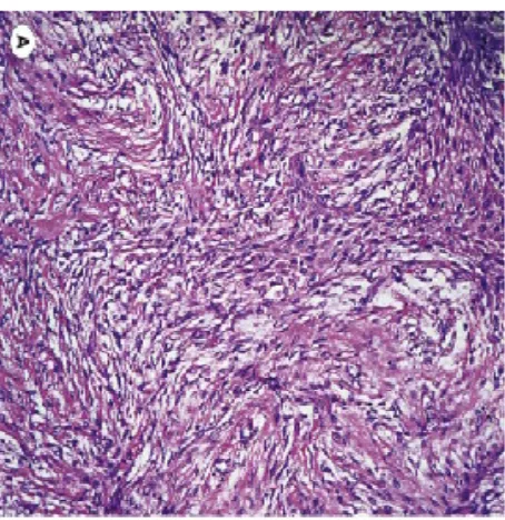
METAPLASTIC CARCINOMA:
Metaplastic carcinoma represents tumor predominantly with cell type
other than epithelial and glandular component. It includes many categories
but which overlap with each other. Metaplastic carcinoma is more aggressive
than invasive ductal carcinoma.Metastasis is usually hematogenous rather
than lymph node metastasis.(7-8)
The tumors are circumscribed, firm to hard in consistency.
Degenerated cystic areas can be seen in cases with squamous metaplasia.
Some of the tumors may have infiltrative borders.
A.Squamous cell carcinoma:
34
In pure squamous cell carcinoma the central cystic cavity is lined by
malignant squamous cells. Most cases represent squamous metaplasia.
Other two variants which can be seen will be acantholytic squamous
cell carcinoma and adenosquamous carcinoma. Low grade adenosquamous
carcinoma is said to have a favourable prognosis whereas acantholytic
squamous cell carcinoma have a aggressive behavior.
B.Carcinosarcoma:
Microscopically the sarcoma like component can be malignant fibrous
histiocytoma, osteosarcoma, chondrosarcoma, angiosarcoma or a
combination of various components.When the transition between
sarcomatous and carcinomatous component is gradual and sharp, it is termed
[image:43.612.213.440.426.660.2]carcinosarcoma.
35
When the transition to osseous or cartilaginous elements is direct without any intervening spindle cell component or osteoclastic giant cells,it
is called matrix producing carcinoma.
Molecular studies suggest that the epithelial and sarcoma like
components originate from same stem cell.
The sarcoma like elements acquire vimentin and other mesenchymal
features.It is referred to as the phenotypic switch.The cells are occasionally
positive for epithelial markers.
PAGETS DISEASE:
Pagets disease was originally described by Sir James Paget in 1874. It
is a crusted lesion of nipple caused by underlying breast carcinoma. About
1-2% of patients with mammary carcinoma show pagets disease. The
accompanying breast carcinoma is usually a intraductal carcinoma.It may be
associated with or without stromal invasion. The epidermis of the nipple
shows characteristic of Paget's cells in the keratinizing epithelium. The cells
may be singly scattered in the superficial epidermis. They may also form
clusters in the basal portions of the epidermis. Individual cells have a pale or
clear cytoplasm and their nuclei have a prominent nucleoli.
Intraductal carcinoma is usually of comedo or solid growth
pattern.About 10 % of the cases show cribriform or papillary carcinoma and
36
79-100 % the cases are strongly positive for Her 2/neu.The underlying
[image:45.612.142.481.136.364.2]insitu lesions are frequently Her2 /neu.(7-8)
Figure 12- Pagets Disease Of Nipple
INFLAMMATORY CARCINOMA :
The diagnosis of inflammatory carcinoma is essentially based on
clinical criteria. Clinically, the entire breast is red, warm. The skin shows
widespread edema which resembles that of mastitis. It has been believed that
clinical appearance is due to widespread carcinomatosis of dermal lymphatic
vessels. Skin biopsy is usually performed to reveal dermal lymphatic
permeation. Histopathological examination of some of the cases show a
37
The prognosis is usually bad. Studies done by Charafe-Jauffret et al.
found most inflammatory carcinoma are negative for estrogen and positive
for MIB1,E-Cadherin and Her2 neu.(7-8).
RISK FACTORS
Several risk factors have been established for the development of
breast carcinoma. The common denominator for most of the factors will be
strong and prolonged estrogen stimulation in a genetically susceptible
background.
AGE:
Majority of the breast cancers are detected during the reproductive age
group.(28)
RACE AND ETHNICITY:
The incidence is high in northern Europe and North America (91.4
new cases per 100 000 women/year), intermediate in southern European and
low in African and asian countries. In the United States due to increased
mammographic screeningthere has been a increase in the detection of breast
carcinoma.29 Due to earlier diagnosis and improved therapy the mortality has
began to fall in some regions like North America, western Europe, and
Australia.29
FAMILY HISTORY:
The risk is 2-3 times higher than general population if the first degree
38
MENSTRUAL HISTORY:
Early menarche and late menopause is associated with increased risk
of carcinoma.30
PARITY:
There will be a increased risk in case of Nulliparity and late age at first
child birth. It has been documented that there is a reduced risk of breast
carcinoma in premenopausal women who have lactated.29
HORMONE REPLACEMENT THERAPY:
Many studies showed that there is an increased risk of breast
carcinoma in women under hormone replacement therapy than women using
estrogen alone. (32)
RADIATION:
An increased risk of breast cancer has been documented on exposure
to ionizing radiation particularly when the exposure is at the time of breast
development.(36)
PRECANCEROUS LESION:
Complex fibroadenoma,florid hyperplasia without atypia,solitary
papilloma without atypical hyperplasia, sclerosing adenosis are associated
with a risk of 1.5-2 times than that of general population.Atypical ductal
hyperplasia and atypical lobular hyperplasia are associated with a risk of
39
BREAST DENSITY :
Breast density solely and strongly contributes to the risk of breast
cancer(32).
GENETIC PREDISPOSITION:
About 5-10 % of breast cancers are familial.33 In various studies it has been reported that the risk of developing breast carcinoma due to BRCA1
mutation will be 56% to 90%.Women carrying BRCA2 mutation have a risk
of 37% to 84%.BRCA1 mutation may account for about 45% of cases of
hereditary breast carcinoma and they are usually of poorly differentiated type
with high proliferative rate.33
MODIFIABLERISK FACTORS
BODY MASS INDEX(diet)
Western diet (High caloric diet rich in proteins and animal
fat),obesity30,,sedentary lifestyle are associated with increased risk of breast carcinoma34.
ALCOHOL CONSUMPTION:
Increased alcohol consumption associated with increased risk of breast
carcinomas30,35.
PROGNOSTIC AND PREDICTIVE FACTORS:
A predictive factor is the one that gives information about the
40
A prognostic factor gives information about the recurrence risk of the
tumour in the absence of the adjuvant therapy, that is a prognostic factor is a
factor that helps to predict the natural history of a tumour.
Many prognostic and predictive factors help in the process of
estimation of risk for recurrence and in selection of additional therapies.
AGE :
Women less than 50 years have the best prognosis. Older patients
have a higher rate of recurrence and distant metastasis7.
PREGNANCY:
Carcinoma breast manifesting during pregnancy may enhance tumour
angiogenesis, over expression of Her 2 neu, and low expression of hormone
receptors. The tumors are generally aggressive in these cases.7
GENE:
Studies showed that BRCA1 mutation carriers have a low survival
rate.
SKIN AND NIPPLE INVASION:
Invasion of overlying skin is associated with decreased survival rate.
Nipple involvement is associated with high incidence of axillary node
metastasis
PRESENCE OR ABSENCE OF INVASION:
The single most important prognostic determinator is the presence of
invasive component. In case of a tumor with both an invasive and in situ
41
metastasis.The in situ component is directly related to the incidence of
multicentricity and indirectly with occult metastasis.
HISTOLOGICAL TYPE OF TUMOUR:
Variants of invasive ductal carcinoma like tubular carcinoma,
cribriform carcinoma, medullary carcinoma, papillary carcinoma, pure
mucinous carcinoma and secretoty carcinoma have a more favorable
prognosis than Invasive ductal carcinoma,NOS type. Signet ring cell
carcinoma have a bad prognosis.
NECROSIS:
Spontaneous tumor necrosis is associated with tumors showing high
histological grade and increased incidence of lymph node metastases.7
LYMPHOVASCULAR INVASION:
Lymphatic emboli within the breast is associated with higher tumor
recurrence. 58Vascular emboli is highly correlated with tumor size, tumor type, histological grade, lymph status and have a poor prognosis.
LYMPH NODE STATUS AND TUMOUR SIZE:
For a woman with operable breast cancer the presence of lymph node
metastases is the strongest prognostic factor [37]. The pN staging is a qualitative parameter.
The prognostic value of lymphnode status can be increased by
counting the number of lymph nodes. This has been shown in an study done
42
quality of the procedure by counting the number of examined lymph nodes
depends on both the surgeon and the pathologist [40].
The tumour size is also another useful prognostic factor and can be
obtained in almost all cases. Thus, recurrence risk has been correlated with
tumour size [41, 42,].
TUMOUR GRADE:
The current grading system in use was first proposed by Bloom and
Richardson[43] and thereafter it was reclassified by Elston and Ellis [44].
HISTOLOGICAL GRADING SYSTEM:
Tumor grade is describing the tumor on the basis of how much
abnormal the tumor cells and the tumor tissue look under a microscope.
Bloom and Richardson histological(43) grading method is most commonly used for grading the breast cancer. This method is very simple and gives
very good prognostic implications. This system grades breast cancer into
three types on the basis of the following three criteria:
i. measurement of tubular differentiation
ii. nuclear pleomorphism, and
TNM N0 Stage 1 No node involvement
TNM N1 Stage 2 Upto 3 axillary/single internal mammary node
involved
TNM N2 Stage 3 4 or more axillary node involved
43
iii. proliferative index.
TABLE -1: MODIFIED BLOOM AND RICHARDSON GRADING
SYSTEM
PARAMETER CRITERIA SCORE
Tubule formation >75% 1
10-75% 2
<10% 3
Nuclear pleomorphism Small and uniform 1
Moderate variation 2
Marked variation 3
Mitotic count/hpf 0-5 1
6-10 2
[image:52.612.126.493.156.481.2]10-15 3
TABLE-2: BLOOM RICHARDSON GRADING SYSTEM
GRADES SCORE
Grade -1(well differentiated) 3-5
Grade-2(moderately
differentiated)
6-7
[image:52.612.127.496.530.719.2]44
Added score Tumour grade Degree of differentiation
• 3-5 Grade I Well differentiated
• 6-7 Grade II Moderately differentiated
• 8-9 Grade III Poorly differentiated
According to Elston and Ellis tumour grade is a well developed
prognostic indicator for overall survival of breast cancer patients [44, 45]. The estimation of histologic grade is simple and cheap, so that it can be done in
almost all woman with breast carcinoma. One of the disadvantages is that the
estimation of histologic grade usually varies among the observers.
Boiesen and co-writers46 were performed histologic grading on ninty three breast carcinomas patients in seven pathologic departments, results
showed an mean kappa value of 0 and 54, which indicates a moderate level
of reproducibility.
ESTROGEN- AND PROGESTERONE RECEPTOR STATUS :
Studies found that a vast majority of breast cancer tumours expressed
oestrogen receptors and progesterone receptors. The first study on the
prognostic importance of estrogen receptor was done almost 20 years ago [49].
However, estrogen receptor and progesterone receptor are not strong
prognostic indicators with longer follow-up time, though women with
positive receptor breast carcinomas have a good prognosis compared to the
45
Only in the late 1970ies, the value of estrogen receptor and
progesterone receptor as predictive factors was demonstrated [51].
Tumours with both estrogen and progesterone receptor positive are
benefit more from hormonal therapy than those who have either estrogen or
progesterone receptors [52].
Approximately 50-60% of women with positive receptor late stage
breast cancer will have better response from hormonal therapy than receptor
negative women [53].
Currently, the biochemical analyses of estrogen receptor and
progesterone receptor are largely replaced by immunohistochemistry.
Immunohistochemistry gives superior prognostic information and it is more
valuable for small tumours [53-54].
DNA CONTENT AND CELL PROLIFERATION MEASUREMENT:
DNA-PLOIDY:
DNA-ploidy is a measure of the deoxy ribonucleic acid content within
the nucleus of somatic and germ cells. The normal cells are either diploid or
euploid.
Measurement of DNA content within tumour cells has been applied to
evaluation of number of tumour types. Bagwell et al showed that ploidy
analysis may be useful prognostic test for node negative breast carcinoma47. Flow cytometry is the most common method used to measure the
46
Due to genetic instability most of the cancerous cells have a abnormal
amount of DNA which may greater or lesser than that of actual amount of
DNA.It is usually associated with a worse prognosis and this is called
aneuploidy [47].
S-PHASE FRACTION (SPF):
The S-phase of DNA-synthesis takes place before cell division and
hence it measures the proliferative activity of the cell. The number of cells in
S-phase can be estimated by means of flow cytometry [48].. Most of the studies revealed that high SPF associates with greater recurrence risk of
breast cancer, and is considered as aprognostic factor.
NEW PROGNOSTIC AND PREDICTIVE FACTORS:
The recently recognised prognostic and predictive markers provides
more reliable information on prognosis and treatment selection.
A prognostic factor provides information on the outcome, irrespective
of the different therapies employed. Whereas, a predictive factor gives
valuable information on the likely benefit from a particular treatment.
So far, the sex-hormone receptor status is the only well established
predictive factor useful in the management of patients with breast carcinoma.
Therefore,an increase in need for other markers, especially prognostic-
and predictive factors employing at the individual level.
Indeed, many studies were conducted to identify different risk groups
47
In recent years, there are three emerging predictive and prognostic
factors of special interest as follows,
• c-erbB-2 gene,
• p53 gene and
• angiogenesis.
One of the markers, c-erbB-2 oncogene is a routinely used predictive
factor, while p53 tumour suppressor gene and angiogenesis in the future may
be of value in management of breast cancer.
c-erbB-2:
c-erbB-2 is an oncogene situated on the q arm of 17th chromosome [55]., which encodes a tyrosine growth factor receptor belongs to EGFR(epidermal
growth factor receptor). [56].
In a Swedish population based study showed that the prevalence of
overexpression of c-erbB-2 was 19% among the breast cancer patients.[57]. Most of the studies revealed that overexpression of c- erB-2 gene has been
associated with bad prognosis(57,58),,though some studies have found that there is a strong association between over expression of c-erB-2 and reduced
sensitivity to chemotherapy and tamoxifen.(60-61)
As a result, evaluation of c-erbB-2 status with immunohistochemistry
on tumour sections has become a routine analysis for women with breast
48
p53:
p53 is a tumour 48uppressor gene situated on the p arm of 17th chromosome [59,62]. Because of its crucial role in control of cell cycle, apoptosis and DNA repair mechanism, p53 is rightly known as “the
policeman of the genome” [59].
P53 Mutation is identified nearly 20-25% of breast carcinomas 63.
The commonest method of determining p53 protein is
immunohistochemistry on tumour sections. This method detects
accumulation of abnormal p53 protein with in the cell and also estimates the
levels of normal p53 protein.
The potential use of p53 in the future seems to be as a predictive
factor. Patients with p53 mutations found to reduced response to tamoxifen
[64],
whereas taxane based chemotherapy have been associated with greater
efficacy in people with p53 mutated breast carcinomas. [65].
Hence, in future, evaluation of p53 status has one of the powerful
markers to ameliorate the management of breast carcinoma.
ANGIOGENESIS:
The ability of a tumour to induce formation of new microvessels
called angiogenesis, more than 30 years ago its importance for tumour
growth was reported [69]..
Angiogenesis induces the growth of both primary and metastatic
49
• Tumour growth more than 1-2 mm2 size is depends on tumour
angiogenesis [64]..In the absence of tumour microvessels, tumour
growth is restricted by reduction of nutrients and accumulation of
waste products.
• The tumour microvessels permits tumour cells to invade in to blood
stream[69, 70], paved an initial pathway for distant metastases.
• Endothelial cells in the tumour microvessels releases growth factors
which induce further growth of the tumour [68].Tumour
neovascularisation also involved in the synthesis of various proteolytic
enzymes [74]
Tumour angiogenesis is regulated by large number of pro angiogenic
and antiangiogenic factors, vascular endothelial growth factor(VEGF) is one
of the most important regulating factor 75.
VEGF act as a mutagen for tumour endothelial cells.
High levels of vascular endothelial growth factors are associated with
50
[image:59.612.109.538.128.503.2]The factors that regulating the angiogenesis are tabulated as follows:
TABLE-3 FACTORS REGULATING ANGIOGENISIS:
PROANGIOGENIC FACTORS ANTI ANGIOGENIC FACTORS
Vascular endothelial growth factor angiopoeitin
Fibroblast growth factor angiostatin
platelet derived growth factor endostatin
Plasminogen activator inhibitor thrombospondin
Angiogenin Platelet factor 4
Plasminogen activators Prolactin
Transforming growth factor osteopontin
Interestingly, Linderholm and co-workers, revealed that p53 mutation
associated with high levels of vascular endothelial growth factors. so, the
p53 gene exerts a inhibitory effect on tumour angiogenesis, wild type p53
protein found to be inhibit the formation of VEGF( vascular endothelial
growth factor). [82,83].
It has been showed that wild-type p53 protein upregulates the
51
So, when mutant p53 protein is present, causes inhibitory effects on
thrombospondin 1 and subsequently increased tumour angiogenesis [83, 84].
Evaluation of tumour angiogenesis by a histological grading system
and microvessel density grade has been correlates with certain clinico
pathological entities like tumour size, tumour grade was described 30 years
ago [85].
Weidner and co-workers were the first proposed the method for
estimation of tumour angiogenesis [17].. They revealed that tumour
microvessels can be highlighted with help of immunohistochemistry, by
staining the tumour sections with an antibody against antigen expressed by
tumour endothelial cells. The microvessel density is then evaluated by
counting these identified microvessels within the region with the most
intense new blood vessels, also called hotspots.
Most of the studies proved that microvessels density can predict bad
prognosis in breast carcinoma. [ 86-90] including lymph-node negative patients [90-96]
.Whereas, some researchers could not find any prognostic significance
of tumour microvessels density [97-101]. The reason for this results variation
may be due to diferrent methods were used to estimate microvessels density.
Indeed, some studies reported contradictory results in the association
between microvessels density and prognosis. However, most of the studies
showed that there is a strong association between high microvessels density
52
Although some studies reported poor correlation between microvessel
density and lymph node metastases [102-103],, this relationship is of interest as the factors that associated with lymph-node metastases could be used for
prediction of lymph-node metastases.
Thus a predictive factor is useful in identifying woman with low risk
of metastases to avoid unnecessary axillary surgery and also defining the
woman with high risk of lymph node metastases.
Moreover, the interest in the therapeutical aspect of angiogenesis has
been focused on the development of specific anti-angiogenic drugs [104, 105]
although there have been some reports on the predictive value of
angiogenesis [106-110] but in search for a new class of antiangiogenic drugs is still to be awaited.
TYPES OF ANGIOGENESIS:
There are 2 types of angiogenesis, they are
1. Intussusceptive angiogenesis
2. Sprouting angiogenesis
INTUSSUSCEPTIVE ANGIOGENESIS:
Intussusceptive angiogenesis is otherwise known as splitting type of
angiogenesis because it splits the vessels in two by direct extension of the
53
It is very fast and efficient compared with sprouting angiogenesis
because it requires only rearragement of already pre existing endothelial
cells and does not depends on endothelial cell migration and proliferation.
Intussusceptive angiogenesis plays an essential role in formation of
new vessels in embryos where resources are limited and growth is fast [111-112].
Its occurrence is based upon the formation of tissue pillars.
Scanning electron microscopes and three-dimensional reconstruction
of serial microscopes are useful in identification of transcapillary tissue
pillars.
This type of angiogenesis was discovered in postnatal lungs of rats
and humans [113,114), but it also occurs in other tissues and organs like choroid of the eye, intestinal mucosa, kidney, ovary, and uterus [115,116]. It also occurs in skeletal muscle, heart, and brain.
It plays an important role in the formation of vein and artery
bifurcations and also pruning of larger microvessels. Intussusceptive
angiogenesis can be identified by determining the frequency of tissue pillars
from scanning electron micrographs of vascular casts[116].. SPROUTING ANGIOGENESIS:
The first discovery of sprouting angiogenesis in growth of the tumour
was reported by Ausprunk and Folkman, 1977, which consists of following
54
i. The basement membrane is locally degraded on the side of the
peritumoral postcapillary venule situated nearer to the angiogenic
stimuli;
ii. Cell to cell contacts between endothelial cells are loosened and
endothelial cells transmigrate into the surrounding extracellular
connective tissue matrix;
iii. Endothelial cells in the extracellular connective tissue matrix form
solid cord;
iv. Finally, The formation of lumen takes place infront of the migrating
portion, newly tubular spuds contact each other and joins to form
functional capillaries, along basement membrane synthesis and
rimming of pericytes around basement membrane.
Ausprunk and Folkman were first to proposed that tumour vessel
spuding can advanced without cell division in the early stage of angiogeneis,
while further outgrowth and prolonged sprouting requires proliferation of
endothelial cells.
A large number of in vivo and in vitro models of angiogenesis have
been introduced with the advancement in this field. In in vitro assays, the
most common methods in use are,
i. rabbit cornea by Muthukkaruppan and Auerbach, 1979117,
55
iii. intersegmental vessel growth in zebrafish by Lawson and Weinstein,
2002119,120.
Cimpean et al in 2010121 has studied selected aspects of the
angiogenesis with an isolated cell lines which includes endothelial cell
migration,endothelial cell proliferation, degradation of the connective tissue
matrix and formation of capillaries.
Grant et al122, first demonstrated the 3 dimensional structure of capillary networks and highlighted the membranes of endothelial cells with
help of light and electron microscopes.
In1991, Paku and Paweletz 123, they proposed steps in sprouting
angiogenesis by integrated with the studies of Ausprunk and Folkman, as
follows:
1. A structural change in the basement membrane takes place, there is a
loss of polarity along the whole surface of the dilated blood vessel,
followed by a loss of the basement membrane at site where endothelial
cell processes are protruding into the extracellular connective tissue;
2. Endothelial cells are lined up in parallel manner, forming a small
lumen and maintaining their basal-luminal