Copyright © 2002, American Society for Microbiology. All Rights Reserved.
Both Mucosal and Systemic Routes of Immunization with the Live,
Attenuated NYVAC/Simian Immunodeficiency Virus SIV
gpe
Recombinant Vaccine Result in Gag-Specific
CD8
⫹
T-Cell Responses in Mucosal
Tissues of Macaques
Liljana Stevceva,
1Xavier Alvarez,
2† Andrew A. Lackner,
2† Elzbieta Tryniszewska,
1Brian Kelsall,
3Janos Nacsa,
1Jim Tartaglia,
4Warren Strober,
2and Genoveffa Franchini
1*
Basic Research Laboratory, National Cancer Institute, Bethesda, Maryland 208921; Division of Comparative Pathology, New
England Regional Primate Research Center, Harvard Medical School, Southborough, Massachusetts 01772-91022;
Laboratory of Clinical Investigation, National Institute of Allergy and Infectious Diseases, Bethesda, Maryland
20892-18903; and Aventis-Pasteur, Toronto, Ontario, Canada M2R 3T44
Received 11 April 2002/Accepted 19 August 2002
As most human immunodeficiency virus (HIV) infection occurs via mucosal surfaces, an important goal of vaccination may be the induction of virus-specific immune responses at mucosal sites to contain viral infection early on. Here we designed a study in macaques carrying the major histocompatibility complex class I Mamu-Aⴱ01 molecule to assess the capacity of the highly attenuated poxvirus NYVAC/simian immunodefi-ciency virus (SIV) SIVgpevaccine candidate administered by the intranasal, intramuscular, or intrarectal route
to induce mucosal immunity. All macaques, including one naive macaque, were exposed to SIVmac251by the
intrarectal route and sacrificed 48 h after infection. The kinetics of immune response at various time points following immunization with NYVAC/SIVgpeand the anamnestic response to SIVmac251at 48 h after challenge
were assessed in blood, in serial rectal and vaginal biopsy samples, and in tissues at euthanasia with an SIVmac
Gag-specific tetramer. In addition, at euthanasia, antigen-specific cells producing gamma interferon or tumor necrosis factor alpha from the jejunum lamina propria were quantified in all macaques. Surprisingly, antigen-specific CD8ⴙT cells were found in the mucosal tissues of all immunized macaques regardless of whether the
vaccine was administered by a mucosal route (intranasal or intrarectal) or systemically. In addition, following mucosal SIVmac251 challenge, antigen-specific responses were mainly confined to mucosal tissues, again
regardless of the route of immunization. We conclude that immunization with a live vector vaccine results in the appearance of CD8ⴙT-cell responses at mucosal sites even when the vaccine is delivered by nonmucosal
routes.
Transmission of human immunodeficiency virus (HIV) oc-curs predominantly via mucosal surfaces. Effective neutraliza-tion of the virus at this anatomic site depends on several components. Preexisting virus-specific immunoglobulin A (IgA) antibodies in rectal secretions are associated with de-creased viral burden but are not sufficient to prevent infection (52). HIV-1/simian immunodeficiency virus (SIV)-specific CD8⫹ cytotoxic T lymphocytes are another such component
and have been shown to be present during the early weeks following infection, before a neutralizing antibody response is demonstrable (8, 26, 27). Such cell-mediated immunity (40) and direct killing by cytotoxic lymphocytes from the vagina and colon lamina propria may be an important factor in containing viral infection at the site of primary infection (35), and it may explain the fact that, despite the rapid heterosexual
dissemi-nation of HIV-1 in the world, mucosal transmission of HIV-1 appears to be relatively inefficient and is estimated to occur only once in 300 or more high-risk exposures (47).
While many studies have investigated the effect of the route of immunization on antibody production at the local or sys-temic level, few have directly investigated the induction of cell-mediated responses at mucosal sites. Studies in mice im-munized with synthetic, multideterminant HIV peptides plus cholera toxin adjuvant have shown that intrarectal immuniza-tion induces cytotoxic T lymphocyte responses in Peyer’s patches, the spleen, and lamina propria of the intestine (4) and that these responses were able to reduce the viral titer in the ovaries and the colorectal tissue upon intrarectal recombinant vaccinia virus challenge. In contrast, subcutaneous immuniza-tion induced cytotoxic T lymphocytes only in the spleen and not in the Peyer’s patches or lamina propria of the intestinal tract and was not able to reduce the viral titers (4).
Similar results were obtained with an modified vaccinia virus Ankara-based recombinant expressing the simian-human im-munodeficiency virus (SHIV) strain SHIV89.6 gp160 (6), and more recently, it has been demonstrated that mucosal
immu-* Corresponding author. Mailing address: National Cancer Institute, Basic Research Laboratory, 41/D804, Bethesda, MD 20892. Phone: (301) 496-2386. Fax: (301) 496-2386. E-mail: [email protected].
† Present address: Tulane Regional Primate Research Center, Cov-ington, LA 70433.
11659
on November 8, 2019 by guest
http://jvi.asm.org/
nization of macaques afforded better protection than subcuta-neous immunization following intrarectal exposure to SHIVku2
(5). These studies suggest that cutaneous or intramuscular immunization generates antigen-specific cells that may not travel to mucosal sites (3, 11, 22, 32, 49).
A somewhat different conclusion was suggested by studies in which macaques were immunized intramuscularly with the highly attenuated poxvirus vector NYVAC encoding the
SIVmak6w Gag, Pol, and Env products (7, 19). In this case
immunization resulted in long-term viremia containment fol-lowing intravenous or intrarectal challenge with SIVmac251(7,
18a). Since, in these studies, all macaques were immunized by the intramuscular route, the results suggested that even in the absence of priming at mucosal sites, systemic immunization can lead to virological control following SIVmacmucosal
chal-lenge exposure (7, 18a, 19).
To further explore these results, we designed a study to directly assess the extent of mucosal immune responses to a Gag immunodominant SIVmac251epitope following
immuniza-tion of macaques by either the intramuscular, intranasal, or intrarectal route. Accordingly, two female macaques express-ing the Mamu-Aⴱ01 major histocompatibility complex class I molecule were used for each route of immunization, and the extent and kinetics of the immune response following both immunization and SIV challenge were measured in the blood
and in serial mucosal biopsy samples with Gag-specific tet-ramer-staining reagents (1). Importantly, the size and specific-ity of the anamnestic virus-specific CD8⫹T-cell responses to
the virus were investigated within 48 h of a SIVmac251
intra-rectal challenge exposure to determine whether it differed in various anatomical compartments according to the route of immunization.
MATERIALS AND METHODS
Animals and procedures.Seven Mamu-Aⴱ01-positive macaques were involved in this study. Animals 536 and 815 were immunized with three inoculations of 108 PFU of NYVAC/SIVgpeby the intramuscular route. Animals 582 and 816 were immunized intranasally and animals 814 and 818 were immunized intrarectally with three inoculations of 5⫻108PFU of NYVAC/SIVgpeeach. The first boost was given 1 month after the first immunization, and the second boost was given 3 months after the second immunization. All six immunized macaques and a control naive macaque (no. 654) were challenged intrarectally with 30 mucosal infectious doses of the SIVmac251(561)isolate (37) and sacrificed 48 h after infection.
Blood samples were collected at several time points during the immunization schedule as well as on the day of SIV exposure and sacrifice. Similarly, multiple vaginal and rectal pinch biopsy samples were taken prior to and during the immunization course.
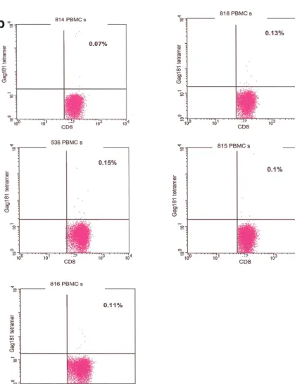
At the time of sacrifice, blood, spleen, mesenteric, iliac, and pararectal lymph nodes, vagina, cervix, jejunum, colon, and rectum samples were collected. The tissues were placed in RPMI supplemented with 10% bovine serum and peni-cillin/streptomycin. Rectal tissue was also placed in phosphate-buffered saline (PBS)-bovine serum albumin solution and used for in situ tetramer staining. FIG. 1. Control staining for tetramers. Staining of isolated lamina propria lymphocytes from vaginal and rectal biopsy samples obtained from infected Mamu-Aⴱ01-negative and naive Mamu-Aⴱ01-positive macaques and macaque 654 before viral challenge, and staining with unrelated tetramer on isolated vaginal and rectal lamina propria lymphocytes from immunized, infected Mamu-Aⴱ01-positive macaques (a) was used to control for the specificity of the staining. Nonspecific binding accounted for up to 0.13% of the CD3⫹CD8⫹lymphocytes. Therefore, values above
this percentage could be considered positive. Tetramer staining of peripheral blood mononuclear cells (PBMCs) isolated prior to immunization was negative in the five macaques examined (b).
on November 8, 2019 by guest
http://jvi.asm.org/
FIG. 1—Continued.
on November 8, 2019 by guest
http://jvi.asm.org/
Isolation of tissue lymphocytes.Mononuclear cells were isolated from periph-eral blood mononuclear cells, lymph nodes, spleen, intestines, vagina, and cervix. Mononuclear cells from spleen and lymph nodes were isolated by mechanical dissociation of the tissue and consecutive Ficoll gradient centrifugation. Tissues from jejunum, colon, rectum, vagina, and cervix were isolated by a modification of a previously published method (9, 21). Tissues were treated with 1 mM dithiothreitol (ICN Biomedicals Inc., Aurora, Ohio) for 30 min, followed by incubation in calcium- and magnesium-free Hanks’ balanced salt solution (Life Technologies, Baltimore, Md.) three (intestines) to four (vagina and cervix) times for 1 h with stirring at room temperature to remove the epithelial layer. At this stage, pieces of tissue were fixed in 10% neutral formalin and embedded in paraffin, and sections were cut and stained with hematoxylin and eosin. Micro-scopic examination was performed to ensure that all of the epithelium was removed and the lamina propria was intact.
Intraepithelial lymphocytes were then purified by Percoll gradient density centrifugation.
Tissue sections were cut into smaller pieces and incubated at 37°C in Iscove’s medium supplemented with 10% fetal calf serum and penicillin/streptomycin containing 400 U of collagenase D (Boehringer GmbH, Mannheim, Germany) and 25 U of DNase (Worthington Biochemical Corporation, Lakewood, N.J.) per ml for 2 to 3 h. Vaginal and cervical tissues were digested with 0.5 mg of collagenase type IV (Sigma Chemical, St. Louis, Mo.) (50). The mononuclear cells were isolated from the supernatant containing dissociated cells by Percoll gradient centrifugation.
Flow cytometry.Fresh cells were directly stained with phycoerythrin-conju-gated tetrameric complexes folded with the immunodominant Gag181-189 CM9 (p11c) peptide. Fluorescein isothiocyanate-conjugated anti-CD3⌭(Pharmingen, San Diego, Calif.) and peridinin chlorophyll protein (PerCP)-conjugated anti-CD8 (Becton Dickinson, San Jose, Calif.) were used in conjunction with the tetrameric complex.
As confirmation of results obtained from freshly isolated lymphocytes, lym-phocytes were also cultured at a concentration of 3⫻106/ml in RPMI enriched with 10% human serum, with addition of 1g of the appropriate peptide and 20 U of interleukin-2 per ml for 7 days (data not shown). Staining with tetrameric complexes was done afterward as described above. Staining with unrelated tet-ramer and of cells isolated from Mamu-Aⴱ01-negative or naive animals was used as a negative control. Staining with phycoerythrin-CD4 (Becton Dickinson) was used as a positive control.
In addition, cells were stained with fluorescein isothiocyanate-conjugated ac-tivation markers CD25, CD69, and HLA-DR. Briefly, 5⫻105lymphocytes isolated by Ficoll diatrizoate or Percoll gradient centrifugation were incubated with 2g of tetrameric complexes and/or selected antibodies for 30 min at room temperature. After washing the cells twice in Dulbecco’s phosphate-buffered saline supplemented with 2% fetal calf serum and fixation in 1% paraformalde-hyde (pH 7.4), samples were analyzed by flow cytometry with CellQuest and the FACScalibur (Becton Dickinson) instrument.
Tetramer staining in situ.In situ tetramer staining was performed on fresh tissues as previously described with some modifications (17, 48). Briefly, tissues were collected by pinch biopsy, washed in cold PBS, and cut into small strips. The resulting sections (n⫽4 to 6) were then incubated with 10l of antigen-specific tetramer labeled with indocarbocyanine (Amersham, Piscataway, N.J.) per sec-tion and gently agitated at 37°C for 15 min. The tissue was then rinsed repeatedly at 37°C with PBS and then twice with ice-cold PBS prior to fixation with cold 2% paraformaldehyde for 20 min.
After additional washes, antibodies to CD3 (rabbit polyclonal; Dako) and CD8 (directly conjugated to fluorescein isothiocyanate; Becton Dickinson) were ap-plied singly or together for 1 h. The tissues were then washed and incubated with anti-rabbit immunoglobulin-Alexa 488 (Molecular Probes) when using only CD3 or anti-rabbit immunoglobulin-Alexa 568 when using both CD3 and CD8. Tis-sues were then washed, mounted on a glass slide with antifading medium (Vectashield; Vector Laboratories, Inc., Burlingame, Calif.), and examined by confocal microscopy.
Confocal microscopy was performed with a Leica TCS SP laser scanning microscope equipped with three lasers (Leica Microsystems, Exton, Pa.). Indi-vidual optical slices represent 0.2m, and 20 to 60 optical slices were collected at a 512- by 512-pixel resolution. The fluorescence of individual fluorochromes was captured separately in sequential mode after optimization to reduce bleedthrough between channels (photomultiplier tubes) with Leica software. NIH Image version 1.62 and Adobe Photoshop version 6 software were used to assign colors to each fluorochrome and the differential interference contrast image (gray scale). Colocalization of antigens was indicated by the addition of colors as indicated in the figure legends.
Intracellular cytokine staining.Intracellular cytokine staining was done on jejunal lamina propria lymphocytes isolated at necropsy by a modified previously published method (16, 24, 53). Isolated and previously frozen lamina propria lymphocytes were thawed and washed. Viability was assessed by trypan blue dye exclusion, and viable cells were counted. Cells were incubated for 1 h at 37°C in 5% CO2at a concentration of 106/ml in the presence of 1l of anti-CD28 (Becton Dickinson), 1l of anti-CD49d (Becton Dickinson), and 2g of the SIV Gag181-189 CM9 peptide per ml. Stimulation with 25 ng of phorbol myristate acetate (Sigma-Aldrich Corp., St. Louis, Mo.) and 1g of ionomycin (Sigma-Aldrich Corp.) per ml for gamma interferon (IFN-␥) or staphylococcal entero-toxin B (Toxin Technology, Inc.) for tumor necrosis factor alpha (TNF-␣) was used as a positive control.
After 1 h of incubation, 10g of brefeldin A (Sigma-Aldrich Corp.) was added and cells were incubated for an additional 5 h. Cells were then washed twice with PBS–2% fetal calf serum and stained for surface antigens with fluorescein iso-thiocyanate-labeled anti-CD3 and PerCP-labeled anti-CD8. After incubating them at room temperature for 30 min and washing them twice with PBS–2% fetal calf serum, cells were fixed in Cytofix/Cytoperm (Pharmingen) for 20 min at 4°C. Following two washes with Wash/Perm buffer (Pharmingen), cells were incu-bated with phycoerythrin-conjugated anti-IFN-␥or allophycocyanin-conjugated anti-TNF-␣antibodies for 30 min at 4°C. This was followed with two washes in Wash/Perm buffer and direct reading of the samples on the FACScalibur flow cytometer.
Cytotoxic T-lymphocyte assay.Cytotoxic T-lymphocyte assays were performed as previously described (7). Fresh cells were cultured overnight in the presence of 300 IU of interleukin-2 per ml and then incubated at different effector-to-target cell ratios for 6 h with Mamu-Aⴱ01-positive,51Cr-labeled autologous transformed B cells pulsed overnight with 1g of a specific peptide per ml, without peptide, or with an unrelated peptide.
RESULTS
Induction of CD8ⴙT-cell response to Gag181-189 CM9
im-munodominant peptide in blood and at mucosal sites by vac-cination.Two Mamu-Aⴱ01-positive female macaques each re-ceived three immunizations with NYVAC/SIVgpeby either the
intramuscular, intrarectal, or intranasal route. Virus-specific CD8⫹ T-cell response to the immunodominant Gag181-189
CM9 peptide in the blood and tissues of the immunized ma-caques was quantitated by staining CD3⫹CD8⫹T cells with
the specific Mamu-Aⴱ01/Gag181-189 CM9 tetramer after each immunization. The specificity of tetramer staining was assessed following each immunization in isolated lymphocytes from bi-opsy samples of both Mamu-Aⴱ01-positive uninfected ma-caques and Mamu-Aⴱ01-negative infected macaques. Staining with unrelated tetramer was also performed (Fig. 1a).
Staining of cells from tissue biopsy samples of a Mamu-Aⴱ01-positive, SIVmac251-infected macaque revealed the
pres-ence of a population of tetramer-positive CD3⫹CD8⫹cells.
Many, if not most, of those tetramer-positive cells were found to be larger (that is, to have a higher forward and side profile) than those which would normally be included in more conven-tional lymphocyte gating (Fig. 2) (2, 13, 30, 45). To explore the possibility that this population represents activated CD8 T cells, the same sample was stained with the activation markers CD69 and CD25. By gating on cells of larger size, we demon-strated that, in the tissues of this macaque, these large cells expressed both the CD8 and CD69 markers, suggesting that they might have been recently activated (data not shown). Therefore, a slightly enlarged gate as shown in Fig. 2, upper left panel, that included these cells was used for all further flow cytometric analyses of tetramer-positive cells in the tissues and blood of the immunized and control macaques.
With these gates, the percentage of CD3⫹ CD8⫹ T cells
staining with the Gag181-189 CM9 tetramer in the blood of the
on November 8, 2019 by guest
http://jvi.asm.org/
FIG.
2.
Gating
of
tissue
lymphocytes
for
flow
cytometry
analysis.
Extending
the
lymphocyte
gate
to
include
larger
cells
revealed
a
population
of
CD3
⫹
CD8
⫹
tetramer-positive
cells
that
was
concealed
when
the
conventional
lymphocyte
gate
was
used.
on November 8, 2019 by guest
http://jvi.asm.org/
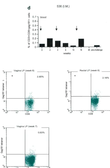
FIG. 3. Tetramer staining during immunization. Immune responses during the immunization procedure were assessed by measuring the percentage of CD3⫹CD8⫹Gag181-189 CM9-positive cells in blood (upper graphs in panels a, b, c, and d; arrows indicate time of immunization)
and in isolated lymphocytes from rectal and vaginal biopsy samples (flow charts in panels a, b, c, and d). Cells were gated through the CD3⫹CD8⫹
gate, and the percentage of Gag181-189 CM9-positive cells was determined by histogram analysis. The flow charts are shown here as dot blots rather than histograms in order to better demonstrate the population of cells. An equal number of CD3⫹CD8⫹cells (104) was acquired for each sample analyzed. Samples in which fewer than 5⫻103CD3⫹CD8⫹cells were acquired are indicated (ⴱ). Times of immunization are indicated
on the graphs with arrows. (a) Findings in intrarectally (I.R.) immunized macaques 814 and 818 shown as a graph for blood and as flow charts for biopsy samples underneath the graph. (b) Findings in intranasally (I.N.) immunized animals 816 and 582. (c) Findings in intramuscularly (I.M.) immunized macaque 815. (d) Findings in intramuscularly immunized macaque 536. LP, lamina propria.
on November 8, 2019 by guest
http://jvi.asm.org/
macaques immunized by the intranasal, intramuscular, and intrarectal routes was determined (top panels of Fig. 3a to d). These studies demonstrated the presence of tetramer-positive cells in the blood of all immunized animals and showed no significant difference among immunization routes. At the end of the immunization period, the percentage of CD3⫹CD8⫹T
cells that were specifically recognized by the Gag-specific tet-ramer in blood was in the range expected with this vaccine modality (19). The cytotoxic T lymphocyte assay performed 2 weeks after the second immunization revealed specific lysis of Gag181-189 CM9-pulsed, Mamu-Aⴱ01-positive, 51Cr-labeled
autologously transformed B cells in some of the macaques (816 and 815) (data not shown).
The presence of Gag181-189 CM9-specific CD3⫹CD8⫹T
[image:7.603.55.539.78.569.2]cells at mucosal sites was assessed on isolated lamina propria lymphocytes from rectal and vaginal biopsy samples (bottom panels of Fig. 3a, b, c, and d). We did not attempt to quantify these cells because often the number of cells obtained from the biopsy samples was insufficient to reliably compare frequen-cies. In both macaques 814 and 818 immunized by the intra-rectal route, a discrete population of cells staining the Gag181-189 CM9 tetramer was detected in serial biopsy samples (Fig.
FIG. 3—Continued.
on November 8, 2019 by guest
http://jvi.asm.org/
FIG. 3—Continued.
on November 8, 2019 by guest
http://jvi.asm.org/
FIG. 3—Continued.
on November 8, 2019 by guest
http://jvi.asm.org/
3a, bottom panels). Similarly, in animals 816 and 582 immu-nized by the intranasal route, a convincing staining for Gag181-189 CM9 tetramer-positive cells in vaginal lamina propria was detected at week 19 and/or week 20 in both macaques (bottom panels of Fig. 3b). At week 20, a discrete population was also found in the rectal lamina propria of macaque 816 (Fig. 3b).
Unexpectedly, in both macaques immunized by the intra-muscular routes (815, bottom panels of Fig. 3c, and 536, bot-tom panels of Fig. 3d), Gag181-189 CM9-positive CD3⫹CD8⫹
T cells were detected as early as week 6 from immunization in both the rectal and vaginal lamina propria. Thus, Gag181-189 CM9-specific CD3⫹CD8⫹cell populations in the
gastrointes-tinal lamina propria and the cervicovaginal compartment were
induced by NYVAC/SIVgpein all of the immunized animals
regardless of the route of immunization.
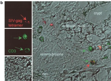
To confirm these results with an independent approach, tetramer-positive cells were visualized by in situ tetramer stain-ing of rectal biopsy samples from a few macaques. Tissues from a lymph node of a chronically infected macaque known to have an extremely high percentage of CD3⫹ CD8⫹ Gag181-189
[image:10.603.59.537.326.499.2]CM9-positive cells as well as cultured spleen cells from the same animal were used as a positive control, and biopsy sam-ples from uninfected, nonimmunized macaques were used as a negative control. Spleen culture cells or lymph node tissue (Fig. 4a) of chronically infected macaques had clusters of dou-ble positive cells for Gag181-189 CM9 and CD8. Single cells
FIG. 4. In situ tetramer staining. Double immunofluorescent staining and image analysis were initially done on positive samples from cultured spleen cells and from lymph nodes taken from a known Gag181-189 CM9-positive animal (a). In immunized macaques, positive staining is shown in the colonic tissue from animal 816 2 weeks after the boost (b). Images for individual channels (CD3, green; SIV-Gag tetramer, red; differential interference contrast, gray scale) are shown on the left side, and a larger merged image containing all three channels is shown on the right (b). Several CD3⫹cells are present, and one is also labeled with the SIV-Gag tetramer. Bar, 10m (b). FITC, fluorescein isothiocyanate; CY3,
indocarbocyanine.
on November 8, 2019 by guest
http://jvi.asm.org/
with similar staining patterns were detected in vaginal and rectal biopsy samples of the immunized macaques. Figure 4b shows tetramer-positive cells in the colonic lamina propria of animal 816 2 weeks after the second immunization.
Influence of route of immunization on anamnestic immune response in systemic and mucosal compartments following SIVmac251 challenge exposure. All seven macaques were ex-posed intrarectally to SIV251and euthanized 48 h
postexpo-sure. The time frame of 48 h postexposure was chosen because, at the time of euthanasia, we wanted to capture and quantitate the local secondary response to Gag181-189 CM9 before the systemic spread of infection. The choice of a time frame of 48 h postexposure was based on the information that mucosal ex-posure to SIVmac251 results in colonization of local lymph
nodes by day 2 to 3 postinfection (20; Chris Miller, personal communication). Since the commitment to proliferation of primed T cells upon encounter with the antigen occurs quickly (0.5 to 2 h) (28) and a cycle of viral replication requires ap-proximately 12 to 18 h, it is therefore reasonable to assume that Gag-specific local memory T cells might be able to un-dergo mitoses within 48 h and/or be recruited to this locale.
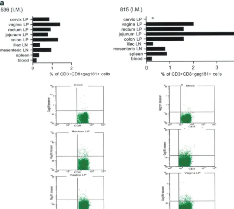
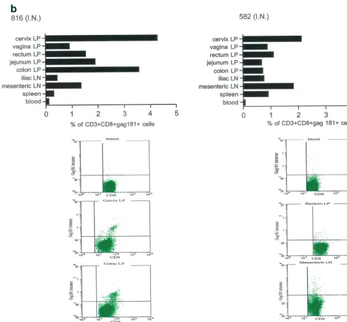
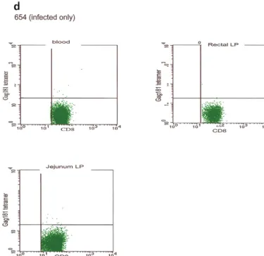
At necropsy, an SIV-specific response, higher than that found after the last immunization in blood to the Gag181-189 CM9 epitope, was found in macaques 815 and 536, immunized intramuscularly, both in some systemic and most mucosal com-partments, and appeared to be higher in the latter (Fig. 5a). In macaques immunized intranasally (macaques 816 and 582), the
response to the Gag181-189 CM9 epitope following challenge exposure was present in most compartments and was generally higher in mucosal compartments (Fig. 5b). However, in the two macaques immunized intrarectally (macaques 814 and 818), this response appeared to be mainly confined to mucosal sites (Fig. 5c). In animal 654, naive at the time of challenge exposure, CD3⫹CD8⫹T cells staining the Gag181-189 CM9
tetramer were not found in any tissues (Fig. 5d).
These findings suggested that early viral replication follow-ing rectal challenge may have preferentially expanded virus-specific immune responses at the mucosal sites, since at 48 h the virus may not be amplified sufficiently to induce a sizable immune response at systemic sites. In fact, in the macaques immunized by the intrarectal route, an expansion of virus-specific responses was evident only at mucosal sites (Fig. 5c). To correlate the observed immune responses after viral chal-lenge to the level of virus in tissues, we isolated RNA either from lymphocytes collected from tissues or from the entire tissue of all seven macaques and quantitated viral RNA by nucleic acid sequence-based amplification (NASBA) assay as previously described. With this assay we can detect as few as 500 copies of RNA per g of total RNA (43). From our estimate, derived from the yield of RNA from a known number of cells cultured in vitro, 1g of RNA corresponds to a total of 104to 105cells. Analysis of RNAs from the spleen, blood,
[image:11.603.73.515.71.394.2]vagina, and rectum of each macaque by NASBA resulted in fewer than 500 copies of viral RNA in all samples (data not
FIG. 4—Continued.
on November 8, 2019 by guest
http://jvi.asm.org/
shown), suggesting that viral expression was still at too low a level to be detected by the technique. We do not believe that this lack of detection of viral RNA was related to an abortive infection, since we infected a total of 34 naive macaques with the same viral stock by the rectal route (37; our unpublished data).
Cytokine production in response to immunization.
Gag181-189 CM9-specific functional responses in the jejunal lamina propria of all seven macaques were assessed by intra-cellular cytokine staining of isolated lymphocytes following in vitro stimulation with the specific peptide, using as a read-out
both TNF-␣and IFN-␥production. In all six immunized ma-caques, considerable numbers of CD8␣/⫹ cells producing
both cytokines were found, and as expected, these cells were absent in macaque 654 (Fig. 6, top panel). Surprisingly, a number of CD8␣/⫺cells also appeared to be producing either
IFN-␥or TNF-␣ (Fig. 6, flow charts). This finding could be related to a downregulation of the CD3 or the CD8␣/ recep-tor on the cell surface during the procedure, as noted by others (29). The graphs on the top of Fig. 6 include only CD8␣/⫹
[image:12.603.56.532.74.499.2]cells that produced IFN-␥(left top panel) or TNF-␣(right top panel).
FIG. 5. Tetramer staining after challenge. Macaques were challenged with SIVmac251 and sacrificed 48 h later. SIV-specific cells were determined as a percentage of Gag181-189 CM9-positive cells of the CD3⫹CD8⫹population in lymphocytes isolated from each tissue. An equal
number of CD3⫹CD8⫹cells (104) was acquired for each sample analyzed. SIV-specific responses in intramuscularly (I.M.) immunized macaques 536 and 815 (a) are shown as a graph (ⴱ, not done). Samples of blood lymphocytes at the time of necropsy and highly positive mucosal tissue samples are shown as flowcharts beneath the graph for each macaque. Tetramer-positive cells in intranasally (I.N.) immunized animals 816 and 582 (only 1.152⫻103CD3⫹CD8⫹cervical lamina propria cells were analyzed for animal 582) (b) and in intrarectally (I.R.) immunized macaques
818 and 814 (c) are shown. Animal 654, which was not immunized but was challenged, is shown (d). LP, lamina propria; LN, lymph node.
on November 8, 2019 by guest
http://jvi.asm.org/
The percentage of Gag-specific CD8⫹ cells expressing
IFN-␥or TNF-␣was derived by subtracting the spontaneous secretion of cytokines in the absence of peptide stimulation (see raw data on the flow charts in Fig. 6).
The extent of this response, although variable among the animals, did not appear to be related to the route of immuni-zation. Overall, the ability of lymphocytes at mucosal sites to express cytokines did not differ among macaques immunized by different routes.
DISCUSSION
The main finding in this study is that immunization of macaques with a live attenuated vaccine, NYVAC/SIVgpe,
by either a mucosal (intrarectal and intranasal) or systemic
(intramuscular) route results in the appearance of Gag181-189 CM9 tetramer-positive CD8⫹ cells in mucosal tissues.
Furthermore, intrarectal challenge of such immunized ma-caques with SIVmac251followed by sacrifice of the animals at
48 h after challenge led to the expansion of tetramer-posi-tive CD8⫹cells in mucosal tissues in all macaques regardless
of the route of immunization. This confirmed that a systemic (intramuscular) immunization led to mucosal CD8⫹T-cell
responses, as did mucosal immunization.
These results are somewhat at odds with studies reviewed above (4–6) that show that the route of induction deter-mines whether or not there is a mucosal immune response and that, in fact, mucosal immunization is necessary for a mucosal response. Thus, intramuscular and intradermal
im-FIG. 5—Continued.
on November 8, 2019 by guest
http://jvi.asm.org/
[image:13.603.45.543.70.531.2]munization with naked DNA generates systemic immune responses but is unable to confer protection at mucosal sites (36). Similarly, subcutaneous immunization in mice with HIV-1 gp160 expressing recombinant vaccinia virus induced cytotoxic T lymphocytes only in the spleen but not in Peyer’s patches or intestinal lamina propria (3).
However, it should be noted that systemic immunization with recombinant vaccinia virus vac-gp160 protected three of four macaques against intrarectal challenge with SIVmnevirus
47 (39) or, in another model, cats against feline immunodefi-ciency virus (38). Similarly, such protection has been achieved with live attenuated virus (41, 42) and whole killed virus (10). Finally, in the case of the NYVAC/SIVgpevaccine,
intramus-cular immunization was previously shown to be able to protect 50% of the macaques that were challenged intrarectally with
SIVmac251from high viremia ((7) and an even higher number
of macaques when a DNA prime/NYVAC/SIVgpeboost was
used (18a).
It should be noted that in the studies where systemic immunization led to at least some degree of mucosal immu-nity, live virus vaccines were used. Such vaccines, as opposed to peptide vaccines, may be more capable of leading to the appearance of immune cells at mucosal sites, either because of antigen circulation or because of the traffic of cells that take up antigen.
[image:14.603.58.524.68.554.2]Several additional conclusions concerning the responses
FIG. 5—Continued.
on November 8, 2019 by guest
http://jvi.asm.org/
elicited by immunization by different routes could be drawn from this study. First, tetramer-positive CD8⫹T cells were
found in the blood in all macaques regardless of the route of immunization. This is in agreement with data obtained in animals infected by different routes (18, 34, 51). Second, intrarectal immunization induced CD8⫹ T cells mainly in
mucosal tissue. Thus, while intramuscular and intranasal immunization led to some level of tetramer-positive CD8⫹
cells in systemic tissues (spleen, iliac lymph node), intrarec-tal immunization led to little or no tetramer-positive CD8⫹
cells at these locations. This finding is in agreement with previous studies showing that intrarectal immunization is less effective in inducing systemic responses (31). Since in-trarectal immunization with a multicomponent-peptide HIV vaccine led to cytotoxic T lymphocyte responses in the spleen only when cholera toxin was used as an adjuvant (23), it may be that systemic responses following mucosal immu-nization require the use of adjuvants.
Intranasal immunization has been proven by numerous reports to be particularly effective in inducing immune re-sponses in the female genital tract (14, 15, 25, 44, 46). Studies done in macaques with attenuated vaccinia virus that express Env and Gag proteins of SIV or recombinant
live attenuated poliovirus expressing the SIV proteins p17gag
and gp41env confirmed these findings regarding antibody
[image:15.603.108.483.74.438.2]responses (12, 33). Studies in macaques that looked for cellular responses in the cervicovaginal tract following in-tranasal immunization, to our knowledge, have not been performed. In this study, intranasal immunization induced T-cell responses in both mucosal and systemic compart-ments. In addition, cytokine production (IFN-␥or TNF-␣) in response to stimulation with the specific peptide Gag181-189 CM9 was high in both animals immunized intranasally. One caveat of the results of this study is that, while responses to immunization and challenge were measured, protection against infection was not. Thus, it may be that mucosal immu-nization is still necessary, even with live virus vaccines, to achieve optimum protection. In a previous study with ALVAC/ SIV, another poxvirus-based vaccine, mucosal/systemic immu-nization was not shown to provide better protection than sys-temic immunization alone, but in this study, the mucosal and systemic routes of immunization alone were not directly com-pared (37). Clearly, studies in larger numbers of macaques investigating protection achieved with different routes of im-munization are now warranted.
FIG. 5—Continued.
on November 8, 2019 by guest
http://jvi.asm.org/
on November 8, 2019 by guest
http://jvi.asm.org/
ACKNOWLEDGMENTS
The base grants for the New England Regional Primate Research Center and the Tulane Regional Primate Research Center are RR00164 and RR00168.
We thank Tatiana Karpova from the Confocal Imaging Facility at the National Cancer Institute for help, Nancy Miller for helpful dis-cussion, John D. Altman for the Gag181-189 CM9 tetramer, Ruth Woodward for animal care, and Steven Snodgrass for editorial assis-tance.
REFERENCES
1. Altman, J. D., P. A. Moss, P. J. Goulder, D. H. Barouch, M. G. McHeyzer-Williams, J. I. Bell, A. J. McMichael, and M. M. Davis.1996. Phenotypic analysis of antigen-specific T lymphocytes. Science274:94–96.
2. Arstila, T., T. P. Arstila, S. Calbo, F. Selz, M. Malassis-Seris, P. Vassalli, P. Kourilsky, and D. Guy-Grand.2000. Identical T-cell clones are located within the mouse gut epithelium and lamina propia and circulate in the thoracic duct lymph. J. Exp. Med.191:823–834.
3. Belyakov, I. M., J. D. Ahlers, B. Y. Brandwein, P. Earl, B. L. Kelsall, B. Moss, W. Strober, and J. A. Berzofsky.1998. The importance of local mu-cosal HIV-specific CD8⫹cytotoxic T lymphocytes for resistance to mucosal viral transmission in mice and enhancement of resistance by local adminis-tration of interleukin-12. J. Clin. Investig.102:2072–2081.
4. Belyakov, I. M., M. A. Derby, J. D. Ahlers, B. L. Kelsall, P. Earl, B. Moss, W. Strober, and J. A. Berzofsky.1998. Mucosal immunization with HIV-1 pep-tide vaccine induces mucosal and systemic cytotoxic T lymphocytes and protective immunity in mice against intrarectal recombinant HIV-vaccinia challenge. Proc. Natl. Acad. Sci. USA95:1709–1714.
5. Belyakov, I. M., Z. Hel, B. Kelsall, V. A. Kuznetsov, J. D. Ahlers, J. Nacsa, D. I. Watkins, T. M. Allen, A. Sette, J. Altman, R. Woodward, P. D. Markham, J. D. Clements, G. Franchini, W. Strober, and J. A. Berzofsky.
2001. Mucosal AIDS vaccine reduces disease and viral load in gut reservoir and blood after mucosal infection of macaques. Nat. Med.7:1320–1326. 6. Belyakov, I. M., L. S. Wyatt, J. D. Ahlers, P. Earl, C. D. Pendleton, B. L.
Kelsall, W. Strober, B. Moss, and J. A. Berzofsky.1998. Induction of a mucosal cytotoxic T-lymphocyte response by intrarectal immunization with a replication-deficient recombinant vaccinia virus expressing human immuno-deficiency virus 89.6 envelope protein. J. Virol.72:8264–8272.
7. Benson, J., C. Chougnet, M. Robert-Guroff, D. Montefiori, P. Markham, G. Shearer, R. C. Gallo, M. Cranage, E. Paoletti, K. Limbach, D. Venzon, J. Tartaglia, and G. Franchini.1998. Recombinant vaccine-induced protection against the highly pathogenic simian immunodeficiency virus SIVmac251: de-pendence on route of challenge exposure. J. Virol.72:4170–4182. 8. Borrow, P., H. Lewicki, B. H. Hahn, G. M. Shaw, and M. B. A. Oldstone.
1994. Virus-specific CD8⫹cytotoxic T-lymphocyte activity associated with control of viremia in primary human immunodeficiency virus type 1 infec-tion. J. Virol.68:6103–6110.
9. Bull, D. M., and M. A. Bookman.1977. Isolation and functional character-ization of human intestinal mucosal lymphoid cells. J. Clin. Investig.59:966– 974.
10. Cranage, M. P., A. Baskerville, L. A. Ashworth, M. Dennis, N. Cook, S. Sharpe, G. Farrar, J. Rose, P. A. Kitchin, and P. J. Greenaway.1992. Intrarectal challenge of macaques vaccinated with formalin-inactivated sim-ian immunodeficiency virus. Lancet339:273–274.
11. Cromwell, M. A., R. S. Veazey, J. D. Altman, K. G. Mansfield, R. Glickman, T. M. Allen, D. I. Watkins, A. A. Lackner, and R. P. Johnson.2000. Induction of mucosal homing virus-specific CD8⫹T lymphocytes by attenuated simian immunodeficiency virus. J. Virol.74:8762–8766.
12. Crotty, S., B. L. Lohman, F. X. Lu, S. Tang, C. J. Miller, and R. Andino.
1999. Mucosal immunization of cynomolgus macaques with two serotypes of live poliovirus vectors expressing simian immunodeficiency virus antigens: stimulation of humoral, mucosal, and cellular immunity. J. Virol.73:9485– 9495.
13. Ferguson, A.1977. Intraepithelial lymphocytes of the small intestine. Gut
18:921–937.
14. Flanagan, B., C. R. Pringle, and K. N. Leppard.1997. A recombinant human adenovirus expressing the simian immunodeficiency virus Gag antigen can induce long-lived immune responses in mice. J. Gen. Virol.78:991–997. 15. Gallichan, W. S., and K. L. Rosenthal.1996. Long-lived cytotoxic T
lympho-cyte memory in mucosal tissues after mucosal but not systemic immuniza-tion. J. Exp. Med.184:1879–1890.
16. Ghanekar, S. A., L. E. Nomura, M. A. Suni, L. J. Picker, H. T. Maecker, and V. C. Maino.2001. Gamma interferon expression in CD8⫹T cells is a marker for circulating cytotoxic T lymphocytes that recognize an HLA A2-restricted epitope of human cytomegalovirus phosphoprotein pp65. Clin. Diagn. Lab. Immunol.8:628–631.
17. Haanen, J. B., M. G. van Oijen, F. Tirion, L. C. Oomen, A. M. Kruisbeek, F. A. Vyth-Dreese, and T. N. Schumacher.2000. In situ detection of virus-and tumor-specific T-cell immunity. Nat. Med.6:1056–1060.
18. Hel, Z., J. Nacsa, B. Kelsall, W. P. Tsai, N. Letvin, R. W. Parks, E.
Trynis-FIG. 6. Intracellular cytokine staining. Isolated jejunum lamina propria lymphocytes at necropsy were incubated with or without Gag181-189 CM9 pep tide and then stained for expression of IFN-␥ and TNF-␣ .The percentage of CD8 ⫹ IFN-␥ /TNF-␣ -positive cells was determined by quadrant analysis as shown on the flow charts. The graphs represent the net population of CD8 ⫹ cells stimulated to express these cytokines, i.e., these percentages were derived after the percentage of CD8 ⫹ cells spontaneously expressing the cytokines was deducted from the percentage of CD8 ⫹ cells expressing them after stimulation with Gag181-189 CM9. The flowcharts show the population of CD8 ⫹ IFN-␥ /TNF-␣ -positive cells before and after stimulation in animal 654 (not immunized) and in some of the highest responders. I.R., intrarectal; I.N., intranasal; I.M., intramuscular; APC, allophycocyanin.
on November 8, 2019 by guest
http://jvi.asm.org/
zewska, L. Picker, M. G. Lewis, Y. Edghill-Smith, M. Moniuszko, R. Pal, L. Stevceva, J. D. Altman, T. M. Allen, D. Watkins, J. V. Torres, J. A. Berzofsky, I. M. Belyakov, W. Strober, and G. Franchini.2001. Impairment of Gag-specific CD8⫹T-cell function in mucosal and systemic compartments of simian immunodeficiency virus mac251- and simian-human immunodefi-ciency virus KU2-infected macaques. J. Virol.75:11483–11495.
18a.Hel, Z., J. Nacsa, E. Tryniszewska, W.-P. Tsai, R. W. Parks, D. C. Montefiori, B. K. Felber, J. Tartaglia, G. N. Pavlakis, and G. Granchini.Containment of SIV infection in vaccinated macaques: correlation with the magnitude of virus-specific pre- and post-challenge CD4⫹and CD8⫹T-cell responses. J. Immunol., in press.
19. Hel, Z., W. P. Tsai, A. Thornton, J. Nacsa, L. Giuliani, E. Tryniszewska, M. Poudyal, D. Venzon, X. Wang, J. Altman, D. I. Watkins, W. Lu, A. von Gegerfelt, B. K. Felber, J. Tartaglia, G. N. Pavlakis, and G. Franchini.2001. Potentiation of simian immunodeficiency virus (SIV)-specific CD4⫹and CD8⫹T-cell responses by a DNA-SIV and NYVAC-SIV prime/boost regi-men. J. Immunol.167:7180–7191.
20. Hu, J., M. B. Gardner, and C. J. Miller.2000. Simian immunodeficiency virus rapidly penetrates the cervicovaginal mucosa after intravaginal inocu-lation and infects intraepithelial dendritic cells. J. Virol.74:6087–6095. 21. James, S. P., C. Fiocchi, A. S. Graeff, and W. Strober.1985.
Immunoregu-latory function of lamina propria T cells in Crohn’s disease. Gastroenterol-ogy88:1143–1150.
22. Kantele, A., J. Zivny, M. Hakkinen, C. O. Elson, and J. Mestecky.1999. Differential homing commitments of antigen-specific T cells after oral or parenteral immunization in humans. J. Immunol.162:5173–5177. 23. Kato, H., H. Bukawa, E. Hagiwara, K. Q. Xin, K. Hamajima, S. Kawamoto,
M. Sugiyama, E. Noda, M. Nishizaki, and K. Okuda.2000. Rectal and vaginal immunization with a macromolecular multicomponent peptide vac-cine candidate for HIV-1 infection induces HIV-specific protective immune responses. Vaccine18:1151–1160.
24. Kern, F., I. P. Surel, C. Brock, B. Freistedt, H. Radtke, A. Scheffold, R. Blasczyk, P. Reinke, J. Schneider-Mergener, A. Radbruch, P. Walden, and H. D. Volk.1998. T-cell epitope mapping by flow cytometry. Nat. Med.
4:975–978.
25. Klavinskis, L. S., C. Barnfield, L. Gao, and S. Parker.1999. Intranasal immunization with plasmid DNA-lipid complexes elicits mucosal immunity in the female genital and rectal tracts. J. Immunol.162:254–262. 26. Koup, R. A., J. T. Safrit, Y. Cao, C. A. Andrews, G. McLeod, W. Borkowsky,
C. Farthing, and D. D. Ho.1994. Temporal association of cellular immune responses with the initial control of viremia in primary human immunode-ficiency virus type 1 syndrome. J. Virol.68:4650–4655.
27. Kuroda, M. J., J. E. Schmitz, W. A. Charini, C. E. Nickerson, M. A. Lifton, C. I. Lord, M. A. Forman, and N. L. Letvin.1999. Emergence of cytotoxic T lymphocyte coincides with clearance of virus during primary simian immu-nodeficiency virus infection in rhesus monkeys. J. Immunol.162:5127–5133. 28. Lanzavecchia, A., and F. Sallusto.2001. Antigen decoding by T lymphocytes:
from synapses to fate determination. Nat. Immunol.2:487–492.
29. Maecker, H. T., H. S. Dunn, M. A. Suni, E. Khatamzas, C. J. Pitcher, T. Bunde, N. Persaud, W. Trigona, T. M. Fu, E. Sinclair, B. M. Bredt, J. M. McCune, V. C. Maino, F. Kern, and L. J. Picker.2001. Use of overlapping peptide mixtures as antigens for cytokine flow cytometry. J. Immunol. Meth-ods255:27–40.
30. Mayrhofer, G.1980. Thymus-dependent and thymus-independent subpopu-lations of intestinal intraepithelial lymphocytes: a granular subpopulation of probable bone marrow origin and relationship to mucosal mast cells. Blood
55:532–535.
31. McCluskie, M. J., C. L. Brazolot Millan, R. A. Gramzinski, H. L. Robinson, J. C. Santoro, J. T. Fuller, G. Widera, J. R. Haynes, R. H. Purcell, and H. L. Davis.1999. Route and method of delivery of DNA vaccine influence im-mune responses in mice and non-human primates. Mol. Med.5:287–300. 32. McGhee, J. R., M. E. Lamm, and W. Strober.1999. Mucosal immune
re-sponses. Academic Press, San Diego, Calif.
33. Moldoveanu, Z., A. N. Vzorov, W. Q. Huang, J. Mestecky, and R. W. Com-pans.1999. Induction of immune responses to SIV antigens by mucosally administered vaccines. AIDS Res. Hum. Retrovir.15:1469–1476. 34. Mothe, B. R., H. Horton, D. K. Carter, T. M. Allen, M. E. Liebl, P. Skinner,
T. U. Vogel, S. Fuenger, K. Vielhuber, W. Rehrauer, N. Wilson, G. Franchini, J. D. Altman, A. Haase, L. J. Picker, D. B. Allison, and D. I. Watkins.2002. Dominance of CD8 responses specific for epitopes bound by a single major histocompatibility complex class I molecule during the acute phase of viral infection. J. Virol.76:875–884.
35. Murphey-Corb, M., L. A. Wilson, A. M. Trichel, D. E. Roberts, K. Xu, S. Ohkawa, B. Woodson, R. Bohm, and J. Blanchard.1999. Selective induction of protective MHC class I-restricted CTL in the intestinal lamina propria of
rhesus monkeys by transient SIV infection of the colonic mucosa. J. Immu-nol.162:540–549.
36. Noll, A., N. Bucheler, E. Bohn, R. Schirmbeck, J. Reimann, and I. B. Au-tenrieth.1999. DNA immunization confers systemic, but not mucosal, pro-tection against enteroinvasive bacteria. Eur. J. Immunol.29:986–996. 37. Pal, R., D. Venzon, N. L. Letvin, S. Santra, D. C. Montefiori, N. R. Miller, E.
Tryniszewska, M. G. Lewis, T. C. VanCott, V. Hirsch, R. Woodward, A. Gibson, M. Grace, E. Dobratz, P. D. Markham, Z. Hel, J. Nacsa, M. Klein, J. Tartaglia, and G. Franchini.2002. ALVAC-SIV-gag-pol-env-based vacci-nation and macaque major histocompatibility complex class I (Aⴱ01) delay simian immunodeficiency virus SIVmac-induced immunodeficiency. J. Virol.
76:292–302.
38. Perkus, M. E., J. Tartaglia, and E. Paoletti.1995. Poxvirus-based vaccine candidates for cancer, AIDS, and other infectious diseases. J. Leukoc. Biol.
58:1–13.
39. Polacino, P., V. Stallard, D. C. Montefiori, C. R. Brown, B. A. Richardson, W. R. Morton, R. E. Benveniste, and S.-L. Hu.1999. Protection of macaques against intrarectal infection by a combination immunization regimen with recombinant simian immunodeficiency virus SIVmne gp160 vaccines. J. Vi-rol.73:3134–3146.
40. Price, D. A., P. J. Goulder, P. Klenerman, A. K. Sewell, P. J. Easterbrook, M. Troop, C. R. Bangham, and R. E. Phillips.1997. Positive selection of HIV-1 cytotoxic T lymphocyte escape variants during primary infection. Proc. Natl. Acad. Sci. USA94:1890–1895.
41. Putkonen, P., B. Makitalo, D. Bottiger, G. Biberfeld, and R. Thorstensson.
1997. Protection of human immunodeficiency virus type 2-exposed seroneg-ative macaques from mucosal simian immunodeficiency virus transmission. J. Virol.71:4981–4984.
42. Quesada-Rolander, M., B. Makitalo, R. Thorstensson, Y. J. Zhang, E. Cas-tanos-Velez, G. Biberfeld, and P. Putkonen.1996. Protection against mucosal SIVsm challenge in macaques infected with a chimeric SIV that expresses HIV type 1 envelope. AIDS Res. Hum. Retrovir.12:993–999.
43. Romano, J. W., B. van Gemen, and T. Kievits.1996. NASBA: a novel, isothermal detection technology for qualitative and quantitative HIV-1 RNA measurements. Clin. Lab. Med.16:89–103.
44. Rosenthal, K. L., and W. S. Gallichan.1997. Challenges for vaccination against sexually transmitted diseases: induction and long-term maintenance of mucosal immune responses in the female genital tract. Semin. Immunol.
9:303–314.
45. Rudzik, O., and J. Bienenstock.1974. Isolation and characteristics of gut mucosal lymphocytes. Lab. Investig.30:260–266.
46. Simmons, C. P., T. Hussell, T. Sparer, G. Walzl, P. Openshaw, and G. Dougan.2001. Mucosal delivery of a respiratory syncytial virus cytotoxic T lymphocyte peptide with enterotoxin-based adjuvants elicits protective, im-munopathogenic, and immunoregulatory antiviral CD8⫹T-cell responses. J. Immunol.166:1106–1113.
47. Simonsen, J. N., D. W. Cameron, M. N. Gakinya, J. O. Ndinya-Achola, L. J. D’Costa, P. Karasira, M. Cheang, A. R. Ronald, P. Piot, and F. A. Plummer.
1988. Human immunodeficiency virus infection among men with sexually transmitted diseases. Experience from a center in Africa. N. Engl. J. Med.
319:274–278.
48. Skinner, P. J., M. A. Daniels, C. S. Schmidt, S. C. Jameson, and A. T. Haase.
2000. Cutting edge: in situ tetramer staining of antigen-specific T cells in tissues. J. Immunol.165:613–617.
49. Stevceva, L., A. G. Abimiku, and G. Franchini.2000. Targeting the mucosa: genetically engineered vaccines and mucosal immune responses. Genes Im-mun.1:308–315.
50. Stevceva, L., B. Kelsall, J. Nacsa, M. Moniuszko, Z. Hel, E. Tryniszewska, and G. Franchini.2002. Cervicovaginal lamina propria lymphocytes: pheno-typic characterization and their importance in cytotoxic T-lymphocyte re-sponses to simian immunodeficiency virus SIVmac251. J. Virol.76:9–18. 51. Stevceva, L., E. Tryniszewska, Z. Hel, J. Nacsa, B. Kelsall, R. Washington
Parks, and G. Franchini.2001. Differences in time of virus appearance in the blood and virus-specific immune responses in intravenous and intrarectal primary SIVmac251 infection of rhesus macaques; a pilot study BMC. Infect. Dis.1:9.
52. Wang, S. W., P. A. Kozlowski, G. Schmelz, K. Manson, M. S. Wyand, R. Glickman, D. Montefiori, J. D. Lifson, R. P. Johnson, M. R. Neutra, and A. Aldovini.2000. Effective induction of simian immunodeficiency virus-specific systemic and mucosal immune responses in primates by vaccination with proviral DNA producing intact but noninfectious virions. J. Virol.74:10514– 10522.
53. Weynants, V., K. Walravens, C. Didembourg, P. Flanagan, J. Godfroid, and J. J. Letesson.1998. Quantitative assessment by flow cytometry of T-lym-phocytes producing antigen-specific gamma-interferon inBrucellaimmune cattle. Vet. Immunol. Immunopathol.66:309–320.
on November 8, 2019 by guest
http://jvi.asm.org/