0022-538X/84/090706-07$02.00/0
Copyright X31984, American Society for Microbiology
Differences Exist Between Viral Transcripts in
Cottontail Rabbit
Papillomavirus-Induced
Benign and Malignant Tumors
as
Well
as
Non-Virus-Producing and Virus-Producing Tumors
MOHAMMAD NASSERI1 AND FELIX 0. WETTSTEINl'2*
DepartmentofMicrobiology and Immunology, School of Medicine1 andMolecutlar Biology Institite, University of
California, Los Angeles, Los Angeles, California 90024 Received 21 November 1983/Accepted 1 June 1984
Five major cottontail rabbit papillomavirus-specificpolyadenylated RNA species with sizes of 4.8, 2.6, 2.0, 1.3,and0.9 kilobases(kb)werefoundinvirus-producing tumorsof cottontailrabbits (the natural host for the virus). Two of the RNA species (sizes, 2.0 and 1.3 kb) are indistinguishable with respect to size and map position from the RNA species detected previously in non-virus-producing benign and malignant tumors (Nasseri et al., J. Virol. 44:263-268, 1982). The 2.0-kb RNA in virus-producing benign tumors is more
abundant than the 1.3-kb RNA. This, together with similar observations of benign non-virus-producing tumors,suggeststhat thepredominance of the 2.0-kbRNA isageneral featureofbenigntumors.The change to a preferential synthesis of the 1.3-kb RNA appears to be a phenomenon of tumor progression from papillomastocarcinomas. Threetranscripts of 4.8, 2.6, and 0.9 kbareuniquetovirus-producingtumors.The RNA molecules were mapped in two steps. First, hybridization of Northern blots with subgenomic probes revealed the approximate map position ofthe transcripts. Second, with nuclease SI and exonuclease VII
mapping procedures and end-labeled probes,the majorexons ofthe4.8-, 2.6-, 2.0-, and 1.3-kb RNAswere mapped precisely, and it is shown that all RNAsaretranscribed from thesameDNA strand. Both 1.3- and 2.0-kb RNAs consist oftwo exons which are separated by an identical 2.45-kb intron. The 5' ends of the
5'-proximalexonsof the 2.0- and 1.3-kb RNAsmaptopositions 0.07 and 0.16, respectively. Some of the 2.0-kb RNA molecules, especiallyin thecarcinoma, havean alternative5'end at position 0.06. The3' endsof both
exons map toposition 0.22, where two ends were found about seven nucleotides apart. The sizes of the 5'-proximal exons of the 2.0- and 1.3-kb RNAsare 1.23and0.48 kb, respectively. The 1.3- and 2.0-kb RNAs
share a common 3'-proximal exonof0.66 (0.61)kb. Thisexon hastwo5' ends 50 nucleotides apart atmap position 0.53 anda3' endatmapposition 0.61.Onlythe3'-proximalpartof the 4.8- and2.6-kbRNAshave beenmapped precisely. Both RNAsshareacommon3' end atposition 0.99. The 2.6-kbRNApartconsistsof
asingle 1.59-kbexonwhich extends tomap position0.79. The 4.8-kb RNAisheterogeneous. Somemolecules have one ortwosmall introns at mapposition 0.79or0.61 or both, whereas in othersthis partconsistsofa
single 3.7-kb exon extending toposition 0.53. Thepositions of the leader sequences for the 2.6- and 4.8-kb RNAsas wellas that of the0.9-kbRNA havenotbeenmapped indetail.
Cottontail rabbit(Shope) papillomavirus (CRPV) induces tumorsin both cottontail and domestic rabbits. Tumors, at
first,
are benign (papillomas), but carcinomas usuallyde-velop at the same site several months later. However, among the tumors induced by CRPV, virus production is
only observed in papillomas of the cottontail rabbit, the natural host forthe virus. Although domestic rabbittumors arevirusnegative,cellsof bothtypesoftumorscontain from
10to morethan 100viralgenecopies (21),and the viral DNA inmost tumorsisexclusivelyextrachromosomal(23,25). So
far, nothingis knownaboutanyviral geneproductpresentin these tumors. Antisera which stain virus-producing tumors do notstain non-virus-producingtumorswhen immunofluo-rescent techniques are employed (21); however, virus-spe-cificRNA of low abundance can be detected(24).
Previously, weshowed thatin
non-virus-producing
rabbit tumors two major spliced colineartranscriptsof 1.3 and 2.0 kilobases (kb) are present. To determine whethersynthesis
of viral structural proteinswasassociatedwith the
synthesis
of different polyadenylated
[poly(A)+]
RNAspecies,
RNA isolated from virus-producing cottontail rabbit papillomas (CPs)was analyzed. Here we show that invirus-producing
* Correspondingauthor.
tumors, three unique transcripts are present in addition to
those found in non-virus-producing tumors. Further, since evidence from previous experiments suggested a
quantita-tive difference between carcinomas or acarcinoma-derived
cell line and a papilloma, RNAs isolated from additional
domestic rabbit tumors were included in theseanalyses. Finally, using hybridization of RNA blots with different
subgenomic probesaswellasS1nuclease
(Si)
and exonucle-ase VII (ExoVII) mapping procedures with 5'- and 3'-end-labeled probes, we have mapped the transcripts present in cottontail rabbit tumors.MATERIALS AND METHODS
Animals and virus.Thesourceof animals and virus and the mode of infection were as described earlier(21).
Isolation of RNA. RNA was isolated from tumors
by
extraction with
guanidium
hydrochloride,
andpoly(A)+
RNA was selected byoligodeoxythymidylate-cellulose
columnchromatography (3) as
previously
described(17).
RNA analysis by RNA transfer(Northern) blot
hybridiza-tion. Poly(A)+ RNA was
glyoxalated
(14) andelectropho-resed as described previously (17). RNAwastransferred to Gene Screen (New England Nuclear
Corp.)
andhybridized
with nick-translated(18),full-genomicorsubgenomic
probes
characterized previously (17, 26). The
[32P]dCTP
used in706
on November 10, 2019 by guest
http://jvi.asm.org/
VIRAL TRANSCRIPTS IN CRPV TUMORS 707
nicktranslations hada
specific activity
of3.000 Ci/mmolandwas
purchased
from AmershamCorp.
or NewEngland
Nuclear
Corp.
Filters werehybridized
with 106 cpm of full-genomicprobe
per ml and washed, andautoradiographs
were
prepared
as describedpreviously
(17). Withsubge-nomic
probes,
theamountof labelwasreducedinrelationto the reducedcomplexity
oftheprobes.
RNA
analysis by
Si and Exo VIImapping.
S1 and Exo VIImapping
wasby
the method of Berk andSharp
(4, 5),employing
end-labeledprobes
(22).
The end-labeledprobes
were
prepared by
thedigestion
of the whole recombinant CRPVpBR322 plasmid
orby
thedigestion
ofpreisolated
subgenomic fragments
with appropriate restriction endonu-cleases. For 5'-endlabeling,
thecut DNA wasdigested
with alkalinephosphatase
and reisolatedby phenol
extraction and ethanolprecipitation.
The ends were labeled with 3'Pusing T4
polynucleotide
kinase and[cx-2PIATP
with a spe-cific activity of5,000
Ci/mmolpurchased
from AmershamCorp.
3' ends were labeled with the Klenowfragment
of DNApolymerase
I with[o-32P]dCTP
orTTPwithaspecific
activity
of2,000 or 3,000 Ci/mmol,respectively,
purchasedfromAmersham
Corp.
orNewEngland
NuclearCorp.
Afterlabeling,
theprobes
were recut with restrictionendonucle-ases to
provide probes
labeledonly
ononeendorseparated
directly
from other labeled DNAfragments by
neutral(Tris-acetate
[20])
or Tris-borate(0.089
M Tris-borate, 0.089 M boricacid,
0.002 MEDTA)
orby
alkaline(0.03
M NaOH, 0.001 MEDTA)
agarosegel electrophoresis.
Probefrag-ments were isolated
by
electroelution in neutral agarose buffer containing 10 ,ug oftRNA per ml,phenol
extraction, and ethanolprecipitation.
The restriction enzymes werepurchased
from NewEngland
BiolabsorBethesdaResearch Laboratories and were used under the conditionsrecom-mended
by
the manufacturers.Hybridization
mixtures contained (per 10Vdl):
ca. 0.02 to 0.05pmol
ofprobe
DNA,
Sor10 1Lgofpoly(A)+
CP RNAor domestic rabbit tumor RNA,respectively,
and tRNA tobring
the total amount of RNA to 25 ,ug. DNA and RNAwere dissolvedin80% deionized formamide-0.4M NaCl-40
mM PIPES
[piperazine-N,N'-bis(2-ethanesulfonic
acid; pH
6.4]-1 mM EDTA.
Samples
(5 or10[LI)
were sealed in10-pli
glass capillaries,
denatured for 10 minat68°C,
andhybrid-ized for 3 h at
50°C.
Standard conditions for S1digestion
were4,000
U/ml(Miles
Laboratories)
for 30 min at37°C.
Exo VII(BethesdaResearchLaboratories)
digestion
wasfor 30 min at37°C
with various concentrations of enzyme.Digestion products
wereethanolprecipitated
with sonicated denaturedcalfthymus
DNAserving
asa carrier.Analysis
of thedigestion products, equivalent
to 2.5pl1
ofhybridization
mixture
containing
1pLg
of carrier DNA, was on neutral(Tris-acetate)
oralkaline agarosegels
or onpolyacrylamide-urea
gels
(13).
Polyacrylamide
gels
wereautoradiographed
directly
with or without anamplifying
screen. Alkaline agarosegels
wereneutralized,
fixed in70%
ethanol-0.1 MNaCl,
dried between filter paper, andautoradiographed
withamplifying
screens. Neutral agarosegels
were fixed,dried,
and
autoradiographed
as alkalinegels.
RESULTS
RNA
(Northern)
blot hybridizations.The Northern blotsofoligodeoxythymidylate-cellulose-selected
RNA isolated from several types of CRPV-induced tumors are shown inFig.
1. Lanes 1 and 2 represent the RNA analysis of a primary domestic rabbit carcinoma (DC), lanes 3 to 8 representduplicate analyses
of three domestic rabbit papil-lomas (DPs),and,
finally,
lanes8 and 9show theanalysis of1
5 6
7 8
9 10
*M"
FIG. 1. RNA transfer (Northern) blot analysis of virus-specific transcripts from CRPV-induced cottontail and domestic rabbit
tu-mors. Poly(A) RNA wasisolated fromaprimary DC(lanes 1 and 2; 5 p.gpertrack). threedifferentnon-virus-producing DPs(lanes 3
to8:5p.gpertrack).and twovirus-producingCPs(lanes 9 and 10; 0.5 and 1 p.g pertrack. respectively). Exposure of the hybridized filters to X-ray films in the presence of amplifying screens as describedpreviously(17)wasfor5days. exceptforCPs. for which exposurewas3days.Thepositionof 28S and 18S RNA isindicated
bythe arrowheads.
two different CPs. Two RNA bands, 1.3 and 2.0 kb in size, are presentin all domestic rabbittumors. In the DCthe 1.3-kb RNA is more abundant than the 2.0-kb band; this is in agreementwithpreviousdata frommalignanttumors(17). In contrast,the three different DPs show apredominanceofthe 2.0-kbRNA. The RNA of the twodifferentCPs(lanes8and 9) is resolved into five bands representing sizes of4.8, 2.6, 2.0, 1.3, and 0.9 kb. Here again, the 2.0-kb bands are more
prominentthanthe1.3-kbbands. Indeed, thehigherrelative
intensityof the 2.0-kb RNA bandcomparedwith that ofthe 1.3-kb RNA band seems to be even more pronounced in
virus-producing
papillomas than in non-virus-producing ones. Thus these results show that virus-producing tumors contain threeunique transcriptsinaddition to twowhichare identical in size to those found in non-virus-producing tu-mors. and further, the higherabundance of the 2.0-kb RNAcompared with the 1.3-kb RNA appears to be a general feature of
benign
tumors.The results ofhybridizationswith six subgenomic probes
(MspI fragments 5. 8, 4. 1, and 6 andBglII fragment II) are showninFig.2B(lanes 1. 3, 4,6,7, and 2, respectively).The
hybridization with whole genomic probe (Fig. 2B, lane 5)
serves as areference. The map locations of the probes are indicated in Fig. 2A. The data show that BglIl fragment Il and MspI fragment 1 hybridized strongly with the 4.8- and
2.6-kbRNAs(lanes2and6). Theseprobes did not hybridize with the major RNAs of non-virus-producing tumors (17). Probes4and 6(lanes 6and7,respectively) hybridized to all except the 2.6-kbRNA.Twoprobes,5and8(tracks1and 3,
respectively) hybridized onlytooneband each, the 2.0- and 4.8-kbRNA, respectively. Hybridization of the Mspl
frag-ment Sto the 2.0-kb RNA was weak, and hybridization of VOL. 51, 1984
fAL#Ak "
w
-imi6"L
W f..,
040
0
16
"-.%. &-..".,in.yr,
.04
::...,:..f
on November 10, 2019 by guest
http://jvi.asm.org/
[image:2.612.313.553.77.293.2]A
Bg9 II
EcoRI
mlI
'91
5 III7 ' BSailI
*g1II I Bglt
4
.1I I
1 4 l 6 8 33
B 1
...
2
5
6
,
this probe with the 2.0-kb RNA of the Vx-7 cell linewasnot
detected previously (17). However, when hybridizations with MspI fragment 5 were repeated with poly(A)' RNA
isolated from theparentVx-7tumor,which containsahigher
concentration of viral RNA than does the derivedcell line, MspI fragment 5 did indeed hybridize to the 2.0-kb RNA (datanot shown).
The results of the hybridizations shown in Fig. 2B and additionalones (datanot shown)aresummarized inFig.2A. Interestingly, two areas of the viral genome are only
rep-resented by a major poly(A)+ RNA isolated from
virus-producing CPs. The firstone mapstoMspI fragment 1, and the second one maps toBglII fragment II. Further, the 0.9-kb RNA also unique to virus-producing papillomas is
coli-nearwiththe3'-proximal exonof the 2.0- and 1.3-kb RNAs.
The data presented also show that the 2.6-kb RNA is spliced.
Si and Exo VIImappingoftranscripts.Inthe second step, themajorexonsof the4.8-, 2.6-, 2.0-, and1.3-kb RNAswere more preciselyexamined by Si mapping procedures (4, 5), in which 5'- and 3'-end-labeled probes were used (22). Labelingsites werechosen within transcribed regions
iden-tified by subgenomic probe hybridization ofNorthern blots shown in Fig. 2. The autoradiograms of Si and Exo VII mapping experimentsareshown inFig. 3, panels Athrough J. The map location of the restriction sites used and the results of the mapping data are summarized in the center
section ofFig.3.Finally,the location ofopenreadingframes of CRPV DNA determined by Giri, I. 0. Danos, and M.
FIG. 2. Mapping of CRPV-specific transcripts in virus-producing CPs. (A) Restriction map of CRPV DNA. The map positions of
BgllI, EcoRI, and Sall restriction endonuclease sitesas wellas the
fragments generated byBglll digestion (l,II,andIII)areindicated
abovethe mapline, and those generated by Msplare shown below the line. At the bottom of A is a schematic presentation of the subgenomic probe hybridization shown in B, and it includes the resultsofsomeprevious experiments with the 1.3- and 2.0-kb RNAs (17). (B) RNA transfer (Northern) blot analysis with full genomicor
subgenomic probes. Lanes: 1, Mspl fragment 5; 2, BglII fragment Il; 3, MspI fragment 8; 4, Mspl fragment4;5,whole genomic probe; 6,MspI fragment 1; 7, Mspl fragment 6. Exposure of the hybridized filters was for 5 days for fragments 5 and 8, 3 days for MspI
fragments 4, 6, and 1andBgllI fragment II,and overnight for the
whole genomic probe. The positions of 28S and 18S rRNA are
marked on both sides of the figure. Not all agarose gels were
electrophoresed tothe same extent, and barsconnectpositions of majorRNAbands.
Yaniv (submitted for publication) (0. Danos, I. Giri, F.
Thierry, and M. Yaniv, J. Invest. Dermatol., in press) are
shownat the bottom of the figure.
Theposition of 5' ends of the left-hand exons of the 1.3-and2.0-kbRNAsweredeterminedby probes 5'-end labeled
at the BamHI (map position, 0.15), EcoRI (map position, 0.18)and HindlIl (mapposition, 0.19)sites. Shown inFig. 3, panel A,are the results ofprobeslabeled atthe HindIll site
as anexample. TheS1 digestsrevealonlyonemajorband of
0.95 kb and several minor bands in all tumors. The minor bandswerealso observed whenhybridizations werecarried out at 45°C or when Si digestion was with 1,000 or 8,000 Miles units of Si per ml. However, few minor bands were
detected when hybridization was carried out at 55°C. With the EcoRI and BamHI 5'-labeled probes, the major bands
were 1.0 and 0.65 kb, respectively (datanot shown). Thus the three differentprobes mappedthe 5' endtoposition0.07. Exo VII digestion of the Hindlll probe hybridizations (Fig. 3, panel A) showed the same major band as Si digestion: however,the CP containedasecondweak band of about 1.4 kb, and the end of the parent RNA maps to
position 0/1.0.
Based on the subgenomic probe hybridizations of the
Northern blots(Fig. 2), wewould haveexpectedto seetwo
bands in Si digestions, at least with the HindlIl andEcoRI labeled probes. This is so since both the 2.0- and 1.3-kb RNAs hybridizedtoprobes representingsegmentstothe left andrightof the EcoRI site(Fig. 3; 17).Whenprobeslabeled
atHinfl siteswereused,the 5' ends of both RNAs couldbe
II
2 l
i I1Kb q
4.8
Kb2.0 1.3
0.9
on November 10, 2019 by guest
http://jvi.asm.org/
[image:3.612.144.482.80.195.2]VIRAL TRANSCRIPTS IN CRPV TUMORS 709
A.
Exo Vil
..
)
-4m~~~~
- n t
Hind III 5'
0.19
B C. D. E. F. G. H
w~~~~~~~~ <
_Q . . Z uz z
HinfIS' Hinf15' HinttdII3' BstE115' BstEIf3' BStE115' BStE 113' BaniRH 3' Barni
0.21 0.12 019 0.54 054 054 0 54 0813 (08
N vW N 4 #:S S; 0f
CO -- N
00 0 00 0 a
01 .=
m: I I11;- _
c
O IN
.
-41
* .._0-
---go5
E
E4 ES
I..."
E6 E7
-L2
AA A
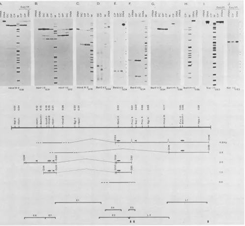
FIG. 3. Mappingof CRPVtranscriptswithend-labeledprobes. Poly(A)- RNA isolated fromCPs.DPs.orDCsortRNAwashybridized todifferent3'-or5-end-labeledprobes. digestedwithnucleaseS1orExoVII, andanalyzedonacrylamide-urea gels (panels A, B,C. F,G, H, andJ)oronneutral (panelsD and E)oralkaline agarose(panel J)gelsasdescribed inthe text. The topsection shows autoradiograms whichwereobtained afterexposurewithamplifyingscreensfor 18h(panels A. B. D. E, F.G. H. andJ) or3 days(panel I)orwithoutan
amplifying screen for 3days (panel C). The map positions of the labeled ends of the hybridizing probe strands are indicated below the
autoradiograms.Theprobesusedinthe autoradiogramsshown were:A,Hindlll 0.19toHinidlll 0.77(thesame resultswereobtained with theproberecut atBa,n1HI 0.15):B(left),Hinfl 0.21toHinifl 0.12 (thesameresultswereobtainedwiththeproberecut atBaInHI): B(right),
Hinfl0.12toHinfl 0.04:C.HinidlIl 0.19toHindlll 0.26: D.BstEII0.54toSall 0.63:E. BstEII(full-sized recombinant DNA with pBR322 insertedatthe EcoRI site): F.same as D:G (left).sameas E:G(right),BainHI 0.86toBglll0.02: H. BainiHI 0.86toEcoRI 0.18: 1 andJ, Sall0.63toEcoRl0.18 andextending650nucleotidestotheSall sitein pBR322.Themolecularweightmarkers inthe trackslabeledMwere end-labeledfragments ofHinidlll-digested ADNA withsizes of23.1,9.41. 6.55. 4.37. 2.32, 2.02. and 0.564 kb:Haelll-digested replicative
form of 4X174with sizes of 1.353. 1.078.0.872,0.603.and 0.310 kb and weak bandsat0.281 and 0.271kb;Mspl-digested pBR322DNA with sizes of622. 527.403. 309 (notresolved from the310-base-pair bandofHaelll-digested replicativeformofd3X174). 243. 238,217. 201,190.
180, 160(double band). 147(double band), 122, 100. 90, and 76 bases. Inpanels A, B. C, G. and 1. both 'bX174 and pBR322 markers are present;the smallestmarkersshownare 122, 147, 122. 147, and 76 bases. respectively. In panelsF andH.onlypBR322markersarepresent, and thesmallestones are76and110bases, respectively.Inpanel D,the markersareXDNAHitdIll digested.In EandJ. theX DNAHindlll
and the fourlargest 4X174replicative form HaellI marker positions are indicated tothe right of the panels. The center portion shows a
restrictionmapofCRPV andamapofCRPVtranscripts.The results of Northern blothybridizations (Fig. 2)werecombined with those of nucleaseS1 and Exo VIl mapping(topof figure). Exons aremarked by heavy lines, andintronsaremarked by light brackets. The mapped ends ofexons aremarkedby vertical bars, and the ends ofexonsnotmappedareindicated by extending theexonlineas adashedline. Dashed
intronbracketsinthe 4.8-kb RNAindicate thatsomeRNAmolecules donothaveintrons. Thearrowheads point from the end of the labeled probeinthe directionofprobeextension. The bottomportiondepictsopenreading frames of CRPVDNA.Thereading directionis fromleft
toright; the vertical arrowheads indicate the positions ofpotential polyadenylation sites (AATAAAsequences) (Giri et al.. submitted for publication).
w .e
c:
--on
goo
VOL. 51, 1984
0
M.
0
.-MM
-m
I.4
O C
on November 10, 2019 by guest
http://jvi.asm.org/
[image:4.612.61.559.73.533.2]mapped. Hybridization with Hinifl fragment 0.04 to 0.12 (Fig. 3, panel B. right side) again identified the 5' end of the 2.0-kb RNA at map position 0.07. Further, in DC particu-larly, a second minor 2.0-kb RNA species exists with a 5' end 70 bases upstream. With Hiuifl fragment 0.12 to 0.21 (Fig. 3, panel B, left side), a major 0.4-kb band is present, mapping the 5' end of the 1.3-kb RNA to position 0.16. In Fig. 3, panel B, left side, the bands visible in panel B, right side, are detectable as weak bands due to a minor contami-nation with the other Hiiifl probe. The detection of the 5' endof the 1.3-kb RNA with the Hi,itlprobe, but not with the HindIII and EcoRI probes. suggests that unusual sequences near the two sites prevent stable hybrid formation.
The 3' ends of the 5'-proximal exons were mapped with probes labeled 3' at the HinidIII (map position, 0.19) and EcoRI (map position. 0.18) sites. A double band of 0.28 kb is present in all tumors (Fig. 3. panel C). The two bands are less than 10 nucleotides different in size. In the CP and DP tumors, the smaller band is more intense, whereas in the DC tumor, both are of about equal intensity. Since the hybridi-zation mixtures appeared to contain about equal amounts of virus-specific RNA, the difference in intensity may be sig-nificant, and a tentative conclusion could be that the smaller band represents the 2.0-kb RNA, which is more prominent in papillomas, and the larger band represents the 1.3-kb RNA. Alternately, the exons of both RNAs could have two 3' ends. The hybridizations with EcoRI labeled probes mapped the ends of the exons to the same position as did the HindIII probes (data not shown).
The endsof the 3'-proximal exons of the 1.3- and 2.0-kb RNAs were mapped with BstE-II labeled probes. The
map-ping experiments with 5'-labeled probes (Fig. 3. panel F) show two bands of 0.14 and 0.09 kb in all tumors. Since the relative intensity of the bands is very similar in papillomas and carcinomas, the two different ends cannot be assigned uniquely to one or the other RNA species. To show that both the 1.3- and 2.0-kb RNAs were mapped, we analyzed Si
digests on neutral agarose gels (Fig. 3, panel D). In this analysis, the domestic carcinoma revealed two well-defined bandsof0.7and 1.4 kb.which are the sizes expected for the segmentsof the 1.3- and 2.0-kb RNAs, respectively. The CP inaddition shows a major band (0.5 kb) and two minor bands
(0.9and 1.1kb). Theseprobably derived from the 4.8-or 0.9-kbRNA orboth since the two species are unique to CPs and
hybridize strongly to the genomic segment (MspI fragment 4) containing the BstE-II site (Fig. 2B). The 3' ends of the exons were mapped with BstE-II 3'-labeled probes, and the
Sidigests revealed one major band of0.52 kb (Fig. 3, panel
G, left side). A weak band with the mobility (but not necessarily the size) of the probe can be seen in all tracks
including the control (tRNA); however, this band is
dis-tinctly more pronounced in the CP. suggesting that in this tumor, some RNA molecules do not terminate at map position0.61. The neutral agarosegelanalysis (Fig. 3,panel
E)of the same
Si
digests revealed one strong bandof about 0.6kb in all tumors; inaddition, theCPcontained aband of 3.6 kb. The relativeintensity ofthe3.6-kbband to the0.6-kb band in the neutral agarose gel is higher than the relative intensity ofthe 'probe-sized" band to the 0.52-kb band in the acrylamide gel. This suggests that some ofthe parent RNAmolecules givingrise tothe3.6-kbband have an intron 0.52 kb downstreamfrom the BstE-II site. Finally, a probe labeled 3' at the BstE-II site and extending leftward to the EcoRl site did not give riseto any bands(data not shown), and this furtherconfirms thatallRNAspecieswhich contain the BstE-II site in their exons (4.8-. 2.0-, 1.3-, and 0.9-kbRNA) have the polarity 5' to 3' from left to right on the map (Fig. 3).
The 3'-proximal exons of the 2.6- and 4.8-kb RNAs were mappedwithprobes labeled3'and 5' at theBa,nHI site(map
position, 0.86). The 3-labeled probe gave rise to a single band of about 1.05 kb (Fig. 3, panel G, right side), mapping the ends of both RNAs to position 0.99. The5'-end-labeled probe revealed one major band of 0.54 kb (Fig. 3, panel H); in addition, in some analyses (data not shown) a minor probe-sized bandcould be detected. The results suggest that the majority of the RNA molecules mapping to this area of the genome contain an exonextending 5' from map position 0.79 to map position 0.99 at the 3' end and that a small minority ofmolecules representing probably a fraction of the 4.8-kb RNA have an exon extending further upstream.
Our Northernblots(Fig. 2)indicated that MspI fragment 8 hybridized only to the 4.8-kb RNA, and therefore, a probe labeled within this segment could be used to map the 4.8-kb RNA. AStill (map position. 0.63) 5'-labeled probe extending upstream to the EcoRI site (mapposition, 0.18) and 650 base pairs into pBR322 sequences was used. The SI digest (Fig. 3,panel I. left side) revealed a majorband of 0.85 kb, which maps the 5' end of the majorexon to position 0.53. A faint minor band has a size of0.23 kb, locating the endof a minor exon at position 0.61. The analysis of Exo VII digests on acrylamide gels showed thesame0.85-kb band, but the 0.23-kb band could not be detected (Fig. 3, panel I, right side). The Exo VIIdigests were also analyzed on agarose gels(Fig. 3, panel J). As can be seen, asecond band of about 3.4 kb is detectable. The 5' end of the largerprotected fragment maps close to the EcoRI site, thus linking the major 3'-proximal portion of the 4.8-kb RNA to sequences also transcribed in the 5'-proximal exons of the 1.3- and 2.0-kb RNAs. The relative intensities of the 0.85- and 3.4-kb RNAs have no true significance. since the Exo VII commercially available also has endonuclease activity. With increasing levels of enzyme. Exo VII digests increasingly resemble Si digests. The presence of this endonuclease activity made it impos-sible to map across large introns. This difficulty and the apparent coline4rity of leader sequences of the 2.6- and 4.8-kb RNAswith the 5' exons of the 1.3- and 2.0-kbRNA have prevented the definitive mapping of the leaders of the 2.6-and4.8-kb RNA.
DISCUSSION
We have analyzed the viral transcripts in CRPV-induced non-virus-producing benign and malignant domestic rabbit tumors and in virus-producing CPs. The non-virus-produc-ing tumors contain two transcripts of 1.3 and 2.0 kb, and virus-producing CPs contain five transcripts of4.8, 2.6, 2.0, 1.3, and 0.9 kb. In virus- and non-virus-producing benign tumors. the 2.0-kb RNA is moreprominent than the 1.3-kb RNA, whereas the opposite is found in malignant tumors. The transcripts were mapped by Northern blot hybridiza-tions with subgenomic probes and by S1 and Exo VII mapping with 3'- and 5'-end-labeled probes, and the results are summarized in Fig. 3.
When the maps of the transcripts present in non-virus-producing tumors are compared with the map of open reading frames, severalfacts can be established. The major 5' end ofthe 2.0-kb RNAmapsjust within E6, whereas the minor 5' end maps to the end or outside E6. Since the transcript continuesuninterrupted byintrons pasttheend of
E6. nosequences inaddition to E6could be translated from this RNA. The significance ofthe increased presence ofa slightly larger transcriptinthecarcinomacannotbeassessed
on November 10, 2019 by guest
http://jvi.asm.org/
VIRAL TRANSCRIPTS IN CRPV TUMORS 711
at present. The 1.3-kb transcript starts toward the 3' end of E6. In both human papillomavirus la (7) and bovine papil-lomavirus type1 (BPV-1) (6) there are no ATG codons in this segment of E6; the next ATG is in E7. and if the situationin CRPV is equivalent, then translation could start here. Since the 3' end of the exon maps to the end of E7. the amino-terminal portion of the proteins translated from the 1.3-kb RNA could be coded for by E7 and the carboxy-terminal portion could be coded for by E4 or E2 orboth.
The 1.3- and 2.0-kb RNA could be generated
by
two mechanisms: differential splicing or transcription from two different sites. In BPV-1 there is only one TATA-like se-quence in this segment ofthe genome. and it is close tothe beginningof E6 (6): in contrast, in humanpapillomavirus la and human papillomavirus 6b there are two such sequences: one again at the beginning of E6 and the other at the beginning ofE7 (7. 19). Ourexperimental evidence suggests thatthe two transcripts are initiatedat different sites. First, in the DP(Fig. 3, panel A) there is no difference between the Si and the Exo VIIdigests, indicating that the transcription for the 2.0-kb RNA starts close to the beginning of E6.Second, preliminary primer extension experiments mapped the 5' ends to the same location asS1 experiments. The notion that the 1.3- and 2.0-kb RNAs may be transcribed from different sites, together with the fact that the 2.0-kb RNA is moreprominent inpapillomas,is particularly intrigu-ing since we have previously found that CRPV DNA in carcinomas is methylated to ahigher degree compared with that in papillomas (26). It is possible then that changes in methylationcould affect the efficiency of transcription from different sites differentially and thus be responsible for the
observed differences in transcription between papillomas and carcinomas.
There are differences between transcripts in
non-virus-producingtumors induced by CRPV and transcripts in
BPV-1-transformed cells(1,
2,
8.9, 11). A most striking difference is that there is no major RNA species inCRPV-induced
tumors which could code for the large open reading frame El, a segmentof the genome which appears to be required inBPV-1-transformed cells for the maintenance of BPV-1 DNA as a plasmid (12, 16). Further, CRPV tumors also do notcontainRNA species which could code for the entire E2 and E4 open reading frames. Finally, it has been proposed that the transcripts in BPV-1-transformed cells have a com-mon5'-leadersequence of at least 150 base pairs and that the 5' end maps close to the beginning of the E6 open reading frame (1),whereas our data suggest that there is no common leader forthe 1.3- and 2.0-kb RNA.
The definitively mapped segments of the 2.6- and 4.8-kb RNAs correspondto 1.59 and 3.7 kb, leaving segments of 0.9 and 1.0 kb, respectively, unmapped. The Northern blots
indicate that these segments are present in a part of the genome which is represented in the 1.3- and 2.0-kb RNAs (Fig. 2B). It was further shown by alkaline agarose gel
electrophoresis of Exo VII digests that the EcoRI site is
contained in 5'-proximal sequences of the 4.8-kb RNA (Fig. 3, panelJ).
There are some similarities between the RNAs unique to
virus-producing
tumors induced by CRPV and BPV-1 (9). The 3' exon of the 2.6-kb RNA is equivalent to an RNA mapped in BPV-1-induced tumors: in both cases it starts close to the 5' end, but within the Li reading frame. andextends beyondthe reading frame to a polyadenylation site. Themain body ofthe 4.8-kb RNA is similar to a BPV-1 RNA inthat itstarts5' within E2 and E4 and coterminates with the othervirus-producing tumor-specific RNA. However, a
dif-ference is that this
segment
here has one or two smallintrons,
at least in some of the molecules. Particularlysignificant may
be the intron located at map position0.79.
since this intron could linkreading
frames Li and L2. Such an RNA could code forproteins
larger than Li or L2, and suchlarger
proteins
have been described in BPV-1 and CRPV (10, 15).Although
themapping
ofpapilloma
transcripts isin-complete,
atthisstage
itappears
that humanpapillomavirus-1
(L. T. Chow and T. R.Broker.
in M. L. Pearson and N. L.Steinberg.
ed.,Gete
Tin.isft'e
(aizd
Cancer, in press) and CRPV resemble each other more closely than they resemble BPV-1.ACKNOVW
LEDGMENTSExcellent technical and
graphic
assistance was providedby
M.Havford.
We thank J. G.Stevens forhelpfuldiscussion
andA.Berk
for advice on
S1
and Exo VII mapping. Wearegreatly indebted to1.
Gir.
0. Danos. and M.Yaniv
for making available before pub-lication amap
of restrictionenzyme
sites and ofopen readingframes
basedon sequencing
data.
This research was
supported
by
Public Health Service grant CA-18151 from the National Cancer Institute and by a grant from theUniversity
of California Coordinating Committee for CancerRe-search.
LITERATURE CITED
1. Ahola, H., A.
Steniund,
J.
Moreno-Lopez,
and U. Pettersson.1983.
Sequences
of bovinepapillomavirus
type1
DNA-func-tional andevolutionary implications.
Nucleic Acids Res. 11:2639-2650.2. Amtmann, E., and G. Sauer. 1982. Bovine papilloma virus
transcription:
polyadenylated
RNA species and assessment of the direction of transcription. J. Virol. 43:59-66.3. Bantle,
J.
A.,1.
H. Maxwell, and W. E. Hahn. 1976. Specificity ofoligo
(dt) cellulose chromatography in isolation of polyad-envlated RNA. Anal.Biochem.
72:413-427.4. Berk,
J.
A., and P. A.Sharp.
1977. Sizingandmapping
of earlyadenovirus
mRNAs bygel
electrophoresis ofS1
nuclease-digested
hybrids.
Cell 12:721-723.5. Berk,
J.
A., and P. A.Sharp.
1978. Spliced early mRNAs of simian virus 40. Proc.Natl.
Acad.Sci.
U.S.A. 75:1274-1278. 6. Chen, E. Y., P. M. Howlev, A. D. Levinson, andP. H. Seeburg.1982.
Primary
structure andgenetic
organization of the bovinepapillomavirus type
1
genome.
Nature(London) 299:529-534. 7. Danos,O.,
M. Katinka, and M. Yaniv. 1982. Humanpapilloma-virus la
complete
DNAsequence:
a noveltype
of genome organization among papovaviridae. EMBO J. 1:231-246.8.
Engel,
L.W.,
C. A.Heilman,
and P. M. Howley. 1982.Virus-specific transcription
in bovinepapilloma-vir-us-transformed
mouse cells. Virology 119:22-35.9.
Engel,
L.W.,
C. A.Heilman,
and P. M. Howlev. 1983.Tran-scriptional
organization
of bovine papillomavirus type 1. J. Virol. 47:516-528.10.
Favre,
MI.,
F.Breitburd,
0. Croissant, and G. Orth. 1975. Structuralpolypeptides
of rabbit. bovine, and human papil-lomaviruses. J. Virol. 15:1239-1247.11. Freese, U. K., P. Schulte, and F. Pfister. 1982. Papilloma
virus-induced tumors contain a virus-specific transcript. Virology 117:251-257.
12.
Lusky,
M.,
and M. R. Botchan. 1984. Characterization of thebovine
papillomavirus
plasmid
maintenance sequences. Cell 36:391-401.13.
Maxam,
A.,
and W.Gilbert. 1980. Sequencingend-labeled
DNAwith base
specific
chemical
cleavages. Methods
Enzymol.65:499-560).
14.
McMaster,
G.K.,
and G. G. Carmichael. 1977.Analysis
ofsingle
and double-stranded nucleic acids onpolyacrylamide andagarose
gels
by
using
glyoxal
and acridin orange. Proc.Natl.
Acad. Sci. U.S.A. 74:4835-4838.VOL. 51, 1984
on November 10, 2019 by guest
http://jvi.asm.org/
15. Meinke, W., and G. Meinke. 1981. Isolation and characteriza-tion of the major capsid protein of bovine papillomavirustype1. J.Gen. Virol. 52:15-24.
16. Nakabayashi, Y., S. K. Chattopadhyay, and D. R. Lowy. 1983. Thetransforming function of bovine papillomavirus DNA. Proc. Natl. Acad. Sci. U.S.A. 80:5832-5836.
17. Nasseri, M., F. 0. Wettstein, and J. G. Stevens. 1982. Two
colinear and spliced viral transcripts are present in
non-virus-producing benign and malignant neoplasms induced by the Shope(rabbit) papilloma virus. J. Virol. 44:263-268.
18. Rigby, P. W., M. Dieckmann, C. Rhodes, and P. Berg. 1977. Labelingdeoxyribonucleic acidtohigh specific activity in vitro by nicktranslation with DNA polymerase 1. J. Mol. Biol. 113:237-251.
19. Schwarz, E., M. Durst, C. Demankowski, 0. Lattermann, R.
Zech, E.Wolfsperger, S. Suhai, andH.zurHausen. 1983. DNA
sequenceandgenomeorganization of genital human
papilloma-virustype6b. EMBO J. 2:2341-2348.
20. Sharp, P. A., B. Sugden, and J. Sambrook. 1973. Detection of
two restriction endonuclease activities in Haeinophilus
par-a-influenzae using analytical agarose-ethidium bromide elec-trophoresis. Biochemistry 12:3055-3063.
21. Stevens, J. G., and F. 0. Wettstein. 1979. Multiple copies of
Shopevirus DNAarepresentin cells ofbenignandmalignant non-virus-producing neoplasms.J. Virol.30:891-898.
22. Weaver, R. F., and C.Weissmann. 1979. Mapping of RNA bya
modification of the Berk-Sharp Procedure: the 5' termini of 15s,B-globin mRNA precursor and mature 10s3-globin mRNA have identicalmapcoordinates. Nucleic Acids Res. 7:1175-1193. 23. Wettstein, F. O., and J. G. Stevens. 1980. Distribution andstate
of viral nucleic acid in tumors induced by Shope papilloma virus, p. 301-307.In M. Essex,G. Todaro, and H. zurHausen
(ed.),Viruses innaturallyoccurringcancers,vol. 7. ColdSpring Harbor Laboratory,Cold Spring Harbor, N.Y.
24. Wettstein, F. O., and J. G. Stevens. 1981. Transcription of the viral genome in papillomas and carcinomas induced by the Shope virus. Virology 109:448-451.
25. Wettstein, F. O., and J. G. Stevens. 1982. Variable size free episomes of Shope papilloma virus DNAarepresentinall
non-virus-producing neoplasms and integrated episomes are
de-tected insome. Proc. Natl. Acad. Sci. U.S.A. 79:790-794. 26. Wettstein, F. O., and J. G. Stevens. 1983.Shope papilloma virus
DNA is extensively methylated in nonvirus-producing
neo-plasms. Virology 126:493-504.