MANNOSE BINDING LECTIN AS A DIAGNOSTIC
MARKER FOR CORONARY ARTERY DISEASE IN
HYPERTENSIVE PATIENTS
Dissertation submitted for
M.D. BIOCHEMISTRY BRANCH – XIII
DEGREE EXAMINATION
THE TAMILNADU DR.M.G.R.MEDICAL UNIVERSITY
CHENNAI – 600 032
TAMILNADU
BONAFIDE CERTIFICATE
This is to certify that this dissertation work entitled “MANNOSE BINDING
LECTIN AS A DIAGNOSTIC MARKER FOR CORONARYARTERY DISEASE IN HYPERTENSIVE PATIENTS” is the original bonafide work done by
DR.M.DILAGAMARI, Post Graduate Student, Institute of Biochemistry,
Madras Medical College, Chennai under our direct supervisionand guidance.
Prof. Dr. V.Amuthavalli, MD., (Guide)
Professor,
Institute of Biochemistry Madras Medical College, Chennai – 600003.
Prof. Dr. K.Ramadevi. MD.,Ph.D Director & Professor,
Institute of Biochemistry Madras Medical College Chennai-600 003.
Dean
Madras Medical College and
DECLARATION
I, Dr. M. DILAGAMARI, Post Graduate , Institute of Biochemistry, Madras Medical College, solemnly declare that the dissertation titled
“MANNOSE BINDING LECTIN AS A DIAGNOSTIC MARKER FOR CORONARY ARTERY DISEASE IN HYPERTENSIVE PATIENTS” is the
bonafide work done by me at Institute of Biochemistry, Madras Medical College
under the expert guidance and supervision of Prof. Dr.V.AMUTHAVALLI,
M.D., Institute of Biochemistry, Madras Medical College. The dissertation is
submitted to the Tamil Nadu Dr. M.G.R Medical University towards partial
fulfillment of requirement for the award of M.D., Degree (Branch XIII) in
Biochemistry.
Place: Chennai
SPECIAL ACKNOWLEDGEMENT
The author gratefully acknowledges and sincerely thanks
Dr.R .JAYANTHI ,MD, FRCP (Glasg ) , Dean, Madras Medical College and Rajiv Gandhi Government General Hospital, Chennai, for granting her
ACKNOWLEDGEMENT
The author expresses her warmest respects and profound gratitude to
Dr. K. Ramadevi, M.D.,Ph.D., Director and Professor, Institute of Biochemistry, Madras Medical College, Chennai, for her able guidance, constant
encouragement, support and valuable time but for which this dissertation could
not have been made possible.
The author expresses her heartfelt gratitude to her guide and supervisor
Dr.V.Amuthavalli, M.D., Professor, Institute of Biochemistry, Madras Medical College, Chennai, for her constant and valuable guidance, unfailing support,
encouragement and inspiration throughout the period of her study.
The author in particular, is extremely thankful to Prof. Dr. S.Mayilvahanan
M.D., Director of General Medicine & Professor Dr.N.Swaminathan M.D.,DM,
Director of Cardiology, Rajiv Gandhi Government General Hospital, Chennai, for
granting permission to obtain blood samples from the patients.
The author expresses her sincere gratitude to the Professors Dr.R.Chitraa
M.D., Dr.K.Pramila M.D., Dr.Sumathy.S. M.D. & Dr. Chelladurai, M.D,
Institute of biochemistry, Madras Medical College, for their guidance and support.
The author expresses her warm respects and sincere thanks to her co-guide,
Madras Medical College for her guidance regarding the practical issues of
research which is beyond the scope of textbooks.
The author expresses her respects and sincere thanks to all other Assistant
Professors, Institute ofBiochemistry, Madras Medical College, for their guidance and support.
The author expresses warm respects to the members of the Institutional
Ethics committee for approving the study.
The author is indebted to the patients and persons from whom blood
samples were collected for conducting the study.
The author expresses her special thanks to Biochemistry Laboratory Staff,
for their timely help and cooperation during sample collection.
The author expresses her special thanks to her colleagues for their
cooperation and genuine support.
The author gratefully acknowledges the help rendered by Mr. Albert, for
the statistical analysis of the study.
The author expresses her special thanks to her parents for the moral support
CONTENTS
SI.
NO TITLE PAGE No.
1 INTRODUCTION 1
2 REVIEW OF LITERATURE 3
3 AIMS & OBJECTIVES 45
4 MATERIALS & METHODS 46
5 RESULTS 62
6 STATISTICAL ANALYSIS 63
7 DISCUSSION 79
8 SUMMARY & CONCLUSION 86
10 SCOPE FOR FURTHER STUDIES 87
11 BIBLIOGRAPHY 88
ABBREVIATIONS
CAD - Coronary artery disease
NFK - Nuclear factor kappa.
SRB - Scavenger rceptor class B
VCAM - Vascular cell adhesion molecule.
ICAM - Intercellular adhesion molecule.
PG - Prostaglandin
CRP - C Reactive protein.
hs-CRP - High sensitive C Reactive protein.
NO - Nitricoxide.
MBL - Mannose Binding lectin
MASP - MBL associated serine proteases
eNOS - Endothelial nitric oxide synthase
ROS - Reactive oxygen species
PDGF - Platelet derived growth factor
FGF - Fibroblast growth factor
TNF α - Tumour necrosis factor alpha
PeCAM - Pericellular cell adhesion molecule
BMI - Body Mass Index
TGL - Triglycerides
PAF-AH - Platelet Activating Factor – Acetyl Hydrolase
ER - Endoplamic reticulum
ANG II - Angiotensin II
MCP - Monocyte Chemoattractant Protein
M-CSF - Monocyte colony stimulating factor
IL - Interleukin
HDL - High density lipoprotein
LDL - Low density lipoprotein
CHD - Coronary Heart Disease
LVH - Left Ventricular Hypertrophy
HMG-CoA - 3-hydroxy-3-methyl glutaryl coenzyme A
ACAT - acylCoA:cholesterol acyl transferase
LCAT - lecithin:cholesterol acyl transferase
CRD - C- terminal carbohydrate recognition domain
CL - Collectin
I/R - Ischemia –reperfusion injury
C1-INH - C1esterase inhibitor
BOS - Bronchiolitis obliterans syndrome
SLE - Systemic Lupus Erythematosus
sMAP - small MBL associated protein
Map - MBL associated protein
DIC - Disseminated intravascular coagulation
MI - Myocardial Infarction
HT - Hypertension
1
INTRODUCTION
Cardiovascular disease is one of the leading cause of mortality and
morbidity worldwide. Epidemiological investigations pointed out that
hypertension is a powerful cardiovascular risk factor. Elevated blood pressure
levels have been found to be highly predictive of cardiovascular events including
ischemic coronary disease, stroke and peripheral arterial disease. Relationship
between elevated blood pressure and risk of cardiovascular events is continuous,
consistent and independent of other risk factors(1).
The cardiovascular events have a predominant vascular origin resulting
from atherosclerosis. Factors contributing to atherosclerosis are multiple and
complex. Multiple theories including role of dyslipidemia , hypercoagulability ,
oxidative stress, inflammation, endothelial dysfunction have been put forth.
Inflammation plays a major role in all phases of atherogenesis from plaque
initiation to plaque rupture(2). Several inflammatory markers such as C-reactive protein (CRP), Secretory phospholipase A2, interleukin (IL)-6 have been
associated with increased risk of atherosclerosis(3,4).In addition , the markers of
innate immunity have been shown to predict the development of coronary artery
disease.
Mannose - binding lectin (MBL) also known as Mannan – binding lectin or
Mannan – (Mannose - ) binding protein (MBP) is an important component of the
2
associated serine proteases (MASPs) has the ability to activate complement via
the lectin pathway. Since innate immunity has been implicated in atherogenesis ,
MBL has been suggested to play a role in the formation of atherosclerotic
plaque(5,6).
However studies examining the relations between serum levels of MBL
and coronary artery disease risk have reported equivocal results.At present , no
conclusive data are available about the relationship between serum MBL levels
and coronary artery disease risk in hypertensive patients. Hence it is proposed to
study the association of serum levels of MBL in the development of coronary
artery disease in hypertensive patients .
HYPOTHESIS
MBL is an important component of innate immune system. Elevated serum
MBL levels are associated with increased risk of future coronary artery disease in
3
REVIEW OF LITERATURE
Hypertensive patients are more prone for cardiovascular disease. MBL
being a component of innate immune system can be used as a marker of
cardiovascular risk in hypertension. This study was conducted in 90 patients to
study the correlation between the serum MBL levels and other risk factors of
coronary artery disease and hypertension.
HYPERTENSION:
Hypertension is one of the most common chronic disease. In India the
prevalence rate is 29.8% . About 23% of men and 22.6% of women above 25
years suffer from hypertension .Coronary Artery Disease is a leading cause of
morbidity and mortality in hypertensive patients.
Hypertension is a multifactorial disorder that involves pathological changes
in neuronal, renal, genetic, hormonal and vascular mechanism. Genetic alterations
may initiate the cascade to permanent hypertension. Genetic contribution have
been estimated to range from 30% to 60%. Polymorphisms of certain genes
involving rennin – angiotensin system , aldosterone synthesis and adrenergic
receptors are more common in hypertensives than normotensives(7).
Hypertension is known to act on the arterial wall to promote both vascular
remodelling and atherosclerosis resulting in diminished arterial wall compliance
and elevated stiffness. Clinical and experimental investigations have shown that
4
AETIOLOGY:
It is of two types;
1. Primary / Idiopathic / Essential
About 90 -95% of hypertension is idiopathic .The pathogenesis of essential
hypertension is not clearly understood. Different investigators have proposed that
the kidney, the peripheral resistant vessels and sympathetic nervous system as the
seat of primary abnormality.
2. Secondary causes
Of the remaining 5 to 10% is secondary to renal disease or less often to
narrowing of renal artery ,usually by an atheromatous plaque (Renal vascular
hypertension). Infrequently , secondary hypertension is the result of adrenal
disorders such as primary aldosteronism, Cushing’s syndrome and
pheochromocytoma.(9)
Regulation of Normal Blood Pressure:
The magnitude of the arterial pressure depends on two fundamental
hemodynamic variables: Cardiac Output and Total Peripheral Vascular
Resistance.
BLOOD PRESSURE = CARDIAC OUTPUT X PERIPHERAL RESISTANCE
Indeed, the blood pressure level is a complex trait that is determined by the
interaction of multiple genetic ,environmental , and demographic factors that
5
Cardiac output is highly dependent on blood volume which itself is greatly
influenced by the whole body sodium homeostasis. Peripheral vascular resistance
is determined mainly at the level of arterioles and is affected by neural and
hormonal factors.
Pathogenesis of Hypertension:
The multiple mechanism of hypertension constitute aberrations of the
normal physiological regulation of blood pressure. Arterial hypertension occurs
when the relationship between cardiac output and total peripheral resistance is
altered.
Genetics of Hypertension :
In studies of twins and family members in which the degree of familial
aggregation of blood pressure levels is compared with the closeness of genetic
sharing, the contributions have been estimated to range from 30 to 60 percent.(7) Single gene disorders cause relatively rare and severe form of hypertension
eg., Gene defects in enzymes involved in aldosterone metabolism and mutations
in proteins like α - adducin that affect sodium reabsorption.
Inherited variations in blood pressure may also depend on the cumulative
effects of allelic forms of several genes that affect blood pressure.eg., variations in
6
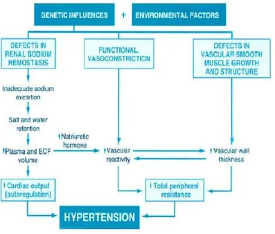
Reduced renal sodium excretion in the presence of normal arterial pressure
may well be the key initiating event in essential hypertension and a final common
pathway for the pathogenesis of hypertension. Decreased sodium excretion
might lead sequentially to an increase in fluid volume, increased cardiac output,
[image:17.595.112.509.235.574.2]and peripheral vascular constriction , thereby elevating blood pressure.(Fig.1)
Figure 1 : Scheme of pathogenesis of essential hypertension(9)
An alternative hypothesis implicates vasoconstrictive influences either the
factors that induce functional vasoconstriction or stimuli that induce direct
7
Environmental factors :
Environmental factors could modify expression of the genetic determinants
of increased pressure.
Stress
Obesity
Smoking
Physical inactivity
Increased intake of salt
have all been implicated as exogenous factors in hypertension.
Essential hypertension results from the combined effect of mutations or
polymorphisms at several gene loci that influence blood pressure, interacting with
a variety of environmental factors. Thus, environmental factors affect the
variables that control blood pressure in the genetically predisposed individual.
Hypertension and Oxidative stress :
Hypertension is considered as a state of oxidative stress(10)
Excessive production of ROS (Reactive Oxygen Species), outstripping
antioxidant mechanism, decreased bioavailability of NO (Nitric Oxide) in the
vasculature and kidneys and ROS mediated cardiovascular remodelling plays an
important pathophysiological role in development of hypertension. Biomarkers of
systemic oxidative stress such as isoprostanes, malondialdehyde and nitrotyrosine
8
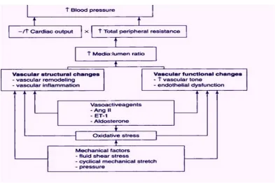
Increased ROS bioactivity leads to endothelial dysfunction , increased
contractility, vascular smooth muscle cell growth, monocyte invasion, lipid
peroxidation, inflammation and increased deposition of extracellular matrix
proteins(11), important factors in hypertension mediated vascular damage (Fig.2). An excessive activity of rennin – angiotensin - aldosterone system leads
to the production of ROS and also contributes to endothelial dysfunction, vascular
inflammation and thrombosis.(12,13)
NO and Hypertension:
Impaired endothelium – mediated vasodilation in hypertension has been
linked to decreased NO bioavailability. This may be secondary to decreased NO
synthesis or to increased NO degradation because of its interaction with
superoxide anion (O2
-
[image:19.595.112.505.454.716.2]) to form peroxynitrite (ONOO- ).
9
Dyslipidemia and Hypertension :
Recent experimental data suggests marked similarities between the effects
of hypertension and hypercholesterolemia on the arterial intima. Both conditions
exert proinflammatory effects on the artery, resulting in the recruitment of
monocytes into the intima. These effects may be due to the production of free
radicals , which in turn stimulate the genes involved in the recruitment of
inflammatory cells into the arterial wall(15)
Endothelial dysfunction is in cross road between hypertension and
dyslipidemia. The key feature in this disorder is the reduced availability of nitric
oxide (NO) due to both decrease in synthesis as well as enhanced degradation.
Hypercholestrolemia plays an important role, as oxidised LDL diminishes the
expression of endothelial NO synthase(12)
Role of complement in hypertension:
Hypertension and hypertensive end organ damage are not only mediated by
hemodynamic but also by innate and adaptive immune responses. Complement
activation may drive the pathology of hypertension through its impact on innate
immunity. Inflammatory responses mediate the development of perivascular
fibrosis and heart dysfunction induced by hypertension. In hypertension induced
cardiac damage, the most important component is the inflammatory
microenvironment including T cells, macrophages, neutrophils, and the
chemokines and cytokines released from these cells which could affect the degree
10
The role of C5a in hypertension has recently been examined. Zhang et
al.(17) reported increased levels of C5a in humans with high blood pressure. Infusion of ANG II-causing arterial hypertension lead to increased systemic
anaphylatoxin generation in mice. C5aR1-deficient mice exhibited markedly
reduced cardiac remodeling and inflammation after ANG II infusion.
Hypertension stimulates structural arterial remodeling, which is
characterized by vascular smooth muscle cell (VSMC) hyperplasia and infiltration
of inflammatory cells. Recent findings demonstrate a key role for complement
C1q induced activation of β-catenin signalling is implicated in VSMC
proliferation during intimal thickening after vascular injury . β-catenin signalling pathway regulates the proliferation and differentiation of smooth muscle cells.
Complement activation causes hypertensive arterial remodelling.(18)
CORONARY ARTERY DISEASE:
Coronary artery disease has been defined as impairment of function of
heart due to inadequate blood supply to the heart compared to its needs. Its being
a multifactorial disease has a complex etiology. Many genetic and environmental
factors act in combination to determine an individual’s risk of developing
coronary artery disease.(19) A large number of studies such as The Framingham heart study(20), The lipid research clinic’s coronary primary prevention trial, The Helsenki heart study, have been conducted to examine the role of risk factors for
11
The risk factors identified by these epidemiological studies include;
Epidemiological risk factor
1. Positive family history
2. Age
3. Gender
4. Cigarette smoking
5. Hypertension
6. Hyperlipidemia
7. Coronary atherosclerosis prone personality
8. Obesity
9. Diabetes Mellitus
Pathological risk factor
1. Atherosclerosis
2. Platelet dysfunction
3. Endothelial dysfunction
Biochemical risk factors
1. Total cholesterol > 240 mg / dL
2. HDL < 35 mg / dL
12
ATHEROSCLEROSIS:
Atherosclerosis is a disease affecting arterial blood vessels. It is a chronic
inflammatory response in the walls of arteries, in large part due to the
accumulation of macrophages promoted by low density lipoproteins (LDL)
without adequate removal of fats and cholesterol from the macrophages by
functional high density lipoproteins (HDL). It is commonly referred to as a
“hardening” or “ furring” of the arteries.(21)
The lesions of atherosclerosis occur principally within the innermost layer
of the artery wall, the intima.(22) They include
Fatty streak
Fibrous plaque
Complicated lesion
Plaque disruption
Atherothrombosis
Fatty streak
The process of atherogenesis begins in childhood with the development of
lipid rich lesions called fatty streaks. They are also found to contain macrophages,
T lymphocytes , smooth muscle cells – each of these cells are found to contain
deposits of cholesterol and cholesterol oleate. The lesions are yellowish and
sessile in appearance and they cause little or no obstruction of the affected artery
13
Fibrous plaque
The fibrous plaques are derived from fatty streaks that continue the process
of cell proliferation , lipid accumulation , and connective tissue formation and the
deep core of lipid and cell debris results from inadequate blood supply,
inflammation, and cell necrosis.
There is a lesion that is accepted as a forerunner of the fibrous plaque –
that is known as fibromusculoelastic or intermediate lesion of the intima, which
consists of proliferated smooth muscle cells surrounded by connective tissue and
contains little or no lipid.
A fully blown fibrous plaque consists of numerous smooth muscle cells
surrounded by connective tissue matrix often intermixed with numerous
macrophages. This cap covers a deep layer of macrophages filled with lipid that
are often intermixed with variable number of T lymphocytes.
Advanced lesions – Plaque disruption and Atherothrombosis
The typical advanced, complicated lesion contains a large necrotic core
with a fibrous core, loaded with macrophages . The macrophages can form
numerous proteolytic enzymes, including metalloproteinases – these enzymes
cause the removal of fibrous cap – thus plaque disruption is found to happen at the
shoulder of the lesion where the cap is thin and concentration of macrophages is
the greatest. The plaque disruption allow the lesion to get involved in thrombotic
14
Two major effects of atherosclerosis are
1. The atheromatous plaques , though compensated by artery enlargement,
eventually lead to plaque rupture and stenosis of the artery and therefore an
insufficient blood supply to the organ it feeds
2. If the compensating artery enlargement process is excessive , then a net
aneurysm results.
These complications are chronic, slowly progressive and cumulative. Most
commonly , soft plaque suddenly ruptures , causing the formation of a thrombus
that will rapidly slow or stop blood flow, leading to death of the tissues fed by the
artery in approximately 5 minutes. This catastrophic event is called an infarction.
The clinical scenarios of this catastrophic event depend on which artery is
affected.
1. Coronary artery – Myocardial Infarction
2. Carotid artery and its branches – Stroke or Transient Ischemic attack
3. Peripheral artery disease
Theories of Atherogenesis:
Long ER et al. has discussed the development of clinicopathological
correlations that evolved during the era when autopsy examination permitted the
formulation of a hypothesis relating the degree of atherosclerosis to the incidence
15
Virchow proposed the idea that some form of injury to the arterial wall
associated with the inflammatory response resulted in the degenerative lesion of
atherosclerosis(25). This idea was subsequently modified by Anitschkow (26) and further included the role of platelets and thrombogenesis in atherosclerosis as
expanded by Duguid(27).
John French noted that the structural integrity of endothelial lining of the
artery represented a key element in the maintenance of normal arterial function(28)
and that alteration in endothelial integrity might precede a sequence of events that
would lead to the various forms of the lesions of atherosclerosis.
Response to endothelial injury:
In normal artery , the endothelial cells form a continuous monolayer that
regulates the passage of substances from the plasma to the underlying wall(29,30), forms a thromboresistance surface that promotes the continuous flow of blood
throughout the vascular tree. Production of certain cytokines by the endothelial
cells regulates the migration and proliferation of smooth muscle cells .
Endothelial cells are capable of transporting plasma lipoproteins of into the
arterial wall (31,32). The endothelium exhibits thromboresistant characters by production of three factors
1. Surface glycoproteins and proteoglycans
2. Prostacyclin
16
Prostacyclins and nitric oxide are potent vasodilatory agents and inhibitors
of platelet aggregation. Injury to the endothelium results in structural and
functional alterations in the endothelial cells, in such a way that they would permit
plasma constituents such as lipoproteins and inflammatory cells to have a ready
access to the arterial wall(33).
Leukocyte adhesion:
The endothelial dysfunction is associated with overexpression of E, L, P
selectin that appear to play a role in inducing rolling and attachment of monocytes
and T lymphocytes to endothelium. This rolling is facilitated by the upregulation
of ICAM 1 and VCAM 1 also. PeCAM 1 , another molecule formed by
endothelium has been involved in interendothelial migration by the adherent
leukocyte into the subendothelial space or intima of the artery. Thus, the earliest
phase of chronic inflammatory response is represented by leukocyte adhesion due
to the formation of these attachment and adherence molecules on the surface of
the endothelium and the leukocytes(34–36).
Transmigration of lipoproteins:
Second event accompanying endothelial dysfunction is transmigration of
lipoproteins particularly LDL particles, this transmigration places the LDL in the
subendothelial space which is virtually devoid of any antioxidant properties of the
circulation , hence it gets oxidised. Oxidised LDL can act as one of the
chemotactic factor and can also induce the endothelial cells and the underlying
17
Chemoattractant Protein 1 ( MCP – 1 ) and a colony stimulating factor (M - CSF).
In this scenario , the monocyte gets activated to macrophages , which express
SR-B1 causing unregulated uptake of LDL particles , forming foam cells. Such a
lesion with foam cells ,activated inflammatory cells is called as fatty streak.
Formation of stable atherosclerotic plaque:
Oxidised LDL, foam cells, the activated macrophages, T- cells produce
various cytokines like IL-1 and TNF-α . Under the influence of these cytokines,
endothelium, macrophages, and T cells produce PDGF and FGF(37).
PDGF stimulates smooth muscle cell migration and proliferation.
FGF stimulates vascular smooth muscle cell to produce collagen and the
various components of extracellular matrix together they form the fibrous
cap.
TNF – α induces apoptosis of foam cells causing exocytosis of its lipid
content, which forms the lipid core.
The lesion with lipid core , surrounded by activated T-cells, macrophages,
platelets lined by a fibrous cap is called as a stable atherosclerotic plaque.
Thus oxidised LDL is not only toxic to the endothelium and the
surrounding cells in the intima but also chemotactic for monocytes and can
activate monocyte derived macrophages to produce growth factors and cytokines.
18
If the injury to the endothelium were a self – limited event and the
endothelial functions were restored , the proliferative lesions may regress and
would be clinically silent. If the injury at focal sites in the artery wall is of long
standing, the lesion would continue to progress .
The atherosclerotic plaque does not only have smooth muscle cells but also
macrophages , which are capable of producing metalloproteinases and TNF α , both of which cause necrosis and digestion of fibrous cap . The loss of fibrous cap
is called plaque rupture.
The plaque rupture exposes the subendothelial extracellular matrix to the
factors of coagulation in the circulation initiating the intrinsic pathway of
coagulation – this is responsible for atherothrombosis.
Effect of Hypertension on Atherosclerosis:
Elevated systolic and diastolic blood pressure have a strong, positive and
graded relationship to CHD (38,39). The risk imposed by hypertension is heightened
substantially when other risk factors are present.
Hypertension clusters with
Insulin resistance
Hyperinsulinemia
Glucose Intolerance
19
Left Ventricular Hypertrophy and
Obesity
and occurs in isolation in fewer than 20% of individuals(40).
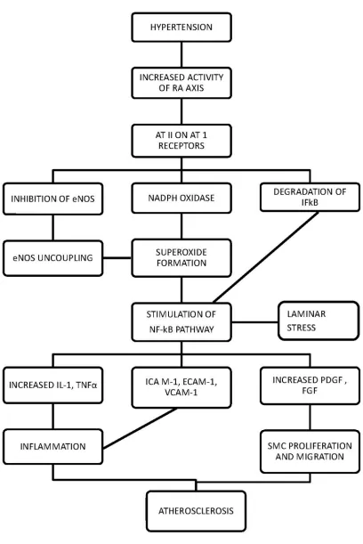
The potential mechanism by which hypertension may cause endothelial
dysfunction include
1. Increased endothelial permeability to lipoproteins,
2. Increased adherence of leukocytes,
3. Increased oxidative stress, and
4. Hemodynamic stress that may trigger acute plaque rupture,
All these are mediated by the activation of NF-kB pathway and
inactivation of eNOS enzyme.
Obesity:
Obesity promotes insulin resistance, hyperinsulinemia, hypertriglyceridemia,
low HDL cholesterol and LVH(41,42). Many observational studies have found that
obesity strongly and positively correlates with the risk of CHD in univariate
analysis. In multivariate analysis , when controlling statistically for risk factors
such as hypertension, diabetes, and dyslipidemia, obesity is not found to be an
20
21
The adverse consequences of obesity are mediated through resultant
metabolic risk factors acting as pathological links in the causal pathway. In
general the greater the degree of overweight , the higher the risk of coronary
mortality(44,45). The central distribution of body fat predicts CHD in men independently of body-mass index and other major risk factors(46). Weight loss improves insulin sensitivity and glucose disposal, reduces blood pressure,
triglycerides and LVH, and increases HDL cholesterol(41,42).
Dyslipidemia:
Plasma cholesterol may arise from the diet or from endogenous
biosynthesis. Cholesterol is transported between tissues in combination with
proteins and phospholipids as lipoproteins. Abnormal levels of plasma lipids is
called dyslipidemia.
Plasma lipoproteins :
Lipoproteins are composed of a neutral lipid core containing
triacylglycerol (TAG) and cholesteryl esters surrounded by a shell of amphipathic
apolipoproteins, phospholipids and unesterified cholesterol. The lipoprotein
particles include
1. Chylomicrons
2. Very low density lipoproteins ( VLDLs )
3. Low density lipoprotein ( LDLs )
22
Metabolism of Chylomicrons(47):
Chylomicrons are assembled in intestinal mucosal cells and carry dietary
TAG, cholesterol, fat soluble vitamins and cholesterol esters to the peripheral
tissues. TAGs account for 90% of lipids in chylomicron. Apo B-48, Apo C-II and
Apo E are the specific apolipoproteins of chylomicrons.
1) Intestinal mucosal cells secrete nascent TAG – rich chylomicrons produced
from dietary lipids.
2) Apo C-II and Apo E are transferred from HDL to the nascent chylomicron.
3) Extracellular lipoprotein lipase , activated by Apo C-II , degrades the TAG
in chylomicrons resulting in the formation of free fatty acids and glycerol.
Insulin enhances lipoprotein lipase synthesis and its translocation to the
luminal surface of capillary.
4) Apo C-II is returned to HDL forming chylomicron remnant.
5) CE – rich chylomicrons remnants bind through apo E to specific receptors
on the liver and are endocytosed.
6) The cholesteryl esters and leftover TGL are metabolised by hepatic lipase.
Metabolism of VLDL and LDL(47):
They are produced in liver. VLDL composed predominantly of endogenous
TAG and their function is to carry this lipid from liver to the peripheral
tissue.LDL particles composed of high concentration of cholesterol and
cholesteryl esters. Their primary function is to provide cholesterol to the
23
1. Liver secretes nascent , TAG – rich VLDL particles.
2. Apo C-II and Apo E are transferred from HDL to the nascent VLDL.
3. Extracellular lipoprotein lipase , activated by Apo C-II, degrades the TAG in
VLDL resulting in the formation of free fatty acids, glycerol and VLDL
remnant. This action of lipoprotein lipase decreases the lipid content of both
Chylomicrons and VLDL by 70-90%. VLDL remnants are also called as IDL.
4. Most of the IDL undergoes conversion to LDL after hydrolysis of all TGL.
LDL principally contains cholesterol and cholesterol esters.
5. LDL binds to LDL receptors specific for Apo B-100 on hepatic and
extrahepatic tissues and are endocytosed.
Regulation of LDL receptor:
LDL (apoB-100, apo-E) receptors are cell surface receptors that are coated
by a protein called clathrin on the cytosolic side of the membrane. It is a
glycoprotein receptor. It spans the membrane with B-100 binding domain at the
extracellularly exposed amino terminal. After binding, LDL is endocytosed. The
endocytosed vesicle fuses with lysosome to form endosome with the receptor
being returned to the surface. The cholesterol esters and apoproteins are
hydrolysed and cholesterol is translocated into the cell. This cholesterol influx
downregulates the transcription of genes encoding HMGCoA synthase, HMGCoA
reductase and other enzymes of cholesterol synthetic pathway. It also inhibits
transcription of genes coding for LDL receptors. It stimulates ACAT enzyme
24
Metabolism of HDL(47)
HDL is synthesized in both liver and intestine.
1. Nascent HDL are disc shaped particles composed mainly of
phosphatidylcholine and apolipoproteins A-1, C and E.
2. They take up unesterified cholesterol with the help of ABCA1 ( ATP-
binding cassette transporters A ) from nonhepatic tissue.
3. Cholesterol is esterified by LCAT ( lecithin: cholesterol acyltransferase )
bound to nascent HDL which is activated by Apo- A1.
4. CETP moves some of the cholesteryl esters from HDL to VLDL in
exchange for TAG, relieving the product inhibition of LCAT.
5. Cholesteryl esters rich HDL binds to liver leading to selective transfer of
cholesteryl esters into the cells mediated by SR-B1 ( scavenger receptor
class B type 1)
6. Then HDL transports cholesterol to liver where it is excreted through bile.
This is called reverse cholesterol transport.
Total Cholesterol and LDL cholesterol in atherosclerosis:
Numerous prospective studies have identified a continuous, graded and
direct relationship between serum cholesterol and CHD incidence(48). The level of
total cholesterol and LDL cholesterol interacts with other risk factors to multiply
25
Elevated LDL cholesterol levels appear to be involved in all stages of
atherogenesis – endothelial dysfunction, plaque formation and growth and plaque
instability and disruption. Elevated cholesterol levels in the plasma lead to an
increased retention of LDL particles in the arterial wall , their oxidation and the
secretion of various inflammatory mediators and chemoattractants(51).
LDL is also a potent mitogen for smooth muscle cells ; progressive growth
of atherosclerotic plaques with a large lipid core and numerous lipid filled
macrophages are prone to rupture(52). Thus the epidemiological evidence strongly
supports LDL- Cholesterol’s role in atherosclerosis.
Small dense LDL are felt to be more atherogenic(53). The two possible reasons for this are
1) When a person has more of small LDL particles, for a given cholesterol
content, the number of LDL particles will be more and an LDL receptor
can accept only one LDL particle at a time and hence the rate of
metabolism of LDL is decreased , causing accumulation of LDL in the
plasma.
2) The endothelium will be more permeable to small LDL particle when
compared to normal LDL.
Triglycerides:
The relationship between triglycerides and CHD has been less clear. In
men , univariate analysis have demonstrated a direct dose-response relationship.
26
HDL cholesterol , obesity and diabetes(54). Hypertriglyceridemia however has been found to be an independent risk factor in women(55).
Several mechanisms have been proposed to explain the triglyceride- CHD
association.
Hypertriglyceridemia have a predominance of small , dense LDL particles
Fasting hypertriglyceridemia may be a marker of exaggerated postprandial
hyperlipidemia, which may promote the uptake of atherogenic triglyceride
rich lipoprotein remnants by endothelial cells(56).
Serum triglyceride levels are strongly related to fibrinogen and factor VII
in numerous epidemiological studies(57).
Therefore , a number of mechanisms act as direct and indirect link
serum triglycerides and CHD.
HDL cholesterol :
Numerous prospective epidemiological studies have demonstrated a
continuous, inverse relationship between HDL cholesterol levels and the
incidence of CHD. The total cholesterol to HDL cholesterol ratio is better
predictor of CHD than the HDL cholesterol level alone(58). Two important
mechanisms by which HDL is thought to play a protective role against
atherosclerosis are;
1. Reverse cholesterol transport and
27
Normal HDL contains several enzymes such as
o Paraoxonase ( PON )(60)
o Lecithin : cholesterol acyltransferase(61)
o Glutathione selenoperoxidase(62)
o Platelet activating factor – Acetyl Hydrolase ( PAF – AH )(63)
that can potentially prevent the formation of oxidized LDL and also
inactivate the inflammatory LDL-derived oxidized phospholipids.
MANNOSE BINDING LECTIN:
Mannose binding lectin is a pattern recognition molecule of the innate
immune system. It is a part of complement cascade and plays an important role in
the first line of defense against pathogenic microorganisms(64,65). MBL has opsonic activity . In association with MBL-associated serine proteases (MASPs) ,
have the ability to activate complement via the lectin pathway(66).
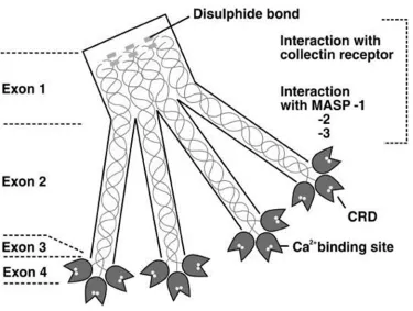
MBL belongs to the collection family, a group of C-type lectin also
possessing a collagen –like helical domain. Human MBL exists as a series of
oligomers of 2-6 subunits, built up from three identical polypeptide chains
(24 KDa, 228 aminoacids each). The apparent molecular weight of these
oligomers range from approximately 1,50,000 to 4,50,000. It forms a bouquet-
28
Four characteristic regions of MBL are
1. The short N- terminal cysteine –rich domain - responsible for arrangement
of subunits in the oligomer , dependent on disulphide bonds ; this region
consist of 21 aminoacids including three Cys residues.
2. The collagen like region interacts with MASPs ; it consists of 59 amino
acids (among them 19 Gly-X-Y triplets); this domain is glycosylated.
3. An α- helical neck region consists of 30 aminoacids stabilizes the polypeptide chain within a structural subunit.
4. The C- terminal carbohydrate recognition domain ( CRD ) is responsible
for pattern recognition and consists of 118 aminoacids(65,67–69)
MBL recognizes sugar patterns on the surface of many pathogens,
phospholipids, immune complexes and apoptotic cells. MBL binds with highest
affinity to D-mannose , N- acetyl – D – glucosamine and L – fucose which allows
the specific recognition of numerous polysaccharides and glycoconjugates like
bacterial lipopolysaccharides, capsular polysaccharides and fungal mannans (70,71). The LPS structure exerts a major influence on MBL attachment to bacteria.
Mannan- binding lectin is synthesized by hepatocytes and secreted into the
blood in an oligmeric form . Moreover , specific mRNA has been found in bone
marrow, fetal lung , small intestine, and testis (72). Its synthesis is controlled by the MBL2 gene located on chromosome 10 (10q11.2) and regulated in a similar
manner to acute phase proteins. The normal serum level of MBL is about 400 –
29
Figure 4 :schematic representation of MBL 2 gene and its genetic polymorphisms(73)
Figure 5 : Structure of tetrameric human mannose binding lectin(74)
[image:40.595.124.499.429.712.2]30
The MBL2 gene contains 4 exons and 3 introns(75).
1. Exon 1 of the MBL2 gene encodes the signal peptide , the cysteine – rich
domain, and part of glycine rich collagen like region.
2. Exon 2 encodes the remaining part of collagen like region.
3. Exon 3 encodes the neck region and
4. Exon 4 encodes CRD.
Most mammals synthesize two forms of MBL : A and C. Human MBL
resembles the C form . The MBL1 pseudogene ( not expressed) , corresponding to
A form , has been found in the human genome(76).
Polymorphism of MBL gene :
Structural mutation in exon 1 of the human MBL 2 gene at codon 52
(allele D), codon 54 (allele B) and codon 57 (allele C) reduce the functional MBL
levels by disrupting the collagenous structure. B,C,D alleles are collectively
known as ‘O’ allele. Several nucleotide substitution at the promoter region like
H/L polymorphism, X/Y polymorphism and P/Q polymorphism affect the serum
MBL concentration(77–79).
MBL dependent complement activation:
MBL forms a complex with MBL-associated serine proteases (MASPs).
This complex becomes enzymatically active and activates the complement. This
facilitates complement - dependent opsonisation and subsequent uptake and
31
MBL was believed to be the sole collectin able to activate the lectin
pathway (LP) of complement. However , several reports indicate that MBL shares
that property not only with ficolins (another family of collagen – related lectins)
but also with the so called “novel collectins” like collectin 11 ( CL-11 or collectin
–kidney 1, CL-K1) and collectin 10 ( CL-10 or collectin – liver 1, CL-L1)(81,82). After binding of the MBL-MASPs complex to the target structure,
conformational changes lead to the activation of MASPs which in consequence
makes the cleavage of C4 and C2 possible and thus the initiation of the
complement cascade.
In spite of the different initiation mechanism , the lectin pathway resembles
the classical pathway (CP), forming the same convertases for C3 and C5
components(83,84).
MASP
Three proteases
1. MASP - 1
2. MASP - 2
3. MASP - 3
MASP – 2 is believed to be the key enzyme responsible for LP activation
as its proteolytic activity against C4 and C2 significantly exceeds the activity of
32
33
MASP – 1 is believed to upregulate lectin pathway activation. However,
recently its crucial role in MASP – 2 activation has been postulated(88,89).
MASP – 3 and nonenzymatic proteins – MAp19 (sMAP ) and Map44 are
believed to play a regulatory role in this process(84).
Involvement of MBL in Coagulation and Kallikrein-Kinin system:
MBL plays an important role in other systemic processes including
coagulation , inflammation and tissue injury. MASP – 1 and – 2 may participate in
activation of the coagulation system.
MASP – 1 – cleaves fibrinogen , factor XIII , and thrombin – activate fibrinolysis
inhibitor (TAFI )(90)
MASP – 2- cleaves prothrombin(91)
Megyeri et al found MASP – 1 to interact with protease activated
receptor-4, a mediator of inflammation and platelet activation(93). Later, Dobo et al. found high molecular weight kininogen to be its additional substrate(94). This activity like that of kallikrein enables release of bradykinin, a highly proinflammatory
mediator of the kinin – kallikrein system. Although MASP – 2 cleaves kininogen,
no bradykinin is released during this process.
The involvement of MBL – MASP complexes in haemostatic processes
was later demonstrated in an animal model by Takahashi et al.(95) who found that
MBL – null mice infected with staphylococcus aureus were predisposed to the
34
[image:45.595.95.549.101.418.2]
Figure 7 : Role of MBL in complement pathway , coagulation system & kinin system(92)
MBL and disease association:
MBL deficiency and increased susceptibility to disease:
1. Infectious disease especially extracellular pathogens(96,97)
2. Autoimmune disease e.g. SLE(98)
MBL deficiency and protection against disease:
35
MBL and modulation of disease severity
1. Infectious disease e.g. HIV , Hepatitis B and C , pulmonary disease in
cystic fibrosis(100,101)
2. Autoimmune disease e.g. Rheumatoid arthritis(102)
Inappropriate activation of MBL – MASP pathway
1. Lectin pathway activation in renal disease(103–105) e.g. Lupus nephropathy
Membranoproliferative glomerulonephritis
Post – streptococcal glomerulonephritis
Henoch – schonlein purpura nephritis
2. Lectin pathway activation on vascular endothelium following oxidative
stress
e.g. Myocardial reperfusion injury(106)
Factors that increase the concentration of MBL :(107–109)
Thyroid hormone
Growth hormone
Dexamethasone
Low levels of MBL(110,111) are seen in
Breast cancer
36
MBL in ischaemic – reperfusion injury:
Innate immune response is a “double – edge sword”, sometimes beneficial
and at other times disastrous for the host. MBL and MBL- dependent complement
activation have been found to be involved in ischaemia-reperfusion (I/R) injury
associated with numerous clinical condition such as graft rejection and other
pathological processes in the gastrointestinal tract or central nervous system.
In kidney:
Significant deposition of MBL - MASP 2 complexes was found in porcine
kidney after I/R injury. Moreover, colocalization of C4d with MBL was
observed(112). Administration of C1esterase inhibitor ( C1-INH ) resulted in inhibition of apoptosis of tubular epithelial cells and tubular damage. Therefore, a
pathological role of both lectin and classical pathway in I/R renal injury was
implicated.
In Van der Pol et al.(113) Rodent model study therapeutic inhibition of MBL was protective against tubular damage , preventing accumulation of
macrophages and neutrophils as well as expression of proinflammatory cytokines
and chemokines . Following reperfusion , MBL was internalized into tubular
epithelial cells, including rapid cell death. Therefore it was concluded that MBL
-mediated cytotoxicity preceded complement activation and was the primary
reason for tubular injury.
MBL deposition in ischaemically injured human kidney was first
37
capillaries and tubular epithelial cells. Berger et al. demonstrated a beneficial
effect of low MBL concentrations and low MBL variants of the corresponding
gene in recipients of kidney or combined pancreas – kidney transplants on graft
and patient survival. Low levels of circulating MBL correlated with improved
long term graft survival(115,116).
Damman and Seelen (117) suggested that under moderate graft injury, lectin
pathway activation may be beneficial due to participation in the clearance of
dying cells. However, under severe injury, MBL might contribute to the renal
tubular epithelium damage. According to Osthoff et al.(118) MBL deficiency could
offer some protection from I/R radiocontrast –induced kidney injury.
MBL in Cerebral Ischaemia:
Several reports demonstrated an association of MBL with cerebral
ischaemia. Ducruet et al. observed deposition of MBL in murine ishaemic
endothelium(119). Even in MBL – null mice, they observed C3 deposition in the cerebral hemisphere during reperfusion, suggesting involvement of other
complement activation pathways in the pathology.
Later, Orsini et al. working with mice confirmed MBL deposition in
ischaemic vessels and a protective effect of genetically determined MBL
deficiency ; they also found an increase in circulating MBL – MASP – 2 complex
38
Elvington et al. demonstrated the involvement of the alternative pathway of
complement activation(121). Interestingly C6 deficiency had no effect, indicating the lack of significance of the common pathway and thus formation of the MAC
(membrane attack complex). Osthoff et al. (122) determined MBL concentrations in 353 patients with ischaemic stroke observed that MBL – deficient patients had
significantly lower risk of unfavourable outcome and showed smaller lesion
volumes. They suggested that inhibition of lectin pathway may be a promising
strategy for reducing I/R associated cerebral damage.
In a recent clinical study Wang et al.(123) assessed serum levels of MBL in patients with acute ischaemic stroke and found that the MBL levels were
significantly higher in stroke patients compared with healthy controls and
increased with increasing severity of stroke. The authors postulated that elevated
MBL levels could be an independent risk factor for stroke.
MBL in Lung transplantation:
Several papers have focused on the role of MBL in lung transplantation. Its
higher levels in plasma of recipients were associated with development of
bronchiolitis obliterans syndrome (BOS) and poorer long- term outcome(124,125). Immunohistochemistry revealed the presence of MBL in lung tissue from patients
with BOS and at the time of ischaemia. Moreover , Carroll et al. observed a
significant increase of MBL concentration in plasma at 3,6 and 12 months after
39
after transplant was associated with later BOS development(126). Low MBL
concentrations were associated with a longer survival.
Gastrointestinal ischaemia:
Zhang et al. found that IgM bound to antigens in ischaemic mesenterium ,
provided a binding site for MBL resulting in complement activation(127).This finding was further confirmed by Lee et al. who detected MBL complexed to
antigen and natural IgM in the intestinal I/ R injury(128). Schwaeble et al. postulated MASP-2 deficiency to protect mice from gastrointestinal ischaemia –
reperfusion damage(129).
Cardiovascular disease:
Involvement of MBL in coagulation and ischaemia – reperfusion injury is
potentially harmful in the development of cardiovascular disease, especially
coronary artery disease and myocardial infarction, as well as in the rejection of a
heart transplant.
Ischaemia changes the expression of surface molecules and leads to
formation of neoantigens. Paradoxically, reperfusion causes harmful
inflammatory response that can counteract the beneficial effects of improved
blood flow. During reperfusion, myocardial cells become the targets of innate
immunity; promoting the release of inflammatory mediators, neutrophil
40
Mechanism causing myocardial ischaemia – reperfusion (MI/R ) injury
was provided by Busche et al. Using MBL and IgM – null mice , they found that
myocardial tissue injury following MI/R associated with complement activation
dependent on both MBL and IgM antibodies(131).
Pesonen et al. found lower serum concentrations of C3, higher levels of
MBL, and a higher frequency of high MBL level – associated MBL2 genotypes in
a cohort of patients with unstable angina pectoris or acute myocardial infarction
compared with healthy controls(132). Similarly , Haahr-Pedersen et al. observed significantly higher MBL and lower soluble C5b-9 (complement membrane attack
complex, MAC) concentrations in sera of patients with ST – elevation myocardial
infarction (STEMI) , undergoing primary percutaneous intervention with left
ventricular ejection fraction (LVEF) <35 % compared with those of LVEF
≥ 35%(133).
Keller et al. noted that an elevated MBL level is a risk factor for future
coronary artery disease (CAD) in apparently healthy men but not in women(2). Later , Schoos et al. have found high plasma levels of MBL and ficolin – 2
(L-ficolin ) to be synergistically associated with increased postinfarct left ventricular
end systolic and diastolic volumes (ESV, EDV) in STEMI patients(134).
Trendelenburg et al. reported that MBL functional deficiency contributed
to the significant reduction of 90- day mortality in patients with acute STEMI,
41
MBL and activation of lectin pathway of complement may be protective
against the development of atherosclerotic lesions by
clearance of apoptotic cells and cell debris from atherosclerotic plaques or
protection from Chlamydia pneumonia infection , known to be associated
with development of atherosclerosis(136).
On the other hand MBL may take part in I/R injury and enhance
thrombosis. The resultant effect may depend on diverse factors, specific for the
individual , including accompanying diseases , life style , age and sex.
C-REACTIVE PROTEIN:
It is an acute phase protein of hepatic origin(137). CRP was so named because it was first identified as a substance in serum of the patients with acute
inflammation that reacted with the somatic – C polysaccharide antigen of the
capsule of pneumococcus.
CRP is a member of Pentraxin family consisting of five identical, non
glycosylated polypeptide subunits, non covalently linked to form a disk shaped
cyclic polymer with a molecular weight of 115 k Da. Apart from the
polysaccharides in many bacteria, fungi and protozoal parasites CRP also binds to
phosphoryl choline such as lecithin and polyanions like nucleic acids in the
presence of calcium ions. In the absence of calcium ions , it binds to polycations
42
FUNCTIONS OF CRP:
C- reactive protein activates classical complement pathway of immune
system via C1q .
Initiates opsonisation, phagocytosis and lysis of invading cells and plays
an important role in body’s defence mechanism
Detoxification of toxic autogenous substances(140).
FACTORS THAT INCREASE THE CONCENTRATION OF CRP:
1) Obesity
2) Smoking
3) Metabolic syndrome
4) Dyslipidemia
5) Steroids
6) Hypertension.
Major elevations of CRP occur in
Myocardial infarction
Infection
Inflammation
Stress
Trauma
43
FACTORS DECREASING THE CONCENTRATION OF CRP:
1) Weight loss
2) Medications:-Statins, Fibrates, Niacin, Aspirin, NSAIDs, Moderate
alcohol consumption.
ROLE OF CRP IN ATHEROGENESIS :
CRP released by action of IL-6, binds directly with oxidised LDL and is
present within Lipid laden plaques(141).
It causes Monocyte adhesion & trans migration into the vessel wall leads to
polarisation of Macrophages, which is a pro inflammatory trigger in plaque
deposition in atherosclerotic lesions
Decreases the NO synthesis by inhibiting endothelial Nitric oxide
synthases, and promotes chemotaxis(142).
Increases the expression of tissue factor which acts as a procoagulant
resulting in DIC.
Production of Plasminogen activating inhibitor inhibits tissue plasminogen
activator, decreases fibrinolysis and results in atherogenesis (143).
High sensitivity CRP:
CRP assays are used to predict recurrent ischemia in patients with unstable
angina(3). These standard assays were unable to identify those patient at risk for acute myocardial infarction, as they lacked low – end specificity. Development of
44
mg / L are referred to as high sensitivity (hs ) or ultrasensitive assays. The value
of hs-CRP as a marker of cardiovascular disease risk is recognized and accepted.
Routine CRP measurements – over the range of 3 mg / L
High sensitivity CRP measurement – including the range upto 3 mg / L
Applications of hs – CRP:
1. Diagnostic and prognostic marker in acute coronary syndrome
2. Predictor of future coronary events(144)
Inflammation plays a major role in all phases of atherogenesis and the role
45
AIM OF THE STUDY
1. To evaluate the risk of coronary artery disease in recently diagnosed
hypertensive individuals by estimating serum Mannose Binding Lectin
levels.
2. To correlate the Mannose Binding Lectin level with CRP for predicting
cardiovascular risk in hypertensive patients
OBJECTIVES
1. To estimate the level of serum mannose binding lectin in hypertensive
patients with coronary artery disease.
2. To estimate the level of serum mannose binding lectin in age and sex
matched healthy subjects.
3. To estimate the level of C-Reactive Protein(CRP) in all the study groups.
4. To correlate the level of serum mannose binding lectin and
C-Reactive Protein (CRP)
Lipid profile
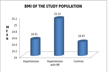
BMI and
46
MATERIALS AND METHODS
The study protocol was approved by the Institutional Ethics Committee of
Madras Medical College, Chennai.
STUDY DESIGN : Cross sectional case- control study
STUDY PERIOD : August 2017 - February 2018
SUBJECT SELECTION :
90 subjects were selected for the study. They were divided into three
groups as follows.
Group A - 30 Recently diagnosed hypertensive patients
Group B- 30 Hypertensive patients who had myocardial infarction recently
Group C - 30 Age and sex matched healthy controls
Inclusion Criteria: GROUP A:-
Recently diagnosed hypertensive patients of less than 6 months duration.
Age – 30 years and above
Outpatients attending hypertension clinic in Rajiv Gandhi Government
General Hospital, Chennai
Hypertension was considered to be present if an individual had a history of
hypertension and was on antihypertensive agents or if the systolic blood
pressure exceeded 140mmHg or the diastolic pressure exceeded 90
47
GROUP B :-
Hypertensive patients who had Myocardial Infarction recently.( < 7 days
duration)
Age – 30 years and above
Inpatients admitted in the cardiology department in Rajiv Gandhi
Government General Hospital, Chennai.
The diagnosis of myocardial infarction was based on
a) a history of characteristic prolonged chest pain or chest discomfort
b) Electro cardiographic evidence of new Q wave or abnormal ST segment
c) Elevated levels of known cardiac biochemical markers like CK-MB.
GROUP C:-
Age and sex matched apparently healthy subjects who were staffs of
Madras Medical College, their relatives and friends.
Exclusion criteria :
Patients with Diabetes mellitus
Patients with renal disorders.
Patients with Liver diseases.
Patient with acute illness / infection.
Chronic Smokers and alcoholics.
48
Blood collection :
5 mL of blood sample was collected from all subjects after overnight
fasting and transferred to serum tubes. The blood was allowed to clot and the
serum was separated after centrifugation at 3000 RPM for 15 minutes. The serum
levels of CRP , total cholesterol , triglycerides , HDL –C, urea , creatinine & uric
acid were measured immediately. About 0.5 mL of serum was stored in eppendrof
at – 20oC for the analysis of mannose binding lectin.
BIOCHEMICAL INVESTIGATIONS:
Analytes Methods
Serum Mannose Binding Lectin ELISA
Serum C-Reactive Protein(CRP) Immunoturbidimetry
Serum Urea , Creatinine & Uric acid Spectrophotometer
Serum Lipid Profile Spectrophotometer
ESTIMATION OF SERUM MANNOSE BINDING LECTIN:
METHOD:
Enzyme linked immunosorbent assay – non competitive , sandwich
PRINCIPLE:
MBL in standard and test samples are captured by anti-MBL
49
Second biotinylated antibody to human MBL is added which binds with
the immobilized MBL thus forming a sandwich of MBL between two anti-
MBL antibodies.
Streptavidin conjugated with enzyme horse radish peroxidase is added.
Streptavidin binds avidly with biotin in the detection antibody.
Substrate to the enzyme horse radish peroxidase is added resulting in the
formation of a coloured complex.
The intensity of colour produced is directly proportional to concentration
of MBL in the sample. The absorbance is measured at 450 nm in an automated
microplate reader.
REAGENTS :
1) Pre coated ELISA plate
2) Standard solution – MBL – 3200 ng / mL
3) Standard diluents
4) Biotin conjugate Anti – human MBL antibody
5) Streptavidin – HRP
6) Wash buffer concentrate
7) Substrate solution A
8) Substrate solution B
9) Stop solution
REAGENT PREPARATION:
All reagents should be brought to room temperature before use.
50
The various concentrations of standard are prepared as follows
Standard 5 1600 ng / mL 120 μL original standard + 120 μL standard diluents
Standard 4 800 ng / mL 120 μL standard 5 + 120 μL standard diluents
Standard 3 400 ng / mL 120 μL standard 4 + 120 μL standard diluents
Standard 2 200 ng / mL 120 μL standard 3 + 120 μL standard diluents
Standard 1 100 ng / mL 120 μL standard 2 + 120 μL standard diluents
ng/ml 3200 1600 800 400 200 100
Wash buffer
Mix the wash concentrate thoroughly until no visible crystals are present.
Then dilute 20 mL of wash concentrate in 480 mL of deionized water to make
500mL of wash buffer.
PROCEDURE:
1. The reagents and samples are brought to room temperature ( 18oC – 25oC)
before use.
2. Label strips as appropriate for the assay.
51
5. Then add 10 μL of anti- MBL antibody to sample wells.
6. Add 50 μL of streptavidin – HRP to sample wells and standard wells. 7. Mix well. Cover the plate with a sealer and incubate at 37oC for 60
minutes.
8. Discard the solution. Wash each well 5 times with 350 μL of reconstituted wash buffer solution in an auto washer. Invert and blot dry the plate against
clean filter paper.Add 50 μL of substrate solution A to each well and then add 50 μL of substrate solution B to all wells. Cover the plate with a new sealer and incubate at 37oC in the dark for 10 minutes.
9. Add 50 μL of stop solution to each well. The blue color will change into
yellow immediately. The absorbance is read at 450 nm within 30 minutes
after adding the stop solution.
STANDARD CURVE :
The standard curve is plotted with standard concentration in ng/mL along
x- axis and absorbance along y- axis. The line of best fit is drawn through the
standard points. The line has to be linear. The concentration of each sample is
calculated from the standard curve.
S. No
Concentration of Std ( ng/mL )
Absorbance (OD)
1 0 0.106
2 100 0.280
3 200 0.450
4 400 0.700
5 800 1.19