0022-538X/93/084923-09$02.00/0
Copyright X 1993, AmericanSociety for Microbiology
Nef from Primary Isolates of Human
Immunodeficiency Virus
Type
1
Suppresses
Surface CD4 Expression
in Human
and Mouse T
Cells
STEPHEN ANDERSON,' DIANE C.SHUGARS,2'3 RONALD SWANSTROM,2'4
ANDJ. VICTOR
GARCIA"*
Departmentof Virology and Molecular Biology, St.Jude Children's Research Hospital, Memphis, Tennessee 38101,1 and Lineberger Cancer Research Center,2 School of Dentistry,3 and Department of Biochemistry
andBiophysics,4 University of North Carolina, Chapel Hill, North Carolina 27599 Received 25 January1993/Accepted 5May 1993
Thehumanimmunodeficiencyvirustype1(HIV-1) nefgene wasoriginallydescribedasanegativeregulator oftranscriptionfrom the viral longterminal repeat promoter. This observation has been disputed, and the functionofNefremainsunclear. In vivo experimentshave indicatedthat an intact nefgene is requiredfor diseaseprogressioninmacaquesinfected with simianimmunodeficiency virus, suggestingarole forNef in the pathogenesisofAIDS. We and others havepreviouslyshown thatexpressionofNef incellsbearing surface CD4 results inasustained decreaseinsurfaceCD4 expression.Thiswasdemonstrated for Nef fromtwolaboratory strains ofHIV-1,Bruand SF2. Because both of these isolateswerepassagedinvitropriortomolecular cloning andin vitropassagecanresult in mutations whichmightalternefgenefunction,wehaveanalyzedtwoprimary isolatesofNef fortheirabilitytosuppresscell surface CD4expression.ThenefgenesofHIV-1 isolatesfrom twopatientswithfewerthan200 CD4+Tcellspermm3ofbloodwereintroducedinto human andmouseT-cell linesbyretrovirus-mediatedgenetransfer.Expressionof Neffrom both isolates correlatedwithadecreasein
surfaceexpressionofboth human andmouseCD4. Todetermine whether theabilitytosuppresssurfaceCD4 expressionisageneralfunction ofNef,wealsotestedanartificiallygeneratedconsensusnefgenederived from
analysisof 54patientisolates of HIV-1.ExpressionoftheconsensusNefproteinalsocorrelated withdecreased cell surface CD4 expression in bothmouse and human T-cell lines. These results suggest thatthe ability to
suppresscell surface CD4expression isan intrinsic feature of HIV-1 Nef.
Thenefgeneofthe humanimmunodeficiencyvirustype1 (HIV-1)codes fora25-to29-kDaproteinwhichisexpressed early after infection alongwithtwoother regulatorygenes, tatandrev(24, 27, 36, 44).Thefunctionsof Tatand Revare nowfairlywellunderstood, butthe function of Nef remains unclear(42). Originally, Nefwas reported tobe a negative regulatoroftranscriptionfrom thevirallongterminalrepeat (LTR) (1, 5, 31, 32, 39),butthishas beendisputed (19, 25). Nefisnotrequired forviralreplication in vitro(11, 50),but evidence from studies withmacaques infectedwith simian immunodeficiency virus (SIV) indicate that an intact nef
gene is necessary for disease progression in vivo (23).
Moreover, rhesus monkeys vaccinatedwith a nefdeletion
mutantofSIVwerefoundtobeprotectedfromsubsequent challengewithpathogenicSIV(8).Theseresultssuggest that Nef isanimportant factor in AIDSpathogenesisandmaybe useful informulatingvaccine strategies.
One of the hallmarks of HIVinfection is thelossofCD4+ Tcells(30). In thisregard, it issignificantthatNef has been shown to suppress CD4 expression at the cell surface (14, 17). This has been demonstrated forNeffromtwolaboratory isolates of HIV-1, Bru and SF2. That Nef reduces CD4 expressionatthecell surface isinterestingsinceCD4 isboth thereceptorfor HIV(7, 26, 33)andacostimulatorymolecule
for T-cell activation (21, 41).
There is significant polymorphism among the nefgenes
fromlaboratoryisolates(43). It ispossiblethattheabilityto
suppressCD4expressionisacharacteristicofNef which has
*Correspondingauthor.
resultedfromserialpassagein vitro (34, 47, 54). Therefore, it is important to determine whether the products of nef
genesfromprimaryclinical isolates of HIV-1 alsohave the
ability to suppress cell surface CD4 expression. In the
present study, we have addressed this question with two
different clinical isolates ofNef. The nefgenes were
intro-duced intomouse and humanCD4+ T-cell lines by
retrovi-rus-mediated gene transfer and tested for their ability to
decreaseCD4expressionatthecell surface. Inaddition,an
artificially generated consensus nef gene was tested. We
found that the productsofall three of thesenefgenes were
ableto suppress surface CD4 expression to a degree
com-parable to the SF2 Nef. Our results demonstrate that the ability to suppress surface CD4 expression is an intrinsic
feature ofNef.
MATERIALSAND METHODS
Isolation ofnefsequencesfromclinicalisolates ofHIV and generation ofretrovirusexpression vectors. Theclinical iso-lates of HIV-1 were obtained from patients 233 and 248.
Thesepatients areunrelated,HIV-1-seropositive males who were participants in the AIDS Clinical TrialsGroup at the University of North Carolina Memorial Hospitals (Chapel Hill, N.C.). At the time ofsample collection, both patients hadsymptoms ofAIDS andwere taking 3'-azido-3'-deoxy-thymidine. CD4+Tcellswere190and 40permm3ofblood forpatients233 and 248, respectively.
Adetailed description of the isolation, amplification, and original cloningofthenefgenesfromthesepatientsappears
elsewhere in this issue (49). Briefly, uncultured peripheral
4923
on November 9, 2019 by guest
http://jvi.asm.org/
4924 ANDERSON ET AL.
blood mononuclear cells were isolated from heparinized blood following separation through lymphocyte separation medium(OrganonTeknika-Cappel, Malvern,Pa.), and total DNA was purified by standard techniques. A 735-bp frag-ment containing the nef gene and flanking viral sequences (the 3' ends of env, tat, and rev and the 5' end of the 3' LTR) wasamplified from cellular DNA via two rounds of 30-cycle nested-primer polymerase chain reaction (PCR) amplifica-tion. The amplified fragment was thencloned into a sequenc-ing vector, and the DNA sequence was determined by the dideoxy chain termination method (48).
Forthe present study, thenef genes from the isolates from patients 233 and 248 (submitted to GenBank as 233-23 and 248-3, respectively [49]) were reamplified to generate PCR products devoid of flanking viral sequences. The primers used for reamplification were Mut5'LXSN (5'
GCA-GTC-
GAA-TTC-GCC-ACC-ATG-GGT-GGC-AAG-TGG-TCA-AAA-CGT-AGT-GTG-AGT-GGA 3')and3'LXSN(5'
ACG-TGC-GAA-TTC-TCA-GCA-GTC-TlT-GTA-GTA-CTC
3'). The Mut5'LXSN primer includes the ribosome-binding se-quence(5'GCC-ACC 3')infront of the initiation codon for enhanced translation in mammalian cells (28) and encodes the first 12 amino acids of the consensus Nef protein sequence (M-G-G-K-W-S-K-R-S-V-S-G-). The 3'LXSNprimer encodes the last six amino acids of the consensus Nef sequence (-E-Y-Y-K-D-C) followed by the termination codon. Both primers include restriction sites for EcoRI
(underlined). Reamplificationwith these primersresulted in thefollowingamino acid changesin the sequence of233-23 Nef: Cys--Ser at position 8,
Val-*Ser
at position 11,Phe- Tyratposition 203,and Asn--Aspatposition205. The nef genes were reamplified by PCR in reaction mixes
con-taining a1,uM concentration of eachprimer; approximately
200 ng ofplasmid DNA; 200 ,uM eachdATP, dCTP, dGTP, anddTTP; 10mMTris-HCl,pH7.5; 1.5mMMgCl2;and 2.5 U of AmpliTaq DNA polymerase (Perkin Elmer Cetus,
Norwalk, Conn.). Amplification was carried out for 16
cycles under thefollowing conditions: denaturation at 94°C for30 s,annealingatramped temperatures(seebelow)for 30 s, and elongation at 72°C for 30 s. Annealing temperatures were 37°C for the first four cycles, 42°C for the nextfour
cycles, and50°C for the final ninecycles. Thenef products
were gel purified and cloned directly with the pT7Blue
T-vector system (Novagen, Madison, Wis.) for DNA se-quenceconfirmation of bothDNAstrands. EcoRIfragments containing the nefsequences were then isolated and sub-cloned into the retrovirus expression vector pLXSN as describedpreviously (35).
Generationof a retrovirus vectorcontaininga consensusnef
gene.A consensus HIV-1Nef sequencewasderivedfroman
alignment of Nef amino acid sequences obtained from 54 noncultured patient isolates ofHIV-1 (49). DNA encoding this consensus Nefprotein was generated by
overlapping-primer PCR amplification (10), using unique regions from four patient-derived nef genes to synthesize a full-length
consensusnef gene. Theprimersdescribedabovewereused in the subsequent amplification of the consensus nef se-quence. Afterthe nucleotide sequence had been confirmed,
the consensus nef gene was cloned into theEcoRI site of pLXSNasdescribedpreviously(14, 35)in both the forward and reverseorientations to generatevectorspLnefSN.cons andpLfenSN.cons, respectively.
Cell lines and culture conditions. HuT-78 cells were cul-tured in RPMI 1640 medium with 25 mM HEPES
(N-2-hydroxyethylpiperazine-N'-2-ethanesulfonic acid) supple-mented with 10% fetal bovine serum (Hyclone, Logan,
Utah), penicillin-streptomycin (50IU and 50 ,ug/ml,
respec-tively), 2 mML-glutamine,and 1mMsodium pyruvate. The human cell lines were maintained at 37°C in a humidified incubator with5% CO2. The murine PE501 (ecotropic) and PA317 (amphotropic) retrovirus-packaging cell lines, NIH 3T3 TK-cells,andAKR1-G1 cells were cultured in Dulbec-co's modifiedEagle's medium with 10%fetal bovine serum andpenicillin-streptomycinat37°Cinahumidified incubator with 10% CO2(13, 35).
Retrovirus-mediated gene transfer. The nefgenes were transducedinto cell lines by retrovirus-mediated gene trans-ferasdescribedpreviously(14, 35). Briefly, 10,ugofplasmid
DNAwas transfected into PE501ecotropic packagingcells (5 x 10⁵ cells) by the calcium phosphate precipitation
method or by electroporation (15). Supernatants from the transfected PE501 cells wereused to infect PA317 ampho-tropic packaging cells. The PA317 cells were selected in G418 (1.5 mg/ml; Geneticin; GIBCO/BRL Life
Technolo-gies,GrandIsland,N.Y.; 50%active),cloned, andexpanded
from individual colonies. The PA317 producer cloneswere thenanalyzed for Nefexpression(seebelow) and virus titer onNIH 3T3 TK- cellsasdescribedpreviously (35). HuT-78 and AKR1-G1 cells (5 x 10⁵) were transduced by addition of virus-containing supernatant (0.25 to 2ml) from the respec-tive producer lines in the presence of 4 ,ug of Polybrene (SigmaChemical Co., St. Louis, Mo.) per ml. Excess virus and Polybrene were removed 16 h later, and the cellswere plated in medium containing 1.5 mg of G418 perml. Popu-lations of cells were examined for Nef and CD4 expression 10 to 14 days later as described below.
Immunoprecipitation and Western immunoblot analyses. For determinationof Nefexpression, 2 x 10⁶to3 x 106cells were lysed in 250
pl
of RIPA buffer (50 mMTris (pH 7.5), 0.15MNaCl,1% Nonidet P-40 [NP-40], 0.5% deoxycholate, 0.1% sodium dodecyl sulfate [SDS]) containing 10 mg of bovine serum albumin, 1 ,ug of leupeptin, and 1 ,ug of aprotinin per ml and 1mMphenylmethylsulfonyl fluoride for 20 min on ice. The lysates were clarified by spinning for 5 min at full speed in a refrigeratedmicrocentrifuge at 4°C. The supernatants were immunoprecipitated with 1 ,ul of rabbit anti-Nef antiserum(kindly provided by L. Ratner, Washing-ton University, St. Louis, Mo.) for 60 min at 4°C on a rotator, after which 30RI
of protein A-containing Staphylo-coccus aureusbacteria (Pansorbin; Calbiochem, San Diego, Calif.)was added, and rotation was continued for another 60 min at 4°C. The precipitates were washed once with RIPA buffer, once with 3% NP-40 buffer (3% NP-40, 20 mM Tris [pH 8], 0.15 M NaCl, 2 mMEDTA), and once with TBS (10 mM Tris [pH 7.2], 0.15 M NaCl) and then resuspended in SDS-polyacrylamide gel electrophoresis (PAGE) sample buffer (50 mM Tris [pH6.8], 2% SDS, 5% 3-mercaptoetha-nol, 20% glycerol, 0.1% bromophenol blue). Alternatively, cells were lysed in 3% NP-40 buffer containing leupeptin, aprotinin, and phenylmethylsulfonyl fluoride for 20 min on ice. The lysates were clarified by spinning for 10 min atfull speed in arefrigeratedmicrocentrifuge at4°C. The superna-tant was transferred to a new tube with an appropriate amount of Sx SDS-PAGE sample buffer to give a final concentration of 106 cells per 20p,l.
Immunoprecipitated proteins or lysates wereseparated on 12% polyacrylamide-SDS minigels (Mini-Protean II; Bio-Rad, Hercules, Calif.)and transferred to nitrocellulose filters (Hybond-C; Amer-sham Life Science, Arlington Heights, Ill.) with a semidry electrotransfer apparatus (Bio-Rad). The filters were blocked with 1% casein in TBS and probed with rabbit anti-Nef serum, 1:500 in TBS-casein. After four washes in J. VIROL.
on November 9, 2019 by guest
http://jvi.asm.org/
TBS, the filters were blotted with alkaline phosphatase-conjugated goat anti-rabbit immunoglobulin G (IgG; Fc
specific; Promega, Madison, Wis.), 1:3,000 in TBS-casein,
washed, anddeveloped with nitroblue
tetrazolium-5-bromo-4-chloro-3-indolylphosphate toluidinium
(Promega)
in alka-line phosphatase buffer (100 mM Tris [pH 9.5], 100 mM NaCl, 5 mM MgCl2).Analysis
of surface CD4expression byflowcytometry. For flow cytometric analyses, 106 cells were resuspended in a solution of the appropriate phycoerythrin(PE)-conjugated
monoclonal antibody diluted in phosphate-buffered saline
(PBS) with 5% calf serum
(CS)
and 0.1% NaN3(PBS/CS/
NaN3) andstainedfor 20 minonice. The cellswerewashed once in 2 ml ofcoldPBS/CS/NaN3 and resuspendedin 1 ml ofPBS/CS/NaN3. The PE-labeled antibody to human CD4 and an isotype-matched control antibody
(both
from Exal-pha, Boston, Mass.) were used at a 1:10dilution,
50 p,lper 106 cells. The PE-labeled anti-mouse CD4antibody
(anti-L3T4; Becton Dickinson, San Jose,
Calif.)
andisotype-matched control antibody (Caltag, South San
Francisco,
Calif.)wereused at1:100dilutions, 100pulper106cells. The
sampleswere analyzed with a Becton Dickinson FACScan instrument equipped with LYSYS II software. Data for PE fluorescencewerecollected in thelogmode.Cells
taking
up propidium iodide (approximately 5,ug/ml
finalconcentra-tion)were excludedby electronic
gating.
Protein sequence analysis. The
predicted
Nefprotein
se-quences of HIV-1 laboratoryisolates SF2 andBru,
clinical isolates 233 and 248, and the consensusnef
gene werealigned with the PileUp program of the
University
of Wis-consin Genetics Computer Group sequenceanalysis
soft-wareprogram (9).RESULTS
Generation of retrovirus producer cell lines
containing
the nef genes from primary isolates ofHIV-1. Thenef
genes of HIV-1 isolates from twopatients
with <200CD4+
T cells permm3wereused in the present studies(49).
Thenef
genes wereinitiallyisolatedbydirectcloningof PCRfragments,asillustratedinFig. 1.The
nef
genesfrom isolates 233 and 248 werethenreamplifiedfrom the originalPCR-generated
frag-ments to remove
flanking
viral sequences, and theintegrity
of the original
nef
sequenceswasconfirmed. Thenef
genes were subsequently subcloned into theretroviralexpression
vector pLXSN to generate the vectors
pLnefSN.233
andpLnefSN.248
(Fig.
1).
PA317amphotropic
retrovirus pro-ducer lineswere established andanalyzed
for virus titerby
using NIH 3T3 TK- cells
(35).
Three PA317 clones of LnefSN.233 and three of LnefSN.248 with virus titers of 106CFU/ml or greater were tested for the
production
of Nefprotein by
immunoprecipitation
and Westernblotting.
Two of three LnefSN.233 clones and three of three LnefSN.248 clones screened for Nefexpression
produced
significant
amounts of Nef
(not
shown).
Twoindependent
PA317 pro-ducer line clones each ofLnefSN.233andLnefSN.248werechosen forsubsequent
experiments.
Expression of Nef from primary clinical isolates of HIV-1 suppresses CD4expression inHuT-78 human T cells.
Super-natants from the LnefSN retrovirus
producer
lines were used totransduce the human T-cell line HuT-78.Following
selection in G418, bulk cell
populations
wereanalyzed
for Nefexpression byimmunoprecipitation
and Westernblot-ting. As shownin
Fig. 2,
HuT-78cells transduced with twoindependently derived
producer
line clones each of LnefSN.233 and LnefSN.248produced
thecorresponding
Nefproteins in amountscomparable tothose in cells
trans-duced withtheSF2 laboratory isolate of Nef (Fig. 2). Cells
transduced inparallelwithacontrolvirus,LN(35), didnot expressa protein recognized bythe anti-Nefserum. These results indicate that the retrovirus producer lines can
effi-cientlytransduce thenefgeneinto human Tcells, leadingto theexpressionof Nefprotein. Interestingly,theNefproteins
encodedby thepatient233 and248 isolatesmigrate slightly
fasteronSDS-PAGE than SF2 Nef. The slowermigrationof the SF2 Nef isprobablyduetofourextraamino acids in this
protein and to the presence of an aspartic acid residue at position 58 (40).
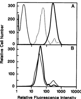
To determine whether the clinical isolates of Nef havean effect on surface CD4 levels, Nef-expressing HuT-78 cell
populations were stained for surface CD4 expression and
analyzed by flow cytometry. Cells expressing the SF2 Nef showed a significant decrease in surface CD4 levels
com-paredwith cellstransduced with the controlvectorLN(Fig. 3A). HuT-78cells transduced with virus from twodifferent LnefSN.233 producer lines and expressing the 233 Nef
protein showed a significant decrease in cell surface CD4
expression (Fig. 3B). Similarly, HuT-78 cells transduced with virus fromtwodifferent LnefSN.248producerlines and
expressing the 248 Nef protein also showed a significant
decrease in CD4 expression (Fig. 3C). In both cases, the reduction in cell surface CD4expressionwassimilartothat inducedby SF2 Nef(Fig. 3A). Thus, the Nefproteinsfrom twodifferentclinicalisolates of HIV have thesameabilityas SF2 Nefto suppress surface CD4 expression in human T cells. These results indicate that theabilityto suppressCD4
expression is not an artifact developed during passage in vitro but isanintrinsic feature of HIV-1 Nef.
Suppression of CD4 expressionin mouse T cellsby clinical isolates of Nef. Recently we have shown that SF2 Nef suppressestheexpressionofmouseCD4(13), indicatingthat theeffect of Nefon CD4isnotspecies specific.As afurther means ofanalyzing the functionalcapability of the clinical isolates ofNef,we transduced amouse T-cell line,
AKR1-Gl, with LnefSN.233 and LnefSN.248. For comparison,
these cellswerealsotransduced inparallelwithLnefSN.SF2
andacontrolvectorwhich doesnotconfer Nefexpression.
To facilitate ourexperiments, NP-40
lysates
oftransduced AKR1-G1 cellswereusedto screendirectlyfor Nef expres-sion. As shown inFig. 4, Westernblotanalysisoflysates
of AKR1-G1 cells reveals the presence of theSF2, 233, and 248 Nef proteins in the appropriate cell lines but not in cells transduced with the controlvector. Omitting theimmuno-precipitation step also eliminated the nonspecific contami-nantband seen in Fig. 2. Therelative levels of expression
andmobilities of the different Nefproteinswerethesame as those observed in the HuT-78 cells, indicating similar
pro-cessingof theseproteins in both human andmouse cells. The AKR1-G1 cells expressing Nef were stained with
PE-conjugated antibodies to mouse CD4 and analyzed by
flow cytometry. As shown in Fig. 5, expressionof the 233 and 248 Nef
proteins
in the AKR1-G1 cells alsocorrelateswith a decrease in cell surface expression ofmouse CD4. ThedecreaseinCD4expressioninducedby233Nef and248 Nef in themouse cellswas similarto the decrease induced
bythe SF2 Nef. These results demonstrate that the clinical isolates of Nefare fullycapable of
suppressing
cell surface CD4expressioninaspecies-independentmanner, asis Nef from theSF2 laboratorystrain of HIV-1.Effect of a consensus nef gene on CD4 expression. By comparing54HIV-1
nef
sequences fromnonculturedpatient
isolates,a consensusnefsequencewasderived
(49).
Inorderon November 9, 2019 by guest
http://jvi.asm.org/
4926 ANDERSON ETAL.
A.
HIV- 1 Provirus
vj,t
I
env1
I|
tat=/
env
ut5'LXSN
I2 roundsof Nested-Primer PCRAmplification and Cloning
net
I PCRre-amplification of selected clones
usingMut5'LXSN and 3'LXSNprimers
,
E.c..RI
_ MGGKWSKRSVSG...
Rb.-net
LgaI
...EYYKNC*I
B.
[ ~ ~ ~~~noopA
pLXSN
ZOoRI
pA
net noo
pLnefSN L s
[image:4.612.143.473.79.539.2]Rib.
FIG. 1. Construction of LnefSN.233 and LnefSN.248 retrovirus vectors. (A) Genome organization of the HIV-1 provirus, PCR amplification,andcloningstrategy.DNAfragmentscontainingthenefgeneandflankingsequenceswithinenv,tat,rev,and the 3' LTRwere
amplified by nested-primer PCR amplification of purified cellular DNAs (see Materials and Methods). The DNAs were prepared from uncultured peripheral blood mononuclear cells obtained from patients 233 and 248. After cloning and sequencing, the nefgeneswere
reamplified with primersMut5'LXSN and 3'LXSNto generatenef-containing PCR products that encode the "Kozak sequence"upstream of theinitiatorcodon and lackflankingviralsequences.AlsoshownarethepredictedNef amino acidsequencesencodedbytheamplification primers (28).(B) Diagram ofthe retrovirusvectorsLXSN and LnefSN. Aftersubcloning the nefgenesinto thepT7BlueT-vector forsequence
determination(not shown), the nefgenes werecloned into the retroviralexpressionvectorpLXSNatthe EcoRIsite. Thesevectorsexpress
the nefgenes from the Moloney murine leukemia virus LTR and the neo gene from the simian virus 40 early promoter (SV). Rib, ribosome-binding site; pA, polyadenylation site.
todetermine whether the suppression of CD4 isa common
propertyofnefgenesingeneral,wetestedthe ability of this consensusNeftosuppresscell surface CD4expression. The
consensus nef gene was cloned in both the sense and
antisense orientations into the retrovirus vector pLXSN (LnefSN.consandLfenSN.cons, respectively). These plas-mids were used to establish retrovirus producer lines as
described above. Pools of PE501 cells transfected with
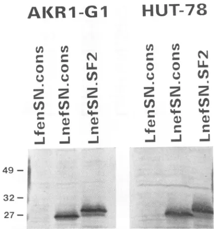
LnefSN.cons and selected in G418were found to produce Nef,while cells transfected with LfenSN.cons didnot(not shown).Allclones of PA317 cells transduced with LnefSN.
consthatwerescreened forproductionof theconsensusNef proteinalsowerepositive (not shown).
LNefSN.cons andLfenSN.cons thenwereusedto trans-duceHuT-78 andAKR1-G1 cells asdescribed above. Bulk populationsofcellswereanalyzedfor Nef andCD4
expres-J.VIROL.
£QRI
A-3'LX5
on November 9, 2019 by guest
http://jvi.asm.org/
PRIMARY ISOLATES OF HIV-1 Nef SUPPRESS CD4 EXPRESSION
HIV-1
Nef
r
N
r
N
2
L/
N
N
N
N
Xi
0
N
NI
N
N
0
C.o
32-
~~~~NE
F
27-FIG. 2. NefexpressioninHuT-78 cells transduced withLnefSN. Lysatesfrom HuT-78 cells transduced with the control vector,LN,
or the indicated LnefSN vectors were immunoprecipitated with rabbitanti-Nef antiserum and analyzed byWestern blotting. The blotswereprobedwith thesameanti-Nefantiserumanddeveloped with alkaline phosphatase-conjugated anti-rabbit IgG(Fc specific) Each lane contains precipitated material from 2 x 10 to3 x 101
cells. Thepositionsofprestained molecular size markersare indi-catedatthe left(inkilodaltons).Arrowsindicate thepositionsof the differentNefspecies.The dark bandjustabove the49-kDa marker is theheavychain from the rabbitIgGused in the immunoprecipi-tation.An unidentified band ofapproximately30 kDaappearsas a
non-specificcontaminant inimmunoprecipitates.
200
100
0 .0
E
_
z
0
0 0
0
200
100
0
200
100
0
1 10 *100 1000 10000
Relative FluorescenceIntensity
FIG. 3. Surface CD4 expression in transduced HuT-78 cells. Representative populationsofcells were stained with a PE-conju-gated anti-human CD4 monoclonal antibody or isotype-matched
controlantibodyandanalyzedbyflowcytometry. Dataarepresented
assingle-color histogramswith PE fluorescence (CD4 expression) along thex axis and relative cell number along they axis. (A) HuT-78/LN (control cells), heavy solid line; HuT-78/LnefSN.SF2,
dotted line; HuT-78/LN with controlantibody, thin solid line. (B) HuT-78 cells transduced with two different LnefSN.233 producer clones. (C)HuT-78 cells transduced withtwodifferentLnefSN.248 producerclones.Allsampleswereanalyzedinthesameexperiment, and the dataareshown in threeseparatepanelsforvisualclarity.
N
X
00
LL
m
(N (N
222
0
cn
0
w 0
~mi -J ~J
49-
32-27-
~
FIG. 4. Nef expression in transduced AKR1-G1 murine thy-momacel!s. The 3% NP-40 lysates from AKR1-G1 cells transduced withacontrol antisenseNef vector(LfenSN.cons)ortheindicated Nefexpressionvectors wereloadeddirectlyonto a12% polyacryl-amide-SDS gel, transferred to nitrocellulose, and analyzed by Western blottingasdescribed inthelegendtoFig.2.Thepositions ofprestained molecular size markers are indicated at the left (in kilodaltons). Each lane contains lysate from 106cells.
300 A
200
E 100
z
*300 -B
cr200 100 0
1 10 io100 00 10000
RelativeFluorescenceIntensity
FIG. 5. SurfaceCD4expressionintransduced murinethymoma cells.Representative populationsofNef-expressing AKR1-G1cells (Fig. 4)werestainedwithaPE-conjugated anti-mouse CD4
mono-clonalantibodyorisotype-matched controlantibody andanalyzed
byflow cytometry.Cells transduced withacontrolantisensevector (LfenSN.cons) were analyzed in parallel. Data are presented as
single-color histograms with PE fluorescence (CD4 expression) along thex axis and relative cell number along they axis. (A) AKRl-G1/LfenSN.cons, heavysolidline;AKR1-Gl/LnefSN.SF2, dotted line; AKR1-G1/LfenSN.cons with control antibody, thin solid line. (B)AKR1-Gl/LnefSN.233,heavysolidline;AKR1-Gl/ LnefSN.248,thin solid line. Allsampleswereanalyzedinthesame
experiment,and the dataareshown intwoseparatepanelsforvisual clarity.
49 1
A
I
IE'~~
I
I~~C
VOL.67, 1993 4927
A.mmgo..
on November 9, 2019 by guest
http://jvi.asm.org/
[image:5.612.59.295.73.252.2] [image:5.612.348.516.77.294.2] [image:5.612.104.246.379.614.2] [image:5.612.366.505.428.597.2]4928 ANDERSON ET AL.
HUT-78
300200
0 0
u
.
4-N
2 2
fin
en
[image:6.612.72.289.69.300.2]a
0j
eFIG. 6. Expression ofa consensus Nef protein in human and mouseT-cell lines. HuT-78 and AKR1-G1 cells transduced with the consensus nef gene in the sense (LnefSN.cons) and antisense (LfenSN.cons) orientations were analyzed for Nef expression by
Westernblotting withanti-Nef antiserumasdescribed in the legend
to Fig. 4. Lysates from cells transduced with LnefSN.SF2 are
includedfor comparison.The positions of prestained molecular size markersareindicatedatthe left(inkilodaltons). Each lane contains lysate from 106 cells.
sion following selection in medium containing G418. As shown in Fig. 6, both the HuT-78 and AKR1-G1 cells transduced withLnefSN.consexpresslevels ofthe
consen-sus Nef protein similar to those in cells transduced with LnefSN.SF2. Interestingly, the consensus Nefhas afaster mobilityinSDS-PAGE than the SF2 Nef, similar tothat of the233Nef and248 Nefisolates. Likethe 233 and 248 Nef proteins,theconsensusNefhas fourfewer amino acids than SF2 Nef andalso hasanalanine residueatposition 54, which togethercouldbe responsible forthedifference in mobility. To test whether the expression of the consensus Nef protein also correlated with a decrease in surface CD4 expression, the transducedHuT-78 and AKR1-G1 cellswere
stainedfor humanor mouse CD4 expression, respectively, and analyzed by flow cytometry. As shown in Fig. 7, the
consensus Nefprotein suppresses CD4 expression in both
human(Fig. 7A)andmouse(Fig. 7B) T-cellpopulations, and thedegreeofsuppressionis similar tothat induced by SF2 Nef. These resultsindicatethat theabilityofNeftosuppress
cell surface CD4 expression is an intrinsic feature ofmost
Nefisolates.
DISCUSSION
In thepresent study,we have shown that nefgenesfrom
twodifferent clinicalisolates of HIV-1 induce the
suppres-sion ofsurface CD4expression in human andmouseTcells. Thisobservationestablishes the fact that primary isolatesof HIV-1havenefgenesthat encode aprotein with afunction
thusfar reported onlyfor two laboratory strains, SF2 (14) and Bru (17),whichwerepassaged invitro prior to molec-ular cloning (47, 54). Moreover, we have shown that an
L..
(D 100
E
z 0
0
L.)
0 > 300
.5..
cr200100
0
r
1 10 100 1000 100
I ~~B
10 100 1000 loC
Moo
DOO
RelativeFluorescenceIntensity
FIG. 7. Decreased surface CD4 expression in cells expressinga
consensus Nefprotein. Representative populations of cells trans-duced with theconsensusNefexpressionvectorLnefSN.conswere
stained forsurface CD4 expression and analyzedby flowcytometry
as described in the legends to Fig. 3 and 5. For controls, cells transduced withthe antisenseconsensus NefvectorLfenSN.cons
wereanalyzed in parallel. (A) HuT-78 human T cells. (B)AKR1-G1
mouseTcells. Heavy solid lines, cells transduced with LfenSN. cons; dotted lines, cells transduced with LnefSN.cons; thin solid
lines, LfenSN.cons cells stained with controlantibody.
artificially generated nefgene that expresses a consensus
Nef protein can also suppress surface CD4 expression, suggesting that this isanintrinsic feature ofmost,ifnotall, nef alleles.
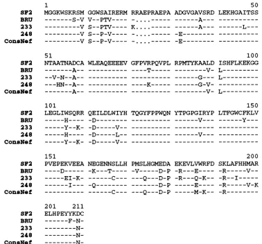
Analignment of the amino acidsequencesof theSF2,Bru, 233, and248 Nefisolates, aswell as theconsensus Nef, is
shown inFig. 8. A significant degree ofsequence identity
amongallfiveof the HIV-1 Nefproteinsis evident.Analysis
ofthese sequencesindicatesthat therearemotifs of partic-ularinterest within thissetof functional Nefproteins.Allof these Nef proteins have a common myristylation signal (M-G-G-X-X-S-) at their N termini. This modification has been showntobeimportantfor the association of Nefwith cell membranes (12, 17, 22). It has beensuggestedthat the K-G-G-L-E-G-motif(residues97to103 of theSF2Nef)has
somehomology withthenucleotide-binding domains ofthe
Ras andSrc proteins (17). Eventhoughthis motif ishighly conserved among the functional Nef proteins, there is no
conclusive evidence that Nef binds ATPorGTPorthatNef haskinaseactivity (2, 22, 37).
The threonine residue at position 15 of the Bru Nef has been shown to be a target for phosphorylation by protein kinase C(17, 18). However, it isclear fromourstudies that
this residue is not critical for the effect of Nef on CD4 expression, since only two of the five isolates have a
threonineatposition 15yetallthe Nefproteinswereable to
suppress CD4expression.
TheSF2 Nefproteinhasaslightly higherapparent molec-ularweight by SDS-PAGEthanthe233,248,andconsensus
Nef proteins (Fig.2, 4,and6).This is due in parttothefour extra amino acids found in SF2 Nef (Fig. 8). However, Obaruetal. (40) recently reported thatisolates of Nef with
an aspartic acid residue at position 54 (position 58 ofSF2 Nef) migrate as 27-kDaproteinson SDS-PAGE, whileNef isolates with an alanine at the same position migrate as
AlR
1
-G
1
N
LL
cn
0a
4-A
r.
0 0
22
49- I
.
432
-27- _
J. VIROL.
I
I
I
:I
0
so
on November 9, 2019 by guest
http://jvi.asm.org/
[image:6.612.366.509.71.260.2]1
MGGKWSKRSM
---S-V
-V_--_--- V
-V_--_--- V
-V_-_-_--V
51
NTAATNADCA
---A--
--V-N--A---
HN--A--GGWSAIRERM V--PTV----
S--PTV----S--P-V
----S--P-V
----RRAEPRAEPA ADGVGAVSRD
_-_____
---A---*K
------
--K....--- - A--- -
E---..---E
WLEAQEEEEV GFPVRPQVPL
-_______--
----T---_______--
----K---RPMTYKAALD50 LEKHGAITSS
100 ISHFLKEKGG ---V- L---
---G---
G-V- L---A-- --- --- ---V-
L---101
LEGLIWSQRR ---H---- __----Y--K-
---H----
___---y--K-151
PVEPEKVEEA
----
D--- ---EI-K-
---I---QEILDLWIYH -D--- -D---V-- -D---L--
-D---V--NEGENNSLLH -K---
T----
---C---
--- ----C---TQGYFPPWQN
PMSLHGMEDA -V---D-P ---Q---D-P
---D-P
---Q---D-P
YTPGPGIRYP
---V--
-EKEVLVWRFD -R---E---R--- Q-K--
---E---
M-K--150 LTFGWCFKLV
______y---200 SKLAFHHMAR -R---V---R--I
---R---V-K
-R---201 211
ELHPEYYKDC
---F-N--
N
- N
-
_---N-FIG. 8. Comparison of the amino acidsequencesof fivefunctional Nefproteins.The HIV-1 Nef amino acidsequencespredictedfrom the nefgenesoflaboratory isolates SF2 and Bru, clinical isolates 233 and 248,andtheconsensusnefgene werealignedwith thePileUpprogram
(9). Only amino acidsdiffering from those intheSF2 Nefareshownforthe otherproteins.
25-kDa proteins. Figure 8 shows that the 233, 248, and
consensusNefproteins all have an alanineatthisposition.
Thechangeinthisamino acid residuefromaspartic acidto alanineispredictedtohaveaneffectontheconformation of the Nefprotein (40). These observations couldexplain the differences inelectrophoretic mobilitybetween SF2 and the 233, 248, and consensus Nef proteins seen in our experi-ments. However, this potential conformational change did notaffectthe abilityof these Nefproteinsto suppress CD4 cell surface levels.
Theabilityof Nefto suppressCD4expressionatthe cell surface is of particular interest for several reasons. The humanCD4 moleculeservesasthecell surfacereceptorfor HIV(7, 25, 33). It issurprising that HIV should have two
geneswhoseproductsarecapableofsuppressingthe
expres-sion of its receptor (4, 6, 14, 17, 20). DuringHIVinfection, the envelope protein precursor gp160 is processed very
inefficientlywithin the host cell(55).Themajorityofgp160is transported to the lysosome and degraded (55). Transient-expression studies have shown that CD4 processing and expression arealso inhibited in thesecells, probablyas the result ofdirectbindingbetweengp160and CD4(6, 56).We have shown that Nef also has the ability to block CD4 expression at the cell surface (13, 14; this study). The mechanismbywhichthisoccursis unknown and iscurrently
underinvestigationinourlaboratories. Incontrast togp160, which does notbind mouse CD4 (29), Nefalso blocks the expressionofmouse CD4 (13). This result is important in view of the structural and functional similarities between
mouseand human CD4(21, 41).Moreover,the interactionof
Nef with CD4 differs from that ofgp160/gp120 in that the effect of Nefon CD4 appears to require the cytoplasmic domain of the CD4 molecule (13), while gp160 and gp120 bindtothe extracellular domain of CD4(29).
Infection with HIV is accompanied by a state ofanergy
within theCD4+ T-cellpopulation, resultingintheeventual depletionof CD4+ T cells (45). The CD4 molecule on the
surface of T cells is important for antigen-driven T-cell triggeringvia the T-cellreceptor(21, 41).CD4 interactswith class II major histocompatibility complex molecules on
antigen-presenting cells, servingasacoreceptorforantigen recognition (21, 41). Engagementof theCD4 moleculealso activates theprotein tyrosinekinasep56k,which hasbeen shown to be important for T-cell activation (16, 21, 46, 51-53). Interference with thecontribution of CD4 toproper
T-cell signalling might have significant consequences for T-cell viability and function. Indeed, it has been demon-strated for bothmouseand human T cells thatremovingCD4 from the cell surfacebycross-linkingwithanti-CD4 antibod-ies, or with gpl20 in the case of human CD4, renders the SF2
BRU 233 248 ConsNef
SF2 BRU 233 248 ConsNef
SF2 BRU 233 248 ConaNef
SF2 BRU 233 248 ConaNef
SF2 BRU 233 248 ConeNef
- -- ---L--
on November 9, 2019 by guest
http://jvi.asm.org/
[image:7.612.120.498.71.423.2]4930 ANDERSON ET AL.
cells unresponsive toT-cell receptor triggering and leadsto cell death byapoptosis(3, 38). Thus, blocking CD4 expres-sion at the cell surface couldcontribute to the depletion of
CD4+ T cells, which is the hallmark ofAIDS.
Although Nef could have deleterious effects on cell func-tion and isnotrequiredfor viralreplication invitro(11, 50), there appears to be selective pressure to maintain a func-tional nefgene in vivo (23), suggesting that it offers some
advantage to the virus. The work ofWilley et al. suggests that the rateofprocessing ofgp160depends onthe ratioof
gp160, CD4, and Vpu inside the cell (55-57). The early
expression
of Nefduring the virus lifecyclemaycontribute totheprocessing
ofgp160togp120and gp4l byinterfering with theexpression
of CD4. Such an effect mightbe more evident at low levels of virus replication. Under such cir-cumstances,thenegativeeffect of NefonCD4 surface levels could result inmoreefficientvirus replication, which corre-lates withthe results obtained invivo withSIV (23).We have shown that suppression of surface CD4 expres-sion is a common function of the Nef protein of HIV-1. While therelevance of Neftoviralpathogenesis anddisease
progressionhasbeenclearlydemonstrated(8, 23), the role of Nef-induced
suppression
ofsurface CD4 expression in thepathogenesis
ofAIDS is at present unclear. However, the fact that the abilityto suppress surface CD4 expression is conserved in both primary and laboratory isolates ofNef suggests theprobable
importanceofthisfunction ofNef.ACKNOWLEDGMENTS
We thank Lee Ratner for kindly providing the rabbit anti-Nef antiserum. Elizabeth Sidell and James Houston forassistancewith the flow cytometric analyses, and Amy Frazier for molecular cloningof thenefconstructs.We alsothankPeterDoherty, Randall Owens,and John Fosterfor critical reviewsof themanuscript.
This workwasfundedin partbygrantRO1-CA59175,grants1405 and 1559 from the American Foundation for AIDS Research (J.V.G.), CORE grant CA-21765, the American Lebanese-Syrian Associated Charities (ALSAC),Public HealthService grants U01-AI25868and RO1-AI25321 (R.S.),and NIH grant RR00046 tothe University of North Carolina General Clinical Research Center. D.C.S. was supported by a Dentist-Scientist Award from the National InstituteforDental Research (K16-DE00165).
REFERENCES
1. Ahmad,N.,and S. Venkatesan.1988.NefproteinofHIV-1isa
transcriptional repressor of HIV-1 LTR. Science 241:1481-1485.
2. Backer, J. M.,C. E.Mendola, J. L.Fairhurst,and I. Kovesdi. 1991. The HIV-1 nefprotein doesnothaveguaninenucleotide binding, GTPase,orautophosphorylating activities. AIDSRes. HumanRetroviruses7:1015-1020.
3. Banda, N. K., J. Bernier, D. K. Kurahara, R. Kurrie, N. Haigwood, R.P. Sekaly, and T. H. Finkel. 1992. Crosslinking CD4byhumanimmunodeficiencyvirusgpl20 primesTcellsfor activation-inducedapoptosis.J. Exp.Med. 176:1099-1106. 4. Bour,S.,F.Boulerice,and M. A.Wainberg.1991.Inhibitionof
gpl60andCD4 maturation in U937 cells afterbothdefective and productiveinfectionsbyhumanimmunodeficiencyvirus type 1. J. Virol. 65:6387-6396.
5. Cheng-Mayer, C.,P.Iannello,K.Shaw,P. A.Luciw, and J. A. Levy. 1989. Differential effects of Nef on HIV replication: implications for viral pathogenesis in the host. Science 246: 1629-1632.
6. Crise, B.,L.Buonocore,andJ.K.Rose.1990. CD4 is retained in the endoplasmic reticulum by the human immunodeficiency virustype 1glycoproteinprecursor. J.Virol. 64:5585-5593. 7. Dalgleish, A. G., P. C. L. Beverly, P. R. Clapham, D. H.
Crawford,M. F.Greaves,andR.A. Weiss. 1984. The CD4 (T4) antigenisanessential component of the receptor for the AIDS
retrovirus. Nature (London) 312:763-767.
8. Daniel, M. D., F. Kirchhoff, S. C. Czajak, P. K. Sehgal, and R. C. Desrosiers. 1992. Protective effects ofa live attenuated SIV vaccine with a deletion inthenef gene. Science 258:1938-1941.
9. Devereux, J., P. Haeberli, and 0. Smithies. 1984.A comprehen-sive setofsequence analysis programs for theVAX. Nucleic Acids Res. 12:387-395.
10. Dillon, P. J., and C. A. Rosen. 1990. A rapid method for the construction of synthetic genes using the polymerase chain reaction. BioTechniques9:299-300.
11. Fisher, A. G., L. Ratner, H. Mitsuya, L. M. Marselle, M. E. Harper, S. Broder, R. C. Gallo, and F. Wong-Staal. 1986. Infectious mutants of HTLV-IIIwith changes in the 3' region and markedlyreducedcytopathic effect. Science233:655-659. 12. Franchini, G.,M.Robert-Guroff, J. Ghrayeb,N. T.Chang, and
F.Wong-Staal.1986.Cytoplasmic localization of theHTLV-III 3' ORFprotein in culturedTcells. Virology155:593-599. 13. Garcia, J. V., J. Alfano,andA. D.Miller. 1993. Thenegative
effect of human immunodeficiency virus type 1 Nef on cell surface CD4 expression isnotspeciesspecific and requiresthe cytoplasmic domainofCD4.J. Virol. 67:1511-1516.
14. Garcia,J. V., and A. D. Miller. 1991. Serine phosphorylation-independent downregulationof cell-surface CD4 by Nef.Nature (London) 350:508-511.
15. Garcia, J. V.,andA. D.Miller. Submitted forpublication. 16. Glaichenhaus,N., N. Shastri, D. R. Littman, and J. M.Turner.
1991.Requirement for associationofp56Ick with CD4in antigen-specific signal transduction inTcells. Cell64:511-520. 17. Guy, B., M. P. Kieny, Y. Riviere, C. Le Peuch, K. Dott, M.
Girard, L. Montagnier, and J.-P. Lecocq. 1987. HIVF/3' orf encodes a phosphorylated GTP-bindingprotein resemblingan oncogeneproduct. Nature (London) 330:266-269.
18. Guy, B., Y. Riviere, K. Dott, A. Regnault, and M. P. Kieny. 1990. Mutational analysis of the HIV nef protein. Virology 176:413-425.
19. Hammes, S.,E.Dixon, M. Malim, B. Cullen, and W. C.Greene. 1989. Nefprotein of human immunodeficiency virus type 1: evidence against its role as a transcriptional inhibitor. Proc. Natl. Acad. Sci.USA86:9549-9553.
20. Jabbar,M.A.,and D. P.Nayak 1990. Intercellular interaction ofhuman immunodeficiency virus type 1 (ARV-2) envelope glycoprotein gpl60 with CD4 blocks the movement and matu-ration ofCD4totheplasma membrane.J. Virol.64:6297-6304. 21. Janeway,C.A., Jr. 1992. TheTcell receptor as a multicompo-nentsignalling machine:CD4/CD8 coreceptors andCD45inT cellactivation.Annu. Rev. Immunol. 10:645-674.
22. Kaminchik, J.,N.Bashan,D.Pinchasi, B. Amit, N. Sarver, M.I. Johnston,M.Fischer,Z.Yavin, M. Gorecki, and A. Panet. 1990. Expression andbiochemical characterizationofhuman immu-nodeficiencyvirustype 1nefgene product. J. Virol. 64:3447-3454.
23. Kestler, H.W.,D. J.Ringler, K. Mori, D. L. Panicali, P.K. Sehgal,M. D.Daniel,and R. C. Desrosiers. 1991. Importanceof the nef gene for maintenance of high virus loads and for developmentofAIDS. Cell65:651-662.
24. Kim, S., R. Byrn, J. Groopman, and D. Baltimore. 1989. Temporalaspects of DNA and RNA synthesis during human immunodeficiencyvirusinfection:evidence for differential gene expression.J. Virol.63:3708-3713.
25. Kim, S.,K.Ikeuchi,R.Byrn, J. Groopman, and D. Baltimore. 1989. Lack ofanegative influenceon viral growth by the nef gene of human immunodeficiency virus type 1. Proc. Natl. Acad.Sci. USA86:9544-9548.
26. Kiatzman, D., E. Champagne, S. Chamaret, J. Gruest, D. Guetard,T.Hercend, J.C.Gluckman, and L. Montagnier. 1984. T-lymphocyteT4moleculebehaves as the receptor for human retrovirusLAV.Nature (London)312:767-768.
27. Klotman,M.E., S. Kim, A. Buchbinder, A. DeRossi, D. Balti-more, and F. Wong-Staal. 1991. Kinetics of expression of multiply splicedRNAin earlyhuman immunodeficiency virus type 1 infection oflymphocytes and monocytes. Proc. Natl. Acad.Sci.USA88:5011-5015.
J. VIROL.
on November 9, 2019 by guest
http://jvi.asm.org/
28. Kozak, M. 1987. At least six nucleotides preceding theAUG initiatorcodonenhancetranslation in mammalian cells. J.Mol. Biol. 196:947-950.
29. Landau, N. R., M. Warton, and D. R. Littman. 1988. The envelope glycoprotein of the human immunodeficiency virus binds to theimmunoglobulin-like domain of CD4. Nature (Lon-don) 334:159-162.
30. Lane, H.C.,andA.S.Fauci.1985.Immunologic abnormalities in theacquired immunodeficiency syndrome. Annu. Rev. Im-munol.3:477-500.
31. Luciw, P. A.,C. Cheng-Mayer, and J. A.Levy.1987.Mutational analysis of thehumanimmunodeficiencyvirus: theorf-b region down-regulates virusreplication. Proc. Natl. Acad. Sci. USA 84:1434-1438.
32. Maitra, R. K., N. Ahmad, S. M. Holland, and S. Venkatesan. 1991. Human immunodeficiencyvirus type 1(HIV-1)provirus expression and LTRtranscriptionarerepressed in nef-express-ingcell lines. Virology 182:522-533.
33. McDougal, J. S., A. Mawle, S. P. Cort, J. K. A. Nicholson, G. D. Cross, J. A. Sheppler-Campbell, D. Hicks, and J. Sligh. 1985. Cellular tropism of the human retrovirus HTLV-III/LAV. I. Role ofTcell activation and expression of the T4 antigen. J. Immunol. 135:3151-3162.
34. Meyerhans, A., R. Cheynier, J. Albert, M. Seth, J. Kwok, J. Sninsky, L. Morfeldt-Mason, B. Asjo, and S. Wain-Hobson. 1989.Temporal fluctuations inHIVquasispecies in vivoarenot reflected bysequentialHIVisolations. Cell 58:901-910. 35. Miller,A.D., andG.Rosman.1989.Improved retroviralvectors
forgenetransfer andexpression. BioTechniques 7:980-990. 36. Munis, J. R., R. S. Kornbluth, J. C. Guatelli, and D. D.
Richman. 1992. Ordered appearance of human immunodefi-ciency virus type 1 nucleic acids following high multiplicity infectionof macrophages. J. Gen.Virol. 73:1899-1906. 37. Nebreda, A.R., T. Bryan, F. Segade, P.Wingfield, S.
Venkate-san, andE. Santos. 1991. Biochemical andbiological compari-sonofHIV-1nef andrasgeneproducts.Virology183:151-159. 38. Newell, M. K., L. J. Haughn,C. R. Maroun, and M. H. Julius. 1990. Deathofmature Tcellsbyseparateligation ofCD4and the T-cellreceptorforantigen.Nature(London) 347:286-289. 39. Niederman, T. M.J., B. J. Thielan, and L. Ratner.1989.Human
immunodeficiency virus type 1 negative factor is a transcrip-tionalsilencer.Proc.Natl. Acad. Sci. USA86:1128-1132. 40. Obaru, K., J. E. deClue, and W. A. Haseltine. 1992. An
amino-terminal amino acid affects theelectrophoretic mobility of theHIV-1Nefprotein.J.AcquiredImmuneDefic.Syndrome 5:308-312.
41. Parnes, J. R.1989.Molecularbiology and functionof CD4 and CD8.Adv.Immunol. 44:265-311.
42. Paviakis,G.N., and B. K. Felber. 1990. Regulationof expres-sion ofhumanimmunodeficiency virus.NewBiol.2:20-31. 43. Ratner, L., B. Starcich,S.F.Josephs, B. H. Hahn, E. P. Reddy,
K.J.Livak, S.R.Petteway, Jr., M.L. Pearson, W. A. Haseltine, S. K.Arya, and F.Wong-Staal. 1985. Polymorphism ofthe 3' openreading frame of the virus associated with the acquired immunodeficiency syndrome, humanT-lymphotropicvirus type III. NucleicAcidsRes.13:8219-8229.
44. Robert-Guroff,M.,M.Popovic,S.Gartner,P.Markham, R.C. Gallo,andM.S. Reitz.1990. Structure andexpression oftat-, rev-, andnef-specific transcripts of human immunodeficiency virus type 1in infectedlymphocytesandmacrophages. J. Virol. 64:3391-3398.
45. Rosenberg, Z. F., and A.S. Fauci.1992.Immunopathogenesis of HIV infection,p.61-76. InV. T.DeVita, S.Hellman, and S.A. Rosenberg (ed.), AIDS: etiology, diagnosis, treatment, and prevention, 3rd ed.J. B. Lippincott Co.,Philadelphia. 46. Rudd, C. E., J. M.Trevillyan,J. D.Dasgupta, L. L. Wong, and
S. F. Schlossman. 1988. The CD4 receptor is complexed in detergent lysates to aprotein-tyrosine kinase (pp58) from hu-manTlymphocytes.Proc. Natl.Acad. Sci. USA85:5190-5194. 47. Sanchez-Pescador, R., M. D. Power, P. J. Barr, K. S. Steimer, M. M.Stempien, S.L.Brown-Shimer, W. W.Gee,A.Renard,A. Randolph,J. A. Levy, D. Dina, and P. Luciw.1985. Nucleotide sequence and expression of an AIDS-associated retrovirus (ARV-2). Science 227:484 492.
48. Sanger, F., S. Nicklen, and A. R.Coulson.1977. DNA sequenc-ing with chain-terminating inhibitors. Proc. Natl. Acad. Sci. USA74:5463-5467.
49. Shugars,D.C.,M.S.Smith,D. H.Glueck,P.V.Nantermet,F. Sellier-Moiseiwitsch, and R. Swanstrom. 1993. Analysis of hu-manimmunodeficiencyvirus type 1nefgene sequences present invivo. J.Virol.67:4639-4650.
50. Terwilliger,E., J. G. Sodroski,C. A.Rosen, and W. A. Hasel-tine. 1986. Effectsofmutations within the 3' orfopen reading frame region of human T-cell lymphotropic virus type III (HTLV-III/LAV)onreplication andcytopathogenicity. J. Virol. 60:754-760.
51. Veillette, A.,M. A. Bookman, E. M. Horak,andJ.B. Bolen. 1988. The CD4 and CD8Tcellsurfaceantigensareassociated with theinternal membranetyrosine-protein kinase p56lck. Cell 55:301-308.
52. Veillette,A., M. A. Bookman, E. M. Horak, L. E.Samelson,and J. B. Bolen.1989.Signal transductionthrough the CD4receptor involves the activation of the internal membrane tyrosine-proteinkinasepS6lck.Nature(London) 338:257-259.
53.
Veiliette,
A., J. C. Zuniga-Pflucker, J. B. Bolen, and A. M. Kruisbeek 1989. Engagement of CD4 and CD8 expressed onimmature thymocytes induces activation of intracellular ty-rosinephosphorylation pathways. J. Exp. Med. 170:1671-1680. 54. Wain-Hobson, S., P. Sonigo, 0. Danos, C. Stewart, and M. Alizon. 1985. Nucleotide sequence ofthe AIDS virus, LAV. Cell40:9-17.
55. Willey, R. L., J. S.Bonafacino,B.J.Potts, M. A. Martin, and R. D.Klausner. 1988. Biosynthesis, cleavage, anddegradation of the humanimmunodeficiency virus1envelopeglycoprotein gpl60.Proc.Natl. Acad.Sci. USA85:9580-9584.
56. Willey,R.L.,F.Maldarelli,M. A.Martin,and K. Strebel. 1992. Human immunodeficiency virus type 1Vpu protein regulates the formation of intracellular gpl6O-CD4complexes. J. Virol. 66:226-234.
57. Willey, R.L.,F.Maldarelli,M.A.Martin,and K.Strebel. 1992. Human immunodeficiency virus type 1 Vpu protein induces rapiddegradation ofCD4. J.Virol. 66:7193-7200.