Copyright C) 1994, American Society forMicrobiology
Human
Immunodeficiency Virus Type
1
Nef-Induced
Down-Modulation of CD4 Is Due to
Rapid
Internalization and
Degradation
of
Surface CD4
SUNG S. RHEE* ANDJON W. MARSH
Laboratory of Molecular Biology, National Institute of MentalHealth, Bethesda, Maryland 20892 Received 1 March 1994/Accepted 18 May 1994
Human immunodeficiency virus type 1 (HIV-1) Nef is a myristylated protein with a relative molecular mass of 27 kDa, is localized to the cytoplasmic surfaces of cellular membranes, and has been reported to down-modulate CD4 in human T cells. To understand themechanismof HIV-1Nef-mediated down-modulation ofcell surface CD4, we expressed Nef protein in human T-cell line VB. Expression of HIV-1 Nefprotein down-modulatedsurface CD4 molecules. In pulse-chase experiments, CD4molecules inNef-expressingcells were synthesizedat normallevels. However, the bulkofnewlysynthesized CD4 proteinwas degradedwith a half-life of approximately 6 h, comparedwiththe24-hhalf-lifein controlcells. ThisNef-inducedacceleration ofCD4turnover was inhibited bylysosomotropic agents NH4Cl and chloroquineaswell asby the protease inhibitor leupeptin. Surface CD4 biotinylation experiments demonstrated that CD4 molecules in Nef-expressing T cells are transported to the plasma membrane with normal kinetics but are then rapidly internalized. Therefore, HIV-1 Nef-induceddown-modulation of CD4isduetorapidinternalization of surface CD4 and subsequent degradation by an acid-dependent process, potentially lysosomal. Additionally, in a Nef-expressing cell, we find accelerated dissociation ofthe T-celltyrosine kinase
p561k"
andCD4butonlyafter thecomplex reaches the plasma membrane. Thisimpliesthat HIV-1Nefprotein might playarole intriggering aseries ofT-cellactivation-like events,which contribute top561ckdissociation and internalization of surface CD4molecules.Human immunodeficiency virus type 1 (HIV-1), the etio-logic agent of AIDS, possesses at least six genes coding for regulatory proteins in addition to the typical retroviral genes
(gag,pol, and env). In the past decade, these gene products have beenintensively studied to understand their rolesduring HIV-1 infection. However,the precise function of these regu-latory proteins in HIV-1 replication and pathogenesis in virus-infected individuals remains unclear, even though our understandinghasgreatly increased.
One of theregulatory genes, thenefgene,wasfirst identified as an open reading frame of 648 bp near the 3' end of the genome, partially overlapping the U3 region (49, 64). It encodesanapproximately 27-kDa protein covalently modified with the amino-terminal myristic acid from multiply spliced mRNAtranscripts (4, 18). After viral infection, Nef proteins are expressed from the most abundant, multiply spliced
mRNA, up to80% of the total early viral transcripts, andare found predominantly in the cytoplasm associated with the plasma membrane (52).Invivo expression of Nef proteinwas supported by detection of antibodies specifictothisprotein in HIV-1- and -2-infected individuals and simian immunodefi-ciency virus (SIV)-infected monkeys (5, 48).
The open reading frame of the nef gene is well retained amongallprimate lentiviruses (HIV-1, HIV-2, and SIV) with highly conserved amino acid sequences, although amino acid variationswithanin-frame termination codon can be as high as 17%inlaboratory isolates of HIV-1 (4, 14, 50). Blumberg et al.
(9), using targeted PCR amplification and DNA sequencing, showed that the nef gene is normally open in clones derived
*Correspondingauthor. Presentaddress: Laboratory of Molecular
Virology, Samsung Biomedical Research Institute, Ilwon-Dong 50, Kangnam-Ku, Seoul,Korea 135-230.
directly from pathologic brain and spleen tissues of HIV-1-infected children and adults. This selective pressure for the functionalnefgenesuggestedthatHIV-1 Nefproteinmayplay
an important roleduring invivoinfection, ashas been noted with the SIV systemby Kestleretal. (24).Inthatstudy, itwas
suggestedthat thenefgeneproductofSIVmac functionsas a
positive effector that is required for a high viral load and diseaseprogressionininfected macaques. Furthermore,it has also been demonstrated thatHIV-1Nefproteinin acell model with primary lymphocytes and macrophages plays a positive role inpromoting viral infection and replication (15, 41, 59).In contrast, in transformed human T-cell lines, Nefprotein did not haveapositive effectonviralreplication (19, 25).
Inadditiontothe in vivobiological function, many biochem-ical activities have been ascribed toNefprotein. The earliest studies suggested that HIV-1 Nef proteins function as a negative regulatory factor by repressing long terminal repeat-driven transcription (2, 32, 34, 43). However, subsequent studiesbyKimet al. (25)and Hammes et al.(19)have failed to support these findings. Additionally, Nef protein was de-scribed as bindingGTP and possessing GTPase activity (18),
which was also subsequently refuted (23, 37, 42). Reproduc-ible, somewhat lessconflictingin vitro studies have shown that HIV-1 and SIV Nefproteins down-regulate the levels of cell surface CD4 in human T-cell lines, such as CEM, HPB-All,
andHUT78,aswellasinthemonocyticcell lineU937 (7, 17, 35, 60). Furthermore, murineand simian CD4 molecules on the cell surfaces of murine T-cell lines (16) aswellasmurine CD4 ofT cells fromtransgenic mice (57) were shown to be down-modulatedbyHIV-1 Nefexpression.These results sug-gested that Nefproteins utilize a mechanism ofaction com-monly shared among different species, although the precise
mechanism remainsundefined. 5156
on November 9, 2019 by guest
http://jvi.asm.org/
The CD4 molecule is a glycoprotein of approximately 55 kDa that consists of extracellular domains homologous to immunoglobulin variable and joining regions, a hydrophobic transmembrane domain, and a highly charged cytoplasmic domain.Thecytoplasmic domain of human CD4, consisting of 38amino acids, is highly conserved among human, murine, and rat CD4 molecules. To better understand the mechanism of HIV-1 Nef-mediated down-modulation of this molecule, hu-manCD4+ T-cell line VB was stably transduced with
ampho-tropicretroviral vectors designed to express HIV-1 Nef. In this report,wedemonstrate that the kinetics of CD4 synthesis and transport to the plasma membrane are unaltered by the presence of Nef. Nef-mediated CD4 down-modulation is found tobe due to rapid internalization of surface CD4 and
subsequentdegradation in an intracellular lysosomal compart-ment. Furthermore, through surface labeling of CD4 mole-cules and assessment of p561Ck association, we show that the Nef-mediated process leading to CD4 down-modulation in-cludes accelerated dissociation of theCD4-p56ck complex and that this dissociation is initiated after the complex reaches the
plasma membrane.
MATERMILS AND METHODS
DNAs andcells. To express HIV-1 Nef protein in human T
cells, a 1.0-kbp nef genefragment of HIV-1 SF2 clone (kindly
provided by Jay A. Levy, University of California, San
Fran-cisco)wascloned into the murine retrovirus expression vector
pLXSN(40). The resulting plasmid was designated pNEF-SF2. Thecontrol plasmidpFEN-SF2 was obtained by inserting the
fragment in an antisense orientation.
BothplasmidDNAs werelinearized and transfected into the
amphotropic retrovirus packaging cell line PA317 (ATCC CRL 9078) by the modified calcium phosphate precipitation method described by Chen and Okayama (11).
Vector-express-ing PA317cell lines were selected in medium containing 0.75 mgof the antibiotic G418 per ml, and infectious amphotropic vector-containing culture media were used to infect human T-cell lymphoma VB (the AIDS Research and Reference
Reagent Program, Division of AIDS, National Institute of
AllergyandInfectiousDiseases, National Institutes of Health) in the presence of 4.0
pLg
of polybrene per ml.Vector-expressing
VBcell lines were grown in medium containing 1.0 mgofG418 per ml and weredesignatedVBCON
and VBNEF.Analysis ofsurface CD4 expression. Flow cytometric analy-sis
(fluorescence-activated
cell sorter [FACS] analysis) wasperformed
on a Becton Dickinson FACScan with Lysis II software to assess CD4 expression on cell surfaces. Half a million exponentially growing cells were labeled withfluores-cein-conjugated anti-CD4 antibody (Olympus
Immunochemi-cals, Lake Success, N.Y.). Debris and nonviable cells were
gated out byforward scatter and the addition of propodium
iodide, respectively.
Radiolabeling,
immunoprecipitation,
and gel electrophore-sis analysis. Exponentially growing cells were starved for 30 min in cysteine- and methionine-free RPMI 1640 (ICNBio-medicals, Costa Mesa, Calif.) and then pulse-labeled for 30 min inlabeling mediumcontaining 3S-labeled methionine and
cysteine (0.1 mCi/ml, 1,000 Ci/mmol) (Tran35S-label; ICN
Biomedicals). Pulse-labeled cells were washed in complete
growthmedium andaliquoted for the chase. Immediately after
labeling orthe chase, individual cell aliquots were washed in coldphosphate-bufferedsaline(PBS) (pH 7.4) and then lysed in PBS containing 0.1% sodium dodecyl sulfate (SDS), 0.5%
deoxycholate, 1.0% Triton X-100, 1 ,ug ofleupeptin per ml, 1
,ug
ofaprotinin
perml,
and 1 mMphenylmethylsulfonyl
fluoride (RIPA buffer).Radiolabeled CD4, Nef protein, and
p561ck
were immuno-precipitated with rabbit anti-CD4 antibody (American Bio-technology, Cambridge, Mass.), rabbit anti-Nef antibody (theAIDS Research and Reference Reagent Program 331, Divi-sion of AIDS, National Institute of Allergy and Infectious Diseases), and rabbit
anti-p56lck
antibody (a gift from Mark Egerton, National Institutes of Health, Bethesda, Md.), re-spectively. The immune complex was precipitated with fixed Staphylococcus aureus cells and then separated by SDS-poly-acrylamide gel electrophoresis (SDS-PAGE) as described pre-viously (51).Radioactivity incorporated into protein was quantified by exposing the dried gel to a storage phosphor screen and scanning the screen with a Fuji
Bas2000.
Cell surface CD4 biotinylation. Biotinylation of cell surface
CD4
was carried out with sulfo-NHS-biotin (Pierce Chemical Co., Rockford,Ill.)
as described previously with some modifi-cations (31). Briefly,VBCON
and VBNEF cells were pulse-labeled for 30min
and chased for 1, 2, and 4 h in complete growth medium as described above. Cells were pelleted, washed once in ice-cold PBS supplemented with 1 mMMgCl2
and 0.1 mM CaCl2 (PBS-plus), and resuspended in freshly prepared 1 mg of sulfo-NHS-biotin per ml in PBS-plus at107
cells per ml. After binding for 1 h at4°C
with constant gentle agitation, cells were collected by centrifugation and washed once in serum-free RPMI 1640 and twice in PBS-plus. Total radiolabeled CD4 molecules were precipitated with rabbit anti-CD4 antiserum, and then biotinylated surface CD4 pro-teins were reprecipitated with streptavidin-linked magnetic beads (Dynabeads; Bioproducts for Science, Indianapolis,Ind.).
Coimmunoprecipitation of
p56Ic"
with surface CD4. To detectp561Ck
associated with cell surface CD4, cells were pulse-chased and incubated in serum-free RPMI 1640 with anti-CD4 monoclonal antibody (HT3.10-28, kindly provided by Peter Rieber, University of Munich, Munich, Germany) for 1 h at4°C
with constant gentle agitation as described above. Cells were lysed in NTNE buffer (1% Nonidet P-40, 20 mM Tris hydrochloride [pH 8.4], 0.15 M NaCl, 2.0 mM EDTA, 1 mM phenylmethylsulfonyl fluoride) on ice, and then excess unlabeled cell lysates were added to the lysates. CD4 com-plexes (CD4 andCD4-associated
proteins) coupled with anti-body were precipitated with protein G-agarose (Genex Corp., Gaithersburg, Md.). The immune precipitates were divided in half and reimmunoprecipitated with fixed S. aureus cells cou-pled with either anti-CD4 antibody or anti-p56 ck antibody.RESULTS
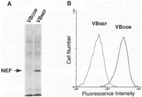
Expression of HIV-1 Nef protein in human T cells. To understand the mechanism by which HIV-1 Nef proteins down-regulate the expression of cell surface CD4, the nef gene fragment was obtained from HIV-1 isolate
SF2
and stably transduced into human T-cell lymphomaVB
with an ampho-tropic retroviral vector as described in Materials and Methods. Two G418-resistant cell populations, expressing either the nef gene-containing vectorpNEF-SF2
or the control vectorpFEN-SF2,
were designated VBNEF andVBCON,
respectively. In order to determine whether Nef protein is synthesized in VBNEF cells, cellular proteins were radiolabeled with [35S]me-thionine-cysteine for 30min
and Nef protein was immunopre-cipitated with rabbit anti-Nef antibody (Fig. IA). A band of approximately 27 kDa, corresponding to Nef protein, could be seen only in the lysates of VBNEF cells, confirming expressionon November 9, 2019 by guest
http://jvi.asm.org/
B
VBNEF
VBCON
E 01
Fluorescence Intensity
FIG. 1. Nef expression and surface CD4down-modulation in Nef-expressing VB cells. To express HIV-1 Nef protein in human T-cell lineVB,cellswerestablytransduced with theHIV-1 SF2nefgeneby using the murine retrovirus expressionvectorpLXSN. (A)In orderto
determine whether Nef proteinwassynthesized,cell linesVBCON and
VBNEF, expressing only theneo gene orboth thenef andneo genes,
respectively, were pulse-labeled for 30 min with
[35S]methionine-cysteine (100
,uCi/ml).
Nef protein was immunoprecipitated withrabbit anti-Nef antiserum and then separated by SDS-PAGE (10% polyacrylamide). InVBNEF cells,abandcorrespondingtoNefprotein
with a relative molecular massof 27 kDa is visible (indicated by an
arrow) while noband is detected in VBCONcells. (B) Surface CD4
down-modulation in VBNEF cellswas determined by FACS analysis.
Cellsweredirectly stained withfluorescein-conjugated CD4 anti-body and then analyzed with aFACScan. A significant reduction in
fluorescence intensity in VBNEF cells with 20 to30% of surface CD4 labeling is observed, compared with that inVBCONcells.
of the 27-kDa HIV-1 Nef protein in VB cells. Pulse-labeled cellswerefurther chasedfor 2to24h (datanotshown). In VB cells, Nef protein isefficientlysynthesizedandverystable, with a half-lifeofapproximately 24 h.
Nef protein has been shown to down-modulate surface expression of CD4 in human T-cell lines. The human T-cell lineVBexpresses high levels of CD4oncellsurfaces and has
beenreportedtobesusceptibletoHIV-1 infection inamanner
that approximates infection in nontransformed human T help-er/inducer cells(29).Itwas,therefore, of interesttodetermine whether Nef protein is functional in VB cells. Cells were
directly stained with fluorescein-conjugated anti-CD4
mono-clonal antibody and analyzed with a FACScan (Fig.
1B)
asdescribed in Materials and Methods. In VBNEF cells, CD4 surface expression is also down-modulated; there is a 70 to
80% drop in fluorescence intensity compared with that of
VBCON
cells. The effect of NefonCD4 inVBcells is specificsincenosignificant reduction in surface expression of CD3 and
HLA-ABCwasobserved (datanotshown).
In Nef-expressing cells, CD4 protein is synthesized at normal levels but degraded rapidly. Previously, Nef protein has been reportedtohavenoeffectonthe steady-statelevelsof
CD4mRNA and CD4 protein (7, 17). However, it isnotclear whether CD4 proteins aresynthesized and turned over under
normalkinetics inNef-expressing cells. Therefore,weanalyzed
the fates of newly synthesized CD4 molecules
by
apulse-chaseexperiment. Cells were pulse-labeled with[3
S]methionine-cysteine for 30
min
andchased for 2, 4, 6, and 24 h. After eachtime point, cells were lysed and then divided in half to
precipitate CD4 and p561Ck with either anti-CD4or
anti-p561ck
antisera, respectively. The results of this experiment areshown
in Fig. 2. Since
p561ck
is stable with a half-life of 32 h andis not modulated in Nef-expressing cells(Fig. 2B),p561ckwas usedas an internal control in this experiment. Quantifying and com-paring the amounts of CD4 andp56lck
at each time pointbyphosphor imaging demonstrated that at the end of the pulse both cell populationshad synthesized equivalent levels of CD4 protein (Fig. 2A, lanes 0).
In control
(VBCON)
cells, no significant proportion of pulse-labeled CD4 proteins were lost during the 6-h chase (Fig. 2A, lanes 0 to 6) and after the 24-h chase, approximately 50%remained (lane 24). Thus, CD4 protein has a half-life of approximately 1 day. In contrast, in VBNEF cells, CD4 degra-dation became evident following a 2-h lag after thepulse (Fig.
2A, lanes 0 and 2) and after that the loss of protein is well described by a single exponential function with anapproximate 6-h half-life (lanes 4 to 24).
These results suggest that Nef expression in VB cellstriggers a rapid degradation of CD4 molecules, which results in down-modulation of CD4 surface expression on Nef-expressing T cells. However, it is important to rule out the possibility that this observed loss of CD4 was due to thesecretion of CD4 into the medium or to a failure to solubilize CD4. In order to demonstrate that this loss can be attributed mostly to lysoso-mal degradation, pulse-chase experiments were carried out in the absence (Fig. 3, lanes 1 and 2) and presenceofinhibitors of
lysosomal function: a protease inhibitor to inhibit lysosomal proteolysis, leupeptin (100
,ug/ml,
lanes 3), and weak-base amines to neutralize lysosomal acidity, 50 mMNH4Cl(lanes4)and 100
,uM
chloroquine (lanes 5). As shown inFig. 3, after the 6-h chase, the loss of radiolabeled CD4 in VBNEF cells (approximately 50%) (lane 2) was suppressed by the addition of individual inhibitors to the chase medium (lanes 3 to 5).Thus, CD4 protein in Nef-expressing cells is routed to a low pH vesicular compartment, presumably the lysosomes, and subsequently degraded.
Newly synthesized CD4 glycoproteins aretransportedto the plasma membrane with normal kinetics but internalized rap-idly. Previous studies by Jabbar and Nayak (22), and Crise et al. (12) showed that CD4 and HIV-1 envelopeglycoproteincan interact in the endoplasmic reticulum (ER) and that this process can lead to the retention of both proteins within the ER. Thus, the loss of surface CD4 in cells infected with HIV-1 has been proposed to be a consequence ofaltered intracellular localization of this protein. Therefore, we examined whether CD4 was retained in the ER of Nef-expressing cells.
Since CD4 is a glycoprotein with two potential N-linked oligosaccharide side chains, the transport ofthisprotein from the ER to the Golgi apparatus wasmonitored byfollowing the acquisition of resistance to digestion by endoglycosidase H (endo H). Both VBNEF and
VBCON
cells were pulse-labeledfor 30min and chased for 2 h, and cell lysates were treated with endo H as described previously (51). A majority of pulse-labeled CD4 molecules from both cells were susceptible to endo H digestion but became resistant within 2 h (data not shown), implying that CD4 transport fromtheER to theGolgi
apparatus is not blocked by thepresence ofNef. Therefore,in VBNEF cells, the loss of CD4 is not due to retention and degradation of the molecule within the ER.
We examined whether CD4glycoproteins in a
Nef-express-ing cell are first transported to theplasmamembraneand then internalized and delivered to thelysosome, as has been noted for several lysosomal membrane proteins (10, 30). Because surface expression of CD4 at any instant in
Nef-expressing
cells is substantially reduced, we performed acombined experiment of pulse-chase labeling and surface proteinlabelingwithbiotin (Fig. 4), as described in Materials and Methods.A
0 ',
NEF
-_
on November 9, 2019 by guest
http://jvi.asm.org/
[image:3.612.59.302.73.239.2]A
VBcON
Hours 0 2 4 6 24
VBNEF
0 2 4 6 24
VBCON
1 2 3 4 5
VBNEF
1 2 3 4 5
B
p56 I Ck *__
FIG. 2. Kinetics of CD4 protein turnover in control and Nef-expressing human T cells. To determine therateof CD4synthesis and itsturnover, VBCONand VBNEF cellswere pulse-labeled for 30 min
with[35S]methionine-cysteine(100,uCi/ml) and chased for 2,4,6, and 24 h incompletegrowth medium. Radiolabeled CD4 andp561ckwere
immunoprecipitated with either rabbit anti-CD4 oranti-p56Ick
anti-serum, respectively, and then separated by SDS-PAGE (10% poly-acrylamide). (A) In control(VBcON) cells, pulse-labeled CD4 (indi-catedbyanarrow) (lane0)is detected withoutsignificantlossduring 2-, 4-, and 6-h chases(lanes 2to6),andfollowing the 24-h chase, 50% is still detectable. In Nef-expressing (VBNEF) cells, the amount of pulse-labeledCD4 is measuredtobeequivalenttothatinVBCONcells (lane0) and remains without anylossduring the 2-h chase (lane 2). However,followinga2-hlag,agradualdecrease in theintensity of the CD4 bandcanbeobserved after4-and 6-h chases(lanes4and6), with one-half of thepulse-labeled CD4 found after the 6-h chase (lane 6). Following the 24-h chase (lane 24), pulse-labeled CD4 is hardly detectable. (B) In both cell lines, p56Ick (indicated by an arrow) is stablyexpressedand turnedoverwithsimilar kinetics: the half-life of theproteinis >32 h. Lanedesignationsarethesame asthosefor panel
A.
Innon-Nef-expressing cells
(VBCON),
about 20% ofnewly synthesized CD4 proteins were already delivered to cell sur-facesduring the 30-minpulse-labeling (Fig. 4A,lane0) and the percentageincreasedoverthe chases(Fig. 4B):40 to45% had reachedthe cellsurface 1 haftersynthesis (Fig.4A, lane1)and 60 to70% afterthe 2-hchase(lane2).Followingthe 4-hchase, no significant increase in surface CD4 was observed and the levelsof radiolabeled CD4weremaintainedat65 to75%(Fig. 4A,lane4).Similar levelsofpulse-labeledCD4 werefoundon thesurface ofVBNEF cells after the 30-min pulse(about20%)(Fig. 4A,lane0) and afterthe 1-hchase (35to40%) (lane 1). However, a significant loss in labeled CD4 was observed following the 2-h chase (about 25% of pulse-labeled CD4) (Fig. 4A, lane 2). After the4-hchase, pulse-labeled CD4was hardly detected onthe surface (less than 10%) (Fig. 4A, lane 4). Thus, Nefexpression resulted in a 60% drop (relative to that of control cells) in surface residency afterthe 2-h chase, and surface residency decreased further, a nearly 90% drop from that of thecontrols, by the 4-h chase.
The implications of the above results are twofold. First, shortly after synthesis, CD4 molecules in VBNEF cells were delivered to cell surfacesand accumulated there with kinetics similar tothose observedinthe absence of Nefprotein. Thus, we can conclude that Nefexpression per se has no effect on CD4 transport from the ER to the plasma membrane.
[image:4.612.352.525.78.218.2]Sec-CD4 -_
_`0
MFIG. 3. Effects of leupeptin, NH4Cl, and chloroquine on CD4
degradation. Pulse-chase experiments were performed to determine whether theloss of CD4during the chase in VBNEF cells, shown in Fig. 2, resulted from lysosomal degradation of the protein. VBCON and VBNEF cells were pulse-labeled for 30 min with [35S]methionine-cysteine(100p.Ci/ml)(lanes 1) and chased for6h in theabsence(lanes 2) andpresenceof 100,ugof leupeptinperml(lanes 3), 50mMNH4Cl (lanes 4),or100 ,uMchloroquine (lanes 5). CD4was immunoprecipi-tated with rabbit anti-CD4 antiserum and separated by SDS-PAGE (10% polyacrylamide).In VBNEFcells, following the 6-h chase (lane 2), theamountof radiolabeled CD4(indicated byanarrow) is reduced
to 50% of the level after the pulse (lane 1). However, this drastic reduction couldnotbe detected inthepresenceof100p.gof leupeptin
per ml (80 to85% of pulse-labeled CD4) (lane 3), 50mM NH4Cl (nearly 100%) (lane 4), or 100 ,uM chloroquine (>90%) (lane 5). Signalsfor CD4 inVBCC)Ncellscanbe seen atequivalent levels in the lanesaftera pulse (lane 1) and 6-h chases without (lane 2) orwith individual inhibitors (lanes 3to5).
ondly,the turnoverof newlysynthesizedcellsurface CD4 in a Nef-expressing cell iseven moredramaticthanthedecrease in half-life for total cellular CD4. HIV-1 Nefproteinappears to induce rapid internalization of cell surface CD4, but this surface phenomenon appears to involve a 1- to 2-h lag, consistentwith theobservation described in thelegendtoFig. 2, in which newlysynthesized CD4 was not subject to degra-dation within the 2-hchase in VBNEF cells.
Prior to internalization in a Nef-expressing cell, surface CD4 molecules dissociate from
p56Ick.
In T lymphocytes, a CD4 molecule is noncovalently associated with the protein tyrosine kinasep56lck
viathe cytoplasmic tail of CD4 andthe amino terminus ofp56/ck
(55, 61, 63). Formation of theCD4-p56Ick
complex has been reported to occur in the ER (13). Afterexposure of CD4+ Tcells toeitherphorbol esters or appropriate antigen-presenting cells, surface CD4 mole-culesarerapidly internalized(1,8,20,56). Furthermore, ithas recentlybeenshown thatdisruption oftheCD4-p56"Ikcomplex precedes internalization of CD4 (21, 58), leading to the proposalthatassociationwithp561ck
mayfunctiontokeepCD4 molecules on cell surfacesand that perturbation of this asso-ciationmayprovideatriggertoinitiate internalization of CD4. However, the mechanism of dissociation of p56/'-k from the CD4moleculeremainsunclear.Therefore,itwasofinterestto determinewhetherNefproteindisrupts association ofsurface CD4withp56lck.
LackofCD4accumulationonthesurfacesofNef-expressing
cells led us to design an experiment in whichwe could trace newly surface-delivered CD4 and its association with
p56lck
before endocytosis occurred. Our previous results, both with VBCONcells and withVBNEFcells, demonstratedthat 1 haftersynthesis40to45% oftotal CD4 moleculesaretransportedto CD4-10- AIMLAbwAim.
0
dbOR4040I*
on November 9, 2019 by guest
http://jvi.asm.org/
[image:4.612.83.271.79.260.2]A
Hours 0 1 2 4
VBcoN
V BNEF... :.
B
4 C
, 3.
0 2-Ca '0 a) .N m 14 0
z 0
P 1 2 3 4
Time (Hours)
FIG. 4. Kinetics of surface CD4 internalization. Endocytosis of surface CD4wasdeterminedbyacombined experiment of pulse-chase
labeling and surface protein labeling with biotin as described in
Materials and Methods. Cells were pulse-labeled for 30 min with
[35S]methionine-cysteine (100 ,uCi/ml), chased for 1, 2, and 4 h in complete growth medium, and then incubated with freshly prepared1
mgofsulfo-NHS-biotinyerml inPBS-plusat107 cellsperml for 1 h
at4°C withconstantgentleagitation. Cellswerelysed, and total CD4 moleculeswereimmunoprecipitated with rabbit anti-CD4 antiserum. CD4wasrecovered from the immune precipitates by boilinginsample
buffer, and biotinylated CD4proteinswerereprecipitated with
strepta-vidin-linked magnetic beads and separated by SDS-PAGE (10% polyacrylamide) (A). Theamountof radiolabeledCD4wasmeasured
by phosphor imaging and normalized by the amountof total pulse-labeled CD4. To simplify thepresentation of data, the initial fraction of CD4onthe surfaceattimezero(pulse period, P)wasassignedan
arbitrary unit of 1. The change in theamountof surface CD4wasthen
plotted against time (B). (A and B) After pulse-labeling, radiolabeled CD4canbedetectedoncell surfaces in both cell lines, which contain
approximately 20%of the totalradiolabeled CD4(lanes 0). During the chases (panel A, lanes 1to4), increasingamountsof CD4canbeseen
inVBCoN cells: 40to45% afterthe 1-h chase (lane 1) and 60to70%
after the 2-hchase (lane 2). Following the 4-hchase, no significant
increaseinsurface CD4 isobserved, but the levels of radiolabeled CD4 remainat65to75% (lane 4).Thelevels ofpulse-labeled CD4 detected
onthe surfaces of VBNEF cells after the 30-min pulsearesimilar(about
20%) (lane 0); furthermore,anincreasingamountof surface CD4can
be observed after the 1-h chase (35 to 40%) (lane 1). However, significant losses of labeled CD4 areobserved following 2- and 4-h
chases:about 25% after the2-hchase (lane 2) and less than 10% after
the 4-h chase(lane 4).
the plasma membrane and retained on the cell surface. No
significant endocytosisof surfaceCD4wasobservedduringthe first 1-h chase inaNef-expressing cell,eventhoughmostwas
internalized within the next 1-h chase. After being pulse-labeled for 30minandchased for 1h,cellswereincubated with
anti-CD4 monoclonalantibodyfor 1 hat4°C, asdescribed in
Materials and Methods. Surface CD4 complexes (CD4 and CD4-associatedproteins) coupledwithantibodywere
precip-itated andresuspended, and then CD4 and p561Ckwere
sepa-rately reimmunoprecipitated with anti-CD4 and anti-p56 ck antibody, respectively.
The results of this experimentareshown in Fig.5. In VBCoN
cells, cell surface CD4 molecules synthesized duringthe pulse
were coprecipitated with pulse-labeled p561ck proteins at a
ratio of almost 1:0.8 to0.9of CD4 to p561ckfor radioactivity incorporated into proteins (Fig. 5, lanes P). After the 1-h chase, more CD4 and p56lck were detected but the ratio
remained unchanged (Fig. 5, lanes 1). Intracellular CD4was
separately coimmunoprecipitated with p561ck to demonstrate thatassociation ofCD4 with p56Ickwasobserved atthesame
ratio asthat seenwith surface CD4 (data notshown). This is well corroborated by the results reported by Crise and Rose
CD4
p561ck
P 1 P 1VBCON
VBNEF
FIG. 5. Coimmunoprecipitation of p561ck with cell surface CD4.To
determine whether Nefprotein disrupts association of surface CD4
with p56Ick, a typical pulse-chase experiment was carried out as
describedinMaterialsandMethods. Afterbeing pulse-labeledfor 30
min and chased for1h,cellswereincubated in serum-free RPMI 1640
with anti-CD4 monoclonal antibody (HT3.10-28)for 1 hat4°Cwith
constant gentle agitation and thensolubilized in NTNE buffer (1% Nonidet P-40, 20mM Trishydrochloride [pH 8.4],0.15 MNaCl,2.0 mM EDTA, 1 mM phenylmethylsulfonyl fluoride). Surface CD4
complexes (CD4andCD4-associatedproteins) coupledwithantibody
wereprecipitatedwithprotein G-agarose and recovered by boilingin
samplebuffer.Samplesweredivided inhalf,andCD4 andpS6lckwere
reimmunoprecipitated with anti-CD4 antibody and anti-p56lck
anti-body, respectively.InVBCONcellsduringthepulse, pulse-labeledcell
surface CD4 molecules are coprecipitated with pulse-labeled p56Ick
proteins at a ratio of almost 1:0.8 to 0.9 of CD4 to
pS61ck
forradioactivity incorporated into the proteins (lanes P).After the 1-h
chase, more CD4 and
p56Ick
canbe detected but the ratio remainsunchanged (lanes 1). In VBNEFcells following thepulse, CD4 and
p56ckcanbe observedataratiosimilartothat inVBCONcells(lanes P). However, followingthe 1-h chase(lanes 1), anincreasingamount
of cell surface CD4canbeseenwithadecreasingamountofpS6Ick, generatingadropintheCD4/p56lckratioto1:0.4.
(13) that CD4 molecules associate withp561ck in the ER and
aretransferredtoandpresentedontheplasmamembrane as
the CD4-p56Ick complex forms.
Shortlyaftersynthesis, CD4inthepresence of Nefprotein interacted withp56lck andwas delivered to the membrane at the normal molar ratio(VBNEgF; Fig. 5,lanesP). Also,internal CD4 was detected with p56ck at the same ratio (data not
shown). These results imply that expression of HIV-1 Nef protein does not interfere with complex formation of CD4-p561ckinthe ERorpresentation of the complexontheplasma
membrane.However, followingthe 1-hchase,a50% reduction
in theCD4/p561ckratiowasobserved(VBNEF; Fig. 5,lanes1). Duringthechase,increasedamountsof CD4weredetectedon
the surface, whereas much less p561ck was coprecipitated,
generating adrop inthe CD4/p561ckratioto 1:0.4. Itappears
that Nefprotein triggerssurfaceCD4 moleculestoberapidly internalized andto dissociatefrom p561ckprior to internaliza-tion.
DISCUSSION
In this article, we have described the mechanismbywhich HIV-1 Nefprotein down-modulates the levels of cell surface CD4 in humanTcells. ThisNef-mediateddown-regulationhas been observed in many CD4+ cells, including human and murine T-cell lines (7, 17, 35, 60). Initial effortstodefine the mechanism demonstrated that expression of HIV-1 Nef
pro-tein doesnot alter the steady-state levels of CD4 mRNA or
CD4protein (7, 17). Morerecently,Garciaet al.(16) showed that this posttranslational process requires the cytoplasmic
-0 VBco
0 VBNEF
on November 9, 2019 by guest
http://jvi.asm.org/
[image:5.612.59.299.71.198.2] [image:5.612.378.483.74.199.2]domainof human CD4 molecules and that Nef functions in a
cell-type- andspecies-independent manner. Nevertheless, the
precise mechanism of this Nef activity has not been defined. Therefore, we designed experiments to determine the fate of newly synthesized CD4 proteins in Nef protein-expressing T cells.
Human CD4+ T-cell line VB, stably transduced with the HIV-1 SF2 nef gene in an amphotropic retroviral vector,
experienced a significant reduction in surface expression of CD4 as evidenced by a 70 to 80% drop in fluorescence intensity compared with that of control cells. This reduction is notduetoinhibition of CD4 synthesis in Nef-expressing cells, since normal levels of CD4 were synthesized during pulse-labeling. This result supports early reports that Nef induces CD4 down-modulation at the posttranslational level (7, 17). Unlike those in control cells, newly synthesized CD4 molecules are rapidly degraded with a half-life of approximately 6 h in Nef-expressing cells. From data shown in Fig. 1 and 3, the loss ofpulse-labeled CD4, seen over the chases in Nef-expressing
cells,islargelydue todegradation by a lysosomal process since this losscanbesuppressed in the presence of drugs (leupeptin,
NH4Cl, and chloroquine) that inactivate lysosomal enzymes with or without raising intravesicular pH (28, 54). It is of interest that CD4 degradation was observed after a 2-h lag after synthesis. During the 2-h lag, CD4 became resistant to endo H digestion, implying that proteins were transported to the Golgi apparatus without being retained in the ER.
Fur-thermore, surfaceCD4labeling with biotin showed that newly
synthesized CD4 wastransported to the plasma membrane in
Nef-expressing cells with kinetics similar to those of control cells. Newly delivered CD4 in Nef-expressing cells was then internalized with a short half-life on the plasma membrane,
following an approximately 1-h lag after synthesis. Therefore, weconclude thatHIV-1 Nef protein triggers rapid endocytosis of cell surface CD4 followed by degradation in the lysosomal compartment, resulting in reduced levels of surface CD4. These results are corroborated well by the recent work of Aiken et al. (3). However, the mechanism described here for Nef-induced CD4 down-modulation is different from those observed with HIV-1 envelope glycoproteingpl60 (12, 13, 22) andVpu proteininconjunctionwithgpl60(65). These studies demonstrated that thereduced surface expression of CD4 was achieved by trapping the molecule in the ER. Envelope
glycoprotein gpl60appears to form stable complexes of
CD4-gpl60
in the ERwhich block transport of CD4 from the ER. Furthermore, ER-retained CD4 is subject to rapid degradation in the presence of Vpuproteins.The human CD4 molecule is a glycoprotein of
approxi-mately55 kDa with twopotential N-linked glycosylation sites. It is expressed primarily on a subset of T lymphocytes and appears to function in the development of immature T
lym-phocytes inthethymus and in theactivation of mature T cells in theperiphery (39, 44, 66). In addition, CD4 serves as the
primary
receptor for HIV (26, 33, 38), and as such, it has beenproposedthat Nef-mediated CD4down-modulation may pre-vent superinfection (7). Soon after HIV-1 infection, Nef
expression
canachieve thisprocess most effectively and rapidlyby internalizing surface CD4 molecules. Later in viral
infec-tion,
this can be reinforced by expression of an envelopeglycoproteinandVpu,whichprevents newly synthesized CD4 from leaving the ER. It has been reported (6, 53) that the interaction of CD4+ T cells with
gpl20-expressing
cells or crossed-linked gp120,followed by T-cell receptor stimulation, leads to programmed cell death (apoptosis). Nef-mediated down-modulation of CD4 in newlyinfected cells would inhibitpotentially
lethal interaction with gpl20. Of course, even inlight of in vitro activities, the in vivo role of Nef remains entirely unknown.
The normal role of CD4 in T-cell activation is highly dependent on its interaction with
p56lck.
In T cells, most of thep56ICk
is associated with CD4 and its enzymatic activity isregulated by CD4 association (62). Thus, Nef-mediated CD4 down-modulation would increase the amount of free
p561ck,
which could lead to alterations in intracellular signaling. Recent studies with transgenic mice expressing Nef in T cells have demonstrated that Nef appears to enhance the T-cell receptor response (57), suggesting that in the presence of Nef theseTcellsbecome sensitized. Such a positive process would be important for HIV infectivity in vivo since viral replication requires the activation of host lymphocytes. The relevance of this positive effect of Nef is supported by ex vivo studies of HIV-1 in which Nef protein has enhanced the ability of primary, quiescent human lymphocytes to replicate HIV-1 (15, 41, 59).During T-cell activation or activation of the protein kinase C pathway by phorbol esters such as phorbol-12-myristate-13-acetate, CD4 and CD8 are rapidly phosphorylated on cytoplas-mic serine residues (1, 8, 20, 56). Subsequently, CD4 molecules are rapidly internalized from the surfaces of cells, whereas CD8 surface expression is not affected. Both CD4 and CD8 physically associate with lymphocyte-specific protein tyrosine kinase,
p561ck,
an essential participant in antigen-specific signal transduction and thymic maturation of T cells. Studies of phorbol-12-myristate-13-acetate-induced internalization of CD4 (21, 58) demonstrated that dissociation of CD4 fromp56lck
occurred prior to rapid internalization of CD4. In contrast, no disruption of the CD8-p56ck complex was ob-served. In resting lymphocytes, endocytosis of surface CD4 is remarkably slow, whereas in nonlymphoid cells, which do not expressp561ck,
it occurs much faster, at rates of 2 to 3% of the cell surface pool per minute (36, 45, 46). Therefore, it was proposed that association withp56lck
may prevent CD4 mole-cules on cell surfaces from being internalized. This was sup-ported by expression ofp56Ick
in nonlymphoid cells, which slows the rate of endocytosis of CD4 (47). Furthermore, disruption of this association may initiate the endocytic path-way of CD4. However, the mechanism of the dissociation ofp561ck
from a CD4 molecule is unknown. Interestingly, disso-ciation was observed prior to Nef-induced CD4 endocytosis (Fig. 5). Recently, Aiken et al. (3) showed that two adjoining leucine residues in the CD4 cytoplasmic domain play a critical role in Nef-mediated CD4 internalization. This dileucine-based motif was previously identified in the CD3 -y and8chains as an endocytosis and lysosomal targeting signal (27). There-fore, one can hypothesize that Nef-inducedp561ck
dissociation exposes the dileucine-based endocytosis motif in CD4 to the endocytic apparatus, which results inCD4
internalization and lysosomal degradation. It raises the question of how HIV-1 Nef selectively triggersp561ck
dissociation from surface CD4. Previously, Garcia and Miller (17) showed that Nef down-regulates surface CD4 in a serine phosphorylation-indepen-dent manner. In this system, a mutant CD4, in which three serine residues in the cytoplasmic domain (potential phosphor-ylation sites by protein kinase C) were mutated to alanine, was not down-modulated by phorbol ester but was down-modu-lated by Nef expression. Thus, in Nef-expressing cells, dissoci-ation ofp56Ick
from a CD4 molecule occurs bya mechanism different from that for phorbol ester-induced serine phosphor-ylation.We report here that HIV-1 Nef induces rapid internalization of surface CD4 followed by degradation in a lysosomal com-partment. These processes, together with dissociation of CD4
on November 9, 2019 by guest
http://jvi.asm.org/
from p561ck prior to internalization, are similar to those observed during T-cellactivation and indicate a potential role for Nef in T-cell activation. However, the biochemical func-tions of Nef inTcellshavenotbeen defined. Recent studieson thebiologicalroleofNef in HIVpathogenesis have indicated that Nef playsan essential role in HIV replication.Given the fact that shortly after viral induction by T-cell activation, receptor molecules for HIV are down-modulated, one can speculate that NefmaysustainstableHIVinfection in vivoby protecting virus-producing CD4 cells before they can be su-perinfected or be led toapoptotic cell death. In addition, as a consequence of CD4 endocytosis, higher levels of p56 ck are no longer regulated by CD4 association. This might enhance T-cellreceptor-mediated signaling, leading to a maximal virus burst from infected CD4 cells. These speculations should be addressed in future studies.
ACKNOWLEDGMENTS
Wethank K. Rigaut and C.Sharp forhelpful discussionsandcritical reading of the manuscript. We also thank Didier Trono forsharing unpublished work.
This work was supported in part by a grant to J.W.M. from the Intramural AIDS Targeted Antiviral Program administered by the Office of the Director, National Institutes of Health.
REFERENCES
1. Acres, R. B., P. J. Conlon, D. Y. Mochizuki, and B. Gallis. 1986. Rapid phosphorylation and modulation of the T4 antigen on cloned helper T cells induced by phorbol myristate acetate or antigen. J. Biol. Chem. 261:16210-16214.
2. Ahmad, N., and S. Venkatesan. 1988. Nef protein of HIV-1 is a transcriptional repressor of HIV-1 LTR. Science 241:1481-1485. 3. Aiken, C., J. Konner, N. R. Landau, M. E. Lenburg, and D. Trono.
1994. Nef induces CD4 endocytosis: requirement for critical di-leucine motif in the membrane-proximal CD4 cytoplasmic domain. Cell 76:853-864.
4. Allan, J. S., J. E. Coligan, T. H. Lee, M. F. McLane, P. J. Kanki, J. E. Groopman, and M. Essex. 1985. A new HTLV-III/LAV encoded antigen detected by antibodies from AIDS patients. Science 230:810-813.
5. Ameisen, J.-C., B. Guy, S. Chamaret, M. Loche, Y. Mouton, J.-L. Neyrinck, J. Khalife, C. Leprevost, G. Beaucaire, C.Boutillon,H.
Gras-Masse, M. Maniez, M.-P. Kieny, D.Laustriat, A.Berthier, B. Mach, L. Montagnier, J.-P. Lecocq, and A. Capon. 1989. Antibodies to the nef protein and to nef peptides in HIV-1-infectedseronegative individuals. AIDS Res. Hum. Retroviruses 5:279-291.
6. Banda, N. K., J. Bernier, D. K.Kurahara, R. Kurrle, N. Haigwood, R. P. Sekaly, and T. H. Finkel. 1992.CrosslinkingCD4by human immunodeficiency virus gpl20 primes T cells for activation-in-duced apoptosis. J. Exp. Med. 176:1099-1106.
7. Benson, R. E., A. Sanfridson, J. S. Ottinger, C. Doyle, and B. R. Cullen. 1993. Downregulation ofcell-surface CD4 expressionby simianimmunodeficiency virusNef prevents viral superinfection. J. Exp. Med. 177:1561-1566.
8. Blue, M.-L., D. A. Hafler, K. A. Craig, H. Levine, and S. F. Schlossman. 1987. Phosphorylation of CD4 and CD8 molecules followingT celltriggering.J. Immunol. 139:3949-3954.
9. Blumberg, B. M., L. G. Epstein, Y. Saito, D. Chen, L. R. Sharer, and R. Anand. 1992. Human immunodeficiency virus type 1 nef quasispecies in pathological tissue. J. Virol.66:5256-5264. 10. Braun, M., A. Waheed, and K. von Figura. 1989. Lysosomal acid
phosphatase is transported to lysosomes via the cell surface. EMBO J. 8:3633-3640.
11. Chen, C., and H. Okayama. 1987. High-efficiency transformation of mammalian cellsby plasmid DNA. Mol. Cell. Biol. 7:2745-2752.
12. Crise, B., L. Buonocore, and J. K. Rose. 1990. CD4 is retained in theendoplasmic reticulum by the humanimmunodeficiencyvirus type 1glycoprotein precursor. J.Virol. 64:5585-5593.
13. Crise, B., andJ. K. Rose. 1992. Human immunodeficiencyvirus type 1glycoproteinprecursorretainsaCD4-p56Ickcomplex in the endoplasmicreticulum. J. Virol. 66:2296-2301.
14. Delassus,S., R. Cheynier, and S. Wain-Hobson. 1991. Evolution of humanimmunodeficiencyvirus type 1nefandlongterminal repeat sequencesover4 yearsin vivo and invitro. J. Virol. 65:225-231. 15. deRonde, A., B.Klaver,W.Keulen, L. Smit,andJ. Goudsmit.
1992.Natural HIV-1 NEFaccelerates virus replication inprimary humanlymphocytes.Virology188:391-395.
16. Garcia, J. V., J. Alfano,andA. D. Miller.1993. Thenegative effect of humanimmunodeficiencyvirus type 1 Nefoncell surfaceCD4 expression is not species specific and requires the cytoplasmic domain of CD4. J. Virol. 67:1511-1516.
17. Garcia, J. V., and A. D. Miller. 1991. Serine phosphorylation-independent downregulation of cell-surface CD4by nef. Nature (London)350:508-511.
18. Guy, B., M. P. Kieny, Y. Riviere, C. LePeuch, K. Dott,M. Girard, L. Montagnier, and J. P. Lecocq. 1987. HIVF/3' orf encodes a
phosphorylated GTP-binding protein resembling an oncogene product. Nature (London) 330:266-269.
19. Hammes, S. R.,E. P.Dixon, M. H. Malim, B. R. Cullen, and W. C. Greene. 1989.Nefproteinofhumanimmunodeficiency virus type 1: evidence against its role as a transcriptional inhibitor. Proc. Natl.Acad. Sci. USA 86:9549-9553.
20. Hoxie, J. A., D. M. Matthews, K. J. Callahan, D. L. Cassel, and R. A. Cooper. 1986.Transient modulation and internalization of T4antigen induced by phorbol esters. J. Immunol. 137:1194-1201. 21. Hurley, T. R., K. Luo, and B.M.Sefton.1989. Activators ofprotein kinase C induce dissociation of CD4, butnot CD8, from p56Ick. Science 245:407-409.
22. Jabbar, M. A.,and D. P.Nayak. 1990. Intracellularinteraction of human immunodeficiency virus type 1 (ARV-2) envelope glyco-protein gpl60with CD4 blocks the movement and maturation of CD4totheplasmamembrane. J. Virol. 64:6297-6304.
23. Kaminchik, J., N. Bashan, D. Pinchasi, B. Amit, N. Sarver, M. I. Johnston, M.Fischer, Z. Yavin, M. Gorecki, and A. Panet.1990. Expression and biochemical characterization of human immuno-deficiencyvirus type 1nefgeneproduct.J.Virol.64:3447-3454. 24. Kestler, H. W., D. J.Ringler, K. Mori, D. L. Panicali, P. K. Sehgal,
M. D. Daniel, and R. C.Desrosiers. 1991. Importance of the nef genefor maintenance of high virus loads and fordevelopment of AIDS.Cell 65:651-662.
25. Kim, S., K. Ikeuchi, R. Bryn, J. Groopman, and D. Baltimore. 1989. Lack of a negative influence on viralgrowth by thenef gene ofhuman immunodeficiency virus type 1. Proc. Natl. Acad. Sci. USA86:9544-9548.
26. Klatzmann, D., E. Champagne, S.Chamaret, J. Gruest, D. Gue-tard, T. Hercend, J. C. Gluckman, and L. Montagnier. 1984. T-lymphocyte T4 molecule behaves as the receptor for human retrovirus LAV. Nature (London)312:767-770.
27. Letourneur, F., and R. D. Klausner. 1992. A novel di-leucine motif and atyrosine-based motifindependentlymediatelysosomal tar-geting andendocytosis of CD3 chains. Cell 69:1143-1157. 28. Libby, P., and A. L. Goldberg. 1978. Leupeptin, a protease
inhibitor, decreases protein degradation in normal and diseased muscles. Science 199:534-536.
29. Lifson, J. D., G. R. Reyes, M. S.McGrath, B. S.Stein, and E. G. Engleman. 1986. AIDS retrovirus induced cytopathology: giant cell formation andinvolvement of CD4antigen.Science 232:1123-1127.
30. Lippincott-Schwartz, J., andD.M.Fambrough. 1986. Lysosomal membranedynamics: structure andinterorganellarmovementofa
major lysosomal membrane glycoprotein. J. Cell Biol. 102:1593-1605.
31. Lisanti, M. P., A. Le Bivic, M. Sargiacomo, and E. Rodriguez-Boulan.1989. Steady-state distribution andbiogenesis of endoge-nous Madin-Darby canine kidney glycoproteins: evidence for intracellular sorting and polarized cell surface delivery. J. Cell Biol. 109:2117-2127.
32. Luciw, P. A., C. Cheng-Mayer, and J. A. Levy. 1987. Mutational analysis of the human immunodeficiency virus: the orf-Bregion down-regulates virus replication. Proc. Natl. Acad. Sci. USA 84:1434-1438.
on November 9, 2019 by guest
http://jvi.asm.org/
33. Maddon, P. J., A. G. Dalgleish,J. S. McDougal, P.R. Clapham,
R. A.Weiss, and R. Axel. 1986. The T4gene encodesthe AIDS
virus receptor and is expressed in the immune system and the
brain.Cell47:333-348.
34. Maitra, R. K., N. Ahmad, S. M. Holland,and S. Venkatesan.1991.
Human immunodeficiencyvirus type 1 (HIV-1) provirus
expres-sion andLTRtranscriptionare repressed inNEF-expressing cell lines. Virology182:522-533.
35. Mariani,R.,and J.Skowronski. 1993.CD4down-regulation by nef
alleles isolated from human immunodeficiency virus type
1-in-fectedindividuals. Proc.Nat]. Acad.Sci. USA90:5549-5553. 36. Marsh,M., J. E. Armes,andA. Pelchen-Matthews. 1990.
Endo-cytosisandrecyclingofCD4. Biochem. Soc. Trans. 18:139-144.
37. Matsuura,Y., M. Maekawa,S.Hattori,N.Ikegami,A.Hayashi,S.
Yamazaki, C. Morita, and Y. Takabe. 1991. Purification and
characterizationof humanimmunodeficiencyvirustypeInefgene
product expressed by a recombinant baculovirus. Virology 184:
580-586.
38. McDougal, J. S.,M.S.Kennedy,J.M.Sligh,S. P.Cort, A. Mawle, andJ.K.A.Nicholson.1986.BindingofHTLV-III/LAVtoT4+T cellsbyacomplexof the 110K viralproteinand the T4molecule.
Science231:382-385.
39. Miceli,M.C.,andJ. R. Parnes. 1991.The rolesofCD4 and CD8
in Tcellactivation. Semin.Immunol.3:133-141.
40. Miller,A.D.,andG.J.Rosman.1989.Improvedretroviralvectors
forgene transferandexpression. BioTechniques 7:980-990. 41. Miller,M. D.,M.T.Warmerdam, I.Gaston, W. C.Greene, and
M. B. Feinberg. 1994.The human immunodeficiency virus-I nef
geneproduct:apositivefactor for viralinfection andreplicationin
primarylymphocytesandmacrophages.J.Exp. Med.179:101-113.
42. Nebreda,A.R., T.Bryan,F.Segade,P.Wingfield,S. Venkatesan, and E. Santos. 1991. Biochemical and biological comparison of HIV-1 NEF andrasgeneproducts. Virology 183:151-159. 43. Niederman, T. M.J.,B.J. Thielan,and L. Ratner. 1989.Human
immunodeficiency virustype 1negativefactor isatranscriptional
silencer. Proc. NatI. Acad. Sci. USA86:1128-1132.
44. Parnes, J. R. 1989. Molecularbiologyand function of CD4and CD8. Adv. Immunol. 44:265-311.
45. Pelchen-Matthews, A.,J. E. Armes, G.Griffiths,and M. Marsh. 1991. Differentialendocytosisof CD4 inlymphocyticand nonlym-phocyticcells. J. Exp. Med. 173:575-587.
46. Pelchen-Matthews, A., J. E. Armes, and M. Marsh. 1989. Inter-nalization and recycling of CD4 transfected into HeLa and NIH3T3 cells. EMBO J. 8:3641-3649.
47. Pelchen-Matthews,A.,I.Boulet,D. R.Littman,R.Fagard,and M.
Marsh. 1992. The protein tyrosine kinase p561ck inhibits CD4
endocytosis bypreventing entryofCD4 intocoatedpits. J. Cell Biol. 117:279-290.
48. Ranki, A.,K.Jarvinen,S.-L.Valle,P.Nurmilaakso,and K.Krohn. 1990. Antibodies torecombinant HIV-1 Nef detected in HIV-1 infectionaswell in nonrisk individuals. J.AcquiredImmune Defic.
Syndr.3:348-355.
49. Ratner, L., W. Haseltine, R. Pataraca, K. J. Livak, B. Starcich, S. F.Josephs, E. R.Doran, J.A. Rafalski, E. A.Whitehorn, K.
Baumeister, L. Ivanoff, S. R. Petteway, M. L. Pearson, J. A. Lautenberger, T. S. Papas,J.Ghrayeb,N. T.Chang, R. C. Gallo, and F.Wong-Staal. 1985. Complete nucleotide sequence of the
AIDSvirus,HTLV-III. Nature(London) 313:277-284.
50. Ratner, L., B.Starcich, S. F.Josephs, B. H. Hahn, E. P. Reddy, K.J. Livak,S. R. Petteway,Jr.,M. L.Pearson,W.A. Haseltine,
S. K.Arya,and F.Wong-Staal.1985.Polymorphismof the3'open
readingframe of the virus associated with the acquired immune deficiency syndrome, human T-lymphotropicvirus type III. Nu-cleicAcids Res. 13:8219-8229.
51. Rhee, S. S.,H.Hui,and E. Hunter.1990.Preassembledcapsidsof typeDretroviruses containasignalsufficient fortargeting specif-icallytotheplasmamembrane. J. Virol. 64:3844-3852.
52. Robert-Guroff, M., M. Popovic, S. Gartner, P. Markham, R. C.
Gallo,and M.S. Reitz. 1990.Structure andexpressionof tat-, rev-, andnef-specific transcriptsof humanimmunodeficiencyvirus type 1 in infected lymphocytes and macrophages. J. Virol. 64:3391-3398.
53. Schwartz, O., M. Alizon, J. M. Heard, and 0. Danos. 1994.
Impairment of T cell receptor-dependent stimulation in CD4 lymphocytesaftercontactwithmembrane-boundHIV-lenvelope glycoprotein. Virology 198:360-365.
54. Segle, P. 0. 1983. Inhibitors of lysosomal function. Methods Enzymol.96:737-764.
55. Shaw,A.S.,K. E.Amrein,C.Hammond,D. F.Stern,B. M.Sefton,
andJ.K. Rose.1989.Thelcktyrosineproteinkinaseinteracts with the cytoplasmictail of the CD4glycoprotein through itsunique amino-terminal domain. Cell 59:627-636.
56. Shin, J., C.Doyl,Z.Yang,D.Kappes,andJ.L. Strominger. 1990. Structuralfeatures of thecytoplasmic regionof CD4requiredfor internalization. EMBO J. 9:425-434.
57. Skowronski, J., D. Parks, and R. Mariani. 1993. Altered T cell activation and development in transgenic mice expressing the HIV-1nefgene. EMBO J. 12:703-713.
58. Sleckman, B. P., J. Shin, V. E. Igras, T. L. Collins, J. L.
Strominger, and S. J. Burakoff. 1992. Disruption of the
CD4-p561ck
complexisrequiredforrapidinternalization of CD4.Proc.Natl.Acad. Sci. USA89:7566-7570.
59. Spina,C.A.,T.J.Kwoh,M.Y.Chowers, J.C.Guatelli,and D. D.
Richman. 1994.The importanceofnefin theinduction of human immunodeficiencyvirus type1 replicationfromprimary quiescent CD4 lymphocytes.J.Exp. Med. 179:115-123.
60. Tsunetsugu-Yokota, Y., S. Matsuda, M. Maekawa, T. Saito, T.
Takemori,and Y. Takebe. 1992.Constitutiveexpressionof thenef gene suppresses human immunodeficiencyvirus type 1 (HIV-1) replication in monocyticcell lines.Virology 191:960-963. 61. Turner,J. M., M. H.Brodsky, B. A. Irving,S. D. Levin, R. M.
Perlmutter, and D. R. Littman. 1990. Interaction of the unique N-terminal region of tyrosine kinase
p561ck
with cytoplasmic domains of CD4 and CD8 is mediated by cysteine motifs. Cell 60:755-765.62. Veillette, A., M. A.Bookman, E. M.Horak,L. E. Samelson,and
J. B. Bolen. 1989. Signal transduction throughthe CD4 receptor involvesthe activation of the internal membraneproteintyrosine kinase
p561ck.
Nature (London)338:257-259.63. Veillette,A.,M. A.Bookman,E.M.Horak,andJ.B. Bolen.1988. The CD4 and CD8 T cellsurfaceantigensareassociated with the internal membrane tyrosine-protein kinase
p56Ick.
Cell 55:301-308.64. Wain-Hobson, S.,P. Sonigo, 0. Danos, S. Cole,and M.Alizon. 1985.Nucleotide sequenceof the AIDSvirus,LAV.Cell 40:9-17. 65. Willey,R.L.,F.Maldarelli, M.A.Martin,and K. Strebel. 1992. Humanimmunodeficiencyvirus type 1Vpuproteininducesrapid degradation ofCD4. J.Virol. 66:7193-7200.
66. Zuniga-Pflucker, J. C., L. A. Jones, L. T. Chin, and A. M.
Kruisbeek. 1991. CD4 andCD8actasco-receptorsduringthymic selection of the T cellrepertoire.Semin. Immunol. 3:167-175.