DISSERTATION ON
“A STUDY OF UNDIFFERENTIATED HEAD AND NECK
CANCERS AND THE ROLE OF IMMUNOHISTOCHEMISTRY”
Submitted for
M.S.DEGREE EXAMINATION
BRANCH IV - OTO- RHINO-LARYNGOLOGY
UPGRADED INSTITUTE OF OTO- RHINO-LARYNGOLOGY MADRAS MEDICAL COLLEGE
CHENNAI – 600 003
Submitted to
THE TAMILNADU DR.M.G.R. MEDICAL UNIVERSITY CHENNAI
ACKNOWLEDGEMENT
I sincerely thank my beloved DIRECTOR and PROFESSOR of the Upgraded Institute of Otorhinolaryngology, Madras Medical College,
Dr. S. AMMAMUTHU, MS,DLO., for his encouragement, motivation and guidance from the beginning to the completion of the dissertation.
My sincere thanks to Dr. A.K. SUKUMARAN, MS, DLO, Additional Professor of ENT , Upgraded Institute of Otorhinolaryngology for his valuable guidance and advice.
I also thank Dr. KULASEKARAN, MS, DLO, Additional Professor of ENT, Dr U. VENKATESAN,MS,DLO, Additional Professor of ENT Upgraded Institute of Otorhinolaryngology
My sincere thanks to Dr. P.P.SUNDARAM, MD(Pathology), PROFESSOR of pathology & VICE-PRINCIPAL of Stanley Medical College for his advise and help in the utilization of the facilities in pathology department at Stanley Medical College.
I also thank all the Assistant Professors of Upgraded Institute of Otorhinolaryngology for their advises and support to this study.
I am extremely grateful to Dr. T.P. KALANITHI. M.D ,The Dean , Madras Medical College for having permitted to utilize the facilities in the institution
CONTENTS
CHAPTER No :
TITLE
Page no
1.
Introduction
1
2.
Aims of the study
3
3.
Review of the literature
i. Maligmant tumors of the
head and neck
4
ii. Lymphoma
21
iii. Immunohistochemistry
32
4.
Materials and Methods
42
5.
Results and analysis
47
6.
Discussion
60
7.
Conclusion
66
INTRODUCTION
Cancers of head and neck refers to neoplasm arising from below the skull base to the region of thoracic inlet. They are diverse group of diseases each with distinct epidemiologic, anatomic and pathologic features. They show wide variation in natural history, prognosis and treatment considerations.
India is one of the high incidence zones for head and neck cancers. In India, the most common head and neck cancers are those of oral cavity and pharynx. Age adjusted incidence for these sites in Indian population as follows
Males : 10.8 to 38.8/ 1 lakh population
Females : 6 to 15/ 1 lakh population
These are poorly differentiated or undifferentiated tumors and can occur with relative frequency in the head and neck. They can arise in mucosa as well as in salivary glands, soft tissues (or) lymphnodes.
The diagnosis and classification of such tumors are fundamental because suitable therapy and prognosis for each case depends upon precise histopathological diagnosis.
The introduction of the immunohistochemical method by Coons et al in 1942 has become a powerful complementary tool in tumor analysis. It has increased the possibilities for histogenetic diagnosis of undifferentiated tumors. Through the identification of specific cellular components of cell patterns, using a special panel of monoclonal or polyclonal antibodies, the immunohistochemical method has transformed the diagnosis of these tumors.
Diagnosis that used to be made on the basis of subjective information can now be accomplished using objective criteria. However there are only a few references in the literature to the immunohistochemical technique applied to the identification of undifferentiated head and neck tumors.
AIMS OF THE STUDY
1. To know the distribution of tumors of head and neck with respect to the site of occurrence.
2. Evaluation of undifferentiated head and neck tumors and the way in which they were distributed according to tumor location.
3. Undifferentiated head and neck tumor incidence according to age and sex distribution.
4. To evaluate the results of the Immunohistochemical techniques in undifferentiated head and neck tumors.
REVIEW OF LITERATURE
Malignant Tumors of the Head and Neck
UNDIFFERENTIATED TUMORS:
Malignant neoplasm that composed of undifferentiated cells are said to be anaplastic. Lack of differentiation is loss of the structural and functional differentiation of normal cells. Cancer arise from stem cells in tissues, so that failure of differentiation accounts for undifferentiated tumors.
Anaplastic cells display marked pleomorphism hyperchromatic nuclei and large nuclei. Nuclear cytoplasmic ratio is 1:1 rather than 1:4 and 1:6. Anaplastic nuclei are variable and bizarre in size and shape. The chromatin is coarse and clumped. Mitosis is numerous and atypical. They loss normal polarity, grow in sheets with total loss of communal structures such as gland formation or stratified squamous architecture.
These undifferentiated tumors constitutes 10% of all diagnosed tumors. In the head and neck, the undifferentiated tumor accounts for 3-6% with the exception of the lymphnodes most frequent locations of undifferentiated tumors in the head and neck region is, pharynx, nose and paranasal sinuses.
Immunohistochemistry is the application of immunologic principles and techniques to the study of cells and tissues.
MALIGNANT TUMORS OF NOSE AND PARANASAL SINUSES
HISTOPATHOLOGICAL CLASSIFICATION
• Basal cell carcinoma
• Squamous cell carcinoma
• Tumors of minor salivary glands
• Sarcomas
• Malignant melanoma
• Esthesioneuroblastoma
• Plasmocytoma
• Adenocarcinoma
• Undifferentiated carcinoma
• Malignant neurogenous tumors
Squamous cell carcinoma:
• 50% begin in the maxillay antrum
• Every patient has signs of bony destruction when first seen
• Male preponderance
• Most cases the disease is localized
• Nasal vestibule tumors – indolent course – survival 80%
• Columella carcinoma - most aggressive form of nasal tumor
Adenocarcinoma:
• Uncommon tumor
• Common in people working in hardwood industry
• Histologically adenocarcinoma are best classified into high and low grade adenocarcinoma
Malignant melanoma:
• This comprises 1% of nasal and paranasal sinus cancers.
• Histologically, there is no relationship to clark’s skin classification
• Origin from the sinus mucosa is uncommon – if occurs survival is 0%,
comparatively nasal melanoma has better response rate.
• Amelanotic melanoma quite frequently present as unilateral polyps, emphasizing the importance of sending all polypoidal material for pathological examination
• Biological behavior unpredictable
Esthesioneuroblastoma (olfactory neuroblastoma)
• This is a neuroendocrine tumor, until recently rarely described- only 400 cases reported.
• Resembles an anaplastic carcinoma and may remain undiagnosed unless the pathologist uses special marker
• Tumor arises in the upper part of nasal cavity from stem cells of neural crest origin, which differentiate into olfactory sensory cells.
• It is regarded as one of the primitive neuroectodermal tumors.
• Catecholamines can be demonstrated in olfactory neuroblastoma by fluorescent techniques after formaldehyde vapor (or) glyoxylic acid treatment.
• Biopsy shows nests of characteristic cells separated into compartments with rosette formation
• Sometimes tissue produces only sheets of densely packed uniform round cells and is mistaken for undifferentiated carcinoma
• Differs from sympathetic neuroblstoma as all ages are affected
• Destructive and by its very nature must be regarded as involving the cribrifrom plate.
Tumors of minor salivary glands:
• The adenoid cystic carcinoma, muco epidermoid carcinoma can occur in the nasal sinuses.
• Vascular invasion, perineural invasion distant metastasis are frequent features.
Nasal neoplasms - Immunohistochemistry
Melanoma S-100, Vimentin; HMB45
Esthesioneuroblastoma Sustentacular pattern S-100
Lymphoma Common Leucocytic Antigen(CLA)
Treatment policy of sinonasal malignancy:
• Surgery alone (or) in combination with radiotherapy is required in the majority of case
• Lymphomas are treated by grading and assessment of spread and then by radiotherapy and chemotherapy.
• Chemotherapy
As part of triple therapy
E g: embryonal rhabdomyosarcoma
In combination with radiotherapy
Eg; disseminated lymphoma
As palliation
Eg; Poorly differentiated squamous cell carcinoma with diseminated disease
• Surgery
3 operations :
2. Total maxillectomy,
3. Craniofacial resection
• Esthesioneuroblastoma
This is a radiosensitive tumour
Craniofacial resection with radiotherapy
Malignant Tumors of Nasopharynx
Carcinoma of the nasopharynx is a leading cause of death for large population in southeast asia
The age incidence curve is bimodal with a peak occurring between 15 to 25 years , another 60 to 69 years cases showing familial aggression have been reported.
The accumulated evidence strongly suggests that this tumour results from the combined action of
- Genetic Predisposition
- Environmental factors
The virus can be demonstrated in the tumor tissue with insitu hybridization and immunohistochemical technique.
The virus has been found in all microscopic types of nasopharyngeal carcinoma including cases exhibiting glandular differentiation- although with different frequencies.
TUMOR TYPES
Squamous cell carcinoma
Carcinoma constitutes 85% of all malignant tumors of the nasopharynx. Nasopharyngeal carcinoma is divided into three types in the World Health Organisation(WHO) classification.
All are regarded as varieties of squamous cell carcinoma. The three types are
1. Keratinizing squamous cell carcinoma
Well differentiated
Moderately differentiatied
2. Non-keratinizing carcinoma which is undifferentiated.
3. Undifferentiated carcinoma of nasopharyngeal type.
The nasopharyngeal carcinoma (formerly called transitional cell carcinoma, lymphoepithelioma or lymphoepithelial carcinoma)are now regarded as falling within the category of undifferentiated carcinoma of nasopharyngeal type.
THE LYMPHOCYTE INFILTRATE IN UNDIFFERENTIATED CARCINOMA CAN BE PREDOMINANT AND LEAD TO ERRORS IN DIAGNOSIS UNLESS IMMUNOHISTOCHEMICAL TECHNIQUES WITH LYMPHOCYTE MARKERS AND ANTICYTOKERATIN ANTIBODIES ARE USED TO PREVENT CONFUSION WITH LYMPHOMAS.
It is impossible to say from where the tumor arise. Most of the tumors arising from fossa of rosenmuller
Other malignant tumors
- The second most common adult tumor is lymphoma. In 95%
- Adenocarcinoma
- Adnoid cystic carcinoma
- Plasmacytoma
- Multiple myeloma
- Malignant melanoma can rarely occurs in nasopharynx.
In children Rhabdomyosarcoma is the commonest malignancy in nasopharynx and accounts for 30% of all malignancies in this site.
Treatment policy
The majority of patients are treated radically.
Radiation Therapy
This is the primary treatment modality for nasopharyngeal carcinoma
Radiation field must be large which includes oropharynx, retropharyngel node.
side has detectable lymphnodal enlargement. If the node is clinically involved, cervical node should be irradiated electively.
Chemotherapy
Often with platinum based combination is being used increasingly frequently in conjunction with radiotherapy for nasopharyngeal carcinoma.
Surgery
Has minimal role that too in recurrent tumors.
Immunohistochemical features
Immunohistochemically, nasopharyngeal carcinoma shows reactivity for Keratin(always), epithelial membrane antigen (usually), CEA(occasionally).
Malignant tumors of the Oropharynx
- The oropharynx is lined by squamous epithelium, so squamous carcinoma represents the most common tumor
- There is abundant lymphoid tissues in the palatine tonsil and also the lingual tonsil which can be affected in the head and neck lymphoma
- The soft palate is especially rich in minor salivary glands – minor salivary gland tumors can occur.
Tumor types
- Squamous cell carcinoma-commonest malignancy - 90%
- Non-hodgkins lymphoma - 8%
- Minor salivary gland tumor - 2%
Prognostic factors
- General condition/age
- TNM status
- Histology of the tumor
Tumor Spread
Lateral wall tumors (Tonsil)
These are the commoner, often involves tonsil spread to retromolar trigone, to buccal mucosa, into the muscles of tonguebase. If erode deeply, they involve pterygoid muscles producing trismus. Inferiorly to the lateral pharyngeal wall, pyriform sinus, inferomedially to aryepiglottic fold, paraglottic space and posteriorly to the posterior pharyngeal wall.
Base of tongue tumors
- Symptoms present only in the advanced stage
- Rapidly spreads through genioglossus muscle and across the midline, very quickly involve the entire tongue
- Spreads inferiorly into vallecula, the epiglottis and hence into the
supraglottis and preepiglottic space.
Soft palate tumors
Treatment policy
Treatment options: squamous carcinoma
Curative:
Radiotherapy
Surgery
Surgery and post operative radiotherapy
Palliation:
Radiotherapy
Radiotherapy/chemotherapy
Tracheostomy
Inoperability of oropharyngeal squamous cell carcinoma
Poor general condition
Disease involving nasopharynx, tongue, larynx, direct neck extension.
Distant metastasis
Lymphoma:
Once the diagnosis of lymphoma was made,the disease should be staged and treatment may be with either chemotherapy (or) radiotherapy (or) combined modality of treatment. Most recurrences, if occurs, occur within 18 months. If there is a recurrence . the patient has only 10% chance of long time survival.
Malignant tumors of the Hypopharynx
- Hypopharynx represents the lower most part of the pharynx
- Divided into 3 distinct sites
- Posterior pharyngeal wall – 10%
- The pyriform sinus – 55%
- The post cricoid space – 35%
Squamous cell carcinoma
- Piriform sinus may involve lateral wall (or) medial wall -marginal
- More extensive, than they appear at clinical examination.
- Maximal mural extension is medially 25mm and downward-20mm
- Lymphnodal metastasis more at presentation is seen in post cricoid and posterior pharyngeal wall tumors.
Other malignant tumors
- Pseudosarcoma (or) spindle cell carcinoma,
- Plasmocytoma,
- Leiomyosarcoma,
Lymphoma
Some tumors which are not exclusively in the province of the head and neck surgeon and can occur in many other parts of the body, includes various types of lymphoma .
When they present with head and neck symptoms and signs, the head neck surgeon plays a pivotal role in obtaining tissue for diagnosis and in the treatment .
The lymphoid malignancy divided into
- Hodgkin’s disease
- Non-Hodgkin’s lymphoma
Malignant lymphoma can present initially as a mass in the sinonasal region, Nasopharynx, Tonsil. Nearly all cases are of Non.Hodgkin type. The large majority fall in three categories
- Natural killer (NK) /T-cell type
- B-cell type
NK /T-cell lymphoma
This is a recently delineated distinct clinicopathologic entity which is highly associated with Epstein-Barr virus. Morphologically, it is characterized by a broad cytologic spectrum ranging from small or medium sized to large transformed cells. Necrosis is nearly always present.
Angioinvasion by tumor cells is a very frequent and diagnostically important feature and this is sometimes accompanied by epitherliotropism reminiscent of that seen in mycosis fungoides. An admixture of reactive histocytes some exhibiting erythrophagocytosis is a frequent feature, probably representing as expression of the virus-associated hemophagocytic syndrome. This type of lymphoma is referred to as angiocentric lymphoma.
lymphoma occur. CD56 positivity is a key diagnostic feature. This can be also seen in non-lymphoid neoplasmus eg., Ewings sarcoma.
High percentage of cases of lethal midline granuloma are due to NK/T-cell lymphoma.
B-cell Lymphoma
B-cell lymphoma of the sinonasal region usually presents in a large cell lymphoma with a diffuse pattern of growth and a relatively monomorphic appearance. It is much more common in paranasal sinuses than in the nasal cavity. It constitutes the most common site of sinonasal lymphoma.
B-cell lymphoma with a diffuse undifferentiated appearance are the predominant form of sinonasal lymphoma in the pediatric population.
Peripheral Tcell lymphoma
evaluation and that many cases of NK/Tcell lymphoma go unrecognized for years.70-80% of patients with Tcell lymphoma shows complete response rate of 75%. In case of B cell lymphoma overall survival rate is 52%.
Plasmacytoma
Arising in the nasal cavity (or) nasopharynx may present primarily in the nose as a soft bleeding mass. microscopic examination shows a monomorphic infiltration by immature plasma cells. The majority of the patients with apparently solitary plasma cell tumors of the upper air passages in whom there is adequate follow up develop disseminated myeloma. Local control of disease usually be achieved with radiation therapy.
Angiotropic lymphoma
In this conditioin the neoplastc lymphocytes are predominantly within the lumen of the vessels rather than infiltrating the vessel wall. This unusual type of lymphoma can present initially as an intranasal lesion.
Pseudolymphoma
Hodgkin’s disease
This is very rarely affects the head and neck region isolated occurence reported. Peak age distribution is bimodal. 20 to 30 years one peak. another peak at old age.
Association with Epstein-Barr virus noted in some cases.
Histopathology
The typical lymphnode architecture is affected by a mixture comprising predominantly normal cells and a lesser proportion of abnormal lymphoid cells. The normal cells – lymphocytes, histocytes, eosinophils, plasmacells and the malignant population Reed-sternberg cells, its mononuclear variant called Hodgkin cell.
Reed-sternberg cell: Large cell with eosinophilic cytoplasma. It contains 2, sometimes 4 (or) more mirror-image lobulated nuclei with darkly staining nuclear membrane.
B symptoms in Hodgkin’s disease
The Revised European-American Lymphoma (REAL) classification
B-cell neoplasm
I- Precursor B-cell neoplasm
1. Precursor B-lymphoblastic lymphoma
II – Peripheral B-cell Neoplasms.
1. B cell chronic lymphocytic leukemia/ small lymphocytic lymphoma.
2. Lymphoplasmacytoid lymphoma
3. Mantle cell lymphoma
4. Follicular lymphoma grade I, II, III
5. Marginal zone B-cell lymphoma
.Extranodal , . nodal
6. Splenic marginal zone lymphoma
7. Hairy cell leukemia
9. Diffuse large B cell lymphoma
10. Burkitt’s lymphoma
11.High grade B-cell lymphoma, Burkitt like.
Tcell and postulated natural killer cell (NK) neoplasm
I . Precursor Tcell neoplasm
1.Precursor T-lymphoblastic lymphoma
II . Peripheral T cell and postulated NK cell neoplasm
1. T cell chronic lymphatic leukemia/ lymphoma
2. Large granular cell lymphatic leukemia
. T cell type / natural killer cell type
3. Mycosis fungoides/ sezary syndrome
4. Peripheral T cell lymphoma unspecified
5. Angioimmunoblastic T cell lymphoma
6. Angiocentric lymphoma
8. Adult T cell lymphoma (HTLV-I)
9. Anaplastic large cell lymphoma(T and NK cell type)
10.Anaplastic large cell lymphoma Hodgkin’s like.
Murphy staging system for Non-Hodgkin’s lymphoma
Stage Definition
I A single tumor(extranodal) (or) single anatomical area
(nodal) with the exclusion of the mediastinum or abdomen.
II A single tumor with regional node involvement
Two or more nodal areas on the same side of diaphragm
Two single tumors with/without regional node involvement
On the same side of the diaphragm
III Two single tumors on opposite side of the diaphragm
Two or more nodal areas above and below diaphragm
All primary intraabdominal disease
IV Any of the above with initial CNS/ bone marrow
Involvement.
Rye’s classification of Hodgkin’s disease
1. Lymphocyte predominant (rare) - Nodular,Diffuse
2. Nodular sclerosing (commonent type) –Type I and II
3. Mixed cellularity (less common)
4. Lymphocyte depleted (rare)
Ann Arbor staging classification
Stage Definition
I involvement of single extranodal site (or) single nodal
Region such as spleen, waldeyer’s ring
II 2 or more lymph node region/lymphatic structure on the
Localized involvement of an extranodal organs or site and
Of one or more lymphnode regions on same side of
Diaphragm
III Involvement of lymphnode region/structures on both
Sides of diaphragm
IV Diffuse or disseminated involvement of one or more
Extranodal organs/ tissues with or without associated
Lymphnode involvement
A or B A- Absence B- presence of constitutional symptoms
o More than 10% weight loss in 6 months
o Unexplained fever during previous month
o Recurrent night sweat during previous month
Tumor characterized, classified and staged- the patient’s age, performance status, comorbidity , also should be taken into consideration.
Localized stage I low grade follicular NHL may be cured by
Involved field radical radiotherapy.
Even extensive disease has better cure rate. Simple chemotherapy with oral chlorambusil & prednisolone may be all that is required.
The initial treatment for fit patients with high grade lymphoma nowadays usually chemotherapy and about half of adult patients may be cured.
In children the cure rate is close to three quarters.
The standard regimen CHOP – cyclophosphamide, doxorubicin, vincristine, prednisolone
IMMUNOHISTOCHEMISTRY
Immunohistochemistry or Immunocytochemistry (IHC) is a method for localizing specific antigens in tissues or cells based on antigen-antibody recognition; it seeks to exploit the specificity provided by the binding of the antibody with its antigen at the microscopic level. IHC has a long history, extending more than half a century from 1940 when Coons developed an immunoflourescence technique to detect corresponding antigens in frozen sections.
However only since the early 1990s has the method found general application in surgical pathology. A series of technical developments in IHC have created sensitive detection systems. Among them is the enzymatic label (Horse radish peroxidase) developed by Avrameas and colleagues which in the presence of a suitable colorogenic substrate systems, allowed visualization of the labeled antibody by orthodox light microscopy.
streptavidin methods, together with amplification methods and highly sensitive polymer based labeling systems .
The development of hybridoma technique facilitated the development of IHC and the manufacture of abundant highly specific monoclonal antibodies, many of which found early application in staining of tissues. Only when the IHC became applicable to routinely formalin fixed, paraffin embedded tissue sections did it usher in the “brown revolution”. The critical significance of rendering the IHC technique suitable for routine paraffin sections was illustrated by Taylor and colleagues, who in 1974, showed that it was possible to demonstrate antigens in routinely processed tissue .
standardization provided a powerful incentive for the development of a new technique.
The antigen retrieval (AR) technique was developed by Shi and associates in 1991. In contrast to enzyme digestion the AR technique is a simple method that involves heating routinely processed paraffin sections at high temperatures before IHC staining procedures. An alternative method that did not use heating was developed for celloidin-embedded tissues. The intensity of IHC staining was increased dramatically after AR pretreatment, as demonstrated by various articles.
BASIC PRINCIPLES OF IMMUNOHISTOCHEMISTRY
An antibody is a molecule that has the property of combining specifically with a second molecule, termed the antigen. Antigen-antibody recognition is based on three dimensional structure of protein or antigen, which is a critical issue in the understanding of the effectiveness of IHC as well as the mechanisms of AR. The term epitope corresponds to a cluster of amino acids residues that bind specifically to the paratope of an antibody. An epitope is a functional unit and not structural element of a protein and may be classified as continuous and discontinuous. The former are composed, of a continuum of residues in a polypeptide chain, whereas the latter consist of residues from different parts of a polypeptide chain, brought together by the folding of the protein conformation.
result polyclonal antibodies may give more nonspecific background staining in slides than the staining obtained using monoclonal antibodies.
Comparison of sensitivity and specificity between polyclonal and monoclonal antibodies indicate that polyclonal antibody may be more sensitive but less specific than monoclonal antibody. The reason may be that polyclonal antibody may recognize several different epitopes on a single antigen whereas a monoclonal antibody recognizes only a single type of
BLOCKING NON-SPECIFIC BACKGROUND STAINING
There are two aspects to the blocking of background staining of tissues, nonspecific antibody binding and the presence of endogenous enzymes. Non specific antibody binding is generally more of a problem with polyclonal antibody, because multiple unwanted antibodies may exist in antiserum. The greater the optimal working dilution, the smaller the problem. If necessary it is advisable to pre incubate the tissue sections with normal serum from the same species of animal in order to occupy unwanted binding sites before incubation with primary antibody.
If enzymes similar to those used as a tracer are present in the tissue they may react with the substrate used to localize the tracer and give rise to problems in interpretation. Inhibiting the endogenous enzymes activity prior to staining can eliminate false positive reactions produced in this way. Peroxidase and substances giving a pseudo peroxidase reaction are present in normal and neoplastic tissues, e.g. leucocytes and erythrocytes and various methods have been described for the destruction of their activity.
produce an almost complete abolition of endogenous peroxidase activity without affecting the immunoreactivity of antigens.
There are many types of alkaline phosphatase within the human body and most endogenous alkaline phosphatase activity can be blocked using a 1mM concentration of levamisole in the final incubating medium. The other commonly used enzyme labels glucose oxidase and bacterial beta-2-galactosidase do not present a problem.
IMMUNOCYTOCHEMICAL METHODS
Traditional Direct Technique
New Direct Technique: (Enhanced Polymer one-step staining method)
A large number of primary antibody molecules and peroxidase enzymes are attached to a dextran polymer backbone. The advantages of this technique are it is rapid, especially frozen sections immunohistochemistry and sensitive enough to demonstrate small amount of antigen.
Indirect
Techniques:-The unconjugated primary antibody is applied, followed by a labeled antibody directed against the first antibody. Horse radish peroxidase labeling is most commonly used and with appropriate chromogen substrate is a more sensitive technique than the equivalent direct method.
Avidin-Biotin Techniques
cause further background problems. A similar molecule streptavidin can be extracted from the culture broth of the bacterium Streptomyces avidini. The lack of oligosaccharide residues and neutral isoelectric point is said to give streptravidin advantages over the chicken egg variant.
Biotin (vit H) is easily conjugated to antibodies and enzyme markers. Up to 150 biotin molecules can be attached to one antibody molecule, often with the aid of spacer arms. By spacing the biotins, the larger glycoprotein avidin has room to bind and maximize its strong affinity for biotin.
Variants of avidin-biotin system include peroxidase and alkaline phosphatase either directly bound to avidin or streptavidin. Alternatively the enzymes are biotinylated and 75% of avidin-binding sites are occupied by the biotinylated label forming the avidin-biotin complex. .
Hapten Labeling
Immuno Gold silver staining Technique (IGSS)
The use of colloidal gold as a label for immunocytochemistry was introduced by Faulk and Taylor. It can be used in both direct and indirect methods and has found wide usage in ulltrastructural immunolocalisation. It is not widely used in light microscope IHC even after the advantages of silver development reported by Holgate et al in 1983 (14). In this method the gold particles are enhanced by the addition of metallic silver layers. To produce slow forming metallic silver with a tolerance for natural light, the technique uses silver lactate as the ion supplier and hydroquinone as the reducing agent in a protective colloid of gum Arabic at PH 3.5. The method
Antigen Retrieval
A simple heat induced AR technique is now widely applied in pathology. Successful application of the AR technique for routine IHC staining of formalin fixed tissues has rendered the search for alternative fixatives to replace formalin less urgent.
Heat mediated Ag Retrieval – Commonly employed antigen retrieval methods include microwave oven, pressure cooker, steamer, autoclave.
MATERIALS AND METHODS
A study was made of 30 biopsies performed from the period of April 2006 to march 2007 from the head and neck regions in the ENT department, Upgraded Institute of Otorhinolaryngology, Madras Medical College, Chennai – Histopathologically that were diagnosed as undifferentiated tumors.
Type of study
Prospective study
Period of study
April-2006 to March-2007
The Inclusion critieria
• Tumors located in the head and the neck
• Histopathological diagnosis of undifferentiated tumors in sections stained using hematoxylin – Eosin (HE)
The Exclusion criteria
• Tumors with evident differentiation seen in sections strained using HE
• Specimens with insufficient material for the immunohistochemical technique
• Tumors affecting the skin of head and neck region
• Secondary necknodes with unknown primary lymphnode biopsies.
All the biopsies utilized were fixed in formalin 10%, embedded in paraffin and stained with hematoxylin – eosin (HE). They all had a diagnosis of undifferentiated tumors as seen under optical microscopy.
These biopsies applied to an immunohistochemical panel with monoclonal antibodies.In accordance with the avidin-biotin peroxidase complex method (ABC) and with respect to the patient’s age and tumor location.
IMMUNOHISTOCHEMICAL PANELS EMPLOYED
Epithelial Cytokeratin
Lymphoid CD - 3
CD - 20
CD - 45
Mesenchymal S - 100 protein
IMMUNOCYTOCHEMICAL STAINING TECHNIQUES
Washes
Buffer
0.005M Tris-buffered saline
Distilled water 10 liters
Sodium chloride 80.0g
TRIS (hydroxymethyl methylamine) 6.05g
M HCL 44 ml
If necessary, adjust final pH to 7.6 with either 1M HCL or 0.2M TRIS solution.
After the appropriate pretreatment steps, the following method employed
Avidin-biotin Technique
In these techniques eithr peroxidase or alkaline phosphatase may be used as the enzyme label.
Labelled avidin/avidin-biotn complex technique for monoclonal antibodies
Method
1. Bring sections to TBS.
3. Incubate in optimally diluted primary antibody for 30-40min
4. Gently wash slides with TBS
5. Incubate in optimally diluted biotinylated bridge reagent for 30min
6. Repeat step 4
7. Incubate in optimally prepared labeled avidin/avidin-biotin complex
for 30min.
8. Repeat step 4
9. Incubate in DAB substrate solution
10.Wash in running water, counterstain in hematoxylin, dehydrate clear and mount.
RESULTS AND ANALYSIS
The following are the results and analysis after a study of 30 biopsies of undifferentiated tumors and the application of Immunohistochemical techniques.
AGE AND SEX DISTRIBUTION
Age incidence and sex distribution of the patients with undifferentiated head and neck cancer was tabulated and analysed.
TABLE – I
Age incidence
Sex
Total Percentage
Male Female
20> 1 3 4 13.3%
21 to 30 2 1 3 10%
31to 40 - 2 2 6.5%
41 to 50 5 2 7 23.33%
51 to 60 3 2 5 16.67%
> 61 9 - 9 30%
Total 20 10 30 100%
AGE INCIDENCE
AGE DISTRIBUTION
SEX INCIDENCE
Head and Neck Male Female Total
Lip 4 4 8
Alveolus 18 9 27
Tongue 70 29 99
Floor of mouth 16 4 20
Cheek 64 68 132
Palate 25 7 32
Salivary gland 14 10 24
Oropharynx 82 31 113
Nasopharynx 18 8 26
Laryngopharynx 87 59 146
Larynx 88 30 118
Ear 4 1 5
Nasal cavity 13 8 21
Paranasal sinuses 12 7 19
Other sites 32 19 51
Total 547 284 841
Among Head and neck tumors , undifferentiated tumor constitutes 3.57% , where males constitute 3.66% and females 3.52%
SITE OF OCCURRENCE
CLINICAL CLASSIFICATION OF PATIENTS TABLE - III
SITE Male Female Total Percentage
Oral cavity 1 2 3 10
Oropharynx 7 0 7 23
Nasopharynx 3 3 6 20
Hypopharynx 2 2 4 13
Larynx 1 1 2 6.5
Nose 1 2 3 10
Paranasal Sinuses
3 - 3 10
Others 2 - 2 6.5
Total 20 10 30
• Pharynx constitutes 56% of those, Oropharynx 23% which
PROPORTION OF UNDIFFERENTIATED TUMORS OF HEAD AND NECK
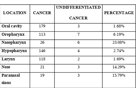
TABLE - IV
LOCATION CANCER
UNDIFFERENTIATED
CANCER
PERCENTAGE
Oral cavity 179 3 1.68%
Oropharynx 113 7 6.19%
Nasopharynx 26 6 23.08%
Hypopharynx 146 4 2.74%
Larynx 118 2 1.69%
Nose 21 3 14.29%
Paranasal sinus
19 3 15.79%
IMMUNOHISTOCHEMISTRY RESULTS TABLE - V
Tumor
Location
Carcinoma Lymphoma Others Inconclusive Total
Oral cavity 3 - - - 3
Oropharynx 2 2 - 3 7
Nasopharynx 2 2 2 - 6
Hypopharynx 3 - - 1 4
Larynx - 1 - 1 2
Nose 1 1 1 - 3
Paranasal sinus
2 1 - - 3
Others - - 1 1 2
Total 13 7 4 6 30
• In case of nasal cavity, S-100 positive olfactory neuroblastoma diagnosed.
TUMOR TYPES IN ACCORDANCE WITH AGE OF PATIENTS
TABLE - VI
AGE Carcinoma Lymphoma Others Inconclusive Total
20> 1 2 1 - 4
21 to 30 1 2 - - 3
31to 40 1 - - 1 2
41 to 50 2 1 2 2 7
51 to 60 1 1 1 2 5
> 61 7 1 - 1 9
Total 13 7 4 6 30
• In younger age – Lymphoma is the dominant group.
PERCENTAGE OF TUMOR TYPES TABLE VII
Type of Tumors No. of cases Percentage
Carcinoma 13 43.33 %
Lymphoma 7 23.33 %
Others 4 13.33 %
Inconclusive 6 20 %
• Carcinoma is the major group followed by lymphoma
• Either conclusive or suggestive diagnosis of undifferentiated tumor was achieved on this study is 80%
• Inconclusive diagnosis – that is not positive for any of the immunohistochemical panel (or) positive for carcinoma, lymphoma and mesenchymal markers, because of multiple epitopes is the main cause in this study.
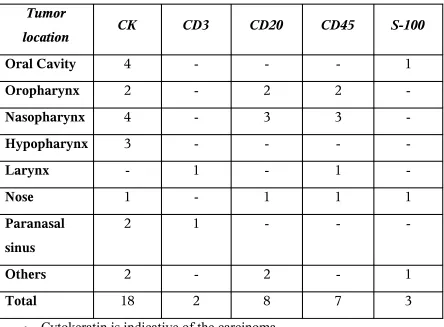
IMMUNOHISTOCHEMICAL MARKER POSITIVITY WITH RELEVANCE TO TUMOR LOCATION
TABLE VIII
Tumor
location CK CD3 CD20 CD45 S-100
Oral Cavity 4 - - - 1
Oropharynx 2 - 2 2
-Nasopharynx 4 - 3 3
-Hypopharynx 3 - - -
-Larynx - 1 - 1
-Nose 1 - 1 1 1
Paranasal sinus
2 1 - -
-Others 2 - 2 - 1
Total 18 2 8 7 3
• Cytokeratin is indicative of the carcinoma
• CD20, CD45 positivity indicates the lymphoma which is of B-cell variety.
[image:63.612.85.529.175.502.2]CONCLUSIVE DIAGNOSIS BY
IMMUNOHISTOCHEMICAL TECHNIQUES ACCORDING TO TUMOR LOCATION
TABLE IX
Location Conclusive Inconclusive Total Conclusive percentage
Oral Cavity 3 - 3 100%
Oropharynx 4 3 7 57.14%
Nasopharynx 6 - 6 100%
Hypopharynx 3 1 4 75%
Larynx 1 1 2 50%
Nose 3 - 3 100%
Paranasal sinus
3 - 3 100%
Others 1 1 2 50%
• 100% conclusive diagnosis was achieved in cases of oral cavity, Nasopharynx, nose and paranasal sinuses in this study
DISCUSSION
The poorly differentiated (or) undifferentiated tumors challenge routine histopathological classification, because of the absence of cell morphological differentiation. The diagnosis and classification of such tumors are fundamental, because suuitable therapy and prognosis for each case depends upon precise hisopathological diagnosis.
The immunohistochemical technique has revolutionized surgical pathology knowledge. The introduction of this method by coons et al in 1942 has become a powerful complementary tool in tumor analysis.
There are only few references in the literature to the immunohistochemical technique applied to the identification of undifferentiated head and neck tumors.
Undifferentiated cancer incidence
Head and neck tumors constitutes about 35% of all diagnosed cancers
Watlter Adriano bianchini (2003) in his study found the undifferentiated cancers constitutes 1.1% of all tumors diagnosed 43 cases out of 3,840.
Sex Incidence
There were 20 male (66%) , 10 female(33%) cases in a proportion of 2:1 was found in this study
which was supported by AlbinaMaria Albermani in his study. Male and female ratio is 67.5% and female is 32.5%. Head and Neck cancers found predominantly in males than in females. Indian text books describe that most of the head and neck cancer ocurs predominantly in males.
Age Incidence
Head and Neck cancers and undifferentiated head and neck cancers were found predominantly in males.
Patients age ranged from 17 years to 72 years
These tumors were most prevalent in 6th decade of life(30%)
followed by 4th decade of life(23%)
Walter adriano bianchini and Jarge Rizzato Paschaal (2003) in their study, the prevalence rate of undifferentiated carcinoma is in the 7th decade of life(34.9%)
Age of the patient ranged from 2 years to 89 years.
Incidence of site of undifferentiated cancer
Abemayor.E; Kessler DJ, Ward PH (1987) in their study the commonest site of occurrence of head and neck tumor was pharynx and neck(21%) , lymphnode(18%), paranasal sinus(14%)
Walter Adriano bianchini (2003) in their study the commonest site of occurrence in Lymphnode (20.9%), pharynx(16.3%), Nose(11.6%) and PNS (9%)
In this study highest incidence was in Oropharynx (23%), Nasopharynx (20%) followed by Nose and paranasal sinuses (20%), hypopharynx(13%)
Proportional incidence of the undifferentiated cancer
Nasopharynx(23%), Paranasal sinus(15.8%), nose(14%). These data was supported by Gallo.O. Graziani et al (1993) – Nasopharynx(18%), Paranasal sinus(21%), nose (18%).
Subsite wise the more number of undifferentiated tumors found in posterior 1/3 of a tongue and nasopharynx.(six patients)
Type of tumors diagnosed by Immunohistochemical methods
The immunohistochemical technique was usseful in conclusive diagnosis of the tumors in the oral cavity (100%), nasopharynx(100%), nose and nasopharynx(100%).
Milroy CM. Ferlito A et al (1995) in his study , the conclusive diagnosis was as follows : oral cavity(100%), Paranasal sinus(100%), Lymphnodes(89.9%) and Nasopharynx(57%).
Galter et al (1985) and Coindre et al (1982) demonstrated greater incidence of lymphomas in undifferentiatied tumors in general.
Darrouzet .V et al (1989) in his study , the most frequent tumor is carcinoma(39%)
In this study, the commonest tumor type diagnosed is carcinoma (43%) followed by lymphoma (23%)
Lymphoma was commonly found in the patients below the age of 30 years (4 cases ) -57%.
Carcinoma found in patients above 40 years of age (10 to 15 cases)
– 76.92%.
Lymphomas commonly found in the oropharynx(Tonsil) and Nasopharynx, except 1 case, all are B cell Lymphoma.
Sensitivity of the Immunohistochemistry
Abemayor et al (1987) in his study using immunohistochemistry, the conclusive diagnosis was achieved in 86.4% patients. It was not possible in 13.6%.
Walter Adriano Bianchini et al (2003) in his study, using immunohistochemistry, the diagnostic guidance was obtained in 81.4% of patients(35 of 43). It was not possible in 18.6%( 8 of 43)
The failure is probably due to limitations of the technique, lesser number of immunohistochemical panel of markers, Antigen changes during tissue fixation, true absence of cellular differentiation.
Immunohistochemical Markers (Panels)
Gallo .O et al (1993) used 19 immunohistochemical markers as panel to study the undifferentiated cancer of nose and paranasal sinuses.
Walter Adriano et al (2003) used about 23 panel of immunohistochemical markers.
In this study, 5 immunohistochemical markers was used. Cytokeratin positive cancers is the common pattern. 18 of 30 (60%), CD3 (8 of 30),CD45 (7 of 30), S-100(3 of 30).
CONCLUSIONS
1. Undifferentiated cancers in the head and neck represents 3.57% of all tumors diagnosed in this study.
2. Undifferentiated head and neck cancers found
predominantly in the males (66%) and highest incidence after the 4th decade of life (68%).
3. Undifferentiated head and neck cancers most frequently
occurs in the nasopharynx (23%), nose (14%) and paranasal sinuses(16%) in this study.
4. Immunohistochemical tehcnique is sensitive enough to conclusively diagnose 80% of the undifferentiated head and neck cancer.
5. The most common tumor type diagnosed in this study is carcinoma(43%) followed by lymphoma(23%)
6. 100% conclusive diagnosis achieved in oral cavity,
BIBLIOGRAPHY
1. Gaveriviere P, AI Saati T, Voigt JJ, Delsol G. Diagnostic of
undifferentiated tumors by means of monoclonal antibodies on
paraffin sections.
2. Gatter KC, AlcockC, Heryer A, et al. The differential diagnosis
of routinely processed anaplastic tumors using monoclonal
antibodies. Am J Clin Pathol 1984;82(10):33-43
3. Goodman MI, Pilch BZ. Tumors of the respiratory tract . In
Fletcher CDM editor . Diagnosis Histopathology of tumors
Edinbuirg; Churchill Livingstone 1995;p.79-126
4. Micheals L Neuroenctodermal tumours. In: Michaels1.ed Ear,
Nose and Throat Histopathology. Berlin: Springer verlag 1987. .
89
5. Abemayor E, Kessler DJ Ward PH, Fu YS. Evaluation of poorly
differentiated head and neck neoplasms. Immunocytochemistry
techniques. Arch Otolaryngol Head neck surg 1987;113(5)
6. Darrouzet v Stoll D,.Deminiere C Apport de
immunohistochemistry to the diagnosis of cervicofacial cancers
Rev Laryngol Otol Rhinol 1989;110(2) 201-4
7. Gallo O, Graziani P, Fini-Storchi O undifferentiated carcinoma
of the nose and paranasal sinuses. An immunohistochemical
and clinical study. Ear Nose Throat J 1993 72(9) 588-90
8. Milroy CM Ferlito A. Immunohistochemical markers in tne
diagnosis of neuroendocrine neoplasms of the head and neck
Ann Otol Rhinol Larngol 1995; 104 (5)413-8
9. Hau Am Raine L, Fanger H, U:se of avidin-biotin-peroxidase
complex(ABC) in immunoperoxidase techniques a comparison
between ABC and unlabeled antibody (PAP) procedures. J
Histochem Cytochem 1981: 29(4); 577-80
10.Vege DS Soman CS Joshi UA Ganesh B, Yadav JN,
undifferentiated tu;mours an immunohistochemical analysis on
11. Azar HA Epsinoza CG Richman AV Saba SR Wanmg T
Undifferentiated Large cell malignancies an ultrastructureal and
immunocytochemical study Hum Pathol,1982 323-33
12.Coindre JM Tanguy F, Merlio JP EDe Mascarel A Trojani The
value of immunohistological techniques in undifferentiated
cancers Tumour 1986 539-44
13.Gatter KC Alcock C Heryet A, Mason DY Clinical importance of
analyzing malignant tumours of uncertain origin with
immunohistological techniques. Lancer 1985;1302-05
14.Walter Adriano Bianchini, Albina Maria Altermani, Jorge Rizzalo
Paschool, Undifferentiated head and neck tumours differential
diagnosis using immunohistochemistry, Medical Journal Sai
Oayki 2003; 244-47
15.Jasani, BN, Wynford- Thomas, D &Williams, E.D 1981 Use of
monoclonal anti-hapten antibodies for immunolocalisation of
tissue antigens. Journal of Clinical Pathology 1000-02
16. Miller K. D Modern methods in immunocytochemistry.
17.Chan JKC. A newq classification of lympohomas sthe Revised
european – Ajmerican Lymphoma Classification Adv Anat
Pathol 1994 543-60
18. Morrison WH , Hoppe RT Weiss LM Small lymphocytic
lymphoma a clinicopathologic analysis of 268 cases. Blood
1989 579-87
19.Baek C H., Kim Ks and Kang Primary mucosal melanoma of
the nasal cavity 582-3
20. Cleary KR and Batsakis JGF Pathology consultation
Sinonasal lymphomas Ann Otol Rhinol Laryngol 103
21. Poon Yf AND Lau WH (1998) Current management of
carcinoma of the nasopharynx in Current Radiation Oncology 3
(JS Tobias and PRM Thomas eds) Arnold London pp 146-76
22.King G(1994) microwave heating as amethod of unmasking
immunoglobin lightchain paraffin section UK NEQAS newsletter
23.Jessup .E.(1994)Antigen Retrival techniques for the
demonstration immunoglobin light chain in paraffin section UK