Copyright © 2000, American Society for Microbiology. All Rights Reserved.
Detachment of Human Immunodeficiency Virus Type 1 from
Germinal Centers by Blocking Complement Receptor Type 2
LACO KACANI,1,2WOLFGANG M. PRODINGER,1* GEORG M. SPRINZL,3MICHAEL G. SCHWENDINGER,1† MARTIN SPRUTH,1,2† HERIBERT STOIBER,1,2SUSANNE DO¨ PPER,1,2SABINE STEINHUBER,1
FRANZ STEINDL,4ANDMANFRED P. DIERICH1,2
Ludwig Boltzmann Institute for AIDS Research,2Institute for Hygiene and Social Medicine,1and Department of Otorhinolaryngology,3University of Innsbruck, A-6020 Innsbruck, and Institute of Applied Microbiology,
University of Agriculture, A-1190 Vienna,4Austria Received 17 February 2000/Accepted 9 June 2000
After the transition from the acute to the chronic phase of human immunodeficiency virus (HIV) infection, complement mediates long-term storage of virions in germinal centers (GC) of lymphoid tissue. The contri-bution of particular complement receptors (CRs) to virus trapping in GC was studied on tonsillar specimens from HIV-infected individuals. CR2 (CD21) was identified as the main binding site for HIV in GC. Monoclonal antibodies (MAb) blocking the CR2-C3d interaction were shown to detach 62 to 77% of HIV type 1 from tonsillar cells of an individual in the presymptomatic stage. Although they did so at a lower efficiency, these antibodies were able to remove HIV from tonsillar cells of patients under highly active antiretroviral therapy, suggesting that the C3d-CR2 interaction remains a primary entrapment mechanism in treated patients as well. In contrast, removal of HIV was not observed with MAb blocking CR1 or CR3. Thus, targeting CR2 may facilitate new approaches toward a reduction of residual virus in GC.
Follicular dendritic cells (FDC) are thought to play a pivotal role in the initiation and maintenance of secondary antibody responses. FDC operate in germinal centers (GC) of lymphoid follicles, where they form a three-dimensional network. Anti-gens complexed with immunoglobulins and complement frag-ments are trapped on the surface of FDC in form of immune-complex-coated bodies or “iccosomes” that contribute to B-cell maturation and B-cell memory (24). FDC express sub-stantial quantities of complement receptor 1 (CR1; CD35) and CR2, in addition to lesser amounts of CR3 (CD11b/CD18) (21). This unique pattern of CR expression allows FDC to interact with all complement C3 fragments: with C3b through CR1, with iC3b through CR2 or CR3, and with C3d via CR2. In the course of human immunodeficiency virus type 1 (HIV-1) infection, most of the virus is produced by CD4⫹T lymphocytes or macrophages and is subsequently stored on the surface of FDC (1, 25). This attachment process was shown to be complement dependent in vitro (16, 23). Quantitative image analysis demonstrated that, after transition to the chronic phase, more than 98% of the viral RNA in lymphoid tissue (LT) is found in virions extracellularly bound to FDC (9, 10, 19). Early histopathological alterations in HIV infection in-clude follicular hyperplasia, destruction of FDC, and lysis of the lymphoid follicles. In the late stage, FDC are destroyed and viral RNA decreases in GC, whereas the level of infectious virions in plasma increases. As FDC are diminished, lymphoid follicles involute and generalized immunosuppression occurs. It was proposed previously that at least part of these effects are mediated by virions trapped in the FDC network (3).
After highly active antiretroviral therapy (HAART) is initi-ated, the amount of HIV trapped on FDC decreases very quickly and plasma HIV-1 RNA becomes undetectable in most
cases (4, 27). After 6 months of HAART, a⬎2,500-fold re-duction in the FDC pool of virions was observed, but HIV RNA on FDC was still detectable in some GC (4). This resid-ual virus probably represents a source of HIV in the case of drug resistance or if therapy is discontinued (9).
Several mechanisms are considered to contribute to the trapping of HIV by FDC (3): (i) virions form immune com-plexes coated with complement C3 fragments and subse-quently attach to CR or Fc receptors on FDC, (ii) gp120 molecules on the viral envelope may specifically interact with immunoglobulins of the VH3 family and thereby constitute immune-complexed virions that bind to FDC, and (iii) the viral envelope acquires adhesion molecules during budding from the host cell which then may interact with their cellular coun-terparts. In this report, tonsillar tissue from HIV-infected in-dividuals was studied to assess the relative contributions of particular CRs to virus trapping in GC and the possibilities of detaching HIV from its reservoir.
MATERIALS AND METHODS
Antibodies.Mouse monoclonal antibodies (MAb) used in this study were 3D9 and 1B4, CR1 blocking the C3b-binding site (18); E11, nonblocking anti-CR1 (Pharmingen, San Diego, Calif.); HB5, nonblocking anti-CR2, obtained from the American Type Culture Collection (ATCC; Manassas, Va.); 4C2, directed against the C3d fragment of complement C3 (17); and AB2 and BB5 (both anti-C3d) and FE8, anti-CR2, developed in our laboratory. FE8 (W. M. Prodinger, M. G. Schwendinger, and M. P. Dierich, 25 March 2000, European Patent Office) is currently the only available MAb that blocks both C3d binding and Epstein-Barr virus binding to CR2 (20). TMG6-5 (26) and TS1/18 (obtained from the ATCC) are directed against human CD11b and CD18, respectively. TMG6-5 blocks the binding of iC3b to CR3 (5). TIB191 (immunoglobulin G1 [IgG1]) and TIB109 (IgG2a), both from the ATCC, were used as isotype con-trols. A single-chain fragment variable (scFv) MAb, scFv-FE8, was generated from the FE8 hybridoma using the Recombinant Phage Antibody System (Phar-macia Biotech, Uppsala, Sweden).
Stripping of C3d-coated beads from Raji cells.B-lymphoblastoid, CR2⫹Raji cells (ATCC) were cultured in complete medium. Fluorescein isothiocyanate (FITC)-labeled, C3d-coated agarose microbeads were prepared as described previously (20). For detachment of complement-coated beads from CR2, 5⫻105
Raji cells were washed with Hanks’ balanced salt solution (HBSS) and incubated for 30 min at room temperature with predetermined saturating concentrations of beads. Thereafter, cells were washed with HBSS and the pellet was resuspended
* Corresponding author. Mailing address: Institut fu¨r Hygiene und Sozialmedizin, Universita¨t Innsbruck, Fritz-Pregl-Strasse 3, A-6020 Innsbruck, Austria. Phone: 43 512 507 3424. Fax: 43 512 507 2870. E-mail: [email protected].
† Present address: Baxter AG, A-1221 Vienna, Austria.
7997
on November 9, 2019 by guest
http://jvi.asm.org/
in 50l of HBSS containing MAb. Samples were incubated for 30 min at 37°C, washed twice with HBSS, and analyzed using a FACScan (Becton Dickinson, Hialeah, Fla.). Nonspecific binding was determined using Raji cells incubated with FITC-microbeads lacking C3 fragments.
Tonsillar tissue.Surgical specimens of palatine tonsils were obtained from HIV-positive individuals by tonsillectomy done for medical reasons unrelated to this study (Table 1). Tonsils were collected in 0.9% saline and processed imme-diately. One half of the tissue was cut into 5-mm blocks, snap frozen in liquid nitrogen, and used for cryosections. The other half was homogenized to obtain a suspension of tonsillar cells (TC).
Preparation of TC.Tonsil tissue was cut into small pieces when immersed in X-VIVO 10 medium (Bio-Whittaker, Verviers, Belgium) and incubated with 1 mg of collagenase type IV (Sigma, St. Louis, Mo.) per ml for 60 min at 37°C. After 30 min, released TC were collected and fresh enzyme solution was added to nondigested tissue. TC were pooled, washed twice with complete medium (RPMI 1640 with 10% fetal calf serum [FCS]), and passed through a 100- m-pore-size cell strainer (Becton Dickinson) to remove cell clumps and nondi-gested tissue. Finally, TC were frozen in RPMI 1640 with 12.5% dimethyl sulfoxide and 20% FCS and stored at⫺70°C until further use.
Stripping of HIV from TC.Frozen aliquots of TC were thawed and washed with complete medium before stripping. TC (2⫻105cells in 100l) were seeded
into 96-well V-bottomed microplates and incubated in the presence or absence of MAb for 1 h at 37°C. To determine the size of the intracellular viral pool, TC were incubated in the presence of different concentrations of proteinase K under the same conditions. After centrifugation, cell pellets were washed three times with complete medium, lysed with 1% NP-40 in RPMI medium, and used for quantification of HIV-1 p24 antigen. Supernatants from stripping experiments were stored at⫺70°C until determination of either p24 or HIV-1 RNA.
p24 capture ELISA.HIV-1 p24 antigen was quantified in TC lysates and for some experiments also in supernatants from stripping experiments by p24 cap-ture enzyme-linked immunosorbent assay (ELISA) as described elsewhere (2).
Stripping from tonsil cryosections.Six-micrometer-thick sections were cut at different levels from frozen tissue blocks and stored at⫺70°C until use. Thawed sections were air dried quickly, and nonspecific binding was blocked by incuba-tion with 10% FCS in HBSS for 30 min at room temperature. The secincuba-tions were allowed to react for 1 h at 37°C with 1g of MAb per ml in 10% FCS–HBSS. Samples were subsequently washed with HBSS and fixed in methanol-acetone (4:1).
Immunohistochemistry.Sections were blocked in 10% FCS in phosphate-buffered saline and incubated with anti-mouse IgG-FITC conjugate (DAKO, Glostrup, Denmark) to stain the stripping MAb. Thereafter, sections were in-cubated with biotinylated anti-p24 MAb (clone 37G12, kindly provided by H. Katinger, Institute for Applied Microbiology, Vienna, Austria) followed by ex-travidin-alkaline phosphatase conjugate (Sigma). The sections were developed using FastRed substrate (Sigma), mounted with DAKO fluorescent mounting medium, and examined with a confocal laser microscope (LSM-510; Carl Zeiss, Jena, Germany).
HIV-1 RNA quantification.HIV-1 virions in supernatants of TC samples were quantified using the AMPLICOR HIV-1 MONITOR test version 1.5 (Roche Diagnostics, Branchburg, N.J.). An ultrasensitive procedure was used according to the manufacturer’s instructions. Linearity for RPMI-FCS culture supernatant specimens was reached between 200 and 50,000 copies per ml (detection limit, 200 copies per ml), as determined in preliminary experiments.
RESULTS
We have recently established a MAb, FE8, directed against CR2 that blocks the binding of C3d-coated ligands (20). Here we demonstrate that FE8 is able to dissociate already-bound C3d-opsonized particles from CR2. Using CR2⫹Raji B cells as a model system, more than 97% of C3d-beads could be re-moved after 1 h of incubation in the presence of ⱖ500 ng of FE8 per ml (Fig. 1A). BB5, a MAb directed against the CR2-binding site in C3d, showed similar effects (Fig. 1B). There-fore, MAb FE8 and BB5 are pertinent tools to assess the
contribution of the CR2-C3d interaction to HIV entrapment in LT.
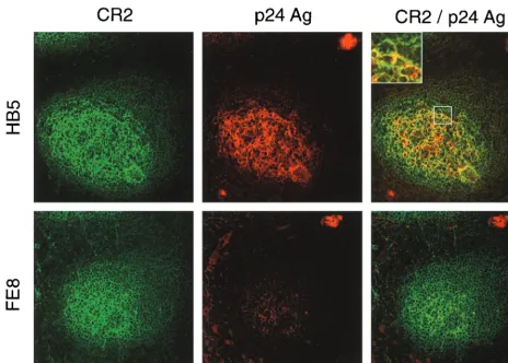
To this end, we performed experiments on tonsillar tissue specimens obtained from three HIV-positive individuals (for patient characteristics, see Table 1). To detach extracellularly bound HIV-1, tonsil cryosections and isolated TC of patient 1 were incubated in the presence of blocking MAb. In sections, most of the virus was localized in the FDC network, whereas virus-replicating cells were found in the T-cell areas of the follicle (Fig. 2). Treatment of cryosections with MAb FE8 led to the disappearance of HIV-1 p24 antigen from GC, while p24 within productively infected cells was not affected. In contrast, nonblocking anti-CR2 MAb HB5 did not affect HIV detach-ment. These results indicated that blocking the CR2-C3d in-teraction allows detachment of HIV-1 from GC.
[image:2.612.53.551.85.140.2]To quantify the removal of trapped virus, TC were treated
[image:2.612.370.495.373.650.2]FIG. 1. Detachment of fluorescent agarose microbeads opsonized with C3d from CR2 of Raji B cells by MAb against CR2 or C3d. (A) Fluorescence intensity of Raji cells loaded with predetermined saturating concentrations of beads and subsequently stripped with 1g of isotype control antibody (grey histogram) per ml or 1g of MAb FE8 (white histogram) per ml. (B) Median fluorescence intensities (MFI) of Raji cells incubated in the presence of anti-CR2 MAb HB5 or FE8 and anti-C3d MAb BB5. Blocking antibodies are shown with closed symbols; nonblocking MAb are shown with open symbols. Values are means from two experiments⫾standard errors of the means.
TABLE 1. Profiles of tonsillectomized HIV-positive individuals
Patient Date of first positiveHIV test result Agea(yr) Stage of
infectiona CD4
⫹T-cell counta
(no. of cells/l) Plasma HIV RNA a
(no. of copies/ml) Antiretroviral drugsb No. of mo receivingHAARTa
1 March 1995 28 Presymptomatic 378 8,912 None None 2 November 1993 35 Late 499 ⬍400 d4T, 3TC, IDV, RTV 19 3 November 1997 33 Late 465 ⬍400 AZT, 3TC, IDV 21
aAt time of tonsillectomy.
bd4T, stavudine; 3TC, lamivudine; AZT, zidovudine; IDV, indinavir; RTV, ritonavir.
on November 9, 2019 by guest
http://jvi.asm.org/
with blocking MAb against CR2, C3d, CR1, or CR3, and cell-associated p24 levels were determined subsequently by capture ELISA. In our hands, blocking with anti-CR2 MAb FE8, as well as blocking with anti-C3d MAb BB5 and 4C2, was able to remove substantial amounts of virus from TC, whereas anti-CR1 and anti-CR3 MAb had no effect (Fig. 3). Further-more, an FE8-derived scFv antibody, although requiring a five-fold-higher concentration on a molar basis, was shown to be as effective as FE8 IgG. Stripping of TC with FE8 and BB5 demonstrated that (ⱕ22.5 ⫾ 3.7)% and (ⱕ38.3⫾ 6.7)% of p24, respectively, remained cell associated, whereas proteinase K-resistant p24 represented less than 10% (Fig. 3C). This suggests that⬎90% of p24 is found within virions trapped on the surface of TC and only a very small part is localized intra-cellularly, which is in good agreement with previous reports (9).
To further distinguish whether liberated p24 was virion as-sociated or virus free, the amounts of p24 in supernatants of TC after detachment were measured in the presence or ab-sence of 1% NP-40 (Fig. 4). The major part of p24 became detectable only after addition of detergent and lysis of virions. Throughout all MAb concentrations used in this experiment, only (5.7 ⫾ 1.2)% of total stripped p24 was virus free. In parallel experiments, release of HIV-1 RNA from TC was determined using quantitative reverse transcription-PCR with
similar results (data not shown), underlining that most of the released p24 was localized within virions.
Further tonsil specimens were obtained from patients 2 and 3 undergoing HAART, whose plasma HIV-1 RNA levels had remained below the detection limit (i.e.,⬍400 copies/ml) for more than 1 year. p24 levels in lysates of 107TC from these two patients were 3.7⫾0.3 and 5.6⫾0.8 pg/ml, respectively, i.e.,
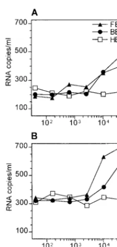
⬎1,000-fold less than those in the presymptomatic patient 1 (7,750⫾ 250 pg/ml). Quantitative reverse transcription-PCR demonstrated that HIV-1 RNA was removed from TC by tar-geting CR2 or C3d (Fig. 5), although⬎10-fold-higher amounts of MAb were required than those for tissue from patient 1.
DISCUSSION
HIV-1 trapped on the surface of FDC constitutes by far the largest viral reservoir in LT, once the infection has reached the chronic phase (9, 10, 19). Both HIV-specific antibody and complement were shown to be indispensable for virus entrap-ment on FDC in vitro (16), although recent reports suggest that HIV starts to accumulate in this pool already in the acute phase (22). Furthermore, virus trapped on FDC was shown to remain infectious for T cells migrating through GC even after being coated with neutralizing antibody (11, 14).
[image:3.612.72.536.72.404.2]Concerning this interplay of HIV and GC, we addressed the
FIG. 2. Double staining for HIV-1 p24 antigen (Ag) and for CR2 of serial cryosections from tonsil tissue of patient 1. Unfixed sections were treated with MAb HB5 (nonblocking anti-CR2; upper panels) or MAb FE8 (blocking anti-CR2; lower panels), subsequently fixed, stained and photographed. Each section shows a typical GC: CR2⫹cells forming a network typical for FDC are stained green, and p24 antigen trapped in GC or found in virus-replicating cells (upper right and lower left, respectively) is stained red. p24 mainly colocalizes (yellow color) with the FDC network within GC (see inset). Overlays were generated by digital superimposition of single color images.
on November 9, 2019 by guest
http://jvi.asm.org/
following issues in this report. With all CRs being expressed in FDC, what are their relative contributions to HIV entrap-ment? Do virions change their anchoring sites in the course of infection or during HAART? If FDC are the major sanctuary for HIV, can targeting of CR reduce this viral pool?
Incubation of tonsillar tissue cryosections and of suspended TC with blocking MAb directed against CR2 or C3d detached HIV-1 p24 from GC, whereas anti-CR1 or anti-CR3 had no effect. From these results, we assume that the interaction of in vivo opsonized HIV within GC is predominantly mediated through binding of the final C3 breakdown product C3d to CR2. This is even more likely, because C3d rather than C3b or iC3b can be expected to be present on the virion after a period of several days (13).
In addition to FDC, activated B cells in GC express CR1 and CR2 and therefore can potentially bind opsonized HIV. How-ever, immunohistochemical staining has shown that HIV pref-erentially associates with the FDC network (4, 9). Since FDC express more CR1 and CR2 than do B cells (21), the density and/or surface distribution of CR seems to be critical for trap-ping of HIV on FDC. On the other hand, B cells could be rather important for disseminating infectious virus throughout the body (15).
Treatment of TC with proteinase K did remove 15 to 20% more extracellular HIV-1 than did either FE8 or BB5. This implies either that not all virions attached to CR2 can be removed by blocking MAb or that other virus-trapping mole-cules do exist on FDC. In this regard, Fc␥receptors (21) or adhesion molecules like intercellular adhesion molecule 1 (ICAM-1) and LFA-1 (8) have been suggested to contribute to virus trapping, but their relative contributions have not been assessed. Further experiments are under way to clarify this issue.
[image:4.612.133.473.75.329.2]FIG. 3. (A, B, D, and E) Stripping of HIV from TC from an untreated, presymptomatic HIV-positive individual (patient 1) by MAb or derived scFv antibodies directed against CR2 (A), C3d (B), CR1 (D), or CR3 (E). Closed symbols represent treatment with blocking antibodies, and open symbols represent treatment with nonblocking MAb. HIV-1 p24 was quantified by capture ELISA in lysates of TC after treatment with MAb or derived scFv antibody. (C) p24 release from TC after treatment with 10g of isotype control, FE8, or BB5 (values taken from panels A and B) per ml compared with proteinase K (Pr-K) digestion (2.5g/ml). p24 was measured in TC lysates by capture ELISA. Data represent means⫾standard errors of the means from experiments done in triplicate. Ag, antigen.
FIG. 4. Discrimination between virion-associated and virus-free p24 de-tached from TC. Total amounts of p24 (A) were measured by capture ELISA in TC supernatants after stripping with FE8 (triangles) or HB5 control MAb (cir-cles) and addition of 1% NP-40 (final concentration). Virus-free p24 (B) was determined in the same way in the absence of detergent. Data represent means⫾standard errors of the means of a representative experiment done in triplicate. Ag, antigen.
on November 9, 2019 by guest
http://jvi.asm.org/
[image:4.612.369.489.414.663.2]To investigate whether the C3d-CR2 interaction remains the main trapping mechanism throughout advanced stages of HIV infection, we tested tonsillar tissue from two individuals under HAART. HIV-1 RNA could still be removed from TC by targeting C3d or CR2, although at much lower efficiency than that for TC of the presymptomatic individual. A recent math-ematical analysis which assumes that virions are trapped in LT exclusively via C3d-CR2 binding points out that during HAART most virions will dissociate from FDC within days or weeks, whereas those coated abundantly with C3d may persist for years (13). Therefore, such extensively opsonized virions can be expected in a larger quantity in LT of patients under HAART. Indeed, our data imply that the effectiveness of HIV detachment through CR2 blockade will be much lower in LT of treated patients than in LT of presymptomatic individuals. So far, the infectivity of viral particles released from FDC has not been investigated in detail. Our data show that natu-rally opsonized virions become available for experimentation. In preliminary experiments, virus capture assays (7) revealed that C3d and IgG are the most abundant proteins detected in the viral envelope of such virions, whereas gp120 and gp41 were hardly accessible for capture antibodies (data not shown). Further studies will have to prove if complement fragments on the surface of HIV acquired in vivo exert significant effects on viral infectivity and tropism.
The data presented here suggest that reduction of the viral reservoir in LT through a blockade of the CR2-C3d interaction is a possible therapeutic approach, albeit it has to take into account several caveats. First, other trapping mechanisms could eventually substitute for CR2. Second, it is unclear at present which time point in the course of HIV infection would be suitable for such an intervention. From the data presented here, release of extracellularly trapped HIV from LT would
appear to be more effective in the presymptomatic stage. On the other hand, direct targeting of nonreplicating virus trapped on FDC in advanced stages of disease and during HAART would be highly desirable. However, in later stages more viri-ons may be coated extensively with C3d and therefore may resist a CR blockade (13). Furthermore, not only HIV but all other immune complexes will be removed from or prohibited from binding to FDC. In this regard, in vivo experiments in mice have shown that blocking CR2 negatively affected, al-though it did not completely abrogate, primary and secondary humoral immune responses (6, 12). These unknowns have to be addressed by forthcoming experimentation.
ACKNOWLEDGMENTS
We thank Brigitte Mu¨llauer and M. Wurm for excellent technical assistance; Heidrun Recheis for help with confocal laser microscopy; R. Zangerle and O. Moling for helpful discussions and support; Mal-gorzata Krych, R. Taylor, V. Koistinen, G. D. Ross, and I. Ando for providing MAb; and B. Sterlacci for reading of the manuscript.
This work was supported by the Austrian Science Fund (grant F202), the Ludwig Boltzmann Society, and the Austrian Ministry of Science (grant GZ 70.028/2-Pr/4/98).
REFERENCES
1.Armstrong, J. A., and R. Horne.1984. Follicular dendritic cells and virus-like particles in AIDS-related lymphadenopathy. Lancetii:370–372.
2.Buchacher, A., R. Predl, K. Strutzenberger, W. Steinfellner, A. Trkola, M. Purtscher, G. Gruber, C. Tauer, F. Steindl, A. Jungbauer, and H. Katinger.
1994. Generation of human monoclonal antibodies against HIV-1 proteins; electrofusion and Epstein-Barr virus transformation for peripheral blood lymphocyte immortalization. AIDS Res. Hum. Retrovir.10:359–369. 3.Burton, G. F., A. Masuda, S. L. Heath, B. A. Smith, J. G. Tew, and A. K.
Szakal.1997. Follicular dendritic cells (FDC) in retroviral infection: host/ pathogen perspectives. Immunol. Rev.156:185–197.
4.Cavert, W., D. W. Notermans, K. Staskus, S. W. Wietgrefe, M. Zupancic, K. Gebhard, K. Henry, Z. Q. Zhang, R. Mills, H. McDade, C. M. Schuwirth, J. Goudsmit, S. A. Danner, and A. T. Haase.1997. Kinetics of response in lymphoid tissues to antiretroviral therapy of HIV-1 infection. Science276:
960–964.
5.Diamond, M. S., J. Garcia-Aguilar, J. K. Bickford, A. L. Corbi, and T. A. Springer.1993. The I domain is a major recognition site on the leukocyte integrin Mac-1 (CD11b/CD18) for four distinct adhesion ligands. J. Cell Biol.120:1031–1043.
6.Fang, Y., C. Xu, Y. X. Fu, V. M. Holers, and H. Molina.1998. Expression of complement receptors 1 and 2 on follicular dendritic cells is necessary for the generation of a strong antigen-specific IgG response. J. Immunol.160:5273– 5279.
7.Frank, I., H. Stoiber, S. Godar, H. Stockinger, F. Steindl, H. W. Katinger, and M. P. Dierich.1996. Acquisition of host cell-surface-derived molecules by HIV-1. AIDS10:1611–1620.
8.Fujiwara, M., R. Tsunoda, S. Shigeta, T. Yokota, and M. Baba.1999. Human follicular dendritic cells remain uninfected and capture human immunode-ficiency virus type 1 through CD54-CD11a interaction. J. Virol.73:3603– 3607.
9.Haase, A. T.1999. Population biology of HIV-1 infection: viral and CD4⫹T cell demographics and dynamics in lymphatic tissues. Annu. Rev. Immunol.
17:625–656.
10. Haase, A. T., K. Henry, M. Zupancic, G. Sedgewick, R. A. Faust, H. Melroe, W. Cavert, K. Gebhard, K. Staskus, Z. Q. Zhang, P. J. Dailey, H. H. Balfour, A. Erice, and A. S. Perelson.1996. Quantitative image analysis of HIV-1 infection in lymphoid tissue. Science274:985–989.
11. Heath, S. L., J. G. Tew, A. K. Szakal, and G. F. Burton.1995. Follicular dendritic cells and human immunodeficiency virus infectivity. Nature377:
740–744.
12. Heyman, B., E. J. Wiersma, and T. Kinoshita.1990. In vivo inhibition of the antibody response by a complement receptor-specific monoclonal antibody. J. Exp. Med.172:665–668.
13. Hlavacek, W. S., C. Wofsy, and A. S. Perelson.1999. Dissociation of HIV-1 from follicular dendritic cells during HAART: mathematical analysis. Proc. Natl. Acad. Sci. USA96:14681–14686.
14. Hufert, F. T., J. van Lunzen, G. Janossy, S. Bertram, J. Schmitz, O. Haller, P. Racz, and D. von Laer.1997. Germinal centre CD4⫹T cells are an important site of HIV replication in vivo. AIDS11:849–857.
15. Jakubik, J. J., M. Saifuddin, D. M. Takefman, and G. T. Spear.1999. B lymphocytes in lymph nodes and peripheral blood are important for binding immune containing HIV-1. Immunology96:612–619.
[image:5.612.113.231.70.319.2]16. Joling, P., L. J. Bakker, J. A. Van Strijp, T. Meerloo, L. de Graaf, M. E.
FIG. 5. Stripping of HIV from TC of patient 2 (A) and patient 3 (B) under-going long-term, effective HAART. Residual amounts of HIV-1 RNA released into the supernatants after stripping of 107cells in 100l with different
concen-trations of nonblocking (open symbols) or blocking (closed symbols) MAb are given (see Fig. 3 legend for MAb specificities). Values represent means from two separate experiments.
on November 9, 2019 by guest
http://jvi.asm.org/
Dekker, J. Goudsmit, J. Verhoef, and H. J. Schuurman.1993. Binding of human immunodeficiency virus type-1 to follicular dendritic cells in vitro is complement dependent. J. Immunol.150:1065–1073.
17. Koistinen, V., S. Wessberg, and J. Leikola.1989. Common binding region of complement factors B, H and CR1 on C3b revealed by monoclonal anti-C3d. Complement Inflamm.6:270–280.
18. Krych, M., R. Hauhart, and J. P. Atkinson.1998. Structure-function analysis of the active sites of complement receptor type 1. J. Biol. Chem.273:8623– 8629.
19. Pantaleo, G., O. J. Cohen, T. Schacker, M. Vaccarezza, C. Graziosi, G. P. Rizzardi, J. Kahn, C. H. Fox, S. M. Schnittman, D. H. Schwartz, L. Corey, and A. S. Fauci.1998. Evolutionary pattern of human immunodeficiency virus (HIV) replication and distribution in lymph nodes following primary infection: implications for antiviral therapy. Nat. Med.4:341–345. 20. Prodinger, W. M., M. G. Schwendinger, J. Schoch, M. Ko¨chle, C. Larcher,
and M. P. Dierich. 1998. Characterization of C3dg binding to a recess formed between short consensus repeats 1 and 2 of complement receptor type 2 (CR2; CD21). J. Immunol.161:4604–4610.
21. Reynes, M., J. P. Aubert, J. H. Cohen, J. Audouin, V. Tricottet, J. Diebold, and M. D. Kazatchkine.1985. Human follicular dendritic cells express CR1, CR2, and CR3 complement receptor antigens. J. Immunol.135:2687–2694. 22. Schacker, T., S. Little, E. Connick, K. Gebhard-Mitchell, Z.-Q. Zhang, J.
Krieger, J. Pryor, D. Havlir, J. K. Wong, D. Richman, L. Corey, and A. T. Haase.2000. Rapid accumulation of human immunodeficiency virus (HIV) in lymphatic tissue reservoirs during acute and early HIV infection: impli-cations for timing of antiretroviral therapy. J. Infect. Dis.181:354–357. 23. Stoiber, H., A. Clivio, and M. P. Dierich.1997. Role of complement in HIV
infection. Annu. Rev. Immunol.15:649–674.
24. Szakal, A. K., M. H. Kosco, and J. G. Tew.1989. Microanatomy of lymphoid tissue during the induction and maintenance of humoral immune responses: structure function relationships. Annu. Rev. Immunol.7:91–109. 25. Tenner Racz, K., P. Racz, M. Bofill, A. Schulz Meyer, M. Dietrich, P. Kern,
J. Weber, A. J. Pinching, F. Veronese Dimarzo, M. Popovic, et al.1986. HTLV-III/LAV viral antigens in lymph nodes of homosexual men with persistent generalized lymphadenopathy and AIDS. Am. J. Pathol.123:9–15. 26. Uciechowski, P., and R. E. Schmidt.1989. Cluster report: CD11, p. 543–551. InW. Knapp, B. Do¨rken, W. R. Gilks, E. P. Rieber, R. E. Schmidt, H. Stein, and A. E. G. von dem Borne (ed.), Leukocyte typing IV. White cell differ-entiation antigens. Oxford University Press, Oxford, United Kingdom. 27. Zhang, L., B. Ramratnam, K. Tenner Racz, Y. He, M. Vesanen, S. Lewin, A.
Talal, P. Racz, A. S. Perelson, B. T. Korber, M. Markowitz, and D. D. Ho.
1999. Quantifying residual HIV-1 replication in patients receiving combina-tion antiretroviral therapy. N. Engl. J. Med.340:1605–1613.