JOURNAL OFVIROLOGY,JUIY 1987, p. 2076-2083 0022-538X/87/072076-08$02.00/0
Copyright© 1987,American Society forMicrobiology
Oligomerization
and
Origin DNA-Binding Activity
of Simian
Virus
40
Large
T
Antigen
ROBERTRUNZLER, SANDRA THOMPSON, AND ELLEN FANNING*
Institutefor Biochemistry der Ludwig-Maximilians Universitat, 8000 Munich2, Federal Republic ofGermany Received 24 September 1986/Accepted 16 March 1987
Simian virus 40(SV40)largetumorantigen (T antigen)exists inmultiplemolecularforms,someof whichare
separablebyzonevelocity sedimentation of soluble extracts from infected monkey cells. Three subclasses ofthis
antigen fromSV40-infected monkeycells have beenseparated and characterized: the5S, 7S, and 14Sforms. Newly synthesized T antigenoccursprimarily inthe 5S form. Chemicalcross-linkingprovidedevidencethat the 14S form is primarily atetramer,whereas the 5S and 7S forms could not becross-linked intooligomers.
TheDNA-bindingproperties of each subclasswereinvestigatedafterimmunopurification.The affinitiesofthe three forms for SV40 DNA and for a synthetic 19-base-pair sequencefrom bindingsite I are very similar (equilibrium dissociationconstant[KD],0.3to0.4nM). The specific activity of DNA bindingwasgreatest for the 5S and 7S subclasses and least for the 14S subclass. Moreover, the specific activity of the 5S and 7S subclasses increased sharply at about 40 h after infection, whereas the activity of the 14S subclass was
maintained ataconstantlow levelthroughoutinfection. A modelrelating oligomerizationand DNAbindingof Tantigen in infected cells is presented.
Thelarge tumor (T) antigen of simian virus 40 (SV40) is the major regulatory protein governing the viral infectious cycle (35, 46). It is a multifunctional protein involved in autoregulation of earlyviral transcription, initiation of viral DNAreplication, and stimulation oflate viral gene
expres-sion. In addition, T antigen affects cellular metabolism directly or indirectly, influencing patterns of cellular gene expression and cellular DNA synthesis, levels ofenzyme
activities, and protein stability. Finally,Tantigenis involved intheestablishment and maintenance of celltransformation (35).
These data raise the fundamental question of how this single protein can fulfill these functions in a particular
orderedsequencewhich culminates in theproductionofnew virions. Severalinterrelated possibilitiesmaybe considered. Extensive genetic and biochemical evidence reveals that
someof the T-antigen functions areassociated withspecific domains of the protein (35). Multiple posttranslational mod-ifications of Tantigen, including phosphorylation, acylation, ADP ribosylation, and adenylylation, may influence T-antigen function (5, 16, 26, 40, 41, 48). Tantigen associates with several different subcellular fractions, e.g., plasma
membrane, nuclear matrix, and chromatin (8, 26, 45). Fi-nally, solubleTantigenoccursin multiple oligomeric forms, which differ in the degree of phosphorylation, and in ATPase, helicase, and DNA-bindingactivity, and thuscould have differentfunctions (35,44).
Sincespecificbinding ofTantigentosequenceswithinand adjacenttotheSV40 origin ofDNAreplicationmediates the controlof viraltranscription and replication (6, 7, 9, 10, 19, 24, 25, 28, 36, 43), this biochemical property has been assayed to search for differences in function among the
various forms ofT antigen. However, investigation of the DNA-binding properties of differentoligomeric forms ofthe protein has led toconflicting conclusions. Results obtained with the T-antigen-related protein D2 suggested that tetrameric T antigen was the active specific DNA-binding
form of the protein, whereas monomers bound only
*Corresponding author.
nonspecifically to DNA (30, 32). Filter binding assays
per-formed with transper-formed cell T antigen separated by zone
velocity sedimentation at high ionic strength were inter-pretedtosuggestthat the 16S and 7to8Sforms,butnotthe 5.5S form, had DNA-binding activity (4). Incontrast,studies carriedoutwithlyticT-antigensubclassesseparatedbyzone
velocity sedimentationorphosphocellulosechromatography showed thatnewly synthesized5to7S T antigen boundmore
efficientlytoSV40 originDNAthana14to16S subclassdid (12, 14, 15, 33, 41). Furthermore, thestoichiometry oflytic T-antigen binding to SV40 DNA was shown to differfrom that ofD2 T antigen; one monomer mass oflytic Tantigen bound to each 5'-GAGGC sequence in the template, i.e., three molecules on site I and fouron site II ofSV40 DNA (29), whereas tetramers of D2 were reported to bind site I
(32).
Thus in an attempt to resolve some of these contradic-tions, we separated and characterized three subclasses of
soluble lytic T antigen from freshly prepared cell extracts and reexamined theiraffinityandspecificactivityofbinding to SV40 DNA by using immunopurified protein under a variety of experimental conditions. Theresultsdemonstrate that allT-antigensubclasses bind withsimilaraffinitytosite
I. However,thespecific activityofbindingdiffers markedly
between the 5Sand 7Sforms, which have high activity, and the tetrameric form, whichhaslittle activity. Moreover, the specific activity of binding of the 5S and 7S forms is dependentontime afterinfection, rising sharplyin
midinfec-tion.
MATERIALS ANDMETHODS
Cells, virus, and antibodies. TC7 monkey cells (37) were
cultured inDulbecco-modified Eagle medium supplemented with 8% newborn calf serum (Boehringer Mannheim Biochemicals) and antibiotics. Propagation of SV40 (strain SVS)andinfection of TC7 cellswere asdescribed previously (3). Hybridoma cell culture and purification of Pab 108 immunoglobulin G (IgG)wasdescribedpreviously(22). Pab
108 binds to a denaturation-resistant epitope located be-tweenSV40mapunits0.62 and0.65 andprecipitates
essen-2076
Vol.61,No. 7
on November 10, 2019 by guest
http://jvi.asm.org/
DNA-BINDING PROPERTIES OF T-ANTIGEN SUBCLASSES tially all subclasses of T antigen (18, 22). Hamster tumor
serum was prepared as described previously (13).
DNA. The plasmid pSV-wt, which carries the SV40 genome in the
BamHI
site of pAT153, was described previ-ously (14). The plasmid pON-wt (38) carries a chemically synthesized 19-base-pair sequence from SV40 T-antigen binding site I (nucleotides 5191 to 5209, BBB numbering [46]), flanked byBamHIlinkers and cloned intothe BamHI site ofpAT153(47). Preparation of DNA, restriction enzyme digestion, and end labeling were described previously (14).Separation of T-antigen subclasses. Labeling of proteins with
[35S]methionine
or32P043-
(Amersham) was performed as described previously (13). Zone velocity sedimentation of whole-cell extracts was carried out as described previously (13), except for the following changes. Extraction was car-ried out for 30min instead of 1 h. Sucrose density gradients were 15 to 30% sucrose in buffer A (10 mM N-2-hydroxyethylpiperazine-N'-2-ethanesulfonic acid [HEPES; pH7.8],
1 mM dithiothreitol, 1 mM phenylmethylsulfonyl fluoride, 0.5 mMMgC92)
containing 150 mMKCl. Centrifu-gation was carried out in a BeckmanSWS5Ti
rotor at 4°C and 45,000 rpm for 16 or 21.5 h. Sedimentation markers wereP-galactosidase
(16S), calf alkaline phosphatase (6.3S), and bovine hemoglobin (4.3S). Fractions of 4 drops (ca. 90Rd)
were collected from the bottom of the gradients. Five fractions of each subclass were combined for DNA-binding assays. The pooled fractions were separated by at least five discarded fractions.Immunoprecipitation of labeled proteins and SDS-PAGE. T antigen was immunoprecipitated either with hamster tumor serum and fixed Staphylococcus aureus cells exactly as described previously (13) or with excess purified Pab 108 IgG instead of tumor serum. Sodium dodecyl sulfate-polyacry-lamide gel electrophoresis (SDS-PAGE) was performed by the method of Laemmli (27), with proteins of known molec-ular weights as markers (13).
Chemical cross-linking. Chemical cross-linking of 32PO43--labeled T-antigen subclasses was performed with 0.5 to 50 mM dimethylsuberimidate (DMS) (Pierce Chemical Co.) in 0.2 ml of 50 mM triethanolamine (pH 8.5)1 mM MgCl2 for 20min at
20°C
and stopped by addition of 2pI
of 1 M Tris (pH 8.5) (34). After immunoprecipitation with hamster tumor serum, cross-linked products were analyzed by electropho-resis in 3.5% Weber-Osborn SDS-polyacrylamide gels (51) and autoradiography.Immune dot blot. Samples ofT-antigensubclasses(usually 5
,u)
were applied to a nitrocellulose filter(BA85;Schleicher &Schull)
in a microfiltration apparatus (Bio-Rad Laborato-ries). The filter was incubated with 3% bovine serum albu-min in phosphate-buffered saline for 1 h at 37°C, washed extensively in phosphate-buffered saline and then treated with Pab 108 IgG (1.5 to 2 pug/ml in phosphate-buffered saline) for 1 h at20°C.
After extensivewashing, thefilterwas incubated for 1 h with biotin-labeled anti-mouse IgG (1:500; Amersham), washed, incubated for 1 h with preformed streptavidin-horseradish peroxidase complexes (1:400; Amersham), washed, and developed with afresh solutionof 0.5 mg of diaminobenzidine per ml-0.02% hydrogen peroxide-0.03% NiCl2 in phosphate-buffered saline for 20 min (1). The stained filters werequantitated by microdensi-tometry (Elscript 400; Hirschmann Geratebau, Unterhach-ing).DNA-binding assays. DNA-binding tests were carried out by using a target-bound assay withT antigenimmunopurified by binding to Pab 108 IgG and S. aureus (20). No quantita-tive or qualitaquantita-tive effect of Pab 108 onSV40 DNA binding
has been observed
(38;
E.Vakalopoulou
and E.Fanning,
unpublished
data). T-antigen subclasses, typically 0.1ml,
wereimmunoprecipitated with 5
pug
of Pab108IgGandfixed S. aureus cells, washed, and suspendedin 0.11 ml of bufferAcontaining80mM
KCl,
1mgof bovineserumalbuminperml,
and 0.2 mg of glycogen per ml. Excess end-labeledpSV-wt HindIII fragments or pON-wt EcoRI-SalI
frag-ments,
typically
0.25pug,
were added, and the mixture wasincubated at
0°C
for2 h to equilibrium. Immunecomplexes
werepelletedand washed, and bound DNA was analyzedby
electrophoresis
in2%agarosegels and autoradiography(14).
RESULTS
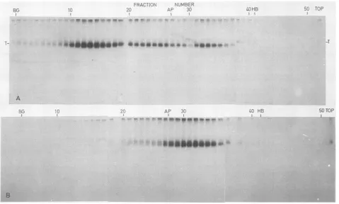
Separation and characterization of T-antigen subclasses. Three subclasses ofT antigen from
32P043--labeled
SV40-infected monkey cells were fractionated by zone
velocity
sedimentation:
5S, 7S,
and 14S(Fig. 1A).The distributionofimmunoreactiveTantigenwasdetermined fromimmunedot
blots ofeach fraction. Most Tantigen was found in the 14S
form;
the7S
form varied in amount from one-fifth to abouthalf as much T antigen as the 14S form (Fig. 1A; data not
shown). The
5S
form contained least T antigen. Similarsubclasses were separated previously by sedimentation and
also by gel filtration (4). These T-antigen subclasses were
reported to be stable at high ionic strength, whereas in
another study, a 16S form of T antigen was shown to
dissociate in 1 M NaCl to a 5 to 7S form (15). Thus, to
characterize the subclasses separated bythe present
proce-dure, zonevelocity sedimentation was also carried out with
1 M NaCl (Fig.
1B).
Most ofthe32P043--labeled
Tantigen
sedimented at
SS,
with a shoulder at 7S (Fig. 1B). Since32P043-
labelingdoes notdistinguish between newlysynthe-sized (young) and accumulated (old) T-antigen molecules (41), we conclude that the bulk of oligomeric T antigen is unstable at high ionic strength.
In previous studies, it was assumed that the three
sub-classes corresponded to T-antigen monomers, dimers, and
tetramers (4, 15, 41). Since protein conformation or
modifi-cationcouldinfluencethe sedimentationorchromatographic
behavior of T antigen, we wished to confirm the identity of these species by chemical cross-linking. Several cross-linkers with different bridge lengths were used: DMS (1.1 nm), dithiobis(succinimidyl propionate) (1.2 nm), and
bis(2-[succinimidooxycarbonyloxy]ethyl)
sulfone (1.3 nm). The highest efficiency of cross-linking was obtained with DMS(Fig. 2). The 14S subclass reacted to yield an SDS-stable
form whose electrophoretic migration corresponded to that predicted for a tetramer (Fig. 2). Neither the 5S nor the 7S subclass was cross-linked in the presenceof DMS(Fig. 2)or the other cross-linking reagents under a variety of experi-mentalconditions (datanotshown). Thusthese dataconfirm thatthe 14S and5STantigensrepresentprimarilyatetramer and a monomer, respectively. Although the cross-linked tetramer was obtained with good recovery, the yield ofSS and 7Sformswasreduced aftercross-linking. Thusthe data
do not distinguish whether the 7S form represents a dimer
notdetectable byimmunoprecipitationoranalteredform of monomeric T antigen.
Oligomerization of newly synthesized T antigen. Pulse-chase experiments have demonstrated that newly synthe-sized Tantigen appearsina5to7Sform, whichthen serves as a precursor for the 14S tetramer (13, 17). However, in these studies the SS and 7S subclasses were not resolved. We thereforeconductedpulse-labeling experiments to deter-mine the form of newly synthesized T antigen (Fig. 3). A
VOL.61, 1987 2077
on November 10, 2019 by guest
http://jvi.asm.org/
2078 RUNZLER ET AL.
".c. .- TO-P
A
J3
SCTOP
FIG. 1. SeparationofT-antigensubclassesbyzonevelocitysedimentation. Anextractof32PO43--labeled SV40-infectedTC7 cells(44h postinfection)wasfractionatedbyzonevelocitysedimentation(SW55, 45,000rpm,16h)insucrosegradientsmadeupin buffer Acontaining 150mMKClasdescribed in Materials and Methods(A)orinagradientalsocontaining1 M NaCl(B).Each fractionwasimmunoprecipitated
with hamstertumorserumand S.aureus,eitherdirectly(A)orafter dilutionto300 mM NaCl(B),and thenanalyzed bySDS-PAGE(27)and
autoradiography. Sedimentation markers inparallel gradientswere,-galactosidase (BG), alkalinephosphatase (AP),andhemoglobin (HB),
asindicated.
prominent peak of T antigen labeled in a 15-min period
sedimented at 5S (Fig. 3A), whereasTantigenlabeledfor 2.5 h wasfound inboth the 5S and 14Sforms (Fig. 3B). Apeak
ofTantigenin the 7Sformwas notobservedafterthe15-min
labeling period, but a trace of 7ST antigen was detectable
after the 2.5-h labeling period (Fig. 3B). These results
demonstrate that newly synthesized T antigen occurs
pri-marily in amonomeric form, which is subsequently
assem-bled intotetramers, but the identityof the 7S form remains
puzzling. Ifthe 7S Tantigenwerean assembly intermediate
with a brief half-life, it might be detected by pulse-chase
experiments. SV40-infectedcells were thus labeled early or
lateafter infectionfor 15 min and chased for 15, 30, or 60 min
(not shown). Although traces of 7S T antigen were detected inthe 60-min chase, no prominent peak of 7S T antigen was observed with any of the chase periods tested. However, it was interesting that oligomerization of T-antigen monomers proceeded significantly faster at 24 h after infection than at
48 h(data notshown), as reported recently (50).
These data could be interpreted to suggest that 7S T
antigenmaybeaverytransient intermediate in the assembly
oftetramers. However, this possibility seems unlikely, be-cause the 7S subclass represents a major fraction of the
32P043--labeled
T antigen and of the total mass ofim-munoreactive Tantigen (Fig. 1A and 4B; data not shown).
The other, more likely, alternative is that the 7S subclass
consistsprimarilyof accumulated T antigen, possibly arising
throughdissociation of 14S tetramers, either in the cell or in
vitro.
Binding ofT-antigen subclassestoSV40 DNA. Some of the
conflictingresults obtained in previousstudies described in
the Introduction can be explained by the use of frozen or
partially purifiedmaterials rather than fresh cellextractsand
bytheuse ofmutant(D2 andSV80) Tantigensrather than thewild-type protein. However, it is also possible that the assays used are responsible for some ofthe disagreement.
Forexample,mostassayswereperformed bythe method of
McKay (30) in crudeextractwithexcessunlabeled nonspe-cific DNA. Binding was generally carried out at 0°C for
periods too short for equilibrium to be reached (49; R.
Runzler and E. Fanning, unpublished data), and T-antigen concentrations differed from one subclass to another.
Fi-nally,therelation between thereported efficiencyof binding
and the commonly used parameters, specific activity of
bindingandbinding affinity, was notdefined.
Thus,torule outtheDNA-bindingassay as the sourceof
the conflicting results, we used a sensitive target-bound
assay carried out in the absence of unlabeled competitor DNA and cellular proteins (20). Equal volumes of each subclasswereimmunopurifiedonPab 108 IgG, an antibody that has no detectable effect on DNase footprints of T
antigen (38; VakalopoulouandFanning, unpublished data).
The amount of SV40 DNA bound at equilibrium in DNA excess wasdeterminedby usingtwotemplates, pSV-wt and
pON-wt DNA, either separately or together (Fig. 4A). A
comparison ofthe amount ofbound origin DNA with the
amountof immunoreactiveTantigenin eachsubclass (Fig. 4B) revealed that 5S T antigen bound considerably more
,..!. A&AMLA&AWK-MIL-Alikffikh,
0 w.wlw: Wwwwassalka:41k loooow
71- AP
.1. .,.::. .. -I1W.am"to Pttomow W...
J. VIROL.
-T
4..H, e.
on November 10, 2019 by guest
http://jvi.asm.org/
[image:3.612.72.551.74.362.2]DNA-BINDING PROPERTIES OF T-ANTIGEN SUBCLASSES
5S 7S 14S
X-LINK + - + +- + +- +
T T NTTNTTN
376
-282
-I
do p
948
-FIG. 2. Chemical cross-linking of separated T-antigen sub-classes. 32PO4--labeled T-antigen subclasses were separated as described for Fig. 1A. Appropriate fractions were combined, di-vided into three samples, and adjusted to 50 mM triethanolamine
(pH8.5)-i mM MgCl2. Two samples (+) were cross-linked with 1
mMDMS as described in Materials and Methods. The samples were immunoprecipitated with nonimmune hamster serum (N) or with hamster tumor serum (T) as indicated and analyzed by SDS-PAGE (50)followed byautoradiography for 15 days.Themolecularmasses (in kilodaltons) of DMS-cross-linked phosphorylase a subunits are indicated on the left.
originDNAthan anequivalentamountofthe 14Sform did.
Thus theresults obtained with the new assay confirm previ-ous reports that monomeric T antigen binds more origin DNA(12, 14, 15, 33, 41). Moreover,pON-wtDNAappeared to competewith pSV-wtDNAforTantigen,suggestingthat their binding affinities may be similar. Differences between
subclasses either in binding affinity or in the fraction of
T-antigen molecules active in origin binding, i.e., specific activity of binding, could account for the observed results.
Affinity of T-antigen binding to SV40 DNA.DNA binding of immunopurified T-antigen subclasses was assayed as a
func-tion ofDNA concentration by using pON-wt and pSV-wt
DNA. The binding data were treated by the method of
Scatchard (39) to determine an equilibrium dissociation
constant (KD) for DNAbinding of each form ofT antigen.
Multiple determination ofKD carried out with subclasses
prepared at 39 to 40 h and at 48 h after infection yielded
similar values, which were averagedtogive aKD of about
0.4 nM for all three subclasses on pSV-wt DNA (Table 1).
Thedissociationconstantsfor the three subclassesmeasured
onpON-wtDNA werealsoverysimilar to each other and to
that determined on pSV-wt DNA (Table 1). Thus within
experimental error, T-antigen binding affinity to site I in
SV40 DNA does notdifferamong subclasses. Theseresults
indicatethat theobserved differencesinDNA-binding
activ-ity ofthesubclasses(Fig. 4)mustderive
primarily
from theirdifferent specific activities of origin binding.
Specific activity of T-antigen DNA binding. A valid
com-parison ofthe specific activities of
origin
binding oftheseforms requiresthat binding experiments be
performed
withequalconcentrations ofTantigen fromeach subclass.
Dilu-FRACTION NUMBER
BG 10 20 AP 30 HB 40 50 TOP
I ~~~~~~I~ ~ , I
1-tA~~~~~~~~~~~~~~~~~~~~~~~~~~~~~~~~~~~~~~~~~~~~~~~~~~~~~~~tn
tt*t,';:S_5^=-';i7s S -Xho wt!,X' C; ;ff ;000 S; ,,7m~~~~~~~~~~~~~~~~~~~~~~~~~~~~~~~~~~~~~~~~~~~~~~~~~~~~~~~~~~~Z.;S,0t
T-f#f;t*#iP%0 6 S l;.; tS 04,1S~~~~~~~~~~~~~~~~~~~~~~~~~~~~~~~~~~~~~~~~~~~~.f?S 0
BG
_ 6%
10 20 AP 30
l
HB 40 50 TOP
...,tsaN\a,
R4 'Mi ,.;*iX; | ^6& t
*...e:voP:'#I,§EM
>>e. .. ._,B;
FIG. 3. Fractionation ofnewly synthesizedTantigen byzonevelocity sedimentation. SV40-infected TC7 cellswerelabeledat42hafter infection with[35S]methionine(250,uCi/ml,800to1,000Ci/mmol)for 15min(A)or2.5h(B). Cellextractswerefractionatedbyzonevelocity sedimentationasinFig. 1A, except that sedimentationwasfor21.5h.Tantigen in each fractionwasimmunoprecipitatedwith hamstertumor
serumandanalyzed by SDS-PAGE (27)andfluorography. The positions of sedimentation markersasinFig. 1Aareindicated.
...-a &
2079 VOL.61, 1987
on November 10, 2019 by guest
http://jvi.asm.org/
[image:4.577.94.232.73.254.2]2080 RUNZLER ET AL.
l)£5 r7511,t -4
JSs
751d4S
psv-.wt [)pSV--wt M .h15S 7$ 14S 5S
....
..
^7S 14Lt-...X
A20- B
C~~~~~~~~~~~~
10
10~~~~~~~
5-~~ ~ ~ ~~~~5
5 10 20
30
50,
volume (utl)
FIG. 4. T-antigen bindingto SV40 DNA. T-antigen subclasses were preparedfrom SV40-infected cellsat 48 hpostinfection. (A) Samples of each subclass (0.1ml)wereimmunopurifiedon Pab 108 andassayed forbindingtoequimolaramountsofpSV-wt,pON-wt, or amixture of thetwo DNAfragments for2 hat0°C asdescribed inMaterials and Methods. Binding assaysincludedpAT153 vector DNAfragments whennecessary tomakeatotal of250 ngofDNA in each assay. MarkerDNA(M) was12.5 ng ofthelabeled input DNA. Arrows indicate fragments containing T-antigen-binding sites. (B) T-antigen subclasses of the indicated volumes were
applied to nitrocellulose and analyzed by immune dot blot and microdensitometry. Symbols: 0, 5S; x, 7S; *, 14S.
tion ofT-antigenoligomers,for example,might be predicted to promote dissociation into subunits more active in DNA
binding. Thus the concentration of T antigen in each
sub-class was first determined by immune blot, and the
oligo-meric forms werediluted to theconcentration of the mono-mer. ImmunopurifiedTantigen from each subclass was then assayedforbinding to SV40 originDNA at several concen-trations ofTantigen.
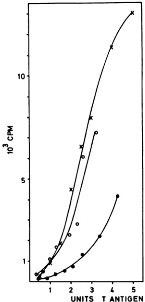
The specific activity of DNA binding was greatest for 5S and7S T antigens at all concentrations tested (Fig. 5), but
therelativespecific activity of binding of the5S and 7S forms
varied slightly among experiments. The 5S and 7S forms
bound five- to sevenfold more origin DNA than an
equiva-lent amount of 14S T antigen did. Similar results were obtained withpON-wtDNA(data not shown). The data are
consistentwith previous results (Fig. 4) (12, 14, 15, 33, 41)
and suggest that the fraction of the 14S T antigen active in
origin
DNA bindingis much smaller than that of the5SandTABLE 1. Affinity of T-antigen subclasses forSV40DNA and a synthetic 19-base-pairsequencefrombindingsite I
KD(nM)a
Template
5S 7S 14S
pSV-wt 0.4 0.1b 0.5 ± 0.3b 0.3 0.2b pON-wt 0.3 ± 0.2c 0.3 ± 0.2c 0.2 ± 0.05c
aThe ratioof boundtounbound DNAwas plotted as afunction ofthe
concentration of bound DNA (39). Slopes(KD1)weredeterminedby using a Hewlett-Packard programfor linearregression analysis. Correlation coeffi-cients were 0.9 orgreater.
bMeanoffiveindependentdeterminations,two at39 h after infectionand three at 48 hafter infection. Theerrorisreportedasthe average deviation from the mean.
CMeanofthree orfourindependent determinations, one at40 h andtwo (7S) or three (5S, 14S) at 48 h after infection. The error isreported as the averagedeviation from themean.
7S subclasses.Interestingly,thebindingcurve with allthree subclasses,butparticularlythe14S subclass, was reproduc-ibly nonlinear when a wide range ofT-antigen concentra-tions was tested; interpretation of this result will require further work.
10l
C)
0"
5
1 2 3 4 5
UNITS T ANTIGEN
FIG. 5. DNA-binding activity of T-antigen subclassesas a func-tion ofT-antigen concentration. T-antigen subclasses preparedas forFig. 4were assayed for immunoreactiveTantigen by immune dot blot (data not shown). Stock solutions of all three subclasses were adjusted to 10 mM HEPES (pH 7.8)-80 mM KCl-0.5 mM MgCl2-1mM dithiothreitol andequalconcentrationsofT antigen. Increasingvolumesofeachsubclasswereimmunopurifiedand then tested for specific binding to pSV-wt DNA fragments in 0.11-ml assays. Bound origin DNAfragments wereexcised from the gels, counted,andplottedas afunction of the amount ofimmunoreactive Tantigenin the assay. One unit of Tantigen isempirically defined
astheamountof Tantigenin 0.1 mlofthestock solutions.Symbols: 0,5S; x, 7S; *, 14S.
J. VIROL.
on November 10, 2019 by guest
http://jvi.asm.org/
[image:5.612.87.275.72.425.2] [image:5.612.315.557.95.149.2] [image:5.612.364.508.310.610.2]DNA-BINDING PROPERTIES OF T-ANTIGEN SUBCLASSES
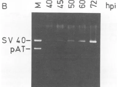
4C 45 Sr 50 72
V 1 2 3 1' 2 3 1 Z'3 ', 2 3 I 2 3
0.~~ ~~~~ -
'7-...~~~~~~~~~~~~E
00
ia
0.*^&^^
^^&
15-i
0
a>
jsg
u cD 5-iCt Un
cI
/~
~~~~~~~~~
"^~~~~~~~~
,."'__,hpi
subclass
B
hpi
SV
40-
pAT-FIG. 6. Specific activity of T-antigen binding toSV40 DNA as a function of time afterinfection (hpi).(A) T-antigen subclasses were preparedatthe indicated times after infection. Samples (0.1 ml) of 5S (lane 1), 7S (lane 2), and 14S (lane 3) T antigen were im-munopurified and assayed for specific binding to pSV-wt HindIlI fragments as for Fig. 5. (B) A parallel dish of infected cells was harvested at each time after infection. The low-molecular-weight DNA was extracted (21), linearized by BamHI digestion, and analyzed by agarose gel electrophoresis. BamHI-cleaved pSV-wt DNAserved as marker (M). (C) TheboundHindIII-C DNA was cut from the gel in panel A, solubilized, and counted. The amount of T antigen inasample of each subclass was determined by immune dot blot inarbitrary units. The countsper minute bound per unit ofT antigen (specific activity) was calculated for each subclass at each time afterinfection.Symbols:0, 5S; x, 7S; *, 14S; A, accumulated SV40DNAdeterminedby microdensitometry of the gel in panel B.
DISCUSSION 6
-4
*I
F._
-T'r-L0 45 50 60 72h
time after infection
Specific activity of T-antigen-DNA binding varies with time afterinfection. TheDNA-binding activity of immunopurified Tantigen subclasses prepared at various times after infec-tion was monitored by using pSV-wt DNA in excess (Fig. 6A).TheamountoforiginDNAboundby duplicate samples of T antigen (equal volumes of each subclass) was deter-mined by scintillation counting of the excisedgel bands (data not shown). The amount of T antigen in each sample estimated by immune blot was then used to calculate the specific activityof DNA bindingas afunctionof time after infection(Fig.6C). Theamountof viral DNA accumulatedat each time afterinfection(Fig. 6B)wasestimatedinarbitrary unitsby microdensitometry (Fig. 6C).
The results demonstrate thatalthoughthespecific activity of DNA binding ofT-antigen tetramers remains at a rela-tively constant low level throughout infection, the specific activityof5STantigenincreasessharplybetween40 and50 h after infection. A similar sharp increase in the specific activity of7STantigenoccurred with adelay ofabout 5 h. The rise in binding activitywas temporallycorrelated with the onset of viral DNA replication and the late phase of infection (Fig. 6C).
Relationship among different subclasses of T antigen. We
havedemonstratedthat Tantigen occursin threeoligomeric
subclasses separable by zone velocity sedimentation: a5S monomer, a 14S tetramer, and a 7S form which could be either a dimer or a modified monomer (Fig. 1 and 2).
PreliminaryresultsontheDNA-binding kineticsand
stoichi-ometry of the 7S form, however, are consistent with a
dimeric mass (R. Runzler, E. Vakalopoulou, and E.
Fan-ning, unpublished data). Newly synthesized T antigen
oc-curs as a monomer which serves as a precursor for a tetrameric form. The bulk of the 7S T antigen, a major fraction by mass and by incorporation of phosphate, is
probablynot anintermediate in theassembly oftetrameric T
antigen (Fig. 1A, 3, and 4B; unpublished data). The
tetrameric form appearstobequitestable in vivo(13, 15, 41) and invitroinsolutions ofneutralpHandmoderate
concen-trations of salt and divalent cations (12, 31, 41). However,
dissociation of thetetramerisobservedathighionicstrength
(15; Fig. 1B), in the presence ofchelating agents (31), and
uponchromatography (12).Takentogether,the data support
the idea that a portion of the highly phosphorylated
tetrameric T antigen may dissociate in vitroor in the cell,
generating 7S and possibly even 5S forms.
Consequently,
the 5S and 7S subclasses each comprise a heterogeneous
population, some representing newly synthesized Tantigen
and some derived by dissociation from tetramers. The
het-erogeneityof the 5Sto7S
T-antigen
phosphorylation
pattern(41) providesfurtherevidence for this
interpretation.
OriginDNAbindingofT-antigensubclasses.The
affinity
ofbinding of each subclass to SV40 DNA wasdetermined
by
measurementof boundDNAas afunction of DNA
concen-tration with two different templates
(Fig.
4; Table1).
The results showclearly
that all threesubclasses, despite
theirVOL.61, 1987 2081
C) Ln CD CD CN
2
1 -4 LO LDon November 10, 2019 by guest
http://jvi.asm.org/
[image:6.612.343.536.74.217.2]2082 RUNZLER ET AL.
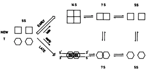
us 7S SS
5S
NEW
e S'4 -==
[image:7.612.62.295.70.183.2]7S SS
FIG. 7. Model for binding of T-antigen subclassestoSV40DNA.
Newly synthesized T antigenassumeseither oftwoconformations,
oneabletobind viral DNA specifically (hexagons)andoneunable to
bindbut which assembles intoatetramer(squares).Sometetramers candissociatetoyield5S and7STantigen, whichcanalsoassume
either of thetwoconformations.A dimermassof Tantigenbindsto
pON-wt DNA, which contains two consensus pentanucleotides (hatched boxes), but the bindingmechanism is unknown(38). The
molecularregulation of T-antigen distributionin thetwo
conforma-tions is thesubject of speculation in thetext.
heterogeneity, have similar affinities for siteIDNA. TheKD measured in our work is very similar to that reported previously forhighly purified wild-type T antigen (23) and T antigen from mKSA SV40-transformed mouse cells(20). A 10-fold-lower affinity calculated for purified lytic T antigen (11) was basedon the assumption that all ofthe protein is
potentially able to bindtoorigin DNA, an assumption that
may not be valid (Fig. 5 and 6) (20, 42; A. Schmid and E. Fanning, unpublished data).
The specific activity was measured for each subclass as the amount of SV40 DNA specifically bound per im-munoreactive unit of T antigen. Under the conditions of the
assay,adifferenceamongsubclassesinthespecific activities of binding may be attributed to a difference among sub-classes inthefraction of the total protein molecules ableto bind the template. The results demonstrate clearly that the specificactivity of origin DNA bindingonthesetemplates is
greatest for 5S T antigen and least for tetrameric T antigen (Fig. 4to 6).
Itis importantto note thatalthough theantibody Pab 108 immunoprecipitates T antigen quantitatively, not all of the N-terminalepitopes in eachtetramerneed be availabletothe antibody. Thus the amount of immunoreactive Tantigen in the 14S form could be an underestimate of the mass ofT antigen in the 14S subclass by as much as a factor of 4. Therefore, the specific activity of the 14S T antigen mea-sured inthis workmustbe regarded as a maximum value.
The specific activity of binding of 5S and 7S T antigen increasedduring the infection, whereas that of thetetramer
remained at a constant low level (Fig. 6). This result could also be accountedfor by increased affinity ofT antigen for SV40 DNA at late times after infection. However, the affinity of T-antigen subclasses for SV40DNAdid notvary
significantly with time after infection (Table 1). Thus the change inbinding activity of the 5S and 7S formsis probably duetoanincrease in the fractionof molecules active in DNA binding atlatetimes after infection, whereas thefraction of material in the tetrameric subclass ableto bind originDNA
remainsapproximately constant.
A modelfor oligomerization and origin DNA binding of T antigen. We have summarized the data available into a
simple model correlating the oligomerization of T antigen with itsorigin-binding activity (Fig. 7). Weproposethat 5S
newly synthesized monomeric Tantigencan take upeither
of twoconformations, onewhich canbindoriginDNA and onewhich cannot. Forsimplicity, thetwoconformationsare shown in equilibrium, although it is likely that their phos-phorylation state, other posttranslational modifications, or the presenceof otherligands determines the distribution of T antigen in the two conformations (5, 41, 42, 49). The stoi-chiometry of binding shown here is that for pON-wt DNA (38), but the kinetic mechanism of binding is not known.
The tetramer is postulated tobe assembleddirectly from newly synthesized monomersandtobe fixed ina conforma-tion unable to bindorigin DNA aslongasit remainsstable. Protein phosphorylation-dephosphorylation reactions, as well as divalent metal cations, may be important in main-taining this stability(2, 31, 41). Dissociation of the tetramer, which is known to occur under a variety of conditions, generates monomers and dimers, which, according to the model, can take up either conformation. Thesemonomers or dimers could be responsible for the observed origin
DNA-binding activity of the tetramer, aprediction which can be
testedexperimentally.
How might the course of the infection influence the
distribution ofmonomers and dimers in the two conforma-tions? It was noted previously that oligomerization of newly
synthesized lytic T antigen was more complete when viral
DNA replication was inhibited (15). More recently it was shown that oligomerization of newly synthesized lytic T
antigen proceeded more rapidly before the onset ofDNA
replication than later in infection (50), a finding which we
have confirmed(datanotshown). We speculateonthebasis ofthese data that the increase in specific activity oforigin DNAbinding observed for monomers and dimers (Fig. 6)is related tothe slowerrate ofoligomerization (Fig. 7). Since the rate ofoligomerization canbereacceleratedbyinhibition of viral DNA synthesis (50), this possibility may be tested experimentally.
Theproposedmodelisconsistentwith the datapresented
here, provides a useful framework to integrate the rather
confusing literature on T-antigen subclasses, and hence
provides a starting point for more detailed structural and biochemical studies.
ACKNOWLEDGMENTS
Wethank AndreaSchmid, Ursula Markau, and Silke Dehde for excellent technical assistance, Jean-Bernard Dietrich for prelimi-naryexperiments on chemicalcross-linking, Monika Westphal for introducingus toimmune dotblots, BradJamesonandHansWolf for help with microdensitometry, and Avril Arthur for helpful
comments onthemanuscript.
Thefinancial support of the Deutsche Forschungsgemeinschaft (Fa 138-1/1 and 138-1/2) and Fonds der Chemischen Industrie is gratefully acknowledged.
LITERATURECITED
1. Adams, J. 1981.Heavymetalintensification ofDAB-basedHRP reactionproduct. J. Histochem. Cytochem. 29:775.
2. Baumann, E. A., and R. Hand. 1982. Phosphorylation and dephosphorylationalter the structureofD2 hybrid T antigen. J. Virol. 44:78-87.
3. Baumgartner, I., C. Kuhn, and E. Fanning. 1979. Identification and characterization offast-sedimenting SV40 nucleoprotein complexes. Virolgy96:54-63.
4. Bradley,M., J.Griffin,and D. M.Livingston. 1982.Relationship ofoligomerizationtoenzymaticand DNAbindingpropertiesof theSV40large Tantigen. Cell28:125-134.
5. Bradley, M. K., J. Hudson, M. Villanueva, and D. M. Livingston. 1984. Specific in vitroadenylylation of the simian virus 40 large T antigen. Proc. Natl. Acad. Sci. USA 81:6574-6578.
J. VIROL.
on November 10, 2019 by guest
http://jvi.asm.org/
DNA-BINDING PROPERTIES OF T-ANTIGEN SUBCLASSES 6. Brady, J., J. Bolen,M.Radonovich,N.Salzman,and G.Khoury.
1984. Stimulation of simian virus 40 late gene expressionby simian virus 40 tumor antigen. Proc. Natl. Acad. Sci. USA 81:2040-2044.
7. Brady, J., G.Khoury.1985.trans-Activation of the simianvirus 40 late transcription unit by T antigen. Mol. Cell. Biol. 5:1391-1399.
8. Covey, L., Y. Choi, and C. Prives. 1984. Association of simian virus 40 T antigen with the nuclear matrix in infected and transformedmonkey cells. Mol. Cell. Biol. 4:1384-1392. 9. DiMaio, D., and D. Nathans. 1980. Cold sensitive regulatory
mutantsof simian virus40. J. Mol. Biol. 140:129-142. 10. DiMaio, D.,and D. Nathans.1982.Regulatorymutantsof simian
virus 40. Effectof mutationsat a Tantigen binding siteonDNA replication and expression of viral genes. J. Mol. Biol. 156:531-548.
11. Dixon, R., and D. Nathans. 1985.Purification of simian virus40 large T antigen by immunoaffinity chromatography. J. Virol. 53:1001-1004.
12. Dorn, A., D. Brauer, B. Otto, E. Fanning, and R. Knippers. 1982. Subclasses of simian virus 40largetumorantigen.Partial purification and DNA binding properties oftwo subclasses of tumor antigen from productively infected cells. Eur. J. Bio-chem. 128:53-62.
13. Fanning, E., B. Nowak, and C. Burger. 1981. Detection and characterization of multiple forms of simian virus 40 large T antigen. J. Virol. 37:92-102.
14. Fanning, E., K.-H. Westphal, D. Brauer, and D. Corlin. 1982. Subclasses of simian virus 40 large T antigen: differential binding of two subclasses of T antigen from productively infected cellstoviral and cellular DNA.EMBOJ. 1:1023-1028. 15. Gidoni, D., A. Scheller, B. Barnet, P. Hantzopoulos, M. Oren, and C. Prives. 1982. Different forms of simian virus 40 large tumor antigen varying in their affinities for DNA. J. Virol. 42:456-466.
16. Goldman, N.,M.Brown,andG.Khoury. 1981. Modification of SV40 Tantigenbypoly-ADP-ribosylation. Cell 24:567-572. 17. Greenspan, D.,and R.Carroll.1981.Complexof simian virus40
largetumorantigenand48,000-daltonhosttumorantigen.Proc. Natl. Acad. Sci. USA 78:105-109.
18. Gurney,E. G.,S.Tamowsky,and W.Deppert. 1986. Antigenic bindingsites of monoclonal antibodiesspecificforsimian virus 40largeT. J. Virol. 57:1168-1172.
19. Hansen, U., D. Tenen, D. Livingston, and P. Sharp. 1981. T antigen repressionofSV40earlytranscriptionfromtwo promot-ers. Cell27:603-612.
20. Hinzpeter, M., E. Fanning, and W. Deppert. 1986. A new
sensitive target-bound DNA binding assay for SV40 large T antigen. Virology 148:159-167.
21. Hirt, B. 1967. Selective extraction of polyoma DNA from infectedmouse cellcultures.J. Mol. Biol. 26:365-369. 22. Huber, B., E. Vakalopoulou, C. Burger, and E. Fanning. 1985.
Identification and biochemical analysis of DNA
replication-defectivelargeT antigensfromSV40-transformed cells. Virol-ogy146:188-202.
23. Jones, K. A., and R. Tjian. 1984. Essential contact residues within SV40largeTantigenbindingsitesIand II identifiedby alkylationinterference. Cell 36:155-162.
24. Keller, J., and J. Alwine. 1984. Activation of the SV40 late promoter: directeffects ofTantigenintheabsenceof viralDNA replication. Cell 36:381-389.
25. Keller, J., and J. Alwine. 1985. Analysis of an activatable promoter: sequences in the simian virus 40 late promoter required for T-antigen-mediated trans-activation. Mol. Cell. Biol. 5:1859-1869.
26. Klockmann,U.,and W.Deppert.1983.Acylatedsimianvirus 40 large T antigen: a new subclass associated with a
detergent
resistant lamina of the plasma membrane. EMBO J. 2:1151-1157.
27. Laemmli,U. K. 1970.Cleavageofstructuralproteins
during
the aasembly of the head ofbacteriophage T4. Nature (London)227:680-685.
28. Margolskee, R.,and D. Nathans.1984.Simianvirus 40mutantT
antigenswithrelaxed
specificty
for the nucleotidesequenceatthe viraloriginof
replication.
J. Virol. 49:386-393.29. Mastrangelo, I.,P.Hough,V.Wilson,J.Wall,J.Hainfeld,and P.Tegtmeyer. 1985. Monomers
through
trimers oflarge
tumorantigenbindinregionIandmonomersthroughtetramersbindin
regionIIof simian virus40originof
replication
DNAasstablestructures in solution. Proc. Natl. Acad. Sci. USA 82:3626-3630.
30. McKay,R. D. G. 1981.
Binding
ofasimian virus40 Tantigen-relatedproteintoDNA. J. Mol. Biol. 145:471-488.
31. Montenarh, M., and R. Henning. 1983.
Disaggregation
and reconstitution ofoligomeric complexesof simian virus40large
Tantigen.J. Gen. Virol. 64:241-246.
32. Myers,R., R. Williams, and R. Tjian. 1981.
Oligomeric
struc-ture ofa simian virus 40 T antigenin free form and boundtoDNA. J. Mol. Biol. 148:347-353.
33. Oren, M.,E. Winocour,andC. Prives.1980. Differential
affin-ities of simian virus40
large
tumorantigen
for DNA. Proc. Natl. Acad. Sci. USA 77:220-224.34. Putney,S.,R. T. Sauer,and P. R.Schimmel. 1981. Purification and
properties
of alanine tRNAsynthetase
from Escherichia coli. J.Biol. Chem. 256:198-204.35. Rigby, P., and D. Lane. 1983. The structure and function of SV40
large
Tantigen.
Adv. Viral Oncol. 3:31-57.36. Rio, D.,A.Robbins,R.Myers,and R.Tjian.1980.
Regulation
ofsimian virus 40
early
transcription
in vitroby
apurified
tumorantigen. Proc.Natl. Acad. Sci. USA77:5706-5710.
37. Robb J., and K. Huebner. 1973. Effect of cell chromosome number on simian virus 40
replication. Exp.
Cell Res. 81:120-126.38. Ryder, K., E. Vakalopoulou, R. Mertz, I.
Mastrangelo,
P.Hough, P. Tegtmeyer, and E. Fanning. 1985. Seventeen base
pairsof
region
Iencodeanoveltripartite binding signal
forSV40T
antigen.
Cell 42:539-548.39. Scatchard,G. 1949. Theattractionsof
proteins
forsmallmole-cules andions. Ann. N.Y. Acad. Sci. 51:660-672.
40. Scheidtmann, K.-H., B. Echle, and G. Walter. 1982. Simian
virus 40 large T
antigen
isphosphorylated
atmultiple
sites clustered intwoseparateregions.
J.Virol. 44:116-133.41. Scheidtmann, K.-H., M. Hardung, B. Echle, and G. Walter.
1984.
DNA-binding activity
of simian virus40large
Tantigen
correlates with a distinctphosphorylation
state. J. Virol. 50:1-12.42. Scheller, A.,L.Covey,B.Barnet,andC. Prives. 1982. Asmall
subclass ofSV40T
antigen
bindstotheviralorigin
ofreplica-tion. Cell 29:375-383.
43. Shortle, D., R. Margolskee, and D. Nathans. 1979. Mutational
analysis
of simian virus 40replicon:
pseudorevertants
of mu-tantswithadefectivereplication origin.
Proc. Natl.Acad. Sci. USA 76:6128-6131.44. Stahl, H., P. Droge, and R.
Knippers.
1986. DNA helicaseactivity
ofSV40large
tumorantigen.
EMBOJ. 5:1939-1944.45. Staufenbiel, M., and W.
Deppert.
1983. Different structuralsystems of thenucleusaretargetsforSV40
large
Tantigen.
Cell 33:173-181.46. Tooze,J. 1981. Molecular
biology
oftumorvirus: part2. DNAtumorviruses, 2nd ed. Cold
Spring
HarborLaboratory,
ColdSpring
Harbor,N.Y.47. Twigg,A., and D. Sherratt. 1980.
Trans-complementable
copynumber mutants of
plasmid
ColEl. Nature (London)283:216-218.
48. VanRoy, F., L.Fransen, and W. Fiers. 1983.
Improved
local-ization ofphosphorylation
sites in simian virus 40large
Tantigen.
J. Virol. 45:315-331.49. Vogt, B., E.
Vakalopoulou,
and E. Fanning. 1986. Allostericcontrol ofsimian virus40
T-antigen binding
toviralorigin
DNA. J. Virol. 58:765-772.50. Wachter, G.Riedle,and R.
Henning.
1985. Functionalimplica-tionsof
oligomerization
ofsimian virus40large
Tantigen during
lyticvirus infection. J. Virol. 56:520-526.
51. Weber, K., and M. Osborn. 1969. The
reliability
ofmolecularweight determinations
by dodecyl
sulfate-polyacrylamide gel
electrophoresis.
J. Biol.Chem. 244:4406-4412.VOL.61, 1987 2083