DISSERTATION ON
COMPARATIVE STUDY OF RISK FACTORS BETWEEN ISCHEMIC AND HEMORRHAGIC STROKE
Submitted in partial fulfilment of Requirements for
M.D. DEGREE BRANCH I GENERAL MEDICINE
Of
THE TAMILNADU DR.M.G.R. MEDICAL UNIVERSITY, CHENNAI.
MADRAS MEDICAL COLLEGE CHENNAI – 600 003.
CERTIFICATE
This is to certify that this dissertation entitled “COMPARATIVE STUDY OF RISK FACTORS BETWEEN ISCHEMIC AND HEMORRHAGIC STROKE” submitted by Dr. G. Ravi appearing for Part II M.D. Branch I General Medicine Degree examination in September 2006 is a bonafide record of work done by him under my direct audience and supervision in partial fulfillment of regulations of the Tamil Nadu Dr. M.G.R. Medical University, Chennai. I forward this to the Tamil Nadu Dr.M.G.R. Medical University, Chennai, Tamil Nadu, and India
Prof.C.Rajendiran., M.D., Unit Chief
Institute of Internal Medicine
Government General Hospital Chennai - 3
Director,
Institute of Internal Medicine,
Government General Hospital,
Chennai – 600 003.
Dean,
DECLARATION
I solemnly declare that the dissertation titled “COMPARATIVE STUDY OF RISK FACTORS BETWEEN ISCHEMIC AND HEMORRHAGIC STROKE” is done by me at Madras Medical College & Govt. General Hospital, Chennai during 2003-2005 under the guidance and supervision of Prof.V.Sundaravadivelu., M.D.,
The dissertation is submitted to The Tamilnadu Dr. M.G.R. Medical University towards the partial fulfillment of requirements for the award of M.D. Degree (Branch I) in General Medicine.
Place: Chennai
Dr.Ravi. G,
M.D. General Medicine Postgraduate Student
Institute of Internal Medicine Madras Medical College Chennai
ACKNOWLEDGEMENT
I am extremely grateful to my Dean Dr.Kalavathy Ponniraivan., Bsc., M.D., for granting me permission to conduct the study in Government General Hospital and Madras Medical College, Chennai - 3
I would like to express my sincere gratitude to my beloved
Professor and Director, Institute of Internal Medicine, Prof. V.Sundaravadivelu M.D., for his guidance and encouragement.
With extreme gratitude, I express my indebtedness to my beloved Chief Prof.C.Rajendiran., M.D., for his motivation, advice and valuable criticism, which enabled me to complete this work.
I thank Mr.Vengatesan, Statistician, Institute of Social and Preventive medicine, Stanley Medical College for his immense help in statistical analysis.
I am also thankful to my family members for their cooperation in finishing my dissertation.
I would always remember with extreme sense of thankfulness for the co-operation and criticism shown by my Postgraduate colleagues.
CONTENTS
Sl. No. Title Page No.
1. Introduction 1
2. Objectives of the study 2
3. Review of Literature 3
4. Materials and Methods 38
5. Statistical analysis 39
6. Observations 40
7. Charts & Graphs
8. Discussion 46
9. Conclusion 51
INTRODUCTION
Stroke or cerebrovascular accident by definition is a syndrome of rapidly developing clinical signs of focal or global disturbance of cerebral circulation with symptoms lasting 24hrs or longer or leading to death with no apparent cause other than of vascular origin. [1] Disorders of cerebral circulation include any disease of the vascular system that causes ischemia or infarction of the brain or spontaneous hemorrhage into the brain or subarachnoid space.
In India, for every 55 seconds one case of stroke has been reported. To presume, for every 5 minutes someone is dying of stroke. [2] Identification and control of modifiable risk factors is the best strategy with excellent choices on the anvil to scale down the incidence of stroke. Also for good measures, the number of cases can be decimated by leaps and bounds.
One large collaborative study did not find a clear association between total cholesterol and risk of stroke, but included only fatal strokes and there was no separation of ischemic from hemorrhagic stroke. [5]
In order to better define the risk factors of ischemic stroke, it is necessary to more precisely diagnose the patients using comprehensive investigations including neuroimaging as well as cerebrovascular and cardiac evaluation.
In Developing countries like India it is very difficult for the patients to afford for the post stroke treatment like taking MRI in the aid of thrombolysis, surgeries for ICH, and other supportive therapies like physiotherapy, speech therapy etc. Because of these shortcomings, we are losing more number of patients than in western countries.
Clinical trails showed that if the risk factors like Systemic hypertension, Diabetes mellitus, Dyslipidemia, smoking etc. are modified and treated well in advance, then both the morbidity and mortality rates due to stroke can be brought down to an amazingly lower level.
OBJECTIVES
To compare the clinical findings and risk factors in cases of hemorrhagic and ischemic stroke comprehensively.
REVIEW OF LITERATURE
Clinically stroke may be caused by either ischemia or hemorrhage of which, ischemia is more common. Some of the entities of stroke are
I. ISCHEMIA
- Transient ischemic stroke (TIA)
- Reversible ischemic neurological deficit (RIND) - Completed Stroke
- Multi infarct dementia II. Hemorrhage
- Intracerebral Hemorrhage - Subarachnoid hemorrhage
• Transient ischemic attack (TIA) is an acute loss of focal cerebral or monocular function with symptoms lasting for less than 24 hours. It is usually due to embolic or thrombotic vascular disease rarely with the persistence of residual signs.
includes TIA’s with mild ischemic stroke with nopersistingneurological disability. This is important for long term management and prevention of future stroke.
• A completed stroke is one in which neurological function is lost suddenly or over a few hours: and the deficit persists for more than a day or two.
• Multi infarct dementia is deterioration of previously normal life and or memory due to repeated clinical or sub clinical episodes of ischemia, infarction or hemorrhage in cerebrum.
• Intracerebral Hemorrhage means evidence of blood in a compatible location in CT / MRI.
• Subarachnoid hemorrhage requires at least one of the following criteria.
o Headache or coma plus evidence of blood in the subarachnoid space by CT / MRI without intraparenchymal blood.
EPIDEMIOLOGY OF STROKE
India is the only country with fair epidemiological data of stroke available. The limitations of epidemiological study of stroke in India are due to their huge population size, poor income and limited health care resources.
[6]
Incidence: Incidence of stroke is reliably assessed by prospective community based study, hospital based study being biased. Generally incidence increases with age. About one fourth of the cases occur below 65 years and one half below 75 years of age. Of them, cerebral infarct forms the major pathological lesion.
Geographical, racial and social influences: Incidence and mortality varies with each place, race etc. In India comparatively, the incidence of young stroke is higher than western countries. In pregnancy, stroke is more common owing to the venous thrombosis.
Mortality: Stroke is the third leading cause of death after Cancer and heart diseases. Stroke mortality increases with age. Figures from various countries vary every year with the incidence in India being one every five minutes. [2]Sudden death is very rarely caused by stroke. One of the causes of sudden death in stroke is intracerebral hemorrhage. Stroke mortality is higher in winter than in summer. Moreover causes of death may be due to other diseases like pneumonia, associated coronary heart diseases, bedsores etc.
RISK FACTORS FOR STROKE
Age:
Age as such is potentially able to influence the occurrence of stroke. It is one of the strongest proponents for infarction, intracerebral hemorrhage, subarachnoid hemorrhage and TIA. The incidence of stroke and age are directionally proportional. 95% of strokes occur in people aged 45 and older
[10]
and two-thirds of strokes occur in those over the age of 65 [11]. The prevalence of stroke in younger (20-40 yrs.) population in India has been reported to be high (18.8 to 32% of all stroke cases) as compared to similar age group in the US and Europe. Various factors like cerebral emboli from cardiac source, thrombosis of internal carotid artery, hyperlipidemias and postpartum venous thrombosis have been attributed to this peculiar problem of stroke in the young in India.
Sex:
disabled than men. [7] Female patients were significantly older (mean age 74.5±12.5 versus 69.2±12.1 years) and more frequently institutionalized before stroke. [12] Men are 1.25 times more likely to suffer CVA's than women [13], yet 60% of deaths from stroke occur in women. Since women live longer, they are older on an average when they have their strokes and thus more often killed [14, 15]. Some risk factors for stroke apply only to women.
Blood Pressure:
Blood pressure stroke association is strong and consistent. One can conclude hypertension at the helm of affairs in the occurrence of stroke. One study highlights that the relative risks of stroke for treated and uncontrolled hypertensive and for untreated hypertensive who needed treatment were 1.30 (95% CI, 0.70 to 2.44) and 1.76 (95% CI, 1.05 to 2.94), [18] respectively.
According to Am J Epidemio: 1995 Dec 15, Systolic BP is stronger predictor than diastolic Bp. A raise of diastolic BP of more than 7.5 mmHg doubles the risk of stroke. In another study, patients with brainhemorrhage had higher diastolic BP than those with other subtypes. [19]
Hypertension which forms the major chunk of risk factors increases the extent and severity of atheroma and also microvascular disease in small penetrating arteries in brain.
Cigarette Smoking:
Cigarette smoking plays a crucial role in the occurrence of stroke but less than that of CAD. The risk of stroke in smokers is 1.5 times higher than that of non smokers. Subarachnoid hemorrhage has a RR of 3 while cerebral infarction has a RR of 2.
But one prospective study witnessed a boom in the number of ICH and SAH among current cigarettesmokers with a graded increase in risk that depended on how many cigarettes were smoked. The effect of smoking on ICH is of about the same magnitude as the effect of smoking on ischemic stroke. [21]
In the male, cerebral blood flow was significantly lower in smokers than in non-smokers. Serum high density lipoprotein cholesterol values in smokers were significantly lower and total cholesterol levels significantly higher than in non-smokers. [22]
Lipid Fraction:
According to Denti – L et al 2003 lipids have their unique privilege of contribution towards stroke irrespective of age. One study showed that HDL – C levels of > 35mg% was more protective even in elderly patients.
[23]
age, body mass index, blood pressure, serum total cholesterol, alcohol consumption and smoking.[24] Higher levels of HDL-C was associated with a decrease incidence in stroke in both smokers and nonsmokers. Also it was protective for both hypertensive and normotensive in a similar way with more marked reduction in hypertensives. [25]
Triglycerides reflect the body’s ability to respond to insulin. When the ability is insufficient, triglycerides levels increase reflecting a characteristic accumulation of atherogenic fatty substances in blood. [26] In another multivariate study TC, HDL, and triglyceride level were not independentrisk factors for ischemic stroke. The TC: HDL ratio did not have a linear association with therisk of ischemic stroke. A suggestion of increased risk of ischemic stroke was limited to those with the highest levels of TC: HDL ratio [27].
Coffee consumption:
Boiled and unfiltered coffee has hyperlipidaemic effect. But it doesn’t have an independent role on vascular events.
Diabetes Mellitus:
Pathogenesis:
o Superoxide levels were increased in the cerebral arterioles in type II diabetics. This superoxide can impair endothelium-dependentvasodilatation of cerebral blood vessels. [30,31]
o Rho-kinaseand PKC have been suggested to play an important role in the regulation of vascular tone. NO may influence vascular contractility through inhibition of Rho-kinase in normal individuals. Increase in superoxide could reduce the inhibition of Rho-kinase by NO, thereby increasing calcium-sensitivity.[32,33]
Plasma Fibrinogen:
There is a strong and consistent relationship between plasma fibrinogen and stroke. Cigarette smoking increases fibrinogen levels and leads to thrombosis. According to American Health Association Journal
dated Aug.17, 1999 hyperglycemic state also increases plasma fibrinogen level.
Coronary Heart Disease:
LV of Myocardial infarction rather than vascular lesion. Rapid heart rate and cardiac failure further increase the risk. Electrocardiograph abnormalities alone can never be a risk factor.
Atrial fibrillation:
AF is the most potent and frequent cardiac source of emboli. Both rheumatic and non – rheumatic AF cause emboli leading to stroke. Lone AF still seems to be a risk factor in very old people. AF in thyrotoxicosis is a very less frequent cause of stroke.
Carotid bruits and supraclavicular Bruits:
Both are risk factors for subsequent stroke. Prediction of stroke and coronary events can be made by follow up of the progression of carotid stenosis.
TIA:
It is a risk factor for stroke. TIA patients have comparatively a higher risk for stroke of about 5 – 10 times.
Hormones:
not raise the risk of stroke. It may have a protective effect. Bilateral oophorectomy with estrogen increases the risk twice.
Hematocrit:
The association between raising hematocrit and the occurrence of stroke alone is less significant. Raising hematocrit due to smoking, hypertension and plasma fibrinogen do have some association. Thick or viscous blood tends to coagulate & form unwanted blood clots that can block blood flow to brain causing stroke. [34]
Alcohol:
Obesity:
Obesity increases the risk of stroke especially when there is increase in weight in middle age. Usually this occurs in patients with associated hypertension and diabetes mellitus.
Eskimo diet:
Eskimos diet is rich in long chain omega – 3 poly unsaturated fatty acids and less of saturated fatty acids. This is said to reduce the incidence of stroke.
Salts:
More than a dozen advanced clinical trials have revealed that high / moderate Nacl intake raises BP. High potassium diet reduces the risk of stroke by reducing BP.
Diet:
Diet which is deficient in fruits and vegetables, selenium and vitamin E increases the vascular risk factors for stroke.
Snoring and sleep apnoea:
They are important risk factors and not causal and consistent. Corneal arcus:
Genetic factors:
Studies have never been reluctant to accept any relation between genetic factors and stroke even in monozygotic twins. They are modified by environmental factors. Better way is to study the genetics of risk factors rather than stroke itself.
THE BLOOD SUPPLY TO THE BRAIN
Brain forms only 2 % of body weight and it receives 15 % of cardiac output, which supplies 25 % of the inspired oxygen.
Blood supply is by two internal carotid and two vertebral arteries which anastomose at the base to form the circle of Wills. Carotids supply anterior and vertebrobasilar arterial system supplies the posterior portion of the brain.
Anatomy of cerebral circulation:
There are two systems of arteries supplying the brain. 1) Carotid system
1) Carotid system of vessels
Common carotid artery in the right side arises from innominate artery while in the left directly from the arch of aorta.
Internal carotid artery starts at the carotid sinus at the bifurcation of the common carotid artery at the level of upper border of thyroid cartilage. It enters the carotid canal and runs through the cavernous sinus in an S shaped curve (Carotid Siphon). Then it divides into anterior cerebral artery and larger middle cerebral artery.
External carotid artery supplies jaw, face, neck and meninges. Ophthalmic artery, the first major branch of internal carotid arises in the cavernous sinus. It passes through the optic canal to supply the eyes and structures of orbit.
Posterior communicating artery arises just before the termination of internal carotid artery. It passes back to join posterior cerebral artery to form circle of Willis.
brain. Occasionally it arises from the proximal middle cerebral artery or posterior communicating artery.
Anterior cerebral artery enters hemispheric fissure and anastomoses with counterpart by anterior communicating artery. It supplies anterior and medial parts of cerebral hemisphere. Small branches supply parts of the optic nerve and chiasm, hypothalamus, anterior basal ganglia and internal capsule.
Middle cerebral artery enters sylvian fissure and divides into 2-4 branches to supply the lateral part of cerebral hemispheres. Medial and lateral group of tiny lenticulo striate arteries and arterioles pass through to supply the basal ganglia and internal capsule, to some extend the corona radiata.
2) Vertebrobasilar system
Vertebral artery ascends ventral to pons to divide into two posterior cerebral arteries. Penetrating branches of brain stem and cerebellum are anterior inferior cerebellar artery, superior cerebellar artery and internal auditory artery.
Meninges are supplied by external, internal carotid arteries and vertebral arteries. Scalp is supplied by external carotid artery.
Collateral blood supply to the brain:
Collateral blood flow develops when major vessel occlusion occurs over weeks or months.
The important collateral is Circle of Willis. It is formed by proximal parts of two anterior cerebral artery connected by anterior
communicating arteries and proximal part of two posterior cerebral arteries connected to internal carotid artery by posterior communicating arteries. About 50% of people have one segment of these as hypoplastic and atheroma is more common in circle of Willis. So the collateral formation is not always good.
Regulation of cerebral blood flow:
A rise of 1mm of Hg of PCo2 in blood increases the blood flow by 5% by means of cerebral vasodilatation. In chronically raised PCO2 it is normal. A fall in PO2 in blood below 50mmHg will increase the cerebral blood flow.
Hematocrit is inversely proportional to blood flow. Increasing regional functional activity increases blood flow while the reverse is
also true. It may be due to metabolically active substances or neural mechanisms that cause these regulations. Cerebral blood flow is dependent on cerebral perfusion pressure and cerebrovascular resistance.
Cerebral Perfusion Pressure (CPP)
= Mean arterial pressure -- Intracranial pressure
unknown etiology may be due to myogenic, metabolic or neurogenic. If the perfusion pressure falls below lower limit of auto regulation the oxygen extraction fraction increases to supply oxygen which is called “misery
Perfusion “. If the perfusion pressure falls further, then it results in ischemia. In hypertensive, the upper and lower limits of auto regulation are shifted to higher levels. If there is a break in the upper limit, it results in intra cerebral hemorrhage. If BP decimates below lower limit, result in ischemia.
Once stroke occurs, auto regulation will be abolished. Since blood flow passively depends on cerebral perfusion pressure, a fall in BP might be symptomatic.
Brain is an obligatory aerobe and depends on glucose to provide energy in form of ATP. When blood flow decimates to 20 ml/100g brain/min. maximum oxygen extraction occurs.
Monitoring intra cranial pressure is very essential as it decreases the cerebral perfusion leading to ischemia.
Cerebral edema may be vasogenic and cytotoxic. Cytotoxic edema occurs within minutes and affects the grey matter more. Vasogenic edema occurs in hours after stroke. It affects comparatively white matter more. Reperfusion exacerbates the edema after 2 hours of stroke.
PATHOLOGICAL TYPES OF STROKE
1. Cerebral infarction
2. Primary intracerebral hemorrhage 3. Subarachnoid hemorrhage
4. Uncertain cause.
ETIOLOGY OF STROKE
Arterial wall disorders
Arterial wall disorders may be caused by atherothromboembolism, intracranial small vessel disease, trauma, dissection, fibro muscular dysplasia, congenital arterial anomalies, Moyamoya syndrome, and embolism from arterial aneurysm, inflammatory vascular diseases, Binswanger disease, Irradiation, Infection etc.
Embolism from the heart
Embolism from the heart may be Paradoxical due to ASD, VSD, PDA, and Pulmonary AV fistula, and from other sources like AF, Sinoarterial disease, Myxoma, Interatrial septum aneurysm, RHD MS/MR, infective endocarditis, Non bacterial thrombotic endocarditis, Libb mann sack endocarditis, prosthetic valve endocarditis, MVPS etc.
Hematological Disorders
Hematological disorders include Polycythemia, essential thrombocythemia, leukemia, sickle cell disease/trait, iron deficiency anemia , paraproteinemia, paroxysmal nocturnal hemoglobinuria, thrombotic thrombo cytopenic purpura, DIC, hypercoagulability etc.
Miscellaneous causes
1 Miscellaneous causes include Pregnancy, Oral Contraceptive Pills, Drug abuse, Cancers, Migraine, Snake bite, Fat embolism, Nephrotic syndrome, Inflammatory bowel diseases, Homocystinemia, and Fabry’s disease.
CAUSES OF SPONTANEOUS INTRACRANIAL HEMORRHAGE
These include trauma, hypertension, and aneurysms like saccular, atheromatous, mycotic, myxomatous, dissecting and vascular malformations like arterio venous, venous, and cavernous, telangiectasia, and cerebral amyloid angiopathy. Other causes are hemostatic failure like hemophilia, thrombocytopenia, thrombotic thrombocytopenic purpura, therapeutic thrombolysis, antiplatelet drugs, polycythemia rubra vera, essential thrombocythemia, paraproteinemias, DIC, renal failure, liver failure, and snake bite.
Pathogenesis of ischemic stroke
On the macroscopic level, ischemic strokes most often are caused by extracranial embolism or intracranial thrombosis. Arterial stenosis (ie, turbulent blood flow), atherosclerosis (ie, ulcerated plaques), and platelet adherence cause the formation of blood clots that either embolize or occlude the artery. On the cellular level, any process that disrupts blood flow to a portion of the brain unleashes an ischemic cascade, leading to the death of neurons and cerebral infarction. Understanding this chain of events is important for understanding current therapeutic approaches.
Ischemic cascade
Within seconds to minutes of the loss of perfusion to a portion of the brain, an ischemic cascade is unleashed that, if left unchecked, causes a central area of irreversible infarction surrounded by an area of potentially reversible ischemic penumbra.
including large quantities of glutamate, which in turn activates N -methyl-D-aspartate (NMDA) and other excitatory receptors on other neurons. These neurons then become depolarized, causing further calcium influx, further glutamate release, and local amplification of the initial ischemic insult. This massive calcium influx also activates various degradative enzymes, leading to the destruction of the cell membrane and other essential neuronal structures.
Free radicals, arachidonic acid, and nitric oxide are generated by this process, leading to further neuronal damage. Within hours to days after a stroke, specific genes are activated, leading to the formation of cytokines and other factors that in turn cause further inflammation and microcirculatory compromise. Ultimately, the ischemic penumbra is consumed by these progressive insults, coalescing with the infarcted core, often within hours of the onset of the stroke.
The ischemic cascade offers many points at which such interventions could be attempted. Multiple strategies for blocking this cascade are currently under investigation. The timing of restoring cerebral blood flow appears to be a critical factor, since initial animal and human imaging studies suggest that reperfusion must occur within 3 hours for the ischemic penumbra to be saved. Time also may prove to be a key factor in neuronal protection. Although still being studied, neuroprotective agents, which block the earliest stages of the ischemic cascade (eg, calcium channel blockers, glutamate receptor antagonists), are expected to be effective only within the earliest window of time.
Pathogenesis of hemorrhagic stroke
CLINICAL FEATURES OF TIA
Unilateral weakness, Heaviness, Clumsiness Unilateral sensory symptoms
Dysarthria
Transient Monocular Blindness Dysphasia
Unsteadiness/Ataxia
Bilateral spontaneous blindness Vertigo
Homonymous hemianopia Diplopia
Bilateral Motor loss Dysphagia
Crossed sensory, motor loss Arterial bruit
Absent carotid/Temporal pulses
Fundus examination – Fibrin platelet emboli of retina
Clinical syndromes of acute stroke
1. Anterior cerebral circulation syndrome 2. Middle cerebral circulation syndrome 3. Posterior cerebral circulation syndrome 4. Lacunar syndrome
5. Watershed infarcts
6. Miscellaneous clinical features
Anterior cerebral artery occlusions primarily affect frontal lobe function, producing altered mental status, impaired judgment, contralateral lower extremity weakness and hypesthesia, and gait apraxia.
Posterior cerebral artery occlusions affect vision and thought, producing homonymous hemianopsia, cortical blindness, visual agnosia, altered mental status, and impaired memory.
Vertebrobasilar artery occlusions are notoriously difficult to detect because they cause a wide variety of cranial nerve, cerebellar, and brainstem deficits. These include vertigo, nystagmus, diplopia, visual field deficits, dysphagia, dysarthria, facial hypesthesia, syncope, and ataxia. Loss of pain and temperature sensation occurs on the ipsilateral face and contralateral body. In contrast, anterior strokes produce findings on one side of the body only.
Lacunar syndrome
1. Pure motor stroke: 50% of lacunae with unilateral motor deficit involving two or three areas (face, arm and legs) due to lesion in posterior limb of internal capsule/ basis pontis.
2. Pure Sensory stroke: 5% of lacunae with purely sensory loss on the face, arm, and leg with or without sensory signs affecting all modalities. The lesion is in venterolateral thalamus.
3. Pure Motor stroke with motor aphasia: 35%of lacunar infarcts. A motor stroke with lesion is in genu and anterior limb of internal capsule with adjacent white matter of the corona radiata.
4. Ataxic hemiparesis: In 10% of cases lesion in the base of pons.
5. Dysarthria and clumsy hand syndrome: Lesion is in the base of the pons or in the genu of the internal capsule.
Clinical features of spontaneous subarachnoid hemorrhage
to acute ICT raise. Sudden death can occur in 15% of cases. Patients are irritable and photophobic and headache lasts for several weeks.
It is possible that long-term smoking can cause formation of an aneurysm as well as increase its size by weakening the vessel walls of the cerebral arteries. The recent study of Baker et al suggests that a serum elastase/ 1-antitrypsin imbalance or increased elastase activity of cigarette
smokers may contribute to either aneurysm formation or SAH. [37] Many patients have warning leaks from aneurysms precipitated by exertion and alcohol. Rarely occurs in sleep. Deterioration occurs with hypoxia, hypotension, seizures, and cardiac failure.
There are four causes for delayed neurological deficits in cases of SAH. [3]
1) Rerupture: It occurs in 30% of cases within one month with the peak in the first 7 days.
2) Hydrocephalus: Acute hydrocephalus can cause stupor and coma. Hydrocephalus may clear spontaneously or require temporary ventricular drainage.
Materials & Methods of study
This prospective study was conducted in Madras Medical College, Government general hsospital representing mostly of low socioeconocmic rural and urban population with low literacy rate.
The present study covered 100cases (57males and 43 females) admitted with the confirmed incidence of stroke defined as rapid onset of a neurological deficit or subarachnoid hemorrhage with deficits persisting for at least 24 hrs. (unless either death ensued within 24 hrs. of symptom onset or CT /MRI showed a lesion consistent with the symptoms) and no underlying brain trauma, tumor or infection to cause the symptoms.
Stroke cases were classified as ischemic or hemorrhagic by trained physicians using all available data. When abstractor could not determine type of stroke based on physical diagnosis or imaging reports a study physician in consultation with neurologist reviewed the documentation to classify the stroke type.
Inclusion Criteria:
1) Patients with age of more than 45 years were included as most of that age group
Exclusion Criteria:
1. Stroke in young that is < 45 years.
2. Stroke patients with cardioemboli and venous thrombosis.
Methods of study
A detailed history in each case regarding onset, predisposing factors, and nature of stroke was recorded. This was followed by a detailed clinical examination to look for the GCS and the presence of seizures. Patients were enquired about the risk factors like SHT, DM, smoking and alcohol.
Then patients were submitted for other investigations like complete hemogram, blood sugar, Urea, creatinine, Serum electrolytes and X ray chest.
A fasting lipid profile was done by automated analyzer in all patients within 48 hrs. onset of stroke. Serum TG level does not change in acute stroke and its measurement within first 48 hours seems to be a goodreflection of usual TG concentrations in individual patients.[38,39]
position, using ALOKA echocardiogram. One senior cardiologist performed the echocardiography. Whenever possible, this cardiologist, who was blinded to clinical details, determined presence of thrombus. Special attention was paid to the structure of the mitral, aortic, tricuspid, and pulmonary valves (different grades of regurgitation and stenosis were assessed) and presence of vegetations.
E.C.G. was taken for all the cases to rule out acute coronary syndrome, arrhythmias, evidence for long standing hypertension.
C.T. scan was taken for all cases within 24 hrs. MRI was taken for patients with posterior circulatory stroke.
Statistical Analysis
Demographic and clinical data wherein Qualitative forms were given frequencies with their percentages.
Demographic and clinical data wherein Quantitative forms were given mean and standard deviation.
Univariate analysis of Chi-square test and ANOVA F – test was used to find the statistical significant difference between hemorrhage and infarct on demographic and clinical variables.
Multivariate analysis of logistic regression was used to find the statistical significance difference between hemorrhage and infarct on demographic and clinical variables.
OBSERVATION & RESULTS
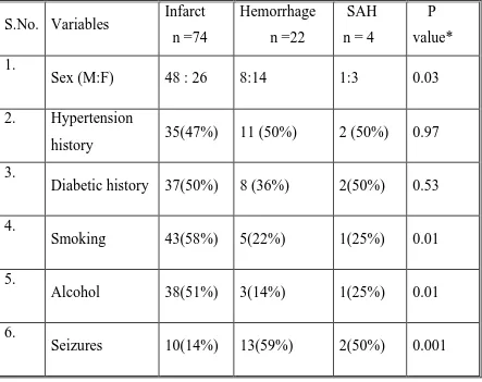
[image:44.612.86.529.238.589.2]The main demographic and clinical features of 100 cases of stroke are summarized in the fig-1.
Figure – 1 Demography and clinical variables of 100 cases of stroke
S.No. Variables Infarct n =74
Hemorrhage n =22
SAH n = 4
P value* 1.
Sex (M:F) 48 : 26 8:14 1:3 0.03
2. Hypertension
history 35(47%) 11 (50%) 2 (50%) 0.97 3.
Diabetic history 37(50%) 8 (36%) 2(50%) 0.53 4.
Smoking 43(58%) 5(22%) 1(25%) 0.01
5.
Alcohol 38(51%) 3(14%) 1(25%) 0.01
6.
Seizures 10(14%) 13(59%) 2(50%) 0.001
* P <0.05 is significant
step forward in the study is that history of hypertension, diabetes; smoking and alcohol were more inclined towards infarct.
Fig – 2. Univariate analysis of association of continuous variables with types of stroke
S.No
Variables Infarct n = 74(M±SD)
Hemorrhage n=22 (M±SD)
SAH
n=4 (M±SD) P value* 1.
Age 66.68±7.54 61.41±7.05 61.41±6.90 0.006 2.
SBP 161.89±17.74 183.73±27.65 172±28.71 0.001 3.
DBP 107.46±15.58 121.09±20.41 116.00±26.3
3 0.005
4.
HDL 35.54±7.11 47.36±4.11 46.00±2.30 0.001 5.
LDL 163.81±29.38 133.27±24.79 137.25±26.3
2 0.001
6.
TC 230.09±23.12 228.15±20.62 226.14±21.1
2 0.71
7.
TGL 251.28±30.59 230.45±30.17 229.25±32.2
4 0.01
* P < 0.05 is significant
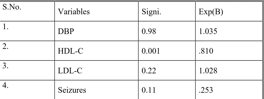
Multivariate analysis of risk factors for stroke by logistic regression shows that the high HDL-C level has inverse correlation with the development of ischemic stroke whereas the high LDL-C has linear relationship.
Fig-3 Multivariate analysis of variables predicting the development of stroke
S.No.
Variables Signi. Exp(B)
1.
DBP 0.98 1.035
2.
HDL-C 0.001 .810
3.
LDL-C 0.22 1.028
4.
Seizures 0.11 .253
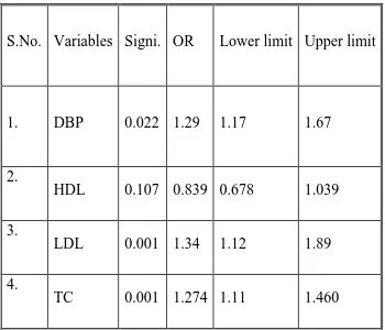
The course of the study demonstrated that variables like diastolic blood pressure, LDL – C and total cholesterol showed significant association with mortality rates only in ischemic stroke.
Fig -4 Variables predicting mortality of stroke patients
S.No. Variables Signi. OR Lower limit Upper limit
1. DBP 0.022 1.29 1.17 1.67
2.
HDL 0.107 0.839 0.678 1.039
3.
LDL 0.001 1.34 1.12 1.89
4.
TC 0.001 1.274 1.11 1.460
DBP – Diastolic blood pressure HDL – High density lipoprotein LDL – Low density lipoprotein TC - Total cholesterol
[image:47.612.84.434.219.519.2]The pie chart below shows the percentage of incidence of stroke subtypes
INCIDENCE
22%
74%
4%
Hemorrhage Infarct
SAH
The bar chart depicting the sex ratio shows a preponderance of ischemic stroke in male gender.
0 10 20 30 40 50
Hemorr Infarct SAH
Sex Pattern
0 10 20 30 40
46-55 56-65 66-75 76-85
Age group pattern
Hemorrhage Infarct
Although the bar chart showed a linear relationship of stroke in the age group of 45-75, a point noteworthy is that the incidence of hemorrhagic stroke decreases in the age group of 66-75.
Relationship between smoking and stroke
0 5 10 15 20 25 30 35 40 45
SMOKING PATTERN
Smoking 43 5
No smoking 5 4
The relationship between lipid profile and stroke subtypes is given below.
Lipid correlation
0 100 200 300
HDL LDL TC TGL
mg/dl
SAH Infarct
DISCUSSION
Until quite recently, studies regarding the problem of stroke were scanty in India. However there is a changing trend now and many prospective and retrospective studies are being conducted to shed more light on the enigma of stroke. An increased risk of ischemic stroke associated with higher cholesterol level has been reported in number of studies [40, 41] while others did not find such an association. [42, 43] Studies focusing on hemorrhagic stroke alone often found an inverse association; with increased risk at the lowest level of total cholesterol. But these studies were not able to formulate an idea on the anvil regarding the risk factors of stroke.
Moreover, most of the studies were conducted in western countries wherein the life style, socio economic status, and health care facilities are in no way comparable to those in India. So, it was thought that studies are to be conducted about the impact of risk factors on stroke pertaining to our set up.
22% (ICH 19%, SAH 3%). Out of the 100 cases in this study, 4 cases (infarct-3, hemorrhage-1) were due to posterior circulatory stroke.
Regarding sex, males (57%) outnumbered females (43%) in the development of stroke. In contrast, one study conducted by Adman Khan et al in Pakistan showed females (52%) outranking males (48%) [44] In this study, hemorrhagic strokes occurred commonly in females (65%) whereas ischemic strokes occupied 63% of all types of stroke occurred in males. This could be attributed to significant sex differences in the effectof triglycerides, smoking, and physical activity on subclinical atherosclerosis like carotid intimal medial thickness in middle-aged men and women. [45]
Mean age for stroke, in both sexes was 65. The mean age in hospital based study conducted in Lady reading hospital on 2004 was 54.38.[44] But if we take subtypes of stroke separately, the age of females slightly over edged that of males. For example, if we take hemorrhagic stroke the mean age for males was 57.4 whereas for females it was 63.7.
in the age group of 56 -65 years. But in cerebral infarction it happens in the age of 66-75 years.
Regarding smoking history, out of 8 hemorrhagic stroke patients 5 patients have accepted smoking with a smoking index of 200(20cigarettes/d for 10 years). The percentage comes around 60% whereas in ischemic patients it was 89.5% (43/48). Not surprisingly, the only male SAH patient also gave smoking history. This prospective study suggests an increased risk of total hemorrhagic stroke, ICH, and SAH in current cigarette smokers with a graded increase in risk that depended on how many cigarettes were smoked. [23]
Regarding alcohol dependence 3 out of 8 hemorrhagic patients gave positive history. Among 48 ischemic patients, 43 accepted significant quantity (150ml 4 times a week) of alcohol intake. The P value is 0.01.
On evaluating the association of history of hypertension in stroke patients, the univariate analysis showed a statistically insignificant association between hypertensive history and occurrence of stroke in both males and females (P values are 0.21 and 0.28) however about 2/3rd of hemorrhagic patients gave history of hypertension.
being 50% and 30% respectively. Also the accumulation of evidence suggested that long standing systemic hypertension and diabetes had insignificant role to play as risk factors in this hospital based study. This may be a bias owing to the fact that long standing hypertension and diabetes are being diagnosed only at the presentation of stroke or MI. This is because in our set up most of the population is ignorant and unaware of these diseases.
The mean systolic BP at the time of arrival to emergency department in cases of hemorrhagic stroke was 183.73, being 161.89 in ischemic stroke and 172 in SAH. One of the studies conducted at Lady reading hospital in 2005 showed a mean systolic BP of 181.25 in cases of ICH & 152.32 in ischemic stroke. The overall systolic BP in our study was 167.10 which is almost comparable to that study (163) [46] (P=0.005). This high systolic blood pressure in hemorrhagic stroke can be explained by the occurrence of cerebral edema in hemorrhagic stroke with the elucidation of Cushing’s reflex.
With regard to the mean HDL level, our study showed a higher trend towards hemorrhage (47.36) than ischemia (35.54). Former study conducted in Western Washington in the year of 1989-2000 demonstrated a similar association. The outcomes were that the levels of HDL in the hemorrhagic stroke was 55 and that in ischemic was 50. There was a suggestion from one another case control study that the association between HDL cholesterol and stroke was more important in atherosclerotic stroke [47] which our data confirm. In a multivariate analysis published in the year 1997, alow concentration of HDL-C appeared to be significantly predictive of ischemic stroke mortality. The relative risk associated witha 5% decrease of HDL was 1.18 (95% confidence interval, 1.03 to 1.34). But a study published in 2000 with a mean follow up period of 16.8 years insisted that higher HDL –C levels were in more association with nonfatal stroke. No association was found for fatal stroke. The possible explanation given was that fatal stroke patients turned out to be heavy drinkers and not surprisingly had a higher HDL level. Serum HDL cholesterol was inversely associated with the risks of subarachnoid hemorrhage and cerebral infarction but not with the risk of intracerebralhemorrhage [48]
The univariate analysis for LDL cholesterol strongly insists that higher level of LDL – C is an important risk factor for atherosclerotic vascular disease.
The mean total cholesterol level in all subtypes was above 200. But among them the mean level for hemorrhagic patients was 226.14±21.1. It was lower than in ischemic stroke [230.09±23.1]. It has been suggested in a study conducted in western Washington that the mean TC level in hemorrhagic stroke was (228) lower than that of ischemic stroke (234) [47] It has been postulated that low serum cholesterol levels could causeweakening of the endothelium of small intracerebral arteries,which, in connection with hypertension, could lead to hemorrhagicstroke.
While taking triglycerides in account, we have come across the fact that our study group had high TGL level in infarct (251.28±30.59) patients comparing to hemorrhagic patients (230.45±30.17). This significant relation of high TGL with ischemia is well supported by the results of the work done by David Tanne et al. Director of the stroke unit, Sheeba medical Centre, Israel which states that high levels of triglycerides are more likely to cause ischemic stroke.
rates. Previously a study done on this subject supports our result stating that there is no significant effect of triglycerides levels on 3 month mortality rate. However they have given a conclusion that low triglyceride levels cause an increase in the 6 month mortality rates. The probable explanation given was that low triglyceride level reflects poor nutritional status which by itself is a supportive factor for poor outcome. Lack of follow up for a period of 6 months is a shortcoming in our study. [48]
In multivariate analysis by logistic regression it was found that there was significant linear association between the development of ischemic stroke and low HDL, and high LDL levels.
15 out of 26 hemorrhagic patients had a seizure (12 – GTCS, 3 partial with secondary generalization) with a percentage of 58 whereas only 13 % of ischemic patients had seizures (8 – GTCS, 2 partial seizures). Ischemic patients who had GTCS had huge infarcts in CT scan. The intracranial tension was found to be raised in patients with GTCS.
Hospital mortality rates revealed that there were a total of 18 deaths of which infarct, hemorrhage, SAH, came to a number of about 9, 7, and 2 respectively. Of these 10 were men and 8 were women. The important significant factor in mortality is presence of High BP at the time of presentation. The course of the study demonstrated that variables like high diastolic blood pressure, (OR 1.29, CI 1.17-1.67) high LDL – C (OR 1.34, CI 1.12-1.89) and high total cholesterol (OR 1.27, CI 1.11 – 1.46) showed significant association with mortality rates only in ischemic stroke. These results were consistent with references given by LL Haheim et al. [49] and Cawl L. Hat et al [50] Moreover; it was found that HDL levels had a reasonable effect on the development of stroke but not on the mortality rates.
The mean systolic BP was 193 in patients who succumbed to death against the overall mean systolic BP of 167.10. Likewise the mean diastolic BP was 131 in them against the overall mean diastolic BP of 110.80.
While looking into smoking as a risk factor for mortality, this study found that 70% of patients who succumbed to death were smokers. Surprisingly, such factors were not found to have any such association regarding mortality of hemorrhagic stroke.
hemorrhagic stroke occurred more in women. The mean age for both sexes was found to be 65. Although the Univariate analysis suggested smoking, alcohol consumption, low levels of HDL and high levels total cholesterol and LDL –C as risk factors for the development for stroke, the multivariate Analysis favored only low HDL and high LDL as the most significant independent risk factors. High levels of diastolic blood pressure at the time of admission along with high levels of low density cholesterol and total cholesterol ascertain their impact on the mortality due to stroke.
There are some shortcomings in our study. With the emergence of diseases like antiphospholipid antibody syndrome, homocysteinemia etc. in the occurrence of stroke, they need to be excluded. However it was not possible due to the lack of laboratory facilities. Also due to economic constraints, MRI to diagnose the site of aneurysms in cases of SAH could not be done in all cases.
This study continues to raise an important issue that public awareness on the risk factors of stroke need to be made for they go a long way in the prevention of stroke.
CONCLUSION
The following are the conclusions of the study:
Ischemic stroke is commoner than hemorrhagic stroke.
Males are commonly affected than females.
Although smoking, alcohol, high LDL and low HDL are strongly associated with ischemic stroke, the independent risk factors for the development of stroke are high LDL and low HDL.
PROFORMA
Study of Stroke cases admitted in MMCH
Name: Age: Sex: I.P: Address:
Admitting Unit:
Symptoms of admission:
Smoking greater than 20 cigarettes/ Beedies /day
Alcohol consumption greater than 150ml four times / week
Past History: IHD/RHD/SHT/DM/TIA/STROKE Family History: SHT/IHD/CVA/DM
Personal History: Smoking/Alcohol/STD exposure
Physical Examination: Pulse- BP –
CVS : Murmur - Yes / No RS : Rales /Rhonchi - Yes / No Abdomen : Organomegaly - Yes / No CNS : Conscious level
Eye signs Fundus Hemi paresis - Yes / No
INVESTIGATIONS
Hemogram
Total cholesterol
HDL cholesterol
LDL cholesterol
VLDL cholesterol
E.C.G.
ECHO
CT scan brain
BIBLIOGRAPHY
1. Davenport R, Denis M et al on Acute Stroke in J neurol Neurosurgery psychiatry 2000;68: 277-88
2. Prof. C. Velmurugendran, Dept. of Neurology, Sriramachandra medical college and research institute, Chennai, TamilNadu, India 3. Goldstein LB, Adams R, Becker K et al. Primary prevention of
ischemic stroke. A statement for health care professionals from the stroke council of the American Heart Association. Stroke 2001; 32: 280-299
4. Ebrahim S, Harwood RH. Stroke : Epidemiology, Evidence and clinical Practice. 2nd eds. Oxford: Oxfore university press,1999
5. Cholesterol, diastolic pressure , and stroke: 13,000 strokes in 450,000 people in 45 prospective cohorts. Prospective studies collaboration. Lancet 1995; 346:1647-53
6. Stroke epidemiological data of nine Asian countries by Asian acute stroke panel J Med Ass. Thai 2000 Jan ;83(1): 1-7
7. Easton DJ, Hauser S, Martin JB in Harrison’s Principle of Internal Medicine 2002; 2325 -38
8. Bogalusa Heart Study Stroke. 2004;35:2782.)
9. K. Anand, D.Chowdhury et al : Estimation of mortality and morbidity in strokes in India : Neuroepidemiology ;2001:20:208-211
10. NINDS 1999, Senelick et al., 1994
11. Jaume Roquer, MD, PhD; Ana Rodríguez Campello, MD Stroke. 2003; 34:1581.)
13. NINDS 1999
14. Villarosa et al., 1993 15. NINDS 1999, NIMH 2002
16. Rajinder K Dhamija et al : Trends in clinico – epi. Correlates of stroke ; Journal of Indian Acadamy of clinical Medicine : Vol.5 No.1
17. Framingham study 2003
18. Olaf H. Klungel, PhD; Bruno H. C. Stricker, PhD: stroke. 1999; 30:1312-1318.)
19. Yuriko Makino, MD; Yuhei Kawano, MD.Stroke2000; 31:48.)
20. Hithito Metoki et al conducted Ohasama study published in Hypertension an American Heart association Journal 2006;47:149 21. Tobias Kurth, MD, MSc; Carlos S. Kase, MD; (Stroke. 2003;
34:1151.)
22. Kubota, T Yamaguchi, Y Abe, T Fujiwara, J Hatazawa and T Matsuzawa Stroke, Vol 14, 720-724
23. Sacco RL, Benson RT et al :Northern Manhatten stroke study ; JAMA 2001 ; June 6 ; 285(21) 2729-35
24. Yoshiyuki Soyama, DDS; Katsuyuki Miura, MD, PhD; Yuko Morikawa, MD, PhD; HDL cholesterol and risk of stroke in Japanese men & women. Stroke 2003;34:863
25. S. Goya Wannamethee, Ph.D. et al : HDL-C, TC and risk of stroke in middle aged British men, Stroke : 2000;31,1882.
26. Tanko, Yu Z, Bagger MD et al in AHA journal report on 18.04.2005 27. Thomas S. Bowman et al : Cholesterol and risk of ischemic stroke ;
28. Centers for Disease Control and Prevention. National Diabetes Fact Sheet; United States; November 2003.
29. Viti Kothari, MSc; Richard J. Stevens, PhD UKPDS 60 Stroke. 2002; 33:1776.
30. Didion SP, Ryan MJ, Didion LA, Fegan PE, Sigmund CD, Faraci FM. Increased superoxide and vascular dysfunction in CuZnSOD-deficient mice. Circ Res. 2002; 91: 938–944
31. Sean P. Didion, PhD; Cynthia M. Lynch, BS; Gary L. Baumbach, MD Frank M. Faraci, PhD Stroke. 2005; 36:342. Sauzeau V, Le Jeune H, Cario-Toumaniantz C, Smolenski A, Lohmann SM, Bertoglio J, Chardin P, Pacaud P, Loirand G, Cyclic GM. P-dependent protein kinase signaling pathway inhibits RhoA-induced Ca++ sensitization of contraction in vascular smooth muscle. J Biol Chem. 2000; 275: 21722–21729.
32. Jin L, Ying Z, Webb RC. Activation of RhoA/Rho kinase signaling pathway by reactive oxygen species in rat aorta. Am J Physiol Heart Circ Physiol. 2004; 287: H1495–H1500
33. Pekka Jousilahti, MD, PhD; Daiva Rastenyte, MD, PhD Jaakko Tuomilehto, MD, PhD Stroke. 2000; 31:1851.)
34. Aaron R. Rolsm MD of the division of epidemiology in the school of Public health at the University of Minnesota, Mineapolis conducted study on thick blood may increase stroke risk.
36. Stefen Kiechl MD Dept. of Neurology, Innsbruck hospital, Austria published a report in American Heart Association Journal dated 12/5/1998
37. Baker CJ, Fiore A, Connolly ES Jr, Baker KZ, Solomon RA. Serum elastase and alpha-1-antitrypsin levels in patients with ruptured and unruptured cerebral aneurysms. Neurosurgery. 1995;37:56–62.
38. Aull S, Lalouschek W, Schnider P, Sinzinger H, Uhl F, Zeiler K. Dynamic changes of plasma lipids and lipoproteins in patients after transient ischemic attack or minor stroke. Am J Med. 1996; 101: 291– 298.
39. Butterworth RJ, Marshall WJ, Bath PMW. Changes in serum lipid measurements following acute ischaemic stroke. Cerebrovasc Dis.
1997; 7: 10–13.
40. Benfante R,Yano K etal : Elevated serum cholesterol is a risk factor for both coronary heart disease and thromboembolic stroke in Hawaiian Japanese men. Implications of shared risk.Stroke1994;25:814-820
41. Dorn-Moreg N, tanne D et al ; Low and high density lipoprotein cholesterol and ischemic cerebrovascular disease: the bezafibtrate infarction prevention registry. Arch Intern Med 2002;162:993-999
43. Shahar E, Chambless LE et al Plasma lipid profile and incident ischemic stroke: the atherosclerosis risk incommunities (ARIC) study.Stroke2003;34:623-631
44. Study at Lady reading hospital done by Adman khan, Akthar sherin et al Peshawar JPMI 2004 Vol.18No.2; 220-224
45. Eva Stensland-Bugge, MD; Kaare H. Bønaa, MD, PhD et al: The Tromsø Study; Stroke. 2000;31:574.)
46. Akthar Sherin, Ghulam Shabbier et al conducted study at Lady reading hospital, Peshawar JPMI 2005 Vol.19 No.2; 220-225
47. D.L.Tirschwell, M.D., N.L. Smith PhD; et al Cholesterol assoc. with stroke risk varies in stroke subtypes and patient subgroups Neuro.J. 2004;63;1868-1875
48. Tomasz Dziedzic, MD; Agnieszka Slowik, MD; et al : Low serum triglyceride level is associated with increased stroke severity ; Stroke.
2004;35:e151.)
49. LL haheim, I Holone etal done a 12 year follow up; Oslo study for the risk factors of stroke incidence and mortality in Stroke 1993;24;1484-1489
MASTER CHART S.No
AGE SEX TYPES SBP DBP smoking alcohol HDL LDL TC TGL SHT Diabetes
Seizures Mortality
1 56 1 H 184 126 1 1 54 102 187 184 1 1 1 1
2 48 1 I 148 96 1 1 34 124 207 244 0 1 0 0
3 66 2 I 154 92 0 0 36 154 255 324 1 0 0 0
4 65 2 I 148 88 0 0 38 152 237 233 1 1 0 0
5 74 1 I 202 142 1 0 52 156 257 246 0 0 1 1
6 68 1 I 172 116 1 1 32 158 235 224 1 1 0 0
7 58 1 H 220 164 0 0 48 99 198 204 0 0 1 0
8 72 2 I 164 112 0 0 28 154 224 212 1 1 0 0
9 46 2 H 168 122 1 0 44 111 220 224 0 0 0 0
10 82 1 I 146 118 1 1 34 166 246 232 0 0 0 0
11 64 1 I 144 98 1 1 34 158 241 246 1 1 0 0
12 72 2 I 156 102 0 0 32 152 222 188 0 1 0 0
13 58 1 SAH 168 112 0 0 44 152 242 220 1 0 1 1
14 64 1 I 136 88 1 1 30 162 237 224 0 1 0 0
15 55 2 I 186 134 0 0 54 93 192 224 0 1 0 0
16 73 1 I 146 98 1 1 32 148 233 265 1 0 0 0
17 64 1 H 196 132 1 0 42 126 220 245 0 0 1 1
18 68 2 I 168 114 0 0 34 146 233 263 1 0 0 0
19 71 1 I 156 104 1 1 28 134 211 247 0 1 0 0
20 63 2 I 174 120 0 0 32 146 235 285 1 1 1 1
21 66 1 I 144 84 1 1 34 134 217 245 0 0 0 0
22 64 2 H 168 108 0 0 46 132 234 273 1 1 0 0
23 58 1 I 158 102 1 0 34 148 231 294 0 1 0 0
24 49 1 I 174 124 1 1 28 153 230 226 1 0 1 0
25 65 2 H 138 84 0 0 44 121 208 206 0 1 1 0
26 59 1 I 182 126 1 1 30 197 280 284 0 1 0 0
27 73 1 I 154 104 0 1 32 146 221 214 1 1 0 0
28 66 2 I 166 100 0 0 44 101 184 195 0 0 0 0
30
63 2 H 160 104 0 0 50 90 177 184 0 0 0 0
31 64 1 I 144 98 1 1 34 154 242 268 1 0 1 0
32 72 2 I 156 102 0 0 32 142 221 233 0 1 0 0
33 58 1 I 168 112 0 0 44 142 235 246 1 0 0 0
34 64 1 I 136 88 1 1 30 162 251 294 0 1 0 0
35 55 2 H 186 134 0 0 54 112 215 245 0 1 1 1
36 73 1 I 146 98 1 1 32 152 235 254 1 0 0 0
37 64 1 I 196 132 1 0 42 154 253 284 0 0 0 0
38 68 2 I 168 114 0 0 34 142 225 245 1 1 0 0
39 71 1 I 156 104 1 1 28 143 216 224 0 1 0 0
40 63 2 I 224 120 0 0 48 187 299 320 1 0 1 1
41 66 1 I 144 84 1 1 34 122 201 225 0 0 0 0
42 64 2 H 168 108 0 0 46 152 253 275 1 1 0 0
43 58 1 I 158 102 1 0 34 132 213 233 0 0 0 0
44 73 1 I 174 124 1 1 28 138 211 223 1 0 0 0
45 51 2 SAH 138 84 0 0 44 152 251 276 0 1 0 0
46 59 1 I 182 126 1 1 30 142 221 245 0 1 1 0
47 73 1 I 154 104 0 1 32 152 227 214 1 0 0 0
48 66 2 H 166 100 0 0 44 144 231 216 1 0 1 0
49 61 1 I 154 96 1 1 32 131 208 219 1 1 0 0
50 72 2 I 160 104 0 0 50 135 234 245 0 0 0 0
51 56 1 H 184 126 1 1 54 117 219 239 1 1 1 1
52 78 1 I 148 96 1 1 34 151 236 257 0 1 0 0
53 66 2 I 154 92 0 0 36 142 228 249 1 0 0 0
54 65 2 I 148 88 0 0 38 162 253 267 1 1 0 0
55 74 1 I 202 142 1 0 52 174 286 302 1 0 1 1
56 68 1 I 172 116 1 1 32 146 231 263 1 1 0 0
57 58 1 H 220 164 0 0 48 135 232 245 0 0 1 1
58 75 2 I 164 112 0 0 28 133 224 316 1 1 0 0
59 68 2 H 168 122 1 0 44 152 246 248 0 0 0 1
60 82 1 I 146 118 1 1 34 164 252 268 0 0 0 0
61 63 2 SAH 174 120 0 0 48 116 208 219 1 1 1 0
63
64 2 I 168 108 0 0 46 142 234 225 1 1 0 1
64 48 1 I 158 102 1 0 34 126 207 235 0 0 0 0
65 73 1 I 174 124 1 1 28 166 252 290 1 0 1 0
66 65 2 H 138 84 0 0 44 152 251 273 0 1 0 0
67 59 1 I 182 126 1 1 30 162 256 321 0 0 0 1
68 73 1 I 154 104 0 1 32 141 222 247 0 1 0 0
69 66 2 I 166 100 0 0 44 142 219 213 1 0 0 0
70 61 1 I 154 96 1 1 32 134 215 246 1 0 0 0
71 64 1 I 144 98 1 1 34 133 211 220 0 1 0 0
72 72 2 I 156 102 0 0 32 182 278 320 1 0 0 0
73 47 1 H 168 112 0 0 44 102 186 200 1 0 1 0
74 64 1 I 136 88 1 1 30 143 219 228 0 1 0 0
75 55 2 I 186 134 0 0 54 114 215 237 0 1 0 0
76 73 1 I 146 98 1 1 32 152 234 249 0 0 0 0
77 64 1 H 242 132 1 0 42 124 216 252 1 0 1 1
78 68 2 I 168 114 0 0 34 151 236 254 0 0 0 0
79 71 1 I 156 104 1 1 28 155 234 256 1 1 0 0
80 63 2 I 174 120 0 0 48 139 236 247 0 0 0 0
81 56 1 H 184 126 1 1 54 98 192 198 1 1 0 0
82 78 1 I 148 96 1 1 34 145 229 248 0 1 0 0
83 66 2 I 154 92 0 0 36 171 254 284 1 0 0 0
84 65 2 I 148 88 0 0 38 152 241 254 0 0 0 0
85 74 2 H 202 142 1 0 52 99 190 196 1 0 1 0
86 68 1 I 172 116 1 1 32 147 226 236 0 1 0 0
87 58 1 I 220 164 0 0 48 152 253 267 0 0 1 1
88 72 2 I 164 112 0 0 28 149 236 245 1 1 0 0
89 64 2 H 238 122 1 0 44 102 188 210 0 0 1 0
90 82 1 I 146 118 1 1 34 155 238 244 0 0 0 0
91 71 2 H 168 106 0 1 46 159 262 275 1 0 0 0
92 69 2 I 156 92 0 0 32 167 246 273 1 0 0 0
93 66 1 I 174 116 1 1 54 158 269 285 0 1 0 0
94 58 2 H 186 124 0 0 50 107 202 224 0 0 0 1
96
67 2 SAH 208 148 0 0 48 107 195 202 0 0 0 1
97 79 2 I 168 118 0 0 44 138 229 234 1 1 1 1
98 66 1 I 160 104 1 0 34 157 240 245 1 0 0 0
99 49 1 I 154 92 1 0 30 158 238 250 0 1 0 0
100 69 2 H 190 122 0 0 48 121 218 254 1 0 1 0
Regarding sex: 1= MALE; 2= FEMALE