0022-538X/83/100229-10$02.00/0
Copyright(C 1983, AmericanSocietyfor Microbiology
Structure
of Simian Virus
40-+X174 Recombinant Genomes
Isolated
from Single Cells
ERNEST WINOCOUR,* VERED LAVIE, ANDILANAKESHET Department ofVirology, The Weiz,nann Institlute of Science, Rehovot, Israel
Received 1 April 1983/Accepted 29 June 1983
Three simian virus (SV40)-+X174 recombinant genomes were isolated from
single BSC-1 monkey cells cotransfected with SV40 and XX174 RF1 DNAs. The
individual cell progenieswereamplified, cloned, andmapped byacombination of
restriction endonuclease and heteroduplexanalyses. In each case, the 600to 1,000
basepairs of 4X174 DNA (derived from different regions of the XX174 genome)
werepresentassingleinserts, located in either the earlyorlateSV40 regions; the
deletion ofSV40 DNA wasgreater than the sizeof theinsert; and the remaining
portions of the hybridgenomewereindistinguishable from wild-type SV40 DNA,
asjudged by both mapping and biological tests. Hence, apart from the deletion
which accommodates the XX174 DNA insert, no otherrearrangements ofSV40
DNA were detected. The restriction map of a SV40-4X174 recombinant DNA
isolate before molecular cloningwasindistinguishable from those oftwo separate
cloned derivatives of thatisolate,indicatingthatthe species clonedwasthemajor
amplifiable recombinant structure generated by a single recombinant-producing
cell. The relative simplicity of the SV40-¢X174 recombinant DNA examined is
consistentwiththe notion thatmostrecombinant-producing BSC-1 cells support
single recombination events generating only one amplifiable recombinant
struc-ture.
Recombination occurs between simian virus 40(SV40)andbacterial virus kX174RF1 DNAs
cotransfected intomonkey BSC-1 cells (14). The
frequencyof this nonhomologous recombination
occurrence has been measured in terms ofthe
quantityofhybridvirus in theyield of a cotrans-fected culture of cells (14) and in terms of the
number of recombinant-producing cells in the
initial cotransfected cell population (4).
Evi-dence has been reported that SV40
nonhomolo-gous recombination events occur in a discrete subpopulationof cells: 1to2%of the successful-lytransfected fraction of cells or 1 in 500 to 1 in
1,000of the total cellpopulation(4). An analysis
of theSV40-¢X174recombinant genomes in the
total yield of a cotransfected cell population
indicated that a variety of recombinant
struc-tures had arisen (14). This variety may result
from thepresence of a few
recombination-profi-cient cells supporting multiple recombination
events; alternatively, cells may support only
single recombination events, each cell giving
rise to a different recombinant structure. We
report here an analysis of three cloned
SV40-XX174 recombinant genomes, each of which
arosefroma single recombinant-producingcell.
The results indicate that most
recombinant-pro-ducing cells, in fact, support single
recombina-tion events generating only one amplifiable
recombinant structure.
MATERIALS AND METHODS
DNAand cells.Theproceduresusedforthe prepara-tion ofwild-typeSV40 DNA I and
qkX174
RFI DNA, andfor the culture ofmonkey BSC-1 cells, were asdescribedelsewhere (4).
Isolation andamplification ofSV40-+X174 recombi-nantprogenyfrom individual transfected cells. Suspen-sions ofBSC-1cellswerecotransfected withSV40and
4X174 RFIDNAs(1,ugofeach DNA per 4x 10⁶ cells
per ml), using DEAE-dextran as the facilitator as
described previously (4). The recombinant yields of individual cotransfected cells were isolated eitherby
cellpartition in microwellplates (15)orfrom the agar
overlay of infectious-centerin situ plaque hybridiza-tionfoci (4). Inthemicrowell method,thetransfected
cells were mixed witha 20-foldexcess of uninfected BSC-1 cells and distributed intomicrowellplates (Mi-crotest II, Falcon Plastics) such that each well re-ceived 1,000 cotransfected cells and20,000uninfected cells in 0.1 ml of medium. When all wells showed
cytopathic effects (after 9 days of incubation), the
plates were subjected tothree cycles of
freeze-thaw-ing, and a 40-,jI sample of each well lysate was
analyzedfor4X174DNAby usingadothybridization procedure(15). Strong autoradiographicsignals, after
hybridizationwith )X174 RF1[32P]DNA,were detect-edin 12 of 192 welllysates.Control wells seeded with
229
on November 10, 2019 by guest
http://jvi.asm.org/
230 WINOCOUR, LAVIE, AND KESHET
cells transfectedwithc>X174RF1 DNAalone (or with SV40 DNA alone) gave rise to noautoradiographic signals afterhybridization withXX174 [32P]DNA. To
amplify the recombinant isolates, 3-cm BSC-1 cell cultures were inoculated with 50-,ul samples of the positive well lysates, andprogenyviruswasharvested
when fullcytopathiceffectsbecame apparent(usually after 5 days ofincubation). These viral yields were then used toinfect15-cmBSC-1cellplates fromwhich
supercoiled progeny DNA was prepared (at 50 h
postinfection) by the Hirt extraction procedure fol-lowed by equilibrium centrifugation in cesium chlo-ride-ethidium bromidedensity gradients (7).Isolates9
and32 (seebelow)werepurified andamplified by the
proceduredescribed above.
To isolate progeny from recombinant
infectious-centerplaques, thecontransfected cells(mixed witha
40-fold excess of uninfected cells) were plated for infectious centers, overlaid with agar, transferred to
nitrocellulose membrane filters, and titrated for the production of SV40-4X174 recombinant plaques as
described previously (4). After hybridization with
,X174 [32P]DNAandautoradiography (4), well-sepa-rated "hybridization plaques" appearingonthe
auto-radiogram were aligned with the agar overlay by
meansofthe systemoforientation marks describedby Villarreal andBerg(12). Aplug ofagar waswithdrawn from the region of the overlay corresponding to the
autoradiographic plaque, suspended in 1 ml of medi-um, and used to inoculate 3-cm cultures of BSC-1
cells. After theappearanceof fullcytopathic effects (9
days post-inoculation), progeny virus was harvested
andused toinfect15-cmBSC-1cellplatesfromwhich
supercoiled viral progeny DNA was isolated as
de-scribedabove. Isolate2 (seebelow)wasprepared and
amplified bythis procedure.
Restriction digestion, gel electrophoresis, and blot hybridization. DNA wasdigestedwith restriction
en-donucleases under the conditions specified by the
supplier (New England Biolabs), and the products were separated by electrophoresis on 1%
agarose-Tris-acetate gels as described previously (4). Gels were stained with 0.5 ,ug of ethidium bromide per ml
and photographed under UV light. In early experi-ments, the DNA was transferred to nitrocellulose
sheets (Schleicher & Schuell Co., BA 85) by the
Southern procedure (11); in later experiments, the
transfer procedure ofSmith and Summers (10) was
utilized. Hybridization withinvitro-labeled[32P]DNA
probes (9) was as described elsewhere (14). Probe concentration andautoradiographyconditions are not-ed elsewhere in the text.
Molecularcloning. The closed-circular DNA yields of individual recombinant-producing cells (amplified asdescribedabove)weredigestedwithEcoRI,ligated to EcoRI-cleaved DNAarms of the X-Charonphage 21A, and packaged in vitro by using previously de-scribed procedures (16).Phageplaques werescreened
by hybridization with SV40 [32P]DNA and with
XAX174 RF1 [32P]DNA. Those which reacted with -X174 RF1 [32P]DNA (0.1% ofthose which reacted with SV40 [32P]DNA) were isolated, plaque purified
bytwoconsecutivecyclesofreplating (at which point 100%of theplaques hybridizedwithbothprobes), and grown tostocks. DNA fromCsCl-purified phage was
digestedwithEcoRl, and the recovered SV40-+X174
DNA wasthen subcloned in theEcoRIsite ofpBR322.
All cloning procedures were carried out under the containment conditions specified by the local bio-hazard safetycommittee.
Heteroduplex analysis and electron microscopy. Wild-type SV40 DNA and SV40-+X174 recombinant
DNA wereexcised from theirpBR322 cloningvectors
by EcoRI digestion, purifiedon 1%agarose gels,and
extracted from the gel by glass powder adsorption (13). For heteroduplex formation between wild-type SV40 DNA and SV40-4X174 recombinant DNA, or
SV40-4X174 recombinant DNAandwild-typeXX174 RF1 DNA,the DNAsamples (final concentration of
eachDNA, 2.6 ,ug/ml)wereheatedat70°C for10min, followed by 60 minat57°C, in thepresenceof 62.6% formamide, 412 mM NaCI, 83 mM piperazine-N-N'-bis(2-ethanesulfonic acid)-NaOH (pH 7.8), 16 mM
Tris-hydrochloride, and 3 mM EDTA (1). Samples
were prepared for electron microscopy (Phillips EM400) by the formamide-cytochrome c monolayer spreading procedure (3).Theconversionfactor (length
measurements to base pair [bp] units) for double-strandedDNAregionswascalculated byusing homo-duplexes derived from linearizedwild-typeSV40 DNA
(5,243 bp) or 4X174 RF1 DNA (5,386 bp); that for
single-strandedDNAregionswasderived from single-strandedfX174 DNA (virionplus strand DNA, 5,386 nucleotides) added at the end of heteroduplex
reac-tions.
RESULTS
Isolation and preliminary characterization of
SV40-4X174
recombinants from individual trans-fected cells. The yields of singlerecombinant-producingtransfected cells were isolatedby the
infectious-center protocol, or by partition of transfected cells in microwells, and were
ampli-fied as described above. The supercoiled DNA of these amplified isolates consisted of 99.9%
wild-type SV40 DNA and 0.1% recombinant
DNA (see below). Despite the overwhelming
background of wild-type SV40 DNA, it was
possible to determine the loss or retention of
SV40restriction sites in theSV40-+X174
recom-binant genomes by digesting the DNA with
restriction enzymes which cut wild-type SV40
DNA but notXX174 DNA, andbyusing4X174
RF1
[32P]DNA
as ahybridizationprobe. Conve-niently, the SV40 single-cut enzymes EcoRI,BglI, BamHI, KpnI, and the SV40 multi-cut
enzymeHindlIl havenorecognitionsitesonthe
XX174 genome (Fig. 1). The digestion products
were separated by agarose gel electrophoresis
andtransferred tonitrocellulosepaper(11), and
those containing 4X174 DNA sequences were
identified by hybridization with 4X174 RF1
[32P]DNA. All five independent SV40-4X174
recombinant isolates (each from a different recombinant-producing cell) examined by this procedure contained single SV40 EcoRI and
BglI restriction sites, asjudged by the
conver-sion of form I or from II to unit-length linear
(form III) DNA. The analysis of two of the five
isolates is shown in Fig. 2. In addition to the
J. VIROL.
on November 10, 2019 by guest
http://jvi.asm.org/
SV40-,X174 RECOMBINANT STRUCTURES
retention of single EcoRI and BglI sites, the
SV40-+X174 recombinant DNA in isolate 2
re-tained single sites for BarnHI and KpnI (lanes 7
and 9); moreover, all of the 4X174 DNA
se-quences in this recombinant DNA isolate were
present within one HinidIII digestion product
which also contained the singleKpnI site (lanes
10 and 11). This preliminary characterization of
the SV40-¢X174 recombinant DNAs derived
from single cotransfected cells was primarily
intended to identify an appropriate restriction
site for molecular cloning. Inaddition, the data
obtained with isolate 2 suggested that all of the
XX174 DNA sequences were located in one
region adjacent to thesingle SV40 KpnI
restric-tion site.
Molecularcloningandstructureofthree
SV40-(X174 recombinant genomes. The SV40-¢X174
DNAs in three single cell yields (isolates 2, 9,
and 32)werecloned byinsertion into the EcoRI
site of the X-Charon phage 21A and were
sub-cloned in the EcoRl site ofpBR322asdescribed
above. The proportion of first-cycle X-Charon
21A plaques (see above) which reacted against
both the SV40
[32P]DNA
and XX174 RF1[32P]DNA
probeswas0.1% of the number whichreacted with the SV40
[32P]DNA
probe alone,indicatingthat the ratio of recombinant to
wild-type SV40 DNA molecules in the three isolates
was 1:1,000. The cloned SV40-¢X174
recombi-nant DNAs were mapped by a combination of
restrictionendonucleasedigestionand
heterodu-plex analyses. Figure 3 shows the maps that
were constructed on the basis of the data from
0.5
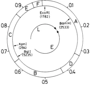
FIG. 1. Wild-typeSV40genome,showing the sites of restriction endonucleases used to map the SV40-4X174recombinantDNAs.Theoutercircle shows the positions of the six HinidlIl fragments labeledAtoFin
descendingorder ofsize. Thebracketedfiguresunder thesingle-cut enzymes arethe SV nucleotidenumbers of these restriction sites (2). BglI cuts within the replication origin sequence. The letters L and E de-note,respectively, theapproximatecodingregions for lateandearly functions. None oftherestriction endo-nucleases noted abovecut 4X174 RF1 DNA.
2 34 5 6 7 8 9 10 I
Ii
IL- II-
_
_I _
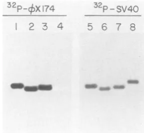
I-FIG. 2. Restriction mapping. before cloning, of
SV40-6X174 recombinant DNA in isolates derived fromsinglerecombinant-producing cells. DNA of iso-lates 2 and 32 (see the text), either undigested or digested with restriction enzymes which do not cut authentic 4X174 RFI DNA, was fractionated on 1% agarose gels, transferred to nitrocellulose paper. and hybridized with 4X174 RFI [132P]DNA.Lanes 1 and 2. isolate 32 DNA undigested or digested with EcoRI. respectively; lanes 3 to7,isolate 2 DNAundigested or
digested with BglI, EcoRI, HindIll, and BarnHl.
re-spectively;lanes 8 to 11, isolate 2 DNAundigested or
digested with KpnI HindlIl, and KpnI-Hinidlll. re-spectively. The arrows point to the positions of wild-typeSV40 DNA, formIandform 11,deducedfromthe ethidium bromide stain of the gel before blot hybrid-ization.Autoradiogramswereexposedat-70°Cfor 40 to 45 h. Althoughthe DNAs of isolates 2 and 32 were subjected to cesium chloride-ethidium bromide densi-ty gradient centrifugation to purify form 1. partial
conversion to form II occurred during storage at -200C.
these two procedures. The essential features of
the recombinant structures are as follows. (i)
The
4X174
DNA sequences (600 to 1,000 bp)were present as single inserts, located within
either theearlyorthe lateSV40regions. (ii)The deletion of SV40 DNA sequences was greater than the sizeof theXX174 DNA insert, resulting
inashortening (by 6 to 10%) of the recombinant
genome relative to wild-type SV40. (iii) Other
than the deletion which accommodated the
XX174 DNA insert, the remainder of the SV40
DNA in the recombinant structure was retained
in an unaltered, intact form. We summarize
below theevidence for these conclusions and for
the maps shown in Fig. 3.
(i) Restriction endonuclease digestion and blot
hybridization. The pBR322 plasmids carrying
the recombinant genomes (or wild-type SV40
DNA as control) cloned in the EcoRI site were
digested with EcoRI, EcoRI-BglI,
E(oRI-BamHI, or EcoRI-HindIII. As noted above,
none of these enzymes cut XX174 DNA. The
products were separated on a 1% agarose gel,
stained with ethidium bromide, photographed
under UV light,andtransferredtonitrocellulose
paper by the method of Smith and Summers
VOL. 1983 231
_.0 di I_
on November 10, 2019 by guest
http://jvi.asm.org/
[image:3.492.246.443.62.191.2] [image:3.492.65.212.443.580.2]232 WINOCOUR, LAVIE, AND KESHET
SV40/0X 2B5
dI-. SV40/OX 32A
dl
SV40/OX 9BA
k- di
-SV40 WT Ori
LATE EARLY LATE
0 0.1 02 Q3 Q4 Q5 0.6 0.7 0.8 0.9 1
FIG. 3. Structure ofthe SV40-¢X174 recombinant genomes. The maps of the recombinant DNAs are
drawn relative to that of the wild-type (WT) SV40 genome and are based upon data from heteroduplex measurements andrestrictioncleavage-blot
hybridiza-tion(seethe text). The cross-hatched boxes denote the
X174 DNA inserts; the open boxes denote SV40 DNA sequences. dl refers to the deletion of SV40
DNA sequences. This deletion is greater than the
'X174
DNA insert; as a consequence, therecombi-nantgenomes are shorter than the SV40WT genome
by an amount corresponding tothe dashed lines(the
sizeorderisWT>2B5>32A>9BA). The WTSV40
map, atthebottom, alsoshowsthesites (inmapunits)
of the restrictionenzymes used toanalyze the struc-tureofthe recombinantgenomes (see alsothe circular map in Fig. 1). Symbols: ,,EcoRI;
1,
BamHI; tHindlII , BglI; 3, KpnI. Ori denotes origin of
replication.
(10), which provided two identical blots from
each gel. One blot was hybridized with SV40
[32PJDNA (5,000 cpm/ml); the twin blot was
hybridized with 4X174 RF1
[32P]DNA
(50,000 cpm/ml). The EcoRI digestion patterns (Fig. 4) showedthateach ofthe intact, linearizedSV40-XX174recombinantgenomesreacted, as
expect-ed, with both probes and was slightly smaller than wild-type SV40 DNA (the orderof size is wild-type SV40 DNA> 2B5 DNA > 32ADNA
> 9BA DNA).
EcoRI-BglI digestionofwild-type SV40DNA
divides the genome into two sectors (Fig. 1): a
3,453-bp fragment (0 to 0.66 map unit) which comprisesalloftheearlyregionandapartof the
late region spanning the EcoRI-BamHI sites;
and a 1,790-bp fragment (0.66 to 1.0 map unit)
which is composed of late region and
origin-flankingsequences.EcoRI-BgIldigestion of 2B5
recombinant DNA shows that the
4X174
DNAsequences are located in the SV40 late region;
oneof thetwoproductswasfoundtocomigrate
with the wild-type SV40 3,453-bp early DNA
fragment and contained only SV40 sequences (Fig. 5; Table 1). The second digestion product
contained both SV40 and (X174 DNA
se-quences and migrated faster than the authentic
1,790-bp SV40late DNAfragment.Asjudged by
gelelectrophoresis, the size oftheproduct con-tainingbothSV40 and4X174DNA sequencesis
J. VIROL
1,450 bp, 340 bp shorter than the wild-type
EcoRI-BglI product.
In contrast to 2B5 recombinant DNA, the 4X174 DNA inserts in recombinant DNAs 32A
and 9BAare located within shortened forms of
the SV40 early region. Of the two products
generated by EcoRI-BglI digestion, the smaller
fragment comigrated with the wild-type 1,790-bp
SV40 late segment and contained only SV40
sequences; the larger digestion product
con-tained both SV40 and 4X174 DNA sequences
and migrated faster than the wild-type 3,453-bp
SV40 early region fragment (Fig. 5; Table 1).
The estimated sizes of the larger products are
3,000 bp for recombinant 32A (453 bp smaller
than the comparable wild-type product) and
2,900 bp for recombinant 9BA (553 bp shorter
than the wild-type fragment). A part of the late
region (the 751-bp EcoRI-BamHI segment) is
contained within the larger EcoRI-BglI product
(Fig. 1). However, EcoRI-BamHI digestion of
9BA and 32A DNA showed that the wild-type
751-bp EcoRI-BamnHI fragment was retained in
both recombinants and did not contain kX174
DNA sequences(Fig. 5; Table 1). The insertsin
9BA and 32Amust therefore be located within
theBamHI-BglI early region ofSV40.
The 2B5 SV40-+X174 recombinant retainsan
intact authentic SV40 early region (the XX174
DNA sequences being located in a truncated
32P-_X 174
32p
PSV40
12 3 4 5 6 7 8
2 3 4 ffA.>
FIG. 4. EcoRIdigestionof clonedSV40-4X174 re-combinants. DNA of plasmids containing wild-type SV40(lanes4and8)ortherecombinants2B5(lanes1 and5), 9BA (lanes2and 6), and 32A(lanes 3and 7)
wasdigestedwithEcoRI. Theproducts were
electro-phoresed on1%agarosegelsandtransferredto
nitro-cellulosepapersbythemethodofSmith and Summers (10), whichprovidedtworeplicateblots from eachgel.
One blot was hybridized with 50,000 cpm ofXX174 RF1[32P]DNAper ml(lanes1 to4);the twin blotwas
hybridized with 5,000cpmofSV40 [32P]DNA per ml
(lanes 5 to 8). The bands on the autoradiograms,
resulting from hybridization with either probe, coin-cided exactly with bands (other than pBR322 DNA
fragments)visualizedbyethidium bromidestainingof thegel before transfer andhybridization.
on November 10, 2019 by guest
http://jvi.asm.org/
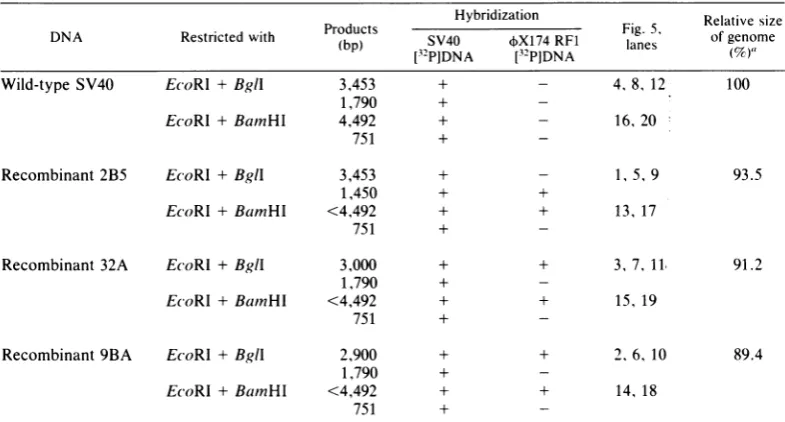
[image:4.492.59.248.69.212.2] [image:4.492.282.426.395.528.2][EcoRl t Bg9l
32P-SV40 32P- 174
2 3 4 5 6 7 8 9 10 11 i2
SV40A (3453)-pBR 322 (2319)-SV43
B(1790)-pBR
322(93°)-
(881)-so an-__mam
32p_
SV40 13 14 15 163000
40 -2900
S - 1450
32P_-
X 174l7 18 19 20
(4492)
(75$)-
-EcoRl + Bamr Hl
FIG. 5. EcoRI-Bgll digestion(upperpanel) andEcoRI-BamlHldigestion(leoerpanel)ofclonedSV40-6bX174
recombinants. Procedural details are as noted in thelegend to Fig. 4. Upperpanel:recombinant 2B5. lanes 1. 5.
9: 9BA. lanes 2, 6. 10: 32A. lanes 3. 7. 11: wild-type SV40 DNA. lanes 4. 8. 12. Lanes 1 to 4 show the
electrophoretic gel patterns, stained with ethidium bromide. before transfer and hybridization: lanes 5 to 8.
autoradiogramsafterhybridizationwithSV4()[32P1DNA; lanes9 to 12.autoradiogramsafterhbridlization with XX174Rl1132P]DNA.The lower panel shows theproducts of 2B5 (lanes 13and 17). 9BA(lanes14and 18). 32A (lanes 15 and 19). and wild-type SV40 DNA (lanes 16 and 20) after digestion with EcoRI-Bum11Hl and hybridization with SV40 [32P]DNA (lanes 13 to 16) or kX174 RFI [32P]DNA (lanes 17 to20). The figures in
parentheses (left side) are the sizes (in bp) of the known pBR322 and wild-typeSV40DNA digestion products which provided the sizemarkers for the gel. The unbracketed figures on the right side are the sizes (in bp) of the recombinant DNAfragments containing4X174sequences, asestimated from an electrophoretic mobility plot of
the stainedgel. Table 1 summarizes these data.
form of the late region segment), whereas the
9BA and 32Arecombinants retainintact
authen-tic SV40 late region segments (the $X174 DNA
sequences being located in truncated forms of
the early region segment) (Table 1). Additional
evidencefor this conclusionwasprovided bygel
blot hybridization patterns with EcoRI-HindIII
(data not shown). 2B5 DNA gave rise to four
products, three of which comigrated with the
wild-type early SV40 fragments and contained
solely SV40 DNA sequences (the HindIll
re-striction sites at0.860 and 0.945 map unit inthe
late SV40 region are missing; Fig. 1 and 3).
EcoRI-HindIII digestion of9BA and 32A DNA
generated products which comigrated with the
wild-type
HindIlll
C, E, and F fragments(span-ning the SV40 replication origin and late region
sector: Fig. I and 3) andcontained solely SV40
sequences.
(ii) Heteroduplex analysis. SV40-tX174
recombinant DNA strands (EcoRI-linearized)
were annealed with wild-type SV40 DNA
strands (EcoRl-linearized) or with tX174 RFI
DNA strands (PstI-linearized) and were
exam-ined by electronmicroscopy as described inthe
legend to Fig. 6. Figure 6depicts the
heterodu-plex segments that were measured, and Table 2
shows the measurements in summaryform.
The single-strand armsofthe loop-out
appar-entineachrecombinant-wild-type SV40
hetero-duplex are of unequal length. Because the
re-striction mapping data indicated that the
deletion of SV40DNAsequences isgreaterthan
the size ofthe4X174 DNA insert(Table 1), the
size estimates of the 4X174 DNA inserts are
derived from the length ofthe shorter loop-out
arm (Table 2, column a). As an independent
check, the values can be compared with the
VOL. 48. 1983
on November 10, 2019 by guest
http://jvi.asm.org/
[image:5.492.113.373.75.357.2]234 WINOCOUR, LAVIE, AND KESHET
TABLE 1. Summary of the gel blot hybridization experiment shown in Fig. 5
Hybridization
DNA Restricted with Products SV40 X174 RF1 g. 5 Relative size
(bp) S4 X14R1 lanes
ofYenm
I3-PIDNA [32PIDNA (7)
Wild-type SV40 E(coRI + Bgll 3,453 + - 4,8, 12 100
1,790 +
-EcoRl + BamHI 4,492 + - 16, 20
751 +
-Recombinant2B5 EcoRl +Bgll 3,453 + - 1, 5,9 93.5
1,450 + +
EcoRI +BamHI <4,492 + + 13, 17
751 +
-Recombinant 32A EcoRI +Bgll 3,000 + + 3, 7, 11, 91.2
1,790 +
-EcoRI +BamnHI <4,492 + + 15, 19
751 +
-Recombinant9BA EcoRl +BglI 2,900 + + 2,6, 10 89.4
1,790 +
-EcoRl + BamnHI <4,492 + + 14, 18
751 +
-a Calculated from the sizes of the
EcoRI-BglI
digestion productsandby
taking
thewild-type
genomesizeas5,243 bp (thus, e.g., 2B5 = [(3,453 + 1,450)/(5,243)] x 100= 93.5%.
estimates of insert size derived from the length ofthedouble-strand regions in the recombinant-XX174RF1 heteroduplexes. The agreement
be-tweenthe two sets of measurements is excellent (Table 2, column a). Estimates of the size of the
SV40 deletion which accommodates the insert
can be derived from the lengths of the longer loop-out arm in the recombinant-wild-type
SV40 heteroduplex (Table 2, column b) or by
subtractingthe sum of the double-strand regions (c and d) from the wild-type SV40 DNA length
of 5,243 bp. By both calculations, the relative
order of recombinant genome size (wild type >
2B5 > 32A > 9BA) is the same as that
estab-lished by restriction mapping (Fig. 4; Table 1).
Examination of 130 recombinant-wild-type SV40 heteroduplex structures also established (i) that the pX174 DNA sequences are present as single insertsin each case and (ii) that other than
thedeletion which accommodates the insert, the
remainder of the SV40 genome is retained intact.
The structuresof the 2B5, 32A, and 9BA
recom-binant genomesarethusrelatively simple in that
they do not exhibit rearrangements outside the region of the substitution.
The distance of the loop-out from either end of the EcoRI-linearized recombinant-wild-type SV40 heteroduplex structure (Table 2, columns
c and d) provides two alternative positions for
the
4X174
DNAinsert relative to theEcoRI siteon thecircular SV40 DNA map. The ambiguity
is resolved by combining the heteroduplex
mea-surements with the restriction mapping data as follows.
(i) Restriction mapping of the 2B5
recombi-nant DNA shows that the XX174 DNA
se-quences are located in the SV40 late region,
somewhere between 0.67 and 1.0map unit. The
loop-out in the 2B5-wild-type SV40 DNA
heteroduplex is 183 + 34 bp from one
EcoRI-cleaved end and 4,108 + 196bp from the other
end. Clearly, to be consistent with the restric-tionmapping,the insertmustbelocated4,108 ± 196 bp clockwise from the EcoRI site (-0.79 map unit) and extend to a point 183 ± 34 bp
distant from the EcoRI site onthe circularmap
(-0.96 mapunit).
(ii)Restrictionanalysisof the 32ADNA
estab-lishes that the
$X174
DNA insert is present inthe early SV40 region, within the BamHI-BglI
segment (0.143 to 0.659 map unit). The
hetero-duplexloop-outis1,000 ±89bpfromoneEcoRI endand 2,922 ± 160 bp from the other end. A
distance of2,922 + 160 bp clockwise from the
EcoRI site on the circular SV40 map would
positionthe
$X174
DNAbetween 0.56and0.80 mapunit,apossibilitywhich isclearlyexcludedby the restriction mapping data (and which is
theoretically impossiblesince it would involvea
deletion of the SV40 origin sequences). The
insert in 32Amusttherefore be located 1,000 ±
89 bpclockwise from the EcoRI
site,
replacing
the wild-type segment from 0.19 to 0.48 map
unit. This determination is also consistentwith
theHindlllrestrictionmappingdatawhich
indi-cate loss of sites at 0.324 and 0.423 map unit.
The heteroduplex and restriction analysis of
recombinant 9BA is similartothat of 32A; using
J. VIROL.
on November 10, 2019 by guest
http://jvi.asm.org/
1. v::; - ..-. b
1n *.)*32(pnl 3.an n 9A(aes n )weeecsdfrmte.pB32vcor, yk(R
N~~~~~~~~~~~~~~~~~~~~~~.
digestion,denatured. and annealed withE(oRI-linearizedwild-typeSV40DNA (panels 1. 3. 5)orP.stl-linearized wild-type kX174 RE1 DNA (panels 2. 4. 6). The annealing conditions and the preparation for electron microscopyare described in the text. The superimposed inset drawings show the heteroduplex segments that
were measured, as described in Table 2.
the same arguments, the position of the XX174
DNA insert in 9BA is estimated to replace the
wild-type region, 0.21 to 0.50map unit.
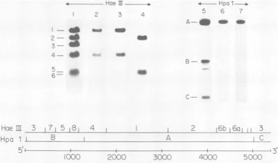
The (X174 DNAsequencesin the recombinant genomes.To determine theregionsof the XX174 genomefrom which the inserts in2B5, 32A, and
9BAwere derived, HaeIII andHpcI fragments
of wild-type (X174 RFI DNA were separated
by gel electrophoresis and hybridized with
32P-labeled SV40-4iX174 recombinant DNAs (Fig.
7). 32P-labeled 2B5 and 9BA DNAs hybridized
solely with the contiguous Haelll fragments
designated I and 4 in Fig. 7, indicatingthat the
inserts in 2B5 and 9BA were derived from a regionwhichstraddles theHaeIIl site at nucleo-tide position 1,776 (within the capsid-coding
region of the kX174 genome). In contrast, the
insert in the 32Arecombinantwas derivedfrom
aportion of the 4iX174 genome closer to the 3'
end, spanning Hoell fragments 2 and 6a/6b
(within the coding region for replication
func-tions).AlthoughHaeIIIfragments5and6a/6b of
(X174 RFI DNA were poorly resolved by gel
[image:7.492.44.442.493.577.2]electrophoresis, the 32A insert cannot contain
TABLE 2. Heteroduplex analysis of three clonedSV40-4X174 recombinants" Heteroduplexsegment(bp)
Heteroduplex
bt
a b c d
2B5-WT-SV40 39 594 + 66 860± 73 183 + 34 4.108 ± 196
2B5,)X174RE1 19 630 + 75
32A-WT-SV40 46 1,010 + 141 1.562 ± 178 2.922 + 160 1,000 + 89
32A4XI74 RFI 38 894 + 71
9BA-WT-SV40 45 949 + 124 1,653 + 176 2.683 + 179 1,097 + 103
9BA-4X174RF1 28 927 + 120
'Recombinants2B5,32A, and 9BADNAs(EcoRI cut)weredenatured and annealed with denaturedwild-type
(WT)SV40 DNA(EcoRIcut) orwith denatured ~X174RF1 DNA(PstIcut) as described in thelegend to Fig. 6 andinthetext.n, Numberof heteroduplexstructuresanalyzed.Theatodheteroduplexsegments measuredare
depictedin Fig.6. Intheheteroduplex structures formed between the recombinant andwild-type SV40DNAs,
segment a is the shorter armof the single-strand loop-out (thedX174DNAinsert);segment bisthelargerarmof thesingle-strand loop-out (the deletion of SV40 DNA); c and d are the double-stranded portions of the structure
(distance of the loop-out from eitherend). Intheheteroduplex formed between the recombinant andwild-type
4X174RF1 DNAs, segmenta is thedouble-strandregion (thejWX174 DNAinsert intherecombinant genome).
Theconversion factor (length measurement to basepairs) for double-stranded DNA was derived from linear measurements of wild-type SV40 homoduplexes (5,243 bp); that for single-stranded DNA was derived from linear measurementsof the 4X174virion plus strand(5,386nucleotides) added before electron microscopy.
48, 1983
on November 10, 2019 by guest
http://jvi.asm.org/
236 WINOCOUR, LAVIE, AND KESHET
4'
..
ir
r;v _ _~~~~~
-lO
_-.i -d
.. _ _
FIG. 7. Theorigin ofthe XX174 DNA inserts in recombinants 2B5. 9BA, and32A. Wild-type XX174RFI
DNA wasdigested with Hpal or with Haelll. The products were separated on 1% agarose and transferred to
nitrocellulosepaper.Replicatenitrocellulose stripswerehybridized with XX174RF1
[32P]DNA
(lanes1 and 5) or with32P-labelDNAs of the clonedSV40-4¢X174
recombinants2B5(lanes 2 and 6). 9BA (lane 3), and 32A (lanes 4and 7).Thelower panel shows theHaelilandHpaI restriction mapsof4X174DNA,linearizedatthe Pstl site
(which isthe zero pointforboth mapunitsandnucleotide numbers).
sequences derived from HaelIl fragment 5
be-cause 32A [32P]DNA did not hybridize with the overlapping Hpal B fragment (Fig. 7). Other
typesofexperiments(4) show that all segments
of the P174 genomerecombine equallywell with
SV40 DNA; indeed, no single portion of the
'bX174
genome appears topossessarecombina-tion advantage.
Comparison of recombinant genomes before
and after molecular cloning. Comparison of the
endonuclease restriction patterns of the
recom-binantstructurebeforeandafter molecular
clon-ing provides information on (i) possible
alter-ations that might have occurred during cloning
and (ii) whether the structure cloned is
repre-sentativeofthe predominanttype presentin the
amplified yield ofa given
recombinant-produc-ing cell. We therefore compared the restriction
patterns of the recombinant DNA yield from a
single
recombinant-producing
cell(isolate 2)
withthose of two independent molecularly cloned
recombinant genomes (2B5aand2B5b) derived
from that yield; 2B5a and 2B5b were isolated
from two separate X-Charon plaques. The
EcoRI, EcoRI-HindIII, and EcoRI-KpnI
diges-tion patterns (Fig. 8) indicate that the
recombi-nants in the single cell yield and the two
inde-pendently cloned derivatives of that yield all
contain
4XX174
DNA sequences withinrestric-tion fragments of identical electrophoretic
mo-bility.The useof three different restriction
endo-nuclease combinations in this experiment
provides strong evidence that these recombinant
structures mustbe verysimilar, ifnotidentical.
The results,therefore, indicate not onlythat no
gross genomic alterations occurred during
mo-lecular cloning, but also, and more importantly,
that the structurescloned represent the
predom-inant types generated by a given
recombinant-producing cell.
DISCUSSION
The SV40-4X174 recombinant DNA struc-tures described in this report originated from single recombinant-producing cells. One isolate
(2B5) was derived from a recombinant
infec-tious-center plaque that arose from plating
co-transfected monkeyBSC-1 cells in the presence
of a large excess of uninfected cells (4). The
frequency of recombinant-producing cells,
un-der the cotransfection conditions used, was
about
10-3
with respectto thetotal cellpopula-tionexposedtoSV40 and XX174RF1DNAs(4),
thusensuring that the plaque sampledwas
initi-ated by the progeny of a single
recombinant-producing cell. Isolates 9BA and 32A were
ob-tained by distributing recombinant-producing
cells in microwells (15) such that only a minor
fraction of the microwells (12 of 192) gave riseto
recombinant progeny. The isolation of
recombi-nants from transfected cells partitioned in
microwells averts adifficultyencountered in the
isolation of recombinants from plaques
identi-fied by in situ hybridization; the successful
samplingof the agar above the plaque requires
rather precise alignment of autoradiogram,
ni-trocellulose filter, and agar overlay (12). It
shouldbe emphasized that successfulisolation,
by either method, alsorequiresthat the
recombi-nant DNA structures be capable ofreplication
and encapsidation, aided by the presence of
J. VIROL.
on November 10, 2019 by guest
http://jvi.asm.org/
[image:8.492.113.399.78.245.2]wild-type SV40 helper virus. These require-ments for recombinant DNA amplification intro-duceanunavoidabledegreeof selection intothe
isolation procedures.
The proportion of recombinant to wild-type SV40DNA molecules,instocksraised from the microwell or plaque progenies, was 1:1,000. Application ofmolecular cloning procedures to
remove the large excess of wild-type DNA
raisedthe questionof whether the recombinant species clonedwasthemajor amplifiable species
generated by a given recombinant-producing
cell. However, comparison of the restriction mapsbefore andaftermolecularcloning (isolate 2 and its cloned derivatives 2B5a and 2B5b) established that the species cloned was in fact the predominant recombinant structure in the amplified progeny of a single recombinant-pro-ducing cell. Recombinant genome mapping be-fore molecular cloning was possible-despite
the presence of a large excess of wild-type
molecules-because oftheavailabilityof a rela-tively large number of restriction enzymes which cutSV40 but notXX174REl DNA.
The structures of the three SV40-+X174
recombinantgenomesexamined are remarkably simple in the sense that the 4X174 DNA
se-quences are present as single inserts and that
other than the deletion of SV40 DNA which accommodates the insert, the remainder ofthe
genome is indistinguishable from wild-type
SV40 DNA, as judged by heteroduplex and
restriction analyses. The retention of an intact SV40 early region in recombinant 2B5 and of intact SV40 late regions in recombinants 9BA
and 32A has also been confirmed by biological
tests. Independent replication of recombinant
2B5 DNA, transfected into BSC-1 cells in the absence ofwild-type SV40 DNA, has been
de-tected by in situ hybridization with
4X174
[32P]DNA.
Incontrast, noreplication of 9BA or32A DNA wasdetected under the same
condi-tions. The retention ofintact SV40late regions
in the 9BA and 32A recombinantgenomes was
confirmed by the observation that these DNAs (butnot2B5DNA)replicated, synthesized SV40 capsidproteins,and underwentencapsidationin theCOS line ofmonkey cells(5) whose chromo-somally integrated SV40 genome expresses T antigen constitutively (data not shown). The relatively simple, unscrambled structures of the
three
SV40-4+X174
recombinant genomesana-lyzed are consistent with the conclusion that
most (but not all) recombinant-producing cells
support only single recombination events (4).
This conclusion, however, should be viewed
within the context that recombinant-producing
cells are themselves a rare occurrence,
appear-ingat afrequency of 1 in 500 to 1 in1,000 cells
(4).
zo 3C~4 r, - 7
-3730
de_-
40em-1353
-;0
o;FIG. 8. Comparisonof a SV40-+X174recombinant genomebefore and after molecularcloning. TheDNAs
of the
SV40-4X174
recombinants in isolate 2 andthe two molecularly cloned derivatives 2B5a and 2B5bwere digested with EcoRI (lanes 1, 2, and 3, respec-tively),EcoRI-HindIII(lanes 4, 5, and 6,respectively)
andEcoRI-KpnI(lanes 7, 8, and 9, respectively). The
products were separated byelectrophoresis and were
blot hybridized with
4XX174
RF1 [32P]DNA as de-scribed in Fig. 2 and 4. 2B5aand 2B5b are derived from two separated X-Charon plaques (see thetext). Size markers are the3,730-bp HpaI fragment A andthe 1,353- and 1,078-bp Haelll products of authentic
4X174RF1 DNA.
A previous investigation (14) of
SV40-4¢X174
recombinant DNA derived from the total viral yield of a cotransfected culture of BSC-1 cells
indicated that avariety of different recombinant
structures hadarisen, many with multiple SV40
origins of replication reminiscent of the host
substituted variants (8). In the three
SV40-4~X174
recombinant DNA structures described herein(each originating from aseparate recom-binant-producing cell),theposition ofthe4X174
DNA insert with respect to the SV40 genome
varied from structure to structure. A fourth
structure, recently defined, contained
4XX174
DNAsequences in yet anotherlocation(datanot
shown). Although the number of recombinant
genomes studied is too small to permit
conclu-sions onthepresence orabsence of recombina-tion "hot
spots"
is SV40 DNA, it is clear that eachrecombinant-producingcell cangenerateadifferent recombinant structure. It is therefore not surprisingthat the total yieldofhybridviral genomes from a mass-cotransfected culture
dis-played ahighly complex pattern ofrecombinant
structures(14);asinglecultureof4 x
106
BSC-1cells would beexpectedtocontain 4,000 to8,000
recombinant-producing cells (1 in 500 to 1 in
1,000) and many, perhaps most, of these
gener-VOL.48,1983
on November 10, 2019 by guest
http://jvi.asm.org/
[image:9.492.275.425.66.256.2]238 WINOCOUR, LAVIE. AND KESHET
ate different recombinant structures. We have
not, so far, detected SV40 multi-origin
SV40-4X174 recombinants in the progenies of single recombinant-producing cells. Most probably, the multi-origin recombinants are generated in
the very rare recombinant-producing cells
sup-porting two independent recombination events
(4). Although the proportion of SV40
multi-origin recombinant genomes would be extremely lowinitially, the growthadvantageensuing from
the presenceofmorethanone replication origin
(6) would be expected to increase their
propor-tion in the total yield of a cotransfected cell
population.
ACKNOWLEDGMENTS
We thank Kenneth 1. Berns and Ann Roman for their critical comments on themanuscript.
This work was supported by a grant from the Leo and Julia ForchheimerCenterforMolecularGenetics.
LITERATURECITED
1. Brack, C. 1981. DNA electron microscopy. Crit. Rev. Biochem. 10:113-169.
2. Buchman, A. R., L. Burnett, and P. Berg.1980. TheSV4t) nucleotidesequence, p. 799-829. In J. Tooze (ed.).The molecularbiologyof tumorviruses, 2nded. Part2, DNA tumor viruses. Cold Spring Harbor Labor-atory, Cold Spring Harbor,N.Y.
3. Davis, R.W., M. Simon, and N. Davidson. 1971. Electron microscopeheteroduIplexmethods formappingregionsof base sequencehomologyinnucleic acids. Methods Enzy-mol.21:413-428.
4. Dorsett, D.L., I. Keshet, and E. Winocour. 1983. Quanti-tationofasimian virus 40nonhomologousrecombination pathway. J. Virol. 48:218-228.
5. Gluzman,Y. 1981. SV40-transformed simian cells support the replication of early SV40 mutants. Cell 23:175-182. 6. Kelly, T. J., Jr., and D. Nathans. 1977.The genome of
simian virus 40. Adv. Virus Res. 21:85-173.
7. Oren, M., E. L. Kuff, and E. Winocour. 1976. The presenceof common host sequences in different popula-tions of substitutedSV40 DNA. Virology 73:419-430. 8. Oren, M., S. Lavi, and E. Winocour. 1978. The structlt-e
of a cloned substituted SV40 genome. Virology 85:404-421.
9. Rigby, P. W. J., M.Dieckmann, C. Rhodes, and P. Berg. 1977. Labelling deoxyribonucleic acid to high specific activity in vitro by nicktranslationwith DNApolymerase I.J.Mol. Biol. 113:237-251.
10. Smith,G. E., and M. D. Summers. 1980. The bidirectional transfer of DNA and RNA to nitrocellulose or diazoben-zyloxymethyl-paper. Anal. Biochem. 109:123-129. 11. Southern, E. M. 1975. Detection of specific sequences
among DNAfragments separated by gel electrophoresis. J. Mol. Biol.98:503-517.
12. Villarreal, L. P., and P. Berg. 1977. Hybridizationinsiti ofSV40 plaques: detection ofrecombinant SV40) viruLs carrying specific sequences ofnonvir-al DNA. Science 196:183-185.
13. Vogelstein, B., and D. Gillespie. 1979. Preparative and analytical purificationof DNA from agarose. Proc. Natl. Acad. Sci. U.S.A. 76:615-619.
14. Winocour, E., and I. Keshet. 1980.Indiscriminate recom-bination in simian virus 40-infected monkeycells. Proc. Natl. Acad. Sci. U.S.A. 77:4861-4865.
15. Winocour, E., M.Singer, and E. Kuff. 1980.Rapid detec-tion,isolation andamplificationofhost-substituted SV40 variants. Cold Spring Harbor Symp. Quant.Biol. 44:621-628.
16. Zehnbauer, B. A., and F. R. Blattner. 1982.Construction and screeningof recombinant DNA libraries with Charon vector phages, p. 249-279. In J. K. Setlow and A. Hollander (ed.). Genetic engineering, principles and methods,vol. 4. PlenumPublishingCorp.,New York.
J. VIROL.