0022-538X/92/041875-09$02.00/0
CopyrightC)1992, AmericanSocietyforMicrobiology
Mutations
in
the
Principal Neutralization Determinant of Human
Immunodeficiency Virus
Type 1
Affect
Syncytium Formation,
Virus Infectivity, Growth Kinetics, and Neutralization
RAYMOND J. GRIMAILA,1 BARBARA A.FULLER,1 PAUL D. RENNERT,1 M. BUDNELSON,2 MARIE LOUISE HAMMARSKJOLD,2 BARBARA
POTTS,'
MICHAELMURRAY,SCOTTD.
PUTNEY,'
ANDGARYGRAY'*
Repligen Corporation, One Kendall Square, Cambridge, Massachusetts 02139,1 and Departments
of
Microbiology andOralBiology, State UniversityofNewYork atBuffalo, Buffalo, New York 14214Received 17 June 1991/Accepted 31 December 1991
The principal neutralization determinant (PND) of human immunodeficiency virus type 1 envelope
glycoprotein
gpl20 containsaconserved GPG sequence. The effects ofa29-amino-acid deletionof mostofthe PND, a 3-amino-acid deletion in the GPG sequence, and 16 single-amino-acid substitutions in the GPG sequenceweredeterminedin atransientexpression assay. All mutantenvelopeglycoproteinswereexpressed at levels comparable to thatof the wild-type envelope, and mutations in the GPG sequence did not affect processing togpl20 or, except for the 29-amino-acid deletion, binding to CD4. Of all of the mutants,only
the GHG and GFG mutantsinduced formation ofsyncytia similar in size and number to those induced by thewild-type envelope. When the envelope expression level was increased 10-fold or more, several additional mutants (APG, GAG, GSG, GQG, GVG, and GPF) also induced syncytium formation. Transfection with infectious proviralmolecularclonescontaining theGHG,GFG, APG, GAG, GSG,orGPF mutations induced production of viral particles; however,
only
the GPG,GHG, and GFG viruses produced active infections in CD4-bearing cells. Furthermore,whereas thewild-type viruswasefficiently
netutralized byPNDpolyclonal
and monoclonalantibodies, the GHG-andGFG-containingviruseswerenot.Theseresultsshow thatmutationsin the GPG sequence found within the PND do notaffectenvelope expressionanddonotsignificantlyaffectCD4 bindingorproduction of viral particlesbut thattheydo affecttheabilityof theenvelopetoinducesyncytiaand those of theviralparticlestoinfect CD4 cellsand beneutralized byPNDantibodies.Thepredominantrouteof HIV-1 infection involves bind-ing of external viral envelope glycoprotein gpl20 to the surface CD4 molecule on human T lymphocytes. A
post-gpl2O-CD4-bindingeventfacilitates fusion of the viral enve-lope with the cellular membrane, thereby permitting viral entry. Viral infectivity and fusion of human
immunodefi-ciency virus type 1 (HIV-1)-infected cells with uninfected CD4 cells can be blocked by antibodies directed to the
principal neutralization determinant (PND) located on the viral envelopeglycoprotein (11, 32, 33, 37).
Several studies have shown that the PND of HIV-1 resides ina36-amino-aciddisulfide loop formed between two invari-ant cysteines (positions 303 to 338) located in the third variable region of gpl20. A comparison of 245 different HIV-1 envelope sequences (19) showed that the PND con-tains conserved sequences (e.g., over 90% of these PND sequences contain the GPG motif) and that the PND con-tains the structural motif
p-strand-type
IIP-turn-13-strand-cX
helix. This suggests that there is selective pressure to maintain these conserved elements. Antibodies elicited by conservedamino acid sequences neutralizedivergentHIV-1 isolates (14).Previous studies(10, 17), using transient envelope expres-sion systems, showed that mutations within the PND can reduce or abolish syncytium formation without affecting
gp160expression,processing,orCD4binding. This suggests that,eventhough thePNDisdistinct from envelope domains
directly involved in CD4binding, elements of the PND are
* Correspondingauthor.
required for the membrane fusion event. These studies did not, however, investigate the effects of PND mutations on virusinfectivity, growth kinetics, or neutralization by PND antibodies. Another study (13) determined the effect of the GPG-to-GAG substitution on virusinfectivity and neutrali-zation. Thissingle change altered the abilityof the virusto replicate in some cell lines and affected the ability of the virus tobeneutralizedbysomePND-specific antibodies.
We made a series of deletions and substitutions in the conserved GPG sequenceatthetip of the PND to study the
importance of the PND in syncytium formation and virus infectivity, growth, and neutralization. Our data show that eventhough all mutant envelopes were expressed and proc-essed as the wild-type GPG, all of the mutations studied except two abolished the ability of the envelope to induce
syncytia. Eachof these two viruses was resistant to neutral-ization by PND antibodies. These results show that certain mutations in thePNDareinfectious and confer the property ofneutralization resistance.
MATERUILSANDMETHODS
Cells, plasmids,and antibodies. Cell lines SW480 (ATCC
CCL228) and COS-7 (ATCC CRL1651) were cultured in Dulbecco modifiedEagle minimal essential medium (GIBCO Laboratories) supplemented with 10% heat-inactivated fetal bovine serum (FBS; Hyclone Laboratories, Logan, Utah).
CD4+celllines CEM and CEM-SS(27)werepropagated and maintained in RPMI 1640 medium (GIBCO Laboratories)
supplemented with 10% heat-inactivated FBS.
Escherichia
coli JM105, HB101, BW313 (18), and TG1 were used for 1875on November 9, 2019 by guest
http://jvi.asm.org/
subcloning, mutagenesis, and amplification of plasmid DNAs and bacteriophage M13. Infectious provirus pNIA-3, an HIV-1 hybrid containing NY-5 and LAV-1 viral DNAs, has been previously described (1). Transient expression plasmid pLOTSA is derived from previouslydescribed plas-mid pSVSX1 (36). Its construction will bedescribed in detail elsewhere (31a). pLOTSA contains 2.1 kB of the HIV-1IIIB envelope gene and expresses a 140-kDa envelope glycopro-tein. Expression is driven by the simian virus 40 late promoter. rev-expressing plasmid pCMVrev has been previ-ously described as pRevl (21). It contains a cDNA copy of HIV-1 rev, and expression is driven by thesimian cytomeg-alovirus immediate-early promoter.
The neutralizing antisera used were goat anti-RP135 (37) and guinea pig anti-RP174c. (RP174 is RP135 with disulfide cross-bridged cysteines on both the amino and carboxyl termini.) AntiserumG1115wasprepared in goatsimmunized with baculovirus-insect cell-expressed gp160 which con-tained a deletion removing the entire PND (gp160 delta loop). Monoclonal antibody (MAb) C13 is a human-murine chimeric antibody constructed from0.5, (23) that binds the PND, including the sequence GPGRAF (a gift from S. Matsushita). MAb
lCl
(Repligen Corporation) recognizes a conserved epitope in thecarboxy terminus ofgpl20. OKT4 was obtained from Ortho Diagnostics.Oligonucleotide synthesis and purification. Oligonucleo-tides were synthesized by using anAppliedBiosystems 380A DNA synthesizer and purified by using 10%polyacrylamide gels followed by Sep-Pak C18 columnchromatography (Wa-ters, Millipore Corp.). Three oligonucleotide mutagenesis primers were synthesized such that each primer contained one degenerate codon in place of that whichencodes amino acid G,
P,
orG. The oligonucleotide primer used to muta-genize the first glycine (XPG) was 5'-GTATCCGTATCCAGAGAXXSCCCGGGAGAGCATT'TG-3',
that used to mu-tagenize the central proline (GXG) was5'-CCGTATCCAGAGAGGAXXSGGGAGAGCATTTGTTAC-3',
and that used to mutagenize the secondglycine (GPX) was 5'-CCGTATCCAGAGAGGACCCXXSAGAGCATTTGTTACAATAGG-3'.
Thedegenerate codon in eacholigonucleotide contains all four nucleotides at the first two codon positions (X) and a roughly equalmixture of G and C at the third position (S). This codon, XXS, cancontain 32 different nucleotidecombinations, which encode all 20 amino acids and onetermination codon. Deleted PNDmutants were prepared by using themutagenic oligonu-cleotides AGPG, 5'-CTAATGTTACAATGTGCGGGTCTT GTACAATTAAT'TC-3', which removed the centralGPG, and APND, 5'-GTAACAAATGCTCT'TCTCTGGATACG
G-3', which deleted most of the 36 amino acids, to leave a 7-amino-acid PND with the sequence CTRPAHC. Two oligonucleotides, VSP-PCR, 5'-GTAGAAATTAATTGTAC
AAGACCC-3', and XBA-PCR, 5'-CCATTl''TTGCTCTAGA AATGTTACAATGTGC-3', were used to sequence PND mutant envelopes from single-stranded M13 DNA and for polymerase chain reaction amplification and sequencing of the entire PND fromvirus-infected cells.
PNDmutagenesis and envelope subcloning. PND mutations were introduced by mutagenesis without phenotypic selec-tion by using themutagenicoligonucleotides (18). The 2.1-kb
KpnI-to-BamHI
fragment frompLOTSA, which encodes all but the carboxy-terminal 104 amino acids of gpl60, was cloned into theKpnI and BamHI sites of M13 vectorsmpl9andmpl8.Template single-stranded DNA containing uracil was obtained by growing these bacteriophage in dUTPase-and deoxyuracil-N-glycosidase-negative E. coli BW313 in thepresence of uridine. The mutagenesis reaction products
were transformed into E. coli to select for bacteriophage progeny derived from the mutagenized strand. Bacterio-phage plaques from the mutagenesis reaction werechosen, and theenvelopemutationswereidentified by dideoxy-chain termination sequenceanalysis. GPG mutant envelopes were subcloned as
KpnI-BamHI
fragments from the double-stranded replicative form of the appropriate bacteriophageinto the corresponding sites of pLOTSA. Followinganalysis of the mutants by transient expression, selected
KpnI-BamHI fragments were cloned into pNL4-3 to generate the proviral plasmids.
Transient expression assay. Rapidly dividing COS-7 cells were trypsinized, washed with RPMI 1640 medium supple-mented with 10% FBS, and collected by centrifugation at 1,000 x g for 5 min. Transfection conditions consisted of 3x
106
to 5 x 10⁶ COS-7 cells in 250 ,ul of ice-cold RPMI supplementedwith 10% FBS to which 125 ng of pCMVREV and 5 ,ug of envelope plasmid DNA were added. The cell-DNAmixture was transferred to a cuvette, and electro-poration was performed by using a Bio-Rad Gene Pulsar set at 0.25 kV, 960 ,uF, and 400Q. Following electroporation, the cuvette was incubated on ice for an additional 10 min and thetransfected cells were plated onto a 10-cm-diameter plate containing 10 ml of Dulbeccomodified Eagle minimalessen-tial medium supplemented with 10% FBS. After overnight incubation, the cells weretrypsinized, washed, and counted. Viable cells
(104)
weremixed with a 10-fold excess of CD4+ CEM cells and placed in a well of a 96-well microtiter tissue culture plate. Syncytia were visualized in the positive con-trol wells as early as 4 h after the cells were mixed, although syncytia were not counted until 24 h aftermixing.CD4-binding assay. The ability of the envelope to bind CD4 was assayed by radioimmunoprecipitation of labeled cell lysates with soluble CD4. COS-7 cells were transfected as described above, except thatincubation proceeded for 40 h after transfection and before labeling. Each 10-cm-diame-ter dish of cells was washed twice with phosphate-buffered saline (PBS) (GIBCO-BRL), starved for methionine by in-cubation in methionine-free medium for 30 min, and meta-bolically labeled overnight with 200
p,Ci
of high-specific-activity [35S]methionine (New England Nuclear). After being labeled, the cells were washed twice with PBS, scraped from the plates,transferred to Eppendorftubes, and washed again with PBS. The cells werelysed by addition of 100pl
ofRIPA-Lite (150 mM NaCl, 10 mM sodium phos-phate, pH 7.2) containing 1% Nonidet P-40 and vigorous vortexing. After dilution with 9 volumes of RIPA-Lite,insoluble material was precipitated by centrifugation for 5 min at 12,000 x g. Labeled cell lysates were incubated with 1 ,ug of soluble CD4 (American Bio-Technologies, Inc.,
Cambridge, Mass.) per ml at22°C for 1 h and then incubated with 1 ,ug of OKT4 per ml at room temperature for 1 h. Immobilized, recombinant protein A-Sepharose (IPA-300;
Repligen Corporation) was pretreated by being washed several times with RIPA-Lite-0.1% Nonidet P-40 and then incubated with an unlabeled COS-7 cell lysate in PBS. One-tenth of a volume of a 1:1 slurry ofpretreated IPA-300 in RIPA-Lite was added to the cell lysate containing sCD4 and OKT4 and incubated at 22°C for 30 min. The IPA-300 immunocomplexes were precipitated by briefcentrifugation
at 12,000 x g,washed three times with RIPA-Litecontaining
0.1% Nonidet P-40, washed three times withRIPA-Lite, and boiled with an equal volume ofgel loading buffer. Proteins were separated on sodium dodecyl sulfate-9%
polyacryl-amide gels and transferred byelectroblottingtoeither nitro-cellulose or Immobilon P (Millipore Corp.) membranes for
on November 9, 2019 by guest
http://jvi.asm.org/
autoradiography. Cell lysates were also radioimmunoprecipi-tated as described above with 1 ,ug of MAb lCl per ml.
Cell staining. COS cells transfected with 5 ,ug of pCM VREV and 5 ,ug of each envelope expression plasmid were
culturedasfor the transient expression assay, and the cells wereharvested byusing EDTA (GIBCO), washed with PBS, and reacted with goat anti-gp160 delta loop (G1115). The cells were stained with fluorescein isothiocyanate-conju-gated anti-goat serum and analyzed in a Becton Dickinson FACScan flow cytometer.
Viral infectivity and growth kinetics. Proviral plasmid DNA was transfected into SW480 cells by calcium phos-phateprecipitation asdescribed previously (1). The infectiv-ity of progeny virions was assessed by infecting CEM-SS CD4+ T cells with cell-free supernatant harvested 48 h after
transfection of the SW480 cells. The infections were per-formed in a 24-well plate by using 0.1 ml of cell-free transfection supernatant and 106 CEM-SS cells in 1.9 ml of RPMI supplemented with 10% FBS. Viral growth was
de-termined by reverse transcriptase (RT) activity by using
[32P]ddTTlP
(47) measured at 2- to 3-day intervals. Cell viability was monitored by using trypan blue exclusion, and the cell concentration was maintained at 106/ml. Infections were terminated when extensive cell death was observed. Cells were taken at day 20 postinfection for polymerase chain reaction amplification and sequencing of the PND region of the mutant HIV-1 envelopes.Western immunoblot analysis was performed on SW480 cells transfected with proviral clones as described
previ-ously, except that the proteins were transferred to Immo-bilon P membranes. Western blots were probed with either human sera from HIV-1-positive individuals or MAb lCl.
V'irusneutralization assay. Theabilities of PND antisera to
neutralizethewild-type andmutantviruses were assessed by
measuring the ability of a fixed amount of antibody to
neutralize virus at 10-fold dilutions. For this assay, MAbs were usedat200,ug/ml and polyclonal antiserum was used at a 1:10 dilution. Viral stocks, stored frozen until use, were serially diluted 10-fold by using RPMI 1640 medium contain-ing 10 mM N-2-hydroxyethylpiperazine-N'-2-ethanesulfonic acid(HEPES). Starting with the highest viral dilution, 0.1 ml of each virus dilution and the undiluted viral stock were transferred to a labeled 96-well plate. A 0.1-ml volume of serumwasdeliveredtoall dilutions of the virus. In addition to the specific antiserum used, controls included virus with
nonimmune antiserum and without antiserum. The control lacking antiserum allowed determination of 50% tissue culture
infective doses
(TCID50)
(40). The 96-well plate was then incubated at 37°C in 5% CO2 for 30 min, after which eachvirus-antiserum mixture was transferred to a 24-well plate containing
106
CEM-SS cells and mixed. The volume of the wells in the 24-well plate was then increased to 2 ml by addition of 0.9 ml of RPMI supplemented with 10% FBS, and theplateswereincubated at 37°C in5% CO2.At3 days, each well containing cells and medium was mixed, 1 ml was transferred to a new well (to obtain a duplicate of the dilution), and 1 ml of complete medium was added to each well. The wells weresplit 1:2 by removing 1 ml and adding 1 ml of fresh medium every 3 days thereafter. Medium was harvested for RT assays, and the cytopathic effect was recorded every 3days.RESULTS
Mutagenesis and selection of PND mutations. Site-directed mutagenesis without phenotypic selection was performed on
the PND ofthe HIV-1 IIIB(BH10) (34)envelopeglycoprotein
to study theeffects of changes in these conservedamino acids on syncytium formation, CD4 binding, and viral infectivity and neutralization. Briefly, uracil-containing single-stranded
template DNAs containing the 2.1-kb
KpnI-BamHI
envelopefragment from the envelope were mutagenized with
oligonu-cleotideswhich generateeitherdeletions orsingle-amino-acid substitutions in the GPG sequence. DNA sequencing of 54 separately isolated bacteriophage clones derived from GPG mutagenesis reactions yielded 49 substitution mutants and 5 nonmutagenized clones for a-mutagenesis efficiency of-91%. Of the 49 mutants obtained, 21 contained unique amino acid substitutions in the GPG sequence. Of the 21 GPG substitu-tions, 16 were chosen for further study. In addition, two deletion mutations, AGPG andAPND, wereconstructed. The 2.1-kb
KpnI-BamHI
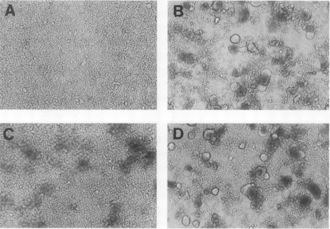
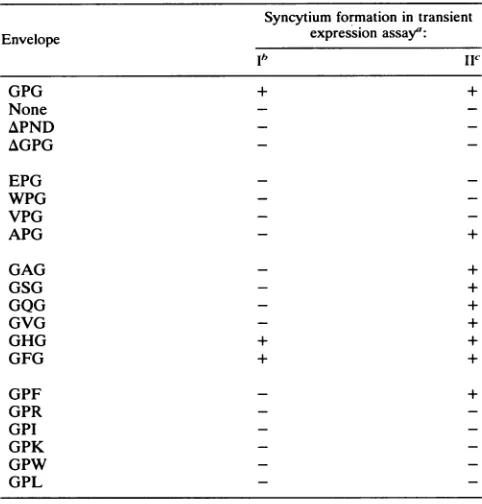
fragments of these 18 mutant envelope genes were cloned into the corresponding sites oftransient expression vectorpLOTSA.Syncytium formation of PND mutations. The effectsof the mutations on syncytium formation were assayed by mixing envelope-expressing COS-7 cells with CD4+ human T-cell cloneCEM. The COS-7 cells were cotransfected with 125 ng of pCMVREV, to provide a source of rev protein required for efficient envelope expression, and 5 ,ug of the envelope plasmid. COS-7 cells expressing the wild-type envelope and the substitution mutant GHG and GFG envelopes each formed equivalent numbers and sizes of syncytia. All other mutants, including the two deletion mutants (AGPG and APND), failed to form syncytia under these transfection conditions (Fig. 1 and Table 1). Although no clearly defined syncytia were generated with the mutants, cells expressing most of the variant envelopes appeared to induce cell association, as visualized by cell clumps (Fig. 1C).
Western blots of transfected COS-7 cells probed with MAb lCl (which recognizes a conserved sequence at the carboxyl terminus ofgpl20) showed that all mutant envelopes were produced at levels equivalent to that of the wild type (GPG) and were processed to the 140- and 120-kDa forms expected in the pLOTSA-based transient expression system (Fig. 2). Since the envelope clones tested contained examples of substitutions at each of the GPG positions, we conclude that anychanges made to these positions affect neither envelope expression nor processing and that the mutant envelopes behave like the GPG envelope. Probing of similar Western blots with a MAbspecific for the amino-terminal half of gp4l (data not shown) showed that all of the mutants tested showed aband at -20 kDa (the size expected from cleavage ofgpl40
togp120 and the transmembrane fragment).
Cell surface expression of envelope substitution mutants wasinvestigated by staining transfected COS cells with goat antiserum (G1115) raised against a HIV-1 gpl60 which lacked aPND loop. This antiserum was used because of the poor reactivity of several of the substitution mutants with antiserum prepared against the entire HIV envelope due to variation in the antigenic PND. Figure 3 shows staining of COS cells transfected with pCMVREV pluspAGPG (panel A) or pGPR (panel B). Both mutants showed clear and reproducible, albeit low, levels of cell staining, as evidenced by the shift in fluorescence intensity. Similar results were observed for substitution mutants pEPG, pGAG, and pGHG, in addition to wild-type pLOTSA. Control COS cells mock transfected and stained with G1115 antiserum or stained with an irrelevant antibody showed cell staining the same asthat of COS cellstransfected with plasmid pLOTSA alone.Although the level of staining of transfected COS cells waslow, all of the mutants tested repeatedly showed shifts
on November 9, 2019 by guest
http://jvi.asm.org/
FIG. 1. Syncytiumformation in the transientexpressionassay. COS-7 cells weremock transfected (A, negative control) or transfected withenvelopeexpression plasmid GPG(B, positivecontrol),WPG(C), or GHG(D).
similartothosepresented in Fig. 3. Most of the transfected COS cells showed little increase inenvelope expression, 10 to20%showedaless-than-10-foldincrease instaining,and 1 to5% showedan up-to-100-foldincrease.
Titration experiments using the GAG envelope in which theamountofpCMVREVwasvaried showed that the level
1
- 2
3
45
6
78 - 9
10
200 kDa
97
68
43~~~~
FIG. 2. Westernblotanalysis ofenvelopesubstitution mutants.
Cells were transfected with pCMVREV plus expression plasmid pLOTSA (lane
3),
pGPF (lane4),
pGPW (lane 5), pGPL (lane 6), pEPG (lane7),
pAPG(lane8),
pGAG (lane9),orpGFG (lane 10).Lane1containedpurifiedrecombinant
gp12O,
andlane2contained mock-transfected COScellsof envelope expression increased approximately 10- to 50-fold when the amount of pCMVREV was increased from 0.125 to 2 ,g (Fig. 4). Transfections using 5 ,ug of pCM VREVresulted inno additionalenvelope expressionabove that found for 2 ,ug (data notshown). Increasing envelope expression by increasing the amount ofpCMVREV in the transient expression assay to 5 ,ug allowed syncytium for-mation formutantsGAG,GSG, GVG, GQG, APG,and GPF
(Table 1). None of the remainingmutantsinduced syncytia
at the elevated level ofenvelope expression.
CD4 binding of mutant envelopes. Previous work has shown thataninsertion(17)oramino acidsubstitutions(10)
in thePNDdidnotaffectgpl20bindingtoCD4. Weassayed
theabilities of thewild-type(GPG), APND, AGPG,and GFG
envelopestobind sCD4byincubatinglabeled COS-7lysates
with sCD4 and determining whether MAb OKT4 could
precipitate the gpl2O-sCD4 complex. Whereas MAb lCl
effectively precipitated each ofthese envelopes, sCD4 and OKT4failedtoprecipitatethe APNDenvelope
(Fig. 5).
This indicates that neither thephenylalanine substitution northe GPG deletion significantly affected the abilityof the enve-lope to bind CD4, but deletion of the bulk of the PND abolished the ability to bind CD4. This could be due to a conformationalchangeingpl20causedbythislargedeletion which alters theCD4-bindingsite.Itis known thatdeletions ofotherregions ofgpl20affect CD4binding (5, 6, 8,22).
Analysisof PND mutations foreffectonvirusinfectivityand growthkinetics.Bothofthe substitutionmutantsthatformed
on November 9, 2019 by guest
http://jvi.asm.org/
[image:4.612.66.302.536.674.2]z
B 1 2 3 4 5 6 7 8
-
200 kDa
-97
- 68
-43
[image:5.612.311.548.77.245.2]FluorescenceIntensity
FIG. 3. Cell staining of COS cells transfectedwithHIV-1
enve-lope variants. Control COS cells were transfected with pLOTSA alone and lacked the pCMVREV plasmid required for envelope expression. (A) COS cells transfected with pLOTSA (gray) or
pA&GPG and pCMVREV (white). (B) COS cells transfected with
pLOTSA (gray)orpGPR andpCMVREV (white).
syncytiainthe transient expression assay underconditions of lower envelope expression (GFG and GHG), aswell as
fourmutantswhichformedsyncytiaunderconditionsofhigh envelope expression (GAG, GSG, GPF, and APG) were
cloned into the pNL4-3 proviral vector to determine the effects of these mutations onviral infectivity and growth. SW480 cellsweretransfected withproviral DNA, and after 40h, supernatantswere assayedfor RT. All of the mutants
were RT positive, indicatingthat transfectionsupernatants contained viral particles (data not shown). These
superna-tantswere used to infect CEM-SS cells. During the subse-quent40days,virusreplicationwasmonitoredbymeasuring thecytopathiceffect and culture supernatantswereassayed
for RT. Viruses NL-GPG (wild type), NL-GHG, and NL-GFG all showed viral replication, while viruses NL-GAG, NL-GSG, NL-GPF,andNL-APGwerenotinfectiousbythe RTandcytopathiceffectassays (Fig. 6).This demonstrated
that only the mutant envelopes inducing syncytia in the transient expression assay underconditions of lower
enve-lope expression giveriseto infectiousvirus.
The growth kinetics of the three viable viruses showed that NL-GHG displays kinetics similar to those of the wild-typeparental NL-GPGvirusin thatbothreached peak RTactivityatday10postinfection. However,thenumberof infectious viralparticlesofNL-GHG(TCID5O,-3 x 10³/ml)
was 1/10 of that for NL-GPG (TCID50, -3 x 10⁴/ml).
NL-GFG exhibited bothdelayed growthkinetics(peakRTat
day
22postinfection)andreduced viral titer(TCID5O, -3 x101/ml). Cellswere taken atday20 postinfection to obtain template DNA forpolymerase chain reaction amplification reactionsusingPNDprimersXBA-PCR andVSP-PCR. The three clones which produced infectious virus, NL-GPG, NL-GFG, and NL-GHG, yielded amplified DNA with the corresponding PND sequence, indicating that growth of NL-GHG and NL-GPG was not due to reversion to the wild-typeGPGsequence. Noneof the noninfectious viruses yielded polymerasechainreaction-amplifiable DNA.
Neutralization ofmutant proviruses. The ability of PND polyclonal antibodiesand MAbstoneutralizevirus infectiv-ityiswelldocumented(11, 14,19, 32, 37).Toinvestigate the effect of PND antibodies on infectivity, the titers of the GPG, GHG, and GFG viruses were determined in the absence or presence of PND polyclonal antibodies and
FIG. 4. Titration ofPCMVREV andexpression of GAG enve-lope variant. COS cellswere transfected with5
pLg
ofpGAG and various amountsofpCMVREV,i.e.,0(lane 1),0.031(lane2),0.062 (lane3),0.125(lane 4),0.250(lane 5), 0.50(lane6),1.0(lane 7),and 2.0(lane8).MAbs known to neutralize IIIB virus. The titers of serial dilutions of virus supernatants takenatpeak RT(day10for GPG and GHG and day 22 for GFG) in the presence or absence of a 1:10 dilution of anti-RP135 or anti-RP174
antiserum or 200 ,ug of MAb
C13l
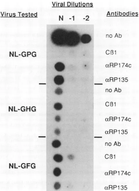
per ml were determined(Fig. 7). Whereas the titer of the wild-type virus was
103
TCID50Iml
in the absenceofantibody,the titerwas10/mlin the presenceofpolyclonal serum and less than 10/mlwith theMAb, indicatingthat99%ormoreof the virusinfectivityTABLE 1. Syncytium formation of PND mutants
Syncytium formationin transient
Envelope expression assay":
ib IIC
GPG + +
None -
-APND -
-AGPG -
-EPG -
-WPG -
-VPG -
-APG - +
GAG - +
GSG - +
GQG - +
GVG - +
GHG + +
GFG + +
GPF _ +
GPR - _
GPI - _
GPK - _
GPW - _
GPL _ _
a -,lack of visiblesyncytia; +,visiblesyncytia.
bTransfection conditions included 125 ng of pCMVREV and 5 ,ug of
envelope expression plasmid.
cTransfection conditions included5
pLg
ofpCMVREVand 5,Lgofenvelopeexpression plasmid.
"-dwolkh,
-.-,-iw
."-: ...
.i
.,:..a'"k
l:
on November 9, 2019 by guest
http://jvi.asm.org/
[image:5.612.48.294.77.225.2] [image:5.612.309.550.437.689.2]1 2 3 4 5 6 7 8 9 10
[image:6.612.328.565.75.326.2]2gpl02
FIG. 5. Radioimmunoprecipitation of mutant envelopes. Cells weretransfected with no DNA (lanes 2 and 7), GPG (lanes 1 and 6), AGPG(lanes 3 and 8), GFG (lanes 4 and 9),orAPND (lanes 5 and 10). The radiolabeled envelope proteins were immunoprecipitated with MAblCl (lanes 1 to 5) or sCD4 and OKT4 (lanes 6 to 10). The location ofgpl20isindicated.
.5
0
E
8_
(U
CoI
0
0.
c
20000
10000
0 was neutralized. In contrast, although the GHG and GFG
mutant viruses grew to a lower titer, neither mutant was neutralized by any of the antibodies. These data show that substitution of theproline at the tip of the PND abolishes the
abilityof the virus to beneutralizedby PND antibodies. DISCUSSION
The HIV-1 PND is the binding site for antibodies that neutralize virus infectivity and prevent fusion of infected cells with uninfected CD4-bearing cells. It is contained in a 36-amino-acid loop formed by a disulfide cross-bridge (20) between two invariant cysteines. The tip of this loop con-tains conservedpeptide sequencesandpredicted secondary structures, suggesting that there is selective pressure to preservethese elements(19). Peptides correspondingtothe
tipofthisloop (e.g., IQRGPGRAF [15] and GPGRAF [14])
elicitneutralizing antibody, showing that these amino acids
comprise theneutralization determinant.
To determine the effects of PND mutations on virus
infectivity, growthkinetics, andsusceptibilityto neutraliza-tion byPND antibodies, weconstructed aseries of substi-tutionmutantsinthehighlyconserved GPGat thetipof the PND. Several of the GPG variants constructed here are found in natural HIV isolates; however, numerous other differences exist with the IIIB-BH10 sequence and those
reported for other envelope genes or the entire envelope
sequenceshavenotbeen determined. Of the 16substitution and 2 deletion envelope mutants tested, only 2, GFG and GHG, induced syncytiawhenexpressed at low levels. The failure of the remaining envelopes to induce syncytium
formationwas notduetoaltered levels ofprotein expression
orenvelope processing, because all of themutantenvelopes tested,including thetwodeletions,wereexpressedatsimilar levels, as measured byWestern blot analysis and
radioim-munoprecipitation with MAb lCl. The mutations (except
APND)
also did not significantly alter theabilities
of the envelopes tested tobind CD4.Similarly, the deletion mutant lacking GPG and mutants withasubstitution at eachposition (EPG, GAG, and GPR) werefoundonthe surface of transfected COScellsatlevels comparable tothatof the wild type, pLOTSA(GPG).
This suggests that the inability of the substitution enve-lopestoparticipate insyncytiumformation isnotdue agross
0
dayspostinfection
FIG. 6. Growth kinetics of substitutionmutantviruses. Virion-associatedRTactivities invirus expressing the wild-typeenvelope (GPG) and two substitution mutants(GHG and GFG)were mea-sured. TheRTactivities of substitutionmutantvirusesGAG, GSG, APG, and GPF were identical to that of the mockcontrol.
defect in eitherexpression level,processing, orcell surface
expression.Freedetal. haverecently shown that mutations to the GPG and adjacent amino acids in the PND of the HIV-1envelope donot alter expression, processing,orcell surface expression in a transient expression assay (10).
Freed's limited number of mutations and the larger group
reportedheredemonstrated thatsingle-amino-acid substitu-tions and deletion of thecore GPG residues has little effect on normal envelope expression but has a large effect on
envelope function, asseenbythe loss ofsyncytium forma-tion. ThatCOS cellsexpressingmost mutantenvelopescan form aggregates with CD4+ cells also suggests surface
expression of the envelopes and points to a block in the fusionevent.
Several of the substitution mutant
envelopes
inducedsyncytium formation when envelope glycoprotein is
ex-pressedat 10-to50-foldhigherlevels. MutantsGSG, GAG, GQG, GVG, GPF,andAPG form
syncytia
whichare com-parablein size and numbertowild-type GPG.Interestingly,
allof themutations of theprolinewetested inducedsyncytiaathigher envelope levels,which isconsistentwith the results of LaRosa et al. (19) that showed more variability in this
position than in the two flanking glycines. The defect in
syncytiumformationcantherefore be overcome,atleast for somePNDmutants,by
elevated
expressionof theenvelope glycoprotein. This suggests that mutations that result in increased envelope expression may be able to compensate for PND mutations that decrease the efficiency of viral infection.Transient expression allowed screening of 18 envelope
mutations for syncytium formation and showed that only
two mutants, GHG and GFG, behaved like the
wild-type
GPG envelope. We tested these envelopes and the GSG,on November 9, 2019 by guest
http://jvi.asm.org/
[image:6.612.80.294.76.216.2]GAG, APG, and GPF envelopes for effects on viral infectiv-ity, growth kinetics, and antibody neutralization. Only the wild-type GPG virus and the two mutants which formed syncytia in the transient expression assay, GFG and GHG, produced infectious virus. Although the other mutants pro-ducedviral particles as judged by the presence of RT in the supernatant aftertransfection, these viruses were not infec-tious in CEM-SS cells as measured by either cytopathic effect or virion-associated RT activity. Although the GHG mutant exhibitedgrowth kinetics similar to those of the wild type and the GFG mutant showed delayed growth kinetics, both mutant viruses grew to lower titers. Because the amountsofviralparticles (as determined by RT activity) for all three viruses were comparable but the viral titers were either 10or100times lower than that of the wild type for the GHG and GFG mutants, it is likely that the number of infectious viral particles is reduced for the two mutants. Both mutants were resistant to neutralization by
PND-specificpolyclonal antibodies and MAbs; this was probably duetoalterationof the sequence of the binding site for these antibodies.
The mechanism by which PND antibodies neutralize in-fectivity is unknown. The PND does not directly participate in CD4
binding,
because PND antibodies do not block binding of gpl20 to CD4 (22,42), and the results presented herein and thoseof others show that small deletions,inser-tions (17), or substitutions (10) do not significantly affect binding of the envelope to CD4. Furthermore, addition of PND antibodies after the virus has bound to CD4 cells
completelyneutralizes viral infectivity (26). This shows that there is a relatively slow post-CD4-binding step that is
blocked byPND antibodies.
The step in the infection pathway blocked by PND anti-bodies is probably the same as that affected by the PND mutations that abolish the ability of the envelope to induce
syncytiaorgenerateinfectious virus. The role of the PND in viral infectivity is not known, but it has been suggested that the PND is cleaved by a cellular proteaseduring virus entry andthatthiscleavage is necessary for syncytium formation.
Cleavage of the PND (between the R andA) by thrombin, tryptase (4), oranendogenous protease in Chinese hamster ovary cells (43) has been shown; antibodies to tryptase inhibit HIV-1-induced syncytium formation (12), and pro-teinase inhibitors block HIV-1 infection (16). It is possible that themutations in the tip of the PND alter the recognition sequenceof the cell surface protease that is required for viral entry, thus making the PND a less efficient substrate for cleavage. This may explain why most mutations do not induce syncytium formation but that some, that may be
recognizedby the protease, enable the envelope to function. Ivanoff et al. (13) showed that aproviral clone containing a GAG mutation was infectious but that the efficiency of infectivity depends on the cell type. This suggests that
differentcelltypeshave proteases with different specificities and further suggests that some of our substitution mutants maybe infections ifother host cells are used.
Theoriginof PNDmutations in vivo is most likely due in part to selective pressure imposed by virus-neutralizing antibody generated during infection. It is possible that, as has been shown for visna virus (29, 30), caprine
arthritis-encephalitis lentivirus (31), and equine infectious anemia virus (2, 24, 39), neutralization-resistant variants that are resistant toneutralization by pre-existing antibody are gen-erated duringHIV-1 infection. Neutralizing antibody-resis-tantvariants ofHIV-1 have been selected both invitro (25,
35)and in vivo(7, 28) with mutations either in- or outside the
Viral Dilutions
VirusTested
N
-1
-2
NL-GPG
Antibodies
no Ab
C01
aRP1
74c
-0
NL-GHG
S
NL-GFG
_
_cRP135
no Ab
C131
czRP1 74c
aRP135
no
Ab
C81
ocRRP1
74c
w
ct~~~RPl 35
FIG. 7. Neutralization assay. Tenfold dilutions of viruses (N, undiluted; -1, 1:10 dilution; -2, 1:100 dilution) GPG, GHG, and GFG were mixed with no antibody (no Ab), 200 ,g of MAb C,1l per ml, 1:10 dilutions of polyclonal sera axRP174c and aRP135 and preincubated at 37°C for 30 min before being mixed with 106 CEM-SS cells. At day 10 postinfection, supernatants were har-vested for RT assay. Labeled products are shown.
PND (25), and our work shows that mutations within the PND that are infectious and resist PND antibody neutrali-zation can be generated. In addition toresistance to
neutral-izing antibodies, there is evidence that virus isolates ob-tained from individuals more advanced in AIDS disease progression are more cytopathic, have increased growth kinetics, and display increased cell tropism (3, 9, 38, 45). Our results show that PND mutations affectvirus growthkinet-ics, and recent data (41, 44, 46) implicate the PND in determination of host cell tropism. It will be important to extend theanalysis of PND mutants to understand the role of the PND in virus evolution and escape from immunological
selective pressure.
ACKNOWLEDGMENTS
WethankSandra Silver, James Rusche, Walter Herlihy, Al Profy, Greg LaRosa, and Kashi Javaherian for helpful discussions; Michael White foroligonucleotide synthesis and assistance with DNA se-quencing; S. Matsushita, S. Tokiyoshi, and Y. Eda for the C,11 antibody; and Malcolm Martin for the proviral vector.
This work was supported in part by NIH grant 1R01 AI 29825-01 toS.D.P.
on November 9, 2019 by guest
http://jvi.asm.org/
[image:7.612.314.551.80.405.2]REFERENCES
1. Adachi, A., H. E. Gendelman, S. Koenig, T.Folks, R. Willey, A. Rabson, and M. A. Martin. 1986.Production ofacquired immu-nodeficiencysyndrome-associated retrovirus in human and non-humancells transfected with an infectious molecular clone. J. Virol. 59:284-291.
2. Carpenter, S., L. H. Evans, M. Sevoian, and B. Chesebro. 1987. Role of the host immune response in selection of equine infections anemia virus variants. J. Virol.61:3783-3789. 3. Cheng-Mayer, C., D. Seto, M. Tateno, and J. Levy. 1988.
Biologic features of HIV-1 that correlatewithvirulence in the host. Science 240:80-82.
4. Clements, G., M. Price-Jones, P. Stephens, C. Sutton, T. Schulz, P. Clapham, J. McKeating, M. McClure, S. Thomson, M. Marsh, J. Kay, R. Weiss, andJ.Moore. 1991. The V3 loops of theHIV-1 and HIV-2 surfaceglycoproteins contain proteolytic cleavage sites: apossible function in viral fusion? AIDS Res. Hum. Retroviruses 7:3-16.
5. Cordonnier, A., L.Montagnier, and M. Emerman. 1989. Single amino acid changes in HIV envelope affect viral tropism and receptor binding. Nature (London)340:571-574.
6. Cordonnier, A., Y. Riviere, L. Montagnier, and M. Emerman. 1989. Effects of mutations in hyperconserved regions of the extracellular glycoprotein of human immunodeficiency virus type 1 on receptor binding. J.Virol. 63:4464-4468.
7. Emini, E. A., P. L. Nara, W. A. Schleif, J. A. Lewis, J. P. Davide, D. R. Lee, J. Kessler, S. Conley, S. Matsushita, S. D. Putney, R. J. Gerety, and J. W. Eichberg. 1990. Antibody-mediated in vitro neutralization of human immunodeficiency virus type 1 abolishes infectivity for chimpanzees. J. Virol. 64:3674-3678.
8. Fennie, C., and L. A. Lasky. 1989. Model for intracellular folding of the human immunodeficiency virus type 1 gpl20. J. Virol. 63:639-646.
9. Fenyo, E. M., L. Morfeldt-Manson, F. Chiodi, B. Lind, A. von Gegerfelt, J. Albert, E. Olausson, and B.Asjo. 1988. Distinct replicative and cytopathic characteristics of human immunode-ficiency virus isolates. J. Virol. 62:4414-4419.
10. Freed, E.O.,D. J.Myers, and R. Risser. 1991. Identification of the principal neutralizing determinant of human immunodefi-ciency virus type 1 as a fusion domain. J. Virol.65:190-194. 11. Goudsmit, J., C. Debouck, R. H. Meloen, L. Smit, M.Bakker,
D. M. Asher, A. V. Wolff, C. J.Gibbs, Jr., and D. C.Gajdusek. 1988. Human immunodeficiency virus type 1 neutralization epitope with conserved architecture elicits early type-specific antibodies in experimentally infectedchimpanzees. Proc. Natl. Acad. Sci. USA85:4478-4482.
12. Hattori, T., A. Koito, K. Takatsuki, H. Kido, and N.Katunuma. 1989. Involvement of tryptase-related cellular protease(s) in human immunodeficiency virus type 1 infection. FEBS Lett. 248:48-52.
13. Ivanoff, L., D. Looney, C. McDanal, J. Morris, F. Wong-Staal, A. Langlois, S.Petteway, and T. Matthews. 1991. Alteration of HIV-1 infectivity and neutralization by a single amino acid replacement in the V3 loopdomain.AIDS Res. Hum. Retrovi-ruses7:595-603.
14. Javaherian, K., A. Langlois,G. LaRosa,A.Profy,D.Bolognesi, W. Herlihy, S. Putney, and T.Matthews.1990.Broadly neutral-izing antibodies elicited by the hypervariable neutralneutral-izing deter-minant of HIV-1. Science250:590-593.
15. Javaherian, K., A. J. Langlois, C. McDanal, K. L. Ross, L.I. Eckler, C. L. Jellis, A. T.Profy, J. R. Rusche, D. P. Bolognesi, S. D. Putney, and T. J. Matthews. 1989. Principal neutralizing domain of the HIV-1 envelope protein. Proc. Natl. Acad. Sci. USA86:6768-6772.
16. Koito, A., T. Hattori, T. Murakami, S. Matsushita, Y. Maeda,T.
Yamamoto, and K. Takatsuki. 1989. Aneutralizing epitope of human immunodeficiency virus type 1 has homologous amino acid sequences with the active site ofinter-a-trypsin inhibitor. Int. Immunol. 1:613-618.
17. Kowalski, M., J. Potz, L. Basiripour, T. Dorfman, W. C. Goh, E. Terwilliger, A. Dayton, C. Rosen, W. Haseltine, and Sodroski. 1987.Functional regions of theenvelopeglycoproteinofhuman
immunodeficiencyvirustype 1.Science 237:1351-1355. 18. Kunkel, T., J. Roberts, and R. Zakour. 1987. Rapid and efficient
site-specific mutagenesiswithout phenotypic selection. Meth-odsEnzymol. 154:367-383.
19. LaRosa, G. J., J. P. Davide, K. Weinhold, J. A. Waterbury, A. T.Profy, J. A. Lewis, A. J. Langlois, G. R. Dreesman, R. N. Boswell, P.Shadduck, L. H. Holley, M. Karplus, D. P. Bolognesi, T. J.Matthews, E. A. Emini, and S. D. Putney. 1990. Conserved sequence and structural elements in the HIV-1 principal
neu-tralizing determinant. Science 249:932-935.
20. Leonard, C. K., M. W. Spellman, L. Riddle, R. J. Harris, J. N. Thomas, and T. Gregory. 1990.Assignment ofintrachain disul-fide bonds andcharacterization of potential glycosylation sites of the type 1 recombinant human immunodeficiency virus envelope glycoprotein (gpl20) expressed in Chinese hamster ovarycells. J.Biol. Chem. 265:10373-10382.
21. Lewis, N., J. Williams, D. Rekosh, and M.-L.Hammarskjold. 1990. Identification of a cis-acting element in human immuno-deficiency virus type 2(HIV-2)that isresponsivetothe HIV-1 rev and human T-cell leukemia virus types [sic] I and II rex proteins. J. Virol. 64:1690-1697.
22. Linsley, P. S., J. A. Ledbetter, E. Kinney-Thomas, and S. L. Hu. 1988. Effects of anti-gpl20 monoclonal antibodies on CD4 receptor binding by the env protein of humanimmunodeficiency virus type 1. J.Virol.62:3695-3702.
23. Matsushita, S., M. Robert-Guroff, J. Rusche, A. Koito, T. Hattori, H.Hosino, K. Javaherian, K. Takatsuki, and S. Putney. 1988. Characterization of a human immunodeficiency virus neutralizing monoclonal antibody and mapping of the neutraliz-ing epitope. J. Virol. 62:2107-2114.
24. McGuire, T., T. Crawford, and J. Henson. 1972. Equine infec-tious anemia:detection of infectiousvirus-antibodycomplexes in the serum. Immunol. Commun. 1:545-551.
25. McKeating, J. A., J. Gow, J. Goudsmit, L. Pearl,C. Mulder, and R. Weiss. 1989. Characterisation of HIV-1neutralizationescape mutants. AIDS 3:777-784.
26. Nara, P. 1989. HIV-1 neutralization: evidence forrapidbinding/ postbindingneutralization from infected humans, chimpanzees, and gpl20-vaccinated animals. Vaccines 89:137-144.
27. Nara, P., W. Hatch, N. Dunlop, W. Robey, L. Arthur, M. Gonda, and P. Fischinger. 1987. Simple, rapid, quantitative, syncytium-forming microassay for the detection of human im-munodeficiency virus neutralizing antibody. AIDS Res. Hum. Retroviruses 3:283-302.
28. Nara, P. L., L. Smit, N. Dunlop, W. Hatch, M. Merges, D. Waters, J. Kelliher, R. C. Gallo, P. J. Fischinger, and J. Goudsmit. 1990. Emergence of viruses resistant to neutraliza-tionbyV3-specific antibodies in experimental human immuno-deficiency virus type IIIB infection ofchimpanzees. J. Virol. 64:3779-3791.
29. Narayan, O., J. E.Clements, D. E. Griffin, and J. S.Wolinsky. 1981. Neutralizingantibody spectrumdetermines the antigenic profiles of emerging mutants of visna virus. Infect. Immun. 32:1045-1050.
30. Narayan, O., D. E. Griffin, and J. E. Clements. 1978. Virus mutation during "slow infection." Temporal development and characterization of mutants of visna virus recovered from sheep. J. Gen. Virol. 41:343-352.
31. Narayan, O., D. Sheffer, D. E.Griffin, J.Clements, andJ.Hess. 1984. Lack of neutralizing antibodies to caprine arthritis-en-cephalitis lentivirus in persistently infectedgoatscan be
over-come by immunizationwith inactivatedMycobacterium tuber-culosis. J. Virol. 49:349-355.
31a.Nelson, M. B., D. Rekosh, and M.-L. Hammarskjold. Unpub-lished data.
32. Palker, T. J., M. E.Clark, A. J.Langlois,T.J.Matthews,K.J. Weinhold, R. R. Randall, D. P. Bolognesi, and B. F. Haynes. 1988. Type-specific neutralization of the human immunodefi-ciency virus with antibodies to env-coded sythetic peptides. Proc. Natl. Acad. Sci. USA 85:1932-1936.
33. Putney, S. D., T. J. Matthews, W. G.Robey, D. L. Lynn, M. Robert-Guroff, W. T. Mueller, A.J.Langlois,J.Ghrayeb,S. R. Petteway, Jr., K. J. Wienhold, P. J. Fischinger, F.Wong-Staal,
on November 9, 2019 by guest
http://jvi.asm.org/
R. C.Gallo, and D. P. Bolognesi. 1986. HTLV-III/LAV-neutral-izing antibodies to an E. coli produced fragmentof the virus envelope.Science 234:1392-1395.
34. Ratner, L., W. Haseltine, R. Patarca, K. J. Livak, B. Starcich, S. F.Josephs,E. R.Doran, A. N. Rafalski, E. A. Whitehorn, K. Baumeister, L. Ivanoff, S. R. Petteway, M. L. Pearson, J. A. Lautenberger, T. S. Papas, J. Ghrayeb, N. T. Chang, R. C. Gallo, and F.Wong-Staal. 1985.Completenucleotide sequence of theAIDS virus, HTLV-III. Nature(London)313:277-284. 35. Reitz, M. S., Jr., C. Wilson, C. Naugle, R. C. Gallo, and M.
Robert-Guroff. 1988. Generation of a neutralization-resistant variant of HIV-1 due to selection for a point mutation in the envelopegene. Cell54:57-63.
36. Rekosh,D., A.Nygren, P. Flodby, M.-L.Hammarskj8ld,and H. Wigzell. 1988. Coexpressionofhuman immunodeficiencyvirus envelopeproteins and tat from asingleSV40 late replacement vector. Proc. Natl. Acad. Sci. USA 85:334-338.
37. Rusche,J.R., K.Javaherian,C. McDanal, J. Petro, D. L. Lynn, R. Grimaila, A. Langlois, R. C. Gallo, L. 0. Arthur, P. J. Fischinger, D. P. Bolognesi, S. D. Putney, and T. J. Matthews. 1988. Antibodies that inhibit fusion ofhumanimmunodeficiency virus-infectedcells bind a 24-amino acid sequenceofthe viral envelope,gpl20. Proc. Natl.Acad. Sci. USA85:3198-3202. 38. Sakai, K., S. Dewhurst, X. Ma, and D. J.Volsky. 1988.
Differ-encesincytopathogenicityandhostcellrange amonginfectious molecular clones of human immunodeficiency virus type 1 simultaneously isolated from anindividual. J. Virol. 62:4078-4085.
39. Salinovich, O., S. L. Payne, R. C. Montelaro, K. A. Hussam, C. J.Issel, and K. L. Schnorr. 1986.Rapid emergence of novel antigenicandgeneticvariants ofequine infectiousanemia virus duringpersistentinfection. J.Virol.57:71-80.
40. Schmidt, N., and R. Emmons. 1989. General principles of laboratory diagnosticprocedures for viral, rickettsial, and chla-mydial infections,p. 1-35. In N. J. Schmidt and R. W. Emmons
(ed.),Diagnostic proceduresforviral, rickettsial,and chlamyd-ialinfections,6th ed.AmericanPublic HealthAssociationInc., Washington,D.C.
41. Shioda, T., J. Levy,and C. Cheng-Mayer. 1991. Macrophage and T cell-line tropisms ofHIV-1 are determined byspecific regionsof theenvelopegpl20 gene. Nature(London) 349:167-169.
42. Skinner,M.A.,A.J. Langlois,C. B.McDanal, J.S.McDougal, D. P.Bolognesi,and T.J.Matthews.1988.Neutralizing antibod-iesto an immunodominant envelopesequence donot prevent gpl20 bindingtoCD4.J. Virol. 62:4195-4200.
43. Stephens,P. E.,G. Clements,G. T.Yarranton,and J. Moore. 1990. Achink in HIV's armour? Nature(London)343:219. 44. Takeuchi, Y., M. Akutsu, K. Murayama, N.Shimuzu, and H.
Hoshino. 1991.Host rangemutantofhumanimmunodeficiency virus type 1: modification of cell tropism by a single point mutationattheneutralizationepitopeintheenvgene.J.Virol. 65:1710-1718.
45. Tersmette, N.,J. M. A. Lange, R. E. Y. DeGoede, F.DeWolf, J. K. M. Eeftink-Schattenkerk, P. T. A. Schellekens, R.A. Coutinho, J. G. Huisman, J. Goudsmit, and F. Miedema. 1989. Association betweenbiological propertiesofhuman immunode-ficiencyvirus variants and risk for AIDS and AIDSmortality. Lanceti:983-985.
46. Westervelt, P.,H. Gendelman,andL. Ratner.1991. Identifica-tionofadeterminant within the humanimmunodeficiencyvirus 1 surface envelope glycoprotein critical for productive infection ofprimary monocytes. Proc. Natl. Acad. Sci. USA 88:3097-3101.
47. Willey, R. L., D. H. Smith, L. A. Lasky, T. S.Theodore, P. L. Earl, B. Moss, D. J.Capon, and M. A. Martin. 1988. In vitro mutagenesis identifies a region within the envelope gene of the humanimmunodeficiencyvirusthat is critical forinfectivity. J. Virol.62:139-147.