Prophage-Mediated
Interference Affecting
the
Development of Bacillus subtilis
Bacteriophage
oe
CARL W. RETTENMIER AND H. ERNEST HEMPHILL
Department ofBiology, Syracuse University, Syracuse, New York 18210
Receivedforpublication 11 October1972
Bacteriophage 4e shows a reduced efficiency of plating on strains of Bacillus
subtilis which arelysogenic for the temperate bacteriophage SP02. Although this
phenomenon resembles prophage-mediated restriction observed in other bacteria, host-controlled modification of 4e was not observed. Mutants of 4e which plated
withhigh efficiencyonthelysogenic hostwereisolated.
Although classical DNA restriction and
modificationsystems have not beendescribedin
BaciUus *ubtilis, it has been observed that some
strains of this bacterium do have mechanisms which can inhibit phage development after the
initialstages of infection arecompleted.
Phenom-enologically, the infection of the nonpermissive
B. subtilis strains resembles occurrences
ob-served in bacteria exhibiting phage restriction;
thephage adsorbtoand even killthe
nonpermis-sive host cells, but few progeny are released
be-cause some cellular mechanism prevents
comple-tionofthenormal lyticcycle (8,9).Theparticular
B. subtilis interference systems described below
also resemble the restriction systems of
Escher-ichia coli in that a certain number of phage
escapethe inhibitingmechanism in thehostcell,
and occasional plaques of the phage appear on
lawns of the normally nonpermissive B. subtilis
strains.
The
phenomenon
observedinB.subtilis,
how-ever,doesshow certaindifferences fromclassical
phage restriction. There is no evidence thatthe
B. subtilis cells contain specific restriction
endo-nucleases similar to those which mediate phage
restriction in other bacteria. Moreover, in the
studies madebyourselvesand others (8),nohost
modification systems have been found to
ac-companyinterference in B.
subtilis.
Because theterm phage restriction has become closely
as-sociated with DNA restriction andmodification,
objections can be raised to referring to the
ob-servations in B. subtil?s asphage restriction. To
meet this criticism, we have chosen to use
another term to describe our observations. B.
subtilis cellswhich permit certain phages to
ad-sorb andpenetrate,butsubsequentlyprevent
de-velopmentof normal yields of progeny, will be
said to exhibit interference. If the interference
systemisclearly associated withaprophage
car-ried bythehost exhibiting interference, we shall
use the term prophage-mediated interference. A
phage which is sensitiveto aninterference system
will bereferredto as aninterference-sensitive
bac-teriophage. It should be noted, however, that
there isatleastoneprecedent in the literaturein
which thetermphagerestriction has been used to
describe a phage-host relationship in B. subtilis
similar to interference (9).
Most strains of B. subtilis are lysogenic for
one or more defective prophages (18); since
prophages are frequently the source of phage
restrictionsystemsinvarious eubacteria (4, 10),
a number of investigators have speculated that
the defective prophages of B. subtilis might be
the source of interference against superinfecting
phages (8, 9). Attempts to investigate this hy-pothesis have beenhampered by difficulties in
transferringthegenomeof the defectiveprophage
fromonestrain of B. subtilisto another.
Never-theless, some circumstantial evidence has been
obtainedwhichsuggeststhat defectiveprophages maybe involvedin interference inthis bacterium.
Gwinnand Lawton (9) reported that B.subtilis
168 cells grown at a high temperature became
permissive hosts for normally
interference-sensitive phages SP10 and
SP20, although
theresultant progeny were not modified. Goldberg
and Bryan (8) found that pretreatment of B.
subtilis 168 with ultraviolet
light
or mitomycinC induced the defective prophage and
simul-taneouslyrendered the cells
permissive
forSP10. We have recently found that B. subtilis cellswhicharelysogenizedwith the temperate phage
SP02 demonstratea reducedefficiency ofplatinig
for certain virulent phages. In the following
372
on November 10, 2019 by guest
http://jvi.asm.org/
reportwe examinesomeof theproperties of this
interference systemandcompareittooneof the
interferencesystemsfound in naturally occurring
strains of this bacterium.
MATERIALS AND METHODS Bacteria and phage strains. B. subtilis strainSBll try-,aderivative of B. subtilis W23, wasobtainedfromE. W.Nester; B. subtilis 168M
and 168 M(SP02)wereprovidedby W. R.Romig;
B. subtilis 168(40105)wasobtained from S. A.
Zah-ler; and Bacillus pumilis was obtained fromthe
stock culture collection of Syracuse Univ. Bac-teriophage ,B22 was supplied by Roy Doi; SP82 was obtained from Charles Stewart; SP02c, was
supplied by W. R. Romig; SPOl andoewere
ob-tained from Clifford Yehle; 0105cr was obtained from S. A. Zahler; and 41 was obtained from
FrankYoung.
Growth and infection of cells. The medium used in thepresentexperiments forgrowing cells and preparing phage lysates was a modified M
mediumasdescribedpreviously (13).Experiments
in liquid medium were carried out at 37 C with culturesgrownin16-mmscrew-captubes. Optical density (OD) at600 nmwasmeasured ina
Spec-tronic 20 colorimeter on cells growing in these
culture tubes.
Phage were titered on M medium made with
1.5%agar.Thephage and theappropriate
indica-torstrain of bacteria were mixed in a 0.7% soft
M agaroverlay and thenpouredonthebase
me-dium.
Determination of relative burst size. The
relative burst size of 0e and SPOI on different
hosts was determined by growing the desired
strain of B. subtilis in modified M broth as
de-scribed above. When the cultures reached an
OD of 0.3at600nm,OeorSPOlwasaddedatthe desired multiplicity of infection (MOI), 1 min
was allowed for adsorptionwithout shaking, and
adsorption was continued with shaking for 5
additional min. The cultures were then
centri-fuged toremoveunadsorbed phage, suspendedin
Mbroth and diluted 1:10,000 in thesamemedium.
After lysis, the number of phage particles
re-leasedwas determined by plating on B. pumilis orB. subtilis168.B. pumiliswasusedasan indi-cator in studies involving B. subtilis 168 (SP02) becausecultures of thislysogenic strain frequently contained considerable numbers ofspontaneously induced SP02 which confused results. SP02 did
notformplaquesonB.pumilis while oe and SPOl
formed plaques at high efficiency on this strain.
Determination of numbers of cells
releas-ing phage. Experiments to determine the
num-ber of infected cells which released any phage were conducted in approximately thesame man-ner as those doneto determine relative yields of
progeny. The desired strain of B. subtilis was
growil toanOD of 0.3at600 nmandinfected, 6
minwas allowed for adsorption, and the cultures were centrifuged to remove unadsorbed phage.
The cells weresuspendedinM medium and then immediately diluted and plated on B. subtilis 168 orB.pumilis. Eachproductivelyinfectedcell was expected to produce one plaque regardless
ofthe numberofprogenyreleased.
Determination of phage adsorption. To determine the ability of oe to adsorb to various Bacillus strains, bacteria were grown to an OD of 0.3 at 600 nm and infected at anMOI of 0.2. One minute was allowed for adsorption without shaking. Adsorptionwas continued withshaking
for 5 additional min, the culture was diluted1: 10,000 and treated with chloroform, and
unad-sorbed phages in the supernatant fraction were
titeredonB. pumilis.
Production of doubly lysogenic strains of B. subtilis. As noted above, strains of B. sub-tilis lysogenic for SP02, or 4105 were obtained from other investigators, although several new B. subtilis 168 (SP02) strainswere made to ascer-tain thatthe SP02-mediated interferencewas not unique to the original strain. AstrainofB. sub-tilisdoubly lysogenic for 4105 and SP02 (B. sub-tilis 168 [SP02][4105]) wasmade byplating spon-taneously induced 4105 phage on B. subtilis 168 (SP02). Cells from turbid4105 plaques onlawns of this bacterium were streaked on M agar, and cultures were made from isolated colonies. Evi-dencethat the newlyprepared strains were doubly lysogenic included the fact that the cultures spontaneously released both 4105 and SP02, and the cultures were resistant to both
0105c1
(a clear-plaque mutant of 4105) and SP02c, (a clear-plaque mutant of SP02) (see Table 4).RESULTS
Comparisons of twointerference systems
in B. subtilis. Table 1 compares the relative
plaque-formingefficiency ofbacteriophages SP82,
SPOl, and
45e
on B. subtilis 168, B. subtilis 168(SP02), (which is a strain of 168 lysogenic for
the temperatephageSP02),B.subtilis SB11, and
B.pumilis. The three phages used in the present
studyarevirulentand share in common the fact
that their DNA contains the base substitution
hydroxymethyluracil (HMU) in place of thymine
(7, 12, 16). It will be noted that these phages
infectedall four Bacillus strains, but they formed
TABLE 1. Relative plaque-forming efficiencya of B. subtilis phages on
variou.s
Bacillusspecies
Bacilus
species SP82 SPOl oeB. subtilis 168 1 1 1
B. subtilis 168 (SP02) 1 1 10-3
B. subtilis SB11 10-4 104 10-4
B. pumilis 1 1 1
aArelative plaque-forming
efficiency
of 1 wasequal toapproximately1010PFU/ml.
on November 10, 2019 by guest
http://jvi.asm.org/
plaques 10,000 times lessefficiently on B.subtilis
SBll than on most other hosts. In addition, 4e
was specifically unable to grow normally on
B. 8ubtili8 168 (SP02) and formed plaques
1,000-fold less efficiently on this strain than on
B. subtilis 168.
The reduced ability of
oe
to form plaques onthe two nonpermissive strains of B. subtilis is
similar to what is observed when certain phage
infect bacteria carrying classical restriction
systems and invites a comparison of these two
interference systems with each other and with true restriction. We first investigated the
pos-sibilitythat4emight have a lowaffinity for the
adsorption sites on the two hosts demonstrating
interference. However, we found that this
inter-ference-sensitive phageadsorbed equally well to B.8ubtili8strains 168, 168(SP02), and SB11, and
wasabletokillandlyseboth the permissive and
nonpermissive hosts.
One differencebetween the interference systems
in B.subtilis SB11and B. 8ubtilis 168(SP02)was
observed in studies comparing the number of
cells (measured asplaque-forming units
[PFUs])
releasing
oe
(Table 2) and the relative yield ofprogeny (Table 3) at various MOI. At very low
MOI, few progeny wereproduced oneither
non-permissivehost; in B.subtilis SB11 thisappeared
TABLE 2. Relative number of cells releasing e at various MOPa
No. ofcellsreleasing4e
Host
MOI 0.02 MOI5
B. subtilis 168 100 100
B. subtilis 168 (SP02) 30 100
B.subtilisSB11 -5 100
aAvalue of100 in the table -35X 101PFU/ml
forMOI of 0.02;and 690 X 105 for anMOI of5.
TABLE3.Relative number of0eprogenyreleased frompermissive and nonpermissive B.
subtilishostsa
No.ofqbeprogeny released Host
MOI MOI5 MOI10 0.02
B. 8ubtilis168 100 100 100
B. subtilis 168 1 10 14
(SP02)
B. subtilis SBll 10 100 100
a Arelative burst size of 100 -25X 107PFU/ml
forMOI of 0.02; and 200 X 108PFU/mlforMOI of 5and 10.
to reflect a marked drop in the number of cells yielding any progeny, but under the same
condi-tions 30 to 50% (and in some experiments as
high as 75%) of the infected B.
subtilis
168(SP02) cells produced at least one progeny
4e.
These results suggest that the SP02-mediated
interference acted to reduce the yield of progeny
rather than to completely prevent productive
infection. Additional support for thishypothesis
can be seen in the infections at higher MOI. It
was possible tooverwhelm the
SB11
interferenceat an MOI of 5; under theseconditions both the
number of
SB11
cells producinggbe and the totalyield of progeny was equal to that obtained in
the B.
subtilis
168 controls. However, when B.subtilis 168 (SP02) cells were infected at this
MOI, the number of plaque-forming units was
equal to that of the controls but the totalyield
of gbe remained low.
There is otherevidencethat the
SP02-mediated
interference may have itsmost
pronounced
effectonthenumberofphage releasedrather than the
abilityto produceanyprogeny. Asnoted earlier,
SP82 and
SPOl
formed plaques with almostequal efficiency on strains 168 and 168
(SP02).
However, wefound that the plaques of SPOl on
lawns of the former were twice the diameter of
those observed on the lysogenic host.
Examina-tion ofthenumberofprogeny released from
SP01-infected B.
msbtilis
168
and 168 (SP02) cells inliquid cultureindicatedthat the burst sizeonthe
latter was one half that on the permissive host.
Growth of
4e
on otherlysogenic
hosts. We next turned our attention to determining whether prophages other than SP02 interferedwith the development of
4ie.
In thesestudies,
temperate B. subtilis
phage
4105
was used toconstructtwolysogenicstrains of thebacterium.
B.
subtilis
168 (4105) and B. subtilis 168(SP02)
(4105)
which islysogenic
for both SP02 and4105.
Table 4 compares the relativeplaque-TABLE 4. Relative
plaque-forming
efficiency of
ke,
SPORci and.kO5ci on
lysogenic
B. subtilisstrainsaBacteriophagetested Hoststrains
SP02c1 105ci 4e
B. subtilis 168 1 1 1
B. subtilis 168 (SP02) 0 1 10-B. subtilis 168 (0105) 1 0 1
B. subtilis 168 (SP02) 0 0 103
(4105)
a Arelative plaque-forming efficiency of 1 was
'80
X10 PFU/mlforSP02c, ;120 X108 PFU/ml
for
0105c1
; and75 X 108PFU/ml
for oe.on November 10, 2019 by guest
http://jvi.asm.org/
[image:3.495.56.251.381.650.2]forming efficiencies of SP02c1, 4 105cl
(respec-tively, clear-plaque mutants of SP02 and 4105),
and4eon several lysogenic B.subtiliU strains. It
can be seen that the strain which was lysogenic
for4105 alonedid notaffect the plaque-forming
efficiency of 45e; cultures doubly lysogenic for
46105 and SP02 did inhibit 4e but the
inter-ference was no greater than that observed on
B. subtilis 168 (SP02) itself.
Interference-resistanthost range mutants
of4pe. We have examined the phages present in
the 4e plaques which did form at low efficiency
on B. subtilU8 168 (SP02). Table 5 demonstrates
that these phages, which are referred to as
leIr, wereadaptedtothe nonpermissivehost and
formed plaques with equal efficiency on B.
UU8btili 168 and the lysogenic strain. More
surprising, the adaptation appeared to be a
genetic onerather than host-controlled
modifica-tion. Reculturing of 46eIr on 168did not
pheno-typicallychangetheadapted phagessothat they
again had a reduced plaque-forming efficiency
on B.8ubtili8168(SP02).Some 20plaquespicked
at random were analyzed and all contained
phages genetically resistant to the SP02-mediated
interference. Table 5 demonstrates, however,
thatthe 4beIrstrains were still sensitive to
inter-ference in B. subtilis SB11.
One troublesome aspect of thisfinding is that
it implies that there was a very high rate of
mutationfrom
OdeI
to 4elr.However, studiesinliquid culture discussed above and observations
on plates suggest a possible explanation. It
appearsthat the interference of4e inB. subtili8
168 (SP02) involved a reduction in burst size
rather thana precipitous drop in the number of
productive infections. Thus, when
q5e
wasplatedon B. subtilis 168 (SP02), there was probably considerable background growth of the normal
phage. Indeed, closeexamination of the lawns of
168 (SP02) onwhich
q6e
had beenplated usuallyrevealed a large number of very tiny plaques
which may have resulted from such background
growth of normal
4e.
Moreover, the plaques of4oelr
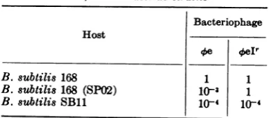
which first formed on the nonpermissiveTABLE 5. Relative plaque-forming efficiency of
0e
and
oeIt
onBacillusstrain8aBacteriophage Host
r/e oeIr
B. 8ubtili8 168 1 1
B. subtilis 168 (SP02) 10' 1
B. 8Ubti1is SBll 1(-4 10-4
aA relativeplaque-forming efficiency of 1 was
-101oPFU/ml.
host were
usually
smaller than those seen whentheisolates of
q5elr
weresubculturedonthesamestrain. Collectively,theseobservations maymean
that the 5eIrmutantswerenot
present
when thephage
wereplated
but arose asspontaneous
mutants ontheplates.
Under these conditions therealmutationratecouldbe much lower than the
apparent rateof mutation.
Phage
were also isolated from the occasionalplaques
of 4e observed on lawns of B. subtilis SB11 to determine whether these phagerepre-sentedasecondclass of
0e
whichwasadaptedtogrow on this nonpermissive host. However, the
phage
obtained from these plaques did not demonstrate an increased plaque-formingeffici-ency on B. subtilis SB11 and thus were
ap-parentlyneither host range mutants nor
pheno-typically modified to grow more efficiently on
B. subtilis SB11. It is possible that they
origi-nated from occasional productive infections of
cells in the lawn of thenonpermissive host,with
the resulting burst of phage multiply
infecting
neighboring cells to produce a plaque. It should be noted, however, that host range mutants of
HMU-containing phage have been isolated
by
other methods. Brodetsky and Romig (5) have
reported the selection of mutants of several such phages which form plaques on B.
subtilis
SB19 at a higher efficiency than do the
wild-types.It isnotunlikelythatmutantsof
q5e
whichhaveanextendedhostrangedo occur, but these
are difficult to distinguish among the normal
background plaques of escape infections.
Sensitivity of other bacteriophages to SP02-mediated interference. In addition to
thebacteriophagesnoted in Table 1, a number of
other phages have been studied to determine if
they were sensitive to the SP02-mediated
inter-ference. Virulent phages
qb29
and 622 were notaffected. However, recently Ronald Yasbin and
Frank Young (personal communication) found
thatbacteriophage
q51
wassensitivetotheinter-ference system in B. subtilis 168 (SP02). Our
examination of 4)1 revealed that this phage was
completely inhibited in B. subtilis 168 (SP02)
and no progeny wereproduced even at very high
MOI. No qbl plaques have been observed on
lawns of the lysogenic host, and our attempts to
isolate interference-resistantmutants of 4)1 have
failed. Like
Oe,
41 was found to be specificallyaffected by SP02 prophage and formed plaques
with high efficiency on strains lysogenic for
4)105. Aside from sharing a common sensitivity
to SP02-mediated interference, however,
q51
isnot related to 4)e. Its DNA does not contain
HMU. Moreover, 41 was not sensitive to the
B. subtilisSBll interferenceand formed plaques
with equal efficiency on B. subtilis strains 168
on November 10, 2019 by guest
http://jvi.asm.org/
[image:4.495.46.240.571.656.2]and SB11. This latter observation further
indi-catesthat thetwo interference systems were not
related.
DISCUSSION
Anumber of situations have been described in
whichabacteriophageisabletoinfect a potential
host cell but isunabletocomplete the lytic cycle
and release progeny.Thefirstcataloging of these
phenomena was made by M. H. Adams (1, 2) who coined the term "abortive infections" to describe these conditions in which there was an
irreversibleloss of theplaque-formingpotentiality of a phage-infected cell. The best understood
abortive infections are those in which DNA
restriction endonucleases selectively degrade the
phage genome into several pieces (4, 15). E. coli
cells which contain F+ or substituted F' sex
factors demonstrate asecond type of inhibitory
mechanism which causes abortive infections of
T7andseveralotherphages.Inthisinstance the
episomeapparentlydirects thesynthesis ofsome
product which interferes with synthesis of
several classes of phage proteins (11).
The two interference systems discussed inthe
present paper should probably be considered
examples ofabortiveinfections. This iscertainly
true of *1 infection of B. subtilis 168 (SPO2) because no progeny were released from the
in-fectedcells. However, these same lysogenic cells
infected with
0e
orSPOl releasedareducedyield rather than no progeny at all,and thus did notpreciselymeetthedefinition ofabortive infection
as given above. The naturally occurring
inter-ference observedinB.
oubtilis
SBllappeared tobe directed specifically against certain
HMU-containing phages, and blocked
phage
develop-ment only at low MOI. The latter interference
system would meet the definition of abortive
infection but only at these low multiplicities of infection.
We donotyetunderstand themolecular basis of either of the B. 8ubtili8 interference systems,
but several preliminary studies should be
con-sidered. Itisimportantto notethatour
observa-tions and those of others
(8)
indicate that theinterferenceinB.subtiliswas not
accompanied
by
modification as is the case in phage restriction
systems whicharemediated
by
DNArestrictionendonucleases.The association of DNA
modifica-tionwith DNA restriction is
thought
toreflectaneed to protect the host's DNA from the same
endonucleaseswhich breakdownthe
heterologous
phage DNA (4). Thelack ofmodification inthe
B.
subtilis
interference systems could then beinterpreted tomeanthat endonucleases weretot involved. The B.
8ubtilis
SBll interferencesystem could include a nuclease without a
modification enzyme, however, if the nuclease
specifically inactivated phage DNA containing
HMU as in SP82,
oe,
and SPOl. In such ahypothetical situation, the cell's DNA would be
protectedby virtue of the fact that it contained
thymine rather thaii hydroxymethyluracil. On
the other hand, the SP02-mediated interference
affected unrelated phages, only one group of which contained HMU. The fact that some
progeny were released when the superinfecting
phage was
0e
or SPOl would suggest that theDNA ofthese phages was not destroyed. Indeed
preliminary studies of DNA synthesis in B.
8ubtilis 168 (SP02) cells infected with *e not
only indicate that
0e
DNAremains intact in thenonpermissive host but that new phage DNA is synthesized.
Two abortive infection systems in
Entero-bacteriaceae show similarities to SP02-mediated
interference in that they are related to lysogeny and neither is accompanied by modification. T2
and T4infections of E. coli W which is lysogenic
forphagewresemble restriction in that the
enter-ingphage DNA is
degraded,
but several lines ofanalysis indicate that the DNAbreakdown may
be secondary to the actual mechanism of
inter-ference. It has been hypothesized that abortive
infections in this bacterium may be related to
changes in the E. coli membrane resulting from
lysogeny (14,17). The T-evenandT5coliphages
give abortive infections in
Shigella
dysenteriaelysogenicfor phage P2. Here again the failure to
supportproductive infection is thought toreside
inchanges in themembranerelatedtoprophage
conversion. It has been suggested that P2
pro-phage may preventresealing of the cellenvelope
after penetration of superinfecting phage (6).
Interference in B. subtilis could be related to
differences in the cellmembrane, but one would
not expect that such a mechanism would be
overwhelmed at high MOI as was the case in
B. subtilis SBll interference. Indeed, one might predict that in such a system the number of
infectious centers would decrease even further
at high MOI. This was not observed in either
B.
subtilis
SB11orB.5subtili
168 (SP02).We arecontinuing our study of possible mechanisms to
explaininterference in B. subtilis.
ACKNOWLEDGMENT
Thisinvestigationwassupported byagrantfrom the
Brown-Hazenfund of the ResearchCorporation. LITERATURE CITED
1. Adams, M.H. 1954.Abortiveinfection withviruses,
p.86-93 In F. W.Hartman,F. L.Horsfall, and J. C. Kidd (ed.), The dynamics of virus and
rickettsial infections. Blakiston Company, Inc.,
NewYork.
on November 10, 2019 by guest
http://jvi.asm.org/
2. Adams, M. H. 1955. Abortive infection with phage T2atlow temperatures.Virology 1:336-346. 3. Arber, W., and S. Linn. 1969. DNA modification
and restriction. Annu. Rev. Biochem. 38:467-500.
4. Boyer, H. W. 1971. DNA restriction and modifica-tion mechanisms in bacteria. Annu. Rev. Micro-biol. 25:153-176.
5. Brodetsky, A. M., and W. R. Romig. 1965. Char-acterization ofBaciUlus subtiliubacteriophages. J. Bacteriol. 90:1655-1663.
6. Fields, K. L. 1969. Comparison of the action of colicins El and K on Escherichia coli with the effects of abortive infection by virulent bacterio-phages. J. Bacteriol. 97:78-82.
7. Green, D. M. 1964. Infectivity of DNA isolated from Bacilus subtilis bacteriophage SP82. J. Mol. Biol. 10:438-451.
8. Goldberg, I. D., andT. Bryan. 1968. Productive infection of Bacilus subtilis 168, with bacterio-phage SP-10, dependent upon inducing treat-ments.J. Virol. 2:805-812.
9. Gwinn, D. D., and W. D. Lawton. 1968. Alteration of host specificity in Bacilussubtili. Bacteriol. Rev. 32:297-301.
10. Lederberg, S. 1957. Suppression ofthe multiplica-tion of heterologous bacteriophages in lysogenic bacteria.Virology3:495-513.
11. Morrison, T. G., and M. H. Malamy. 1971. T7 translational control mechanisms and their
inhibition by F factors. Nature N. Biol. 231:37-41.
12. Okubo, S., B. Strauss, and M. Stodolsky. 1964. The possible role of recombination in theinfectionof competent Bacillus subtiUis by bacteriophage deoxyribonucleic acid. Virology 24:552-562. 13. Palefski, S., H. E. Hemphill, P. E. Kolenbrander,
and H.R. Whiteley. 1972. Dominance
relation-ships in mixedly infected Bacillus subtili. J.
Virol.9:594-601.
14. Pizer, L. I., H. S. Smith, M.Miovic, and L.Pylkas.
1968.Effect of prophage Wonthe propbgation of
bacteriophages T2 and T4. J. Virol. 2:1339-1345. 15. Revel, H. R., and S. E. Luria. 1970.
DNA-glucosy-lation in T-even phage: genetic determination and role in phage-host interaction. Annu. Rev. Genet. 4:177-192.
16. Roscoe, D. H., and R. G. Tucker. 1964. The biosynthesis ofa pyrimidine replacing thymine
inbacteriophage DNA. Biochem. Biophys. Res. Commun. 16:106-110.
17. Smith, H. S., and L.I.Pizer. 1968.Abortive
infec-tion of Escherichia coli strain W by T2 bacterio-phage. J. Mol. Biol. 37:131-149.
18. Subbaiah, T. V., C. D. Goldthwaite, and J. Marmur. 1965. Nature of bacteriophagesinduced inBaciUlus subtilis,p.435-446. In V.Bryson and H. J. Vogel (ed.), Evolving genesand proteins. Academic Press Inc., New York.
on November 10, 2019 by guest
http://jvi.asm.org/