Joint Experimental and Computational
17
O and
1
H Solid State NMR
Study of Ba
2
In
2
O
4
(OH)
2
Structure and Dynamics
R
ı
za Dervis
og
̧
̆
lu,
†,‡,∥Derek S. Middlemiss,
†,‡Fre
de
́
ric Blanc,
́
‡,⊥Yueh-Lin Lee,
§Dane Morgan,
§and Clare P. Grey
*
,†,‡†Department of Chemistry, Stony Brook University, Stony Brook, New York 11794-3400, United States ‡Department of Chemistry, University of Cambridge, Lensfield Road, Cambridge CB2 1EW, U.K.
§Department of Materials Science and Engineering, University of Wisconsin, Madison, Wisconsin 53706, United States
*
S Supporting InformationABSTRACT: A structural characterization of the hydrated form of the brownmillerite-type phase Ba2In2O5, Ba2In2O4(OH)2, is reported using experimental multinuclear NMR spectroscopy and density functional theory (DFT) energy and GIPAW NMR calculations. When the oxygen ions from H2O fill the inherent O vacancies of the brownmillerite structure, one of the water protons remains in the same layer (O3) while the second proton is located in the neighboring layer (O2) in sites with partial occupancies, as previously demonstrated by Jayaraman et al. (Solid State Ionics2004,170, 25−32) using X-ray and neutron studies. Calculations of possible proton arrangements within the partially occupied layer of Ba2In2O4(OH)2 yield a set of low energy structures; GIPAW NMR calculations on these configurations yield1H and 17O chemical shifts and peak intensity
ratios, which are then used to help assign the experimental MAS NMR spectra. Three distinct 1H resonances in a 2:1:1 ratio are obtained experimentally, the most intense resonance being assigned to the proton in the O3 layer. The two weaker signals are due to O2 layer protons, one set hydrogen bonding to the O3 layer and the other hydrogen
bonding alternately toward the O3 and O1 layers. 1H magnetization exchange experiments reveal that all three resonances originate from protons in the same crystallographic phase, the protons exchanging with each other above approximately 150°C. Three distinct types of oxygen atoms are evident from the DFT GIPAW calculations bare oxygens (O), oxygens directly bonded to a proton (H-donor O), and oxygen ions that are hydrogen bonded to a proton (H-acceptor O). The17O calculated shifts and quadrupolar parameters are used to assign the experimental spectra, the assignments being confirmed by 1H−17O double
resonance experiments.
1. INTRODUCTION
Perovskites display a wide range of properties due to their ability to accommodate varying cations, substitutions, non-stoichiometry, and structural defects. They are consequently used in a widespread variety of applications. Of specific interest
to this work is their application, when hydrated, as proton conductors at intermediate to high temperatures (above ca. 200
°C).1,2Initial studies by Iwahara et al. and Nowick et al. led to
the discovery that perovskites such as LaYO3,3 SrZrO3,3 SrCeO3,4 , 5 BaCeO3,6 , 7 KTaO3,8 Sr2GaNbO69 and Ba3CaNb2O910 have high proton conductivities under humid conditions. All of these materials require cation substitution in order to create oxygen vacancies and, upon hydration, the hydroxyl defects responsible for their proton conductivities. In contrast, Ba2In2O5is an inherently oxygen deficient perovskite, and hence cation substitution is not required to allow water uptake.
Compensation of the Ba2+ and In3+ cation charges in
Ba2In2O5requires the removal of one-sixth of the O atoms in the perovskite structure, yielding a high intrinsic concentration of O vacancies along with both tetrahedrally and octahedrally
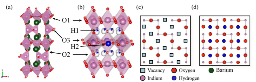
coordinated In3+ ions. The vacancies order at room temper-ature into an orthorhombic structure, resulting in three crystallographically distinct O sites (Figure 1a). The labeling used here has O1 sites at the equatorial positions of In octahedra, O2 sites bridging In octahedra and tetrahedra and O3 sites within the In tetrahedral layer. We use this labeling scheme for both the dry and hydrated structures in contrast to
some earlier work.11,12 The structure type is named
brownmillerite after the original Ca2FeAlO5 mineral which has a similar arrangement of O vacancies.13An understanding
of the water uptake and protonic conduction processes in this system is of particular interest as the extremely large number of vacancies in the brownmillerite structure facilitates a level of hydration rarely possible in related materials.14,15
A TGA study of Ba2In2O5under wet air by Jayaraman et al.11 found a maximum weight gain at around 200°C, indicating the formation of the Ba2In2O4(OH)2phase. Further heating results
Received: January 26, 2015 Revised: May 1, 2015 Published: May 1, 2015
in weight reduction due to H2O loss, and a neutron powder diffraction study under controlled humid air by Yildirim et al.16
showed that only Ba2In2O5is present above 350°C. Studies by Schober et al.17−19and by Yildirim et al.16indicate that there is a metastable, partially hydrated Ba2In2O5phase (ca. 0.5 mol of
H2O per formula unit) with a slight deviation from the
brownmillerite structure with theIma2 space group. Complete hydration of Ba2In2O5 results in a tetragonal structure with
space group P4/mbm where the intrinsic O vacancies of
Ba2In2O5 in the O3 layer are fully occupied by the water O
atoms, yielding the composition Ba2In2O5.H2O (or
Ba2In2O4(OH)2).11,20 The 2cpositions (in the O3 plane) are fully occupied by one of the water protons (H2) while the
second proton (H1) partially occupies (12.5%) the 16l
positions (in the O2 plane) according to the combined X-ray and neutron diffraction analyses of Jayaraman et al.11 (Figure
1b). This model should be contrasted with that proposed in the
earlier studies of Zhang and Smyth23 where the water was
believed to react with (O1) vacancies in the perovskite slabs, the vacancies resulting from p-doping and/or Frenkel defects created by occupancy of vacant O3 sites.
Early force field calculations of the energetics of defect
formation by Fisher and Islam20questioned the role of the O1
vacancies proposed in the Zhang and Smyth model23 and
confirmed that the water oxygen atoms occupy the vacant O3
sites. They suggested, however, that the lowest energy site for the protons were the O1 sites. A subsequent DFT based, explicit full optimization of a large number of possible proton arrangements of the hydrated material Ba2In2O4(OH)2 (two formula units, i.e., 24 ions per unit cell) by Martinez et al.12 identified a set of low energy structures with two protons (H2)
in the O3 layer occupying half of the 4h (x, x̅ + 0.5, 0.5) positions, and two other protons (H1) in one of the two O2
layers occupying one-16th of the 32y positions. The 32y
positions are defined by Martinez et al.12and are derived from
the 4epositions of the O2 site in theP4/mbmspace group by the eight displacements (±0.4 Å, ±0.4 Å, ±0.4 Å). Note that this 32yposition is not found in the International Tables for Crystallography24for theP4/mbmspace group (No. 127) but it
is nevertheless useful for visualization of the proton positions.
Zhang and Smyth23 reported protonic conductivity of
Ba2In2O4(OH)2 under humid air, where three regions of protonic conduction were observed corresponding broadly to
temperatures below 400 °C, between 400 and 925 °C, and
above 925 °C. These authors proposed both Grotthuss
(H-hopping) and vehicle (OH-(H-hopping) mechanisms as being involved in protonic conductivity: the Grotthuss mechanism dominating at low temperature; the vehicle mechanism or a combination of vehicle and Grotthuss mechanisms dominating at elevated temperatures.23
A study of Ba2In2O4(OH)2and of its Ti-doped derivatives11
using 1H and 2H solid state NMR experiments under magic
angle spinning (MAS) identified three distinct proton sites
(giving rise to a higher frequency split resonance and a lower frequency broad resonance), while only two proton sites are anticipated based upon the tetragonal crystal structure obtained by Jayaraman et al.11(Figure 1b). The increased splitting of proton sublattices in the NMR data as compared with the number of crystallographic sites found by neutron diffraction
was interpreted as a result of a further ordering of proton sites with respect to the average structure. The authors postulated that this arises from the actual unit cell being larger than the average unit cell of the P4/mbm space group used in the refinement of the diffraction data (a larger cell having been
observed in their electron diffraction studies), NMR being
sensitive enough to distinguish between variations of different
ordering schemes for at least one of the two environments. However, no clear assignments of these three proton resonances were given.
Our previous paper on Ba2In2O5focused on the dry material and used DFT methods to rationalize the17O spectra seen in this system.22The current work examines hydrated Ba
2In2O5 [i.e., Ba2In2O4(OH)2] using both current solid state17O and 1H NMR spectroscopy techniques and DFT calculations. The
DFT energetics and GIPAW calculations arefirst presented to
describe the various configurations investigated in this work.
We reproduce the general results of the DFT study by Martinez et al.,12although differences in the ground state structure and
the energies of the other low energy structures are observed. First-principles periodic DFT NMR calculations within the gauge-including projector augmented wave (GIPAW)
ap-proach25 are then performed to help interpret the NMR
spectra. High magneticfield strengths are used to obtain NMR
spectra of the hydrated material. This approach allows high resolution solid state NMR spectra of quadrupolar nuclei (such as17O, spinI= 5/2) to be obtained. The individual17O and1H
NMR shifts are assigned to specific oxygen and proton
[image:2.625.89.538.61.205.2]environments, the DFT results allowing us to assign the Figure 1.(a) Room temperature crystal structure of the brownmillerite Ba2In2O5structure in space groupIbm213,21with an···OctTetOctTet′··· staggered O vacancy pattern. (Reproduced from reference 22 by permission of the PCCP Owner Societies.) (b) Room temperature crystal structure of tetragonal Ba2In2O4(OH)2 in space group P4/mbm,11 showing the partially occupied H1 and fully occupied H2 positions. Schematic representations of the O3 layer of (c) Ba2In2O5and (d) Ba2In2O4(OH)2. The interlayer Ba atoms in b, c, and d have been omitted for clarity. Sector
three 1H NMR signals seen at room temperature to specific
local environments. We show that multiple low energy structural configurations are responsible for the experimental 1H NMR spectra. The17O NMR spectra well reproduced with
these multiple configurations, allowing us to assign the proton
donor and acceptor oxygens. Variable temperature 1H NMR
spectroscopy is then used to probe proton motion.
2. MATERIALS AND METHODS
2.1. Experimental Methods. 2.1.1. Sample Preparation.
Ba2In2O5 was prepared according to a literature procedure.22 Ba2In2O4(OH)2was prepared by slow cooling of Ba2In2O5(dried at 400 °C under a flow of dry N2 for 12 h) from 350 °C to room temperature (at a rate of 0.1°C.min−1) under aflow of wet N
2gas. The water vapor pressure corresponds to∼20−30 mbar PH2O(∼2.3% (w/v) H2O) and was controlled by bubbling N2 through water at room temperature.1717O enriched Ba
2In2O4(OH)2was synthesized by heating previously synthesized Ba2In2O4(OH)2in a 50%17O enriched O2gas (Isotec, used as received) atmosphere with∼2.3% (w/v) H2O (nonenriched) in a closed quartz tube, at 1000°C for 24 h. During 17O enrichment with the closed quartz tube, the initial air inside the tube was removed by vacuum while concurrently freezing the H2O inside the tube with liquid N2.
Powder X-ray diffraction patterns were obtained on either a Panalytical Empyrean or Bruker D8-Focus X-ray diffractometer using Cu Kα radiation (λ = 1.5418 Å; Figure S1 in the Supporting Information). Thermogravimetric analyses (TGA) were performed on a Mettler Toledo TGA/SDTA851 thermobalance using an alumina crucible (Supporting Information Figure S2). All measurements were performed underflowing dry nitrogen, in a temperature range of 30− 800°C and at a heating rate of 10°C·min−1. TGA showed that both Ba2In2O4(OH)2 and17O enriched Ba2In2O4(OH)2contain 1 mol of water per formula unit, as anticipated.
2.1.2. Solid State NMR Spectroscopy.Solid state17O MAS NMR experiments on Ba2In2O4(OH)2 were performed on 9.4 T Bruker Avance 400 MHz and 16.4 T Bruker Avance III 700 MHz spectrometers using Bruker 2.5 mm HX probe and Bruker 3.2 mm HXY (in double resonance mode) probe, respectively. Unless otherwise stated, spectra were recorded using a solid ∼π/2 pulse length of 1μs, corresponding to a radio frequency (rf)field amplitude of∼83 kHz, and a MAS frequency of 30 kHz at 9.4 T, and a solid∼π/ 2 pulse length of∼1.7μs, corresponding to a rffield amplitude of∼50 kHz, and a MAS frequency of 20 kHz at 16.4 T. The17O 3QMAS experiment was performed at 9.4 T with 128t1 increments of 1320 scans each. Hard and soft pulses are performed at rffields of 150 and
∼10 kHz, respectively. The 1H−17O cross-polarization (CP) and heteronuclear correlation (HETCOR)26−29 spectra were obtained at
16.4 T with a17O rffield amplitude of∼50 kHz, while the1H rffield amplitude was ramped to obtain maximum signal at∼83 kHz. Small phase incremental alternation with 64 steps (SPINAL64)30 1H heteronuclear decoupling was applied during the acquisition. Contact times for the CP experiments ranged between 5 and 5120μs. All17O NMR data were collected on a freshly17O enriched Ba
2In2O4(OH)2 sample packed in a ZrO2rotor. A recycle delay of 10 s was used for all experiments, with 20480 scans for17O Hahn-echo experiments and 48 t1increments of 256 scans for the HETCOR spectra.
1H MAS NMR experiments were performed on a 16.4 T Bruker Avance III 700 MHz spectrometer equipped with a Bruker 4 mm HXY probe (in double resonance mode). One dimensional (1D) spectra were recorded under MAS using a rotor-synchronized spin echo sequence to suppress the proton background of the probe.31 All 1H spectra were recorded at a rffield amplitude of 100 kHz and a MAS frequency of 12.5 kHz with recycle delays ranging from 4 to 60 s depending on theT1 relaxation times. Temperature calibration was performed using the207Pb resonance of Pb(NO
3)2as a chemical shift thermometer.32,33The sample temperatures quoted subsequently have
all been corrected and have an accuracy of±10°C. Additional fast MAS1H NMR experiments were recorded on the same spectrometer
with a Bruker 1.3 mm HX probe spinning at 60 kHz, using a rotor-synchronized spin echo sequence, an rffield amplitude of 115 kHz, and recycle delay of 8 s.
17O and1H chemical shifts were externally referenced to water at 0.0 and 4.8 ppm, respectively, at 20°C. NMR data were processed using TopSpin 3.034 and MatNMR,35 the latter running within the MATLAB package. Simulations and deconvolutions were performed using the same software and SIMPSON.36
2.2. Computational Methods. 2.2.1. Energetics and Confi g-urations. The first-principles solid state electronic structure calculations used here are similar to those reported in our previous work on Ba2In2O5and in related studies37−43and were all performed within the CASTEP code.44Structural models of Ba2In2O4(OH)2were derived, as described later, from the experimental neutron diffraction model by Jayaraman et al.11following the approach of Martinez et al.12 Full structural optimizations (both cell and atomic positions) of Ba2In2O4(OH)2(two formula units) were performed in the absence of any symmetry operators (i.e., in space groupP1), using a plane wave kinetic energy cutoffof 40 Ry and a linear spacing of 0.04 Å−1 or smaller for the reciprocal space sampling mesh, yielding Monkhorst− Pack meshes of dimension 6×6×4 for the Ba4In4O8(OH)4supercell. Full details of all of these structures are presented in the Supporting Information (SI). The Perdew−Burke−Ernzerhof GGA-type ex-change-correlation functional has been used throughout.45
Con-vergence of total energy with respect to numerical parameters was estimated at 0.2 kJ·mol−1per atom or better. Structural optimizations (both cell parameters and atomic positions) pursued until the energy difference, maximum atomic force, maximum atomic displacement, and maximum stress tensor component fell below tolerances of 1× 10⁻⁶eV, 1×10⁻³eV·Å−1, 1×10⁻³Å, and 5×10⁻³GPa, respectively. The effect of decreasing these listed tolerances by a further order of magnitude was investigated, yielding only minimal changes in geometry and computed NMR parameters. To facilitate our exploration of the complex energy landscape, vibrational free energy contributions were not included in this analysis. However, given the relatively similar bonding sites being considered for the H, we assume that these contributions will largely cancel when comparing the relative energies. Previous work on a series of iron- and aluminum-oxyhydroxides calculated a less than 1.5 kJ/(mol of H) variation in vibrational free energies at room (25 °C) and synthesis (350 °C) temperatures for H across three compounds with both Fe and Al cations.46,47 This error is as small or smaller than that found later
between different DFT approaches, justifying exclusion of these contributions.
The hydration enthalpy of formation for the ground state configuration was calculated for the theoretical reaction of Ba
2In2O5 (staggered configuration)22 with one isolated water molecule H
2O (calculated in a large unit cell with volume of 332 Å3). DFT total energies of geometry-optimized structures were used in this calculation. The experimental enthalpy of hydration is reported to be−0.65±0.08 eV (∼−63 kJ·mol−1) with the Zhang−Smyth model and−0.76 eV (∼−74 kJ·mol−1) (trapped) and−0.3 eV (∼−29 kJ· mol−1) (untrapped) with the trapping model by Schober and Friedrich18at temperatures of 623−1073 K.
Boltzmann distribution weights at 350°C were calculated according to weighti = (exp(ΔEi/RT))/(Σi exp(ΔEi/RT)) where ΔEi is the relative energy of a configuration from the ground state configuration, per mole of hydrogen. Structural models were visualized with the VESTA48and CrystalMaker packages.
2.2.2. NMR Calculations. Fully periodic calculations of NMR parameters within the gauge-including projector augmented wave (GIPAW) approach25,49 have been performed using the CASTEP code, including determination of electric field gradient tensors and associated quadrupolar interaction parameters for 17O sites.50 The
NMR parameters are obtained from single point calculations within the optimized geometry, differing only from the prior optimization runs in that a larger basis set cutoffof 60 Ry was applied. The isotropic shielding was obtained asσiso= (σxx+σyy+σzz)/3, whereσxx,σyy, and
then derived from the computed site shieldingσisoby application of a shielding referenceσrefwith the expressionδiso=σref+mσiso. Bothσref
and the scaling factormfor17O are taken unmodified from previous work by the current authors which obtainedσref= 223.70±3.03 ppm and m = −0.888 ± 0.014 with a mean absolute error (MAE) in computed shifts relative to experiment of 12.1 ppm across a range of phases.37Meanwhile,σ
refandmfor1H are determined from afit of the results of NMR CASTEP calculations on Mg(OH)2(σiso= 30.89 ppm; δiso= 0.5 ppm)51in combination with 55 different proton sites in four
separate organic molecules as computed recently by Yates et al.,52
Webber et al.,53and Sardo et al.,54 yieldingσref= 28.45±0.51 ppm andm=−0.930±0.020 with a MAE (defined similarly to those given earlier) of 0.29 ppm (SI Figure S3). We note that all but one of the proton sites used in deriving the shielding reference arise within organic molecules, and thus we might expect some discrepancy between experimental and calculated 1H chemical shifts in Ba2In2O4(OH)2 as discussed later. Chemical shift anisotropies and asymmetries are also computed, defined asσ
aniso=σzz−(1/2)(σxx−
σyy) andηCS= (σyy−σxx)/(σzz−σiso), respectively. The quadrupole coupling constant for 17O is obtained as C
Q = eQVzz/h and the asymmetry asηQ= (Vxx−Vyy)/Vzz, where an ordering|Vzz|≥|Vyy|≥| Vxx|of the principal components of the traceless electricfield gradient tensor is assumed. The experimental valueQ= −0.02558 barns has been used for the17O nuclear electric quadrupole moment.55
Unless otherwise specified, all of the corresponding simulated17O NMR spectra were obtained by simulation of each individual O site using SIMPSON36 and summation of these spectra, resulting in the final spectra. No attempts were made to include 17O−1H dipolar couplings given that the spectra were acquired under MAS.
3. RESULTS AND DISCUSSION
3.1. DFT Energetics and Configurations.The structural
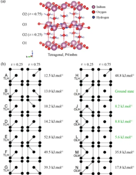
optimization of Ba2In2O4(OH)2was performed with the same approach as that used for Ba2In2O5 in our previous work22 taking theP4/mbmstructure (Figure 1b) of Jayaraman et al. as the starting point.11The cell lengths of the P4/mbm cell are given by √2ap × √2ap × 2ap (where ap is the notional perovskite unit cell length).19,56 Sixteen arrangements of protons are considered. All structures feature full occupancy of the O3 layer proton positions (2c site) but have various proton configurations in the O2 (16l site with fractional
occupancy of one-eigth; 14 arrangements) and O1 (two arrangements) layers, O1 occupancy not being observed by Jayaraman et al.11 but identified theoretically by Fisher and
Islam.20This results in configurations that are similar to those
found in the study by Martinez et al.12The configurations are
then ranked in terms of the calculated total energies (Figures 2 and 3 and Table 1). As was also shown by Martinez et al.,12all configurations undergo significant relaxations from the average
positions suggested by Jayaraman et al.11The O2 layer protons (H1) move offthe 16lpositions [with fractional coordinates (x, y, 0.25)] suggested by neutron diffraction11 into the “32y”
positions (with values close tox±0.07,y±0.07, 0.25±0.04,
e.g., with x = 0.10, y = 0.13) retaining the nomenclature
previously used by Martinez et al.12 The computationally
relaxed ground state forms alternating layers of ···O2−O3− O2−O1··· oxygens, partially occupied, fully occupied, non-occupied, and nonoccupied by protons, respectively (Figure 2b). Thus, the O3 layer O vacancies of Ba2In2O5 are fully occupied by a hydroxyl group (Figures 1c and 2d), while the second water proton occupies one of the two nearby O2 layers. However, only one of the O2 layers is protonated, while the other remains empty (Figure 2b). The optimized In−O3−In bond angles differ significantly from In−O1−In and In−O2−
In, and thus O3 oxygen atoms are in a significantly different
electronic environment (Table 1). Moreover, the bond length
asymmetry of In−O2−In is substantial which may have
implications for the 17O NMR spectra of this phase (Table 1). The optimized structures also indicate that hydrogen bonding constitutes the main interaction driving the formation of a range of distinct chemical environments for both protons and oxygen ions.
The configurations were grouped into three different types.
In the two-formula-units cell of Ba4In4O8(OH)4, two out of four protons are always in the O3 layer (H2) and the other two are each either in a different O2 layer (H1) [two-layer hydroxyl
(TLH), configurations A−H], in the same O2 layer (H1)
[one-layer hydroxyl (OLH), configurations I−L], or present as one
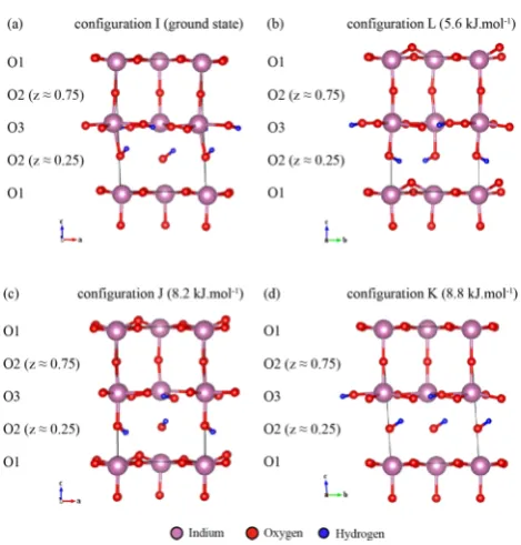
[image:4.625.333.558.70.360.2]water molecule (two protons attached to one oxygen) per layer [one-layer water (OLW), configurations M and N] (Figure 2). Figure 2. (a) Room temperature crystal structure of tetragonal Ba2In2O4(OH)2in space groupP4/mbm11showing full occupancy of the protons in the O3 layer (2csite) and partial occupancy in the O2 layer (16lsite) (denoted, as in Figure 1b, by partiallyfilled white balls). The interlayer Ba cations have been omitted for clarity. (b) Schematic representation of the 14 proton H1 configurations considered, differing in terms of the arrangement of protons in the O2 layers (16lsite). Full and empty circles represent the O atoms and protons, respectively. Eight configurations (A−H) correspond to two-layer hydroxyl (TLH) forms; four configurations (I−L), to one-layer hydroxyl (OLH) forms; and two configurations (M−N), to one-layer water (OLW) forms. H1 protons were placed initially in the 16l positions with fractional occupancy of one-eighth determined by neutron diffraction,11and subsequently moved to the 32ypositions on geometry optimization as in the previous study of Martinez et al.12
The calculated energy per formula unit (containing two H atoms) of each configuration, relative to the ground state structure I, is also shown on the right-hand side of each configuration. A hydration enthalpy (ΔHh) of −79.3 kJ·mol−1 to form the ground state configuration I from dry Ba
OLH configuration I represents the ground state structure
(with hydration enthalpy,ΔHh=−79.3 kJ·mol−1) in which the protons in the O2 layer (H1) point toward two different O3
acceptors. Configurations in which the protons are located
within one O2 layer (H1) (OLH, Eref = 0−8.8 kJ·mol−1) are systematically lower in energy than the OLW (Eref 17.8 and 35.8 kJ·mol−1) and TLH (E
ref = 10.2−48.8 kJ·mol−1) arrangements. Here all energies are given as Eref to denote their being referenced to the energy of the ground state structure I. All of the OLW configurations considered are
unstable, configuration M relaxing to a mixed O1 and O2 layer
protonation and configuration N to an OLH configuration.
Structures where a proton is located in the O1 plane are noticeably higher in energy. For example, two configurations
were also considered where the O3 layer is fully protonated and one O2 site and one O1 site are also protonated, the two structures differing in the relative orientations of the O1 and
O2/O3 protons. Both of these structures are much higher in energy, occurring at 34.6 and 48.3 kJ·mol−1above the ground state structure. The protons in both of these structures are located within the plane of the O1/In layers, in contrast with the structures proposed by Fisher and Islam20 in which the O1−H bonds were oriented perpendicular to the plane.
Note that the previous study of Martinez suggested the present configuration L as the ground state structure (first low
energy state), in which the O2 layer protons point toward the O1 oxygen site (Figures 2b and 3); a structure close to configuration I represented theirfirst excited state.12The fact
that the present and the Martinez et al. studies12 disagree on
the energy ordering of structures at the∼6 kJ·mol−1(or just∼3 kJ/(mol of H)) level suggests that the DFT approach used cannot be regarded as yielding energies any more accurately than to within a few kJ/(mol of H). Thus, it is clear that we should consider more than just the ground state structure when analyzing our NMR data. Bielecki et al.,57 in an inelastic neutron scattering (INS) study of hydrogen bonding in this
material, suggested that the second lowest energy structure of Martinez et al.12 should not exist due to its strong hydrogen
bonds, which would result in higher frequency O−H wag
modes than observed experimentally. However, our related structure (ground state structure I) has a longer hydrogen bond distance (1.819 Å; see Table 1) than that obtained by Martinez et al.12(1.7 Å). Hence, structure I is likely consistent with the lower frequency O−H wag modes observed experimentally and cannot be ruled out on the basis of the INS data.
The thermal energy kBTNA (or RT) at a typical hydration synthesis temperature (350°C) is∼5 kJ. Assuming fast cooling (equilibrium of atomic motion is not reached in the given time frame and temperature), this suggests that configurations with
up to ∼5 kJ/(mol of H) may be present at significant
concentrations at room temperature. We note that H is quite mobile in this system. A previous thermogravimetric study of hydrated Ba2In2O5 by Schober and Friedrich18 suggests an activation energy of 0.3 eV (∼30 kJ·mol−1) for trapping effects in the hydration, as well as a hydrogen diffusion enthalpy
(ΔH*) of 0.34 eV with diffusion coefficient prefactor (D0) of
0.34×10⁻⁵cm2/s. These values all suggest that at least some H can readily diffuse at room temperature (as will be explored
later by 1H NMR spectroscopy). However, we propose that
either some of the H+ions are significantly less mobile or that
the cooperative motion of the H needed to reorder is inhibited, despite the good mobility of individual H+ions. We therefore consider GIPAW calculations on thefirst four lowest energy
structures I, J, K, and L, all of which are of OLH-type and have relative energies in the range of 0−8.8 kJ·mol−1(or 0−4.4 kJ/ (mol of H)), consistent with the possibility of thermal excitations being trapped in the system and the uncertainty in DFT energies and neglected vibrational energies mentioned earlier. We also limit ourselves to these four structures as they are clearly distinct in character from the next highest energy structures C, A, and B [at 10.2, 12.5, and 13.0 kJ·mol−1, respectively (or∼6 kJ/(mol of H))], which all have protons in both O2 layers (z= 0.25 and 0.75).
Assuming all of the structures occur with a Boltzmann distribution established at 350°C, the relative amounts of each configuration are given by the Boltzmann weights 0.25, 0.11,
0.11, and 0.15 for I, J, K, and L, respectively, and 0.09, 0.08, 0.07, 0.06, and 0.05 for the configurations C, A, B, D, and N,
respectively; the remaining seven configurations have weights
of less than 0.01 contributing total weights of approximately 0.02. We discuss the preceding assumptions in the context of the NMR experiments.
3.2. Experimental and GIPAW Calculations Results.
3.2.1. X-ray Powder Diffraction.The X-ray powder diffraction
pattern of Ba2In2O4(OH)2 samples and the 17O enriched counterpart prepared in this study are consistent with previous reports11,19 (Figure S1 in the Supporting Information) and indicate that the structure has tetragonalP4/mbmsymmetry.11
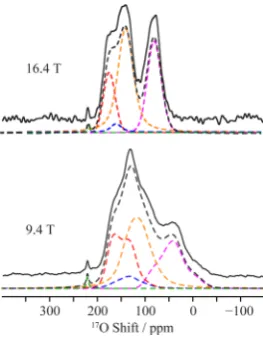
3.2.2. 17O NMR Spectroscopy. The 1D 17O MAS NMR
spectrum of 17O enriched Ba
2In2O4(OH)2 obtained at 9.4 T under conditions where the line shape distortion is minimal (i.e., with short pulse length)58 shows a complicated pattern characteristic of multiple overlapping 17O resonances (Figure 4). Four sites could be resolved in the17O 3QMAS experiment (Figure 5), which could be satisfactorily fit with the NMR
[image:5.625.63.298.61.307.2]parameters given in Table 2. Fitting of the 1D17O MAS NMR spectra with these parameters yields an intensity ratio of 4:1:6:4 for the resonances with chemical shifts of 188, 173, 152, and 97 ppm, respectively (Figure 4). In addition to these resonances Figure 3.DFT optimized geometries of (a) configuration I (ground
there is a fifth peak at 220 ppm which nutates at the same
frequency as water (i.e., it must be associated with a very small or zero quadrupolar coupling) and is tentatively attributed to either a possible H2O molecule on the surface of the material as also discussed in our previous paper,22or to an impurity phase. The17O signals in the 3QMAS show a broadening along the +1 direction (positive slope diagonal), which is attributed to a distribution of chemical shifts arising from a distribution of different local environments.
The 17O isotropic chemical shifts, δ
iso, obtained by GIPAW calculations of the lower energy I, J, K, and L structures of Ba2In2O4(OH)2are also given in Table 2 and are found to span a wide range of values for each O sublattice O1, O2, and O3 (Figure 6). Nevertheless, we may divide the sites into three general types of oxygen atoms, namely, bare oxygens (O), oxygens directly bonded to a proton (H-donor O), and oxygen ions that are hydrogen bonded to a proton (H-acceptor O).
More specifically, we note that O1, acceptor O1, O2,
H-donor O2, H-H-donor O3, and H-acceptor O3 local environ-ments occur in all of the low energy structures I, J, K, and L (see Table 2). A range ofCQare also observed, whereCQ for the acceptor O sites ranges from 3.8 to 4.5 MHz, the H-donor O sites from 5.3 to 6.9 MHz, and the bare O sites from
4.9 to 5.5 MHz. Two distinct ranges for the quadrupolar asymmetry parameter ηQ are obtained: those for bare O sites are approximately 0.1, while those for H-donor and -acceptor O sites are approximately 0.7. Taking the ground state structure I as an example, the O1 (sites 1−4 in Figure 6a, δiso = 170.4 ppm), O2 (sites 5 and 8 in Figure 6a,δiso= 159.6 ppm), and H-acceptor O3 (sites 9 and 12 in Figure 6a,δiso= 156.4 ppm) all contribute to the high frequency resonances in the experimental spectrum (mean, 164 ppm), while the H-donor O2 (sites 6 and 7 in Figure 6a,δiso= 114.4 ppm) and H-donor O3 (sites 10 and 11 in Figure 6a,δiso= 94.5 ppm) contribute to the lower frequency resonances (mean, 104 ppm) (see Table 2).
1H−17O double resonance experiments probe the 1H−17O
heteronuclear dipolar coupling (and hence the 1H−17O
heteronuclear distance) and were therefore carried out to investigate the spatial proximities between the 17O sites and protons. The 17O cross-polarization (CP) spectrum of 17O enriched Ba2In2O4(OH)2 recorded with a very short contact time (τCP= 40μs) is given in SI Figure S4b and reveals a single resonance corresponding to the low frequency signal atδiso=
97 ppm, confirming its assignment to H-donor oxygens.
Additionally, a two-dimensional 1H−17O heteronuclear
corre-Table 1. Cell Constants (a,b, andc, Å;α,β, andγ, deg), In−O and O−H Bond Distances (Å) and In−O−In and O−H···H Bond Angles (deg) for O1, O2, and O3 Environments Obtained from Optimizations of the Initial PerturbedP1 Symmetry Ba4In4O8(HO)4Cells, in the Lowest Energy I, J, K, and L Configurations (Figure 2b), As Compared with Corresponding
Experimental and Calculated Literature Valuesa
present calculations
I struct (P1) J struct (P1) K struct (P1) L struct (P1)
average of I, J, K, and L
structures Martinez et al.calculations12
experimental Jayaraman et al.11
(P4/mbm) cell
a(Å) 5.975 5.951 5.929 5.966 5.955(0.017) 5.915
b(Å) 5.992 6.029 5.984 5.979 5.996(0.020) 5.915
c(Å) 9.308 9.225 9.324 9.247 9.276(0.041) 8.999
α(deg) 93.3 87.9 93.7 89.7 91.2(2.4) 90
β(deg) 93.1 90.2 90.0 90.0 90.8(1.3) 90
γ(deg) 90.3 91.0 90.0 92.1 90.8(0.8) 90
In−O1−In
distance (Å) 2.120 (0.006) 2.138 (0.014) 2.112 (0.006) 2.160 (0.023) 2.133 (0.028) 2.092*
2.132 (0.008) 2.162 (0.008) 2.121 (0.001) 2.162 (0.024) 2.144 (0.026) 2.092*
angle (deg) 169.1 (2.5) 164.0 (11.0) 168.5 (1.3) 160.4 (14.8) 165.5 (18.7) 178.4 In−O2−In
distance (Å) 2.274 (0.162) 2.251 (0.138) 2.299 (0.183) 2.213 (0.076) 2.259 (0.290) 2.192*
2.411 (0.231) 2.391 (0.235) 2.378 (0.172) 2.428 (0.250) 2.402 (0.448) 2.307*
angle (deg) 173.9 (4.1) 169.6 (5.9) 175.6 (3.1) 170.9 (1.7) 172.5 (8.0) 180.0 In−O3−In
distance (Å) 2.231 (0.067) 2.245 (0.085) 2.250 (0.053) 2.253 (0.080) 2.244 (0.145) 2.201 2.293 (0.108) 2.297 (0.128) 2.274 (0.064) 2.262 (0.087) 2.282 (0.199) 2.201 angle (deg) 139.0 (4.4) 138.1 (1.2) 137.5 (3.7) 139.2 (6.0) 138.4 (8.4) 143.6 O3−H···O
distance (Å) 1.011 (0.005) 1.013 (0.001) 1.022 (0.000) 1.018 (0.002) 1.016 (0.006) 1.404 1.750 (0.035) 1.720 (0.020) 1.676 (0.000) 1.680 (0.037) 1.707 (0.055) 1.7 1.404 angle (deg) 173.3 (5.3) 175.3 (0.2) 177.8 (0.0) 176.7 (0.5) 175.8 (5.3) 180.0 O2−H···O
distance (Å) 0.993 (0.003) 0.983 (0.001) 0.995 (0.000) 0.982 (0.001) 0.988 (0.003) 1.00* 0.991 (0.000)*
1.819 (0.005) 1.969 (0.005) 1.844 (0.000) 1.957 (0.014) 1.897 (0.016) 1.90 2.542 (0.051) angle (deg) 153.5 (1.8) 151.0 (1.7) 154.5 (0.0) 150.0 (1.7) 152.2 (3.0) 116.4 (4.2)
aMeans and standard deviations (in parentheses) of the values are shown. For values marked with an asterisk (*), note that the O1 and O2 labels of
[image:6.625.63.564.120.468.2]lation (HETCOR) spectrum, also recorded withτCP = 40μs, shows that this 97 ppm resonance correlates with all of the1H signals (see SI Figure S4a and section 3.2.3). The 17O CP spectrum obtained with a longer contact time of 2.5 ms (SI Figure S4b) still does not contain the signals observed at high
frequencies (δiso = 188 and 173 ppm) in the 17O MAS
spectrum (Figure 4), indicating that the O sites that give rise to these resonances are not in close proximity to the protons (or that the associated protons are too mobile). Instead a weak resonance at δiso= 152 ppm is observed which is tentatively assigned to an H-acceptor oxygen. The17O CP kinetics curve (SI Figure S5) shows the slower CP dynamics associated with the 152 ppm compared to the 97 ppm resonance, consistent with our assignment of the 152 ppm resonance to a H-acceptor O site.
Assuming that all of the low energy configurations, I, J, K,
and L contribute to the spectrum with weights from the Bolztmann distribution at 350 °C (given by 0.25, 0.11, 0.11,
and 0.15, respectively), we combined all of the oxygen resonances obtained from the GIPAW calculations to yield the simulated spectra shown in Figure 6b. A peak intensity ratio of 4.3:1:6.3:4.3 (from high to low frequency) is derived, which is remarkably close to the experimentally derived 4:1:6:4 ratio. On the basis of this reasonable agreement, the experimentally observed high frequency resonance at δiso = ∼188 ppm is, therefore, assigned to the bare O1 sites; the other high frequency weaker resonance at 173 ppm, to the acceptor O1 sites; the next resonance atδiso= ∼152 ppm, to the bare O2 sites and H-acceptor O3 sites; andfinally the lowest frequency
resonance atδiso=∼97 ppm, to the H-donor O2 and H-donor O3 sites, in agreement with the 17O CP experiments, all of these sites corresponding to the 48 O sites in the four Ba4In4O8(OH)4 configurations, I, J, K, and L (Figure 3 and Table 2). In summary, the presentfirst-principles calculations
permit the assignment of the 17O MAS NMR spectrum of
Ba2In2O5(OH)2, yielding chemical shifts and quadrupolar parameters in reasonable agreement with the experimental trends, albeit with some minor differences in the chemical
shifts, quadrupolar parameters, and relative resonance inten-sities. A likely source of error in the intensities is the assumption that only structures I, J, K, and L contribute to the experimental spectra in proportions governed by their relative energies and the synthesis temperature. However, the assignment of the“high”and“low”shift peaks in the17O MAS NMR experimental spectra is consistent; i.e., we expect nondonor oxygen resonances to occur in the high frequency region and a donor oxygen to occur in the low frequency region. Overall, while relative intensities of the experimental peaks may vary, their assignment to these chemical environ-ments is sound.
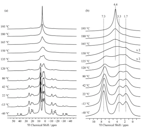
3.2.3.1H NMR Spectroscpy.The room temperature1H MAS
NMR spectrum of Ba2In2O4(OH)2obtained at 16.4 T clearly shows three distinct proton sites at shifts of 7.3, 3.3, and 1.7
ppm in a 2:1:1 intensity ratio (Figure 7). 1H/2H NMR
resonances in approximately the same chemical shift range were observed by Jayaraman et al.11 in their study of this material; however, these authors observed a splitting of the higher frequency resonance (of approximately 1−2 ppm), a broader resonance being observed at lower frequencies (at approx-imately 2.5 ppm) and the high (split) and low frequency resonances occurring with intensity ratios of 1:1. No assign-ment was given in that study. The three resonances observed in our study are fairly broad, and we assign this to inhomogeneous broadening (i.e., a chemical shift distribution) rather than to the effects of strong homogeneous dipolar interactions between
protons, as similar 1H line widths were obtained in spectra acquired under fast MAS conditions (SI Figure S6).59
In order to assign all three resonances observed in the
experimental1H MAS NMR spectrum of Ba
2In2O4(OH)2, we
computed the 1H site shielding tensors in the proton
arrangements discussed earlier (Table 2 and Figure 7 and SI
Figure S7). A simulation of the 1H spectrum using the
calculated NMR parameters (SI Figure S7) of the lowest energy structure I contains three peaks with a 1:1:2 ratio (∼9,∼8, and
∼5 ppm), providing a poor match with the experimental
spectrum (Figure 7a). Thus, in order to generate a spectrum that is closer to the experimental one, other low energy structures were also considered. Inclusion of the first high
energy configuration (L; with Boltzmann weights of 0.25 and
0.11 for I and L) improves thefit to the spectrum in the low
[image:7.625.114.246.67.236.2]part per million region but worsens it in the higher frequency Figure 4.17O MAS NMR spectra of 17O enriched Ba
2In2O4(OH)2 obtained at 9.4 and 16.4 T. Experimental spectra are shown with full lines and total best-fit simulations in black dashed lines. The individual site components are shown as dashed lines in red (site A, O1), blue (site B, acceptor O1), orange (site C, combination of acceptor O2 and acceptor O3), pink (site D, donor O2 and donor O3), and green (small quadrupole coupling impurity site) (see Table 2). Assignments of the O sites are made by comparison with parameters derived from DFT GIPAW calculations (see Figure 6).
[image:7.625.60.301.340.461.2]region (Figure 7b). When all of the low energy configurations,
with respective weights, are considered (Eref< 20 kJ·mol−1), we do not observe a narrow peak at the high shift region in the spectrum (Figure 7c). Concentrating on NMR parameters obtained in the four most stable configurations (structures I, J,
K, and L), the calculations yield O3 layer proton resonances at
δiso of 7.8−9.3 ppm (due to configuration I), 8.6−9.1 (configuration J), 9.7 (configuration K), and 9.1−9.9 ppm
(configuration L) (Figure 7d; spectrum shown with 350 °C
Boltzmann weights). In some proportion, all of these O3 layer
1H signals are likely to contribute to the broad signal (Δν 1/2=
800 Hz (∼1.5 ppm on 16.4 T magnet)) observed
experimentally at approximately 7.3 ppm for Ba2In2O4(OH)2 (Figure 7a). Two distinct sets of shifts are observed, at 5.2± 0.2 ppm from the O2 layer protons (H1) in structures I and K and at 3.9±0.2 ppm arising from structures J and L (Figure 7, Table 2), which are reasonably close to the two experimental resonances observed at 3.3 and 1.7 ppm.
[image:8.625.78.562.96.570.2]Of note, the spectrum simulated with structure I resembles that obtained by Jayaraman et al.,11 predicting the small
Table 2. Experimental and Calculated1H and17O Isotropic Shift (δ1, ppm), Isotropic Chemical Shift (δiso, ppm), Quadrupolar Coupling Constant (CQ, MHz), and Quadrupolar Asymmetry Parameter (ηQ) for Ba2In2O4(OH)2
structure nucleus environmenta δ1(ppm) δiso(ppm) CQ(MHz) ηQb Nc
Experimentald
Ba2In2O4(OH)2 17O O-site A (O1)a 140 188(4) 4.5(2) 0.0(1) 0.25 17O O-site B (H-acc. O1)a 174 173(4) 4.1(2) 0.7(1) 0.0625
17O O-site C (H-acc. O2 and O3)a 192 152(4) 4.2(2) 0.5(1) 0.375
17O O-site D (H-donor O2 and O3)a 207 97(4) 4.8(2) 0.7(1) 0.25
17O O-site E (no match) 223(4) 1.0(2) 0.0(1) 0.0625
1H H-site A (H2, O3 plane)a 7.3(1) 0.5
1H H-site B (H1, O2 plane I,K)a 3.3(1) 0.25
1H H-site C (H1, O2 plane J,L)a 1.7(1) 0.25
Calculatedb
I 17O O1 200.6 (11.1) 170.4 (7.0) −5.0 0.2 0.33 17O donor O2 168.7 (5.2) 114.4 (3.3) −6.0 0.8 0.167 17O O2 193.4 (1.1) 159.6 (0.7) −5.3 0.1 0.167
17O donor O3 136.9 (1.6) 94.5 (1.0) 5.3 0.8 0.167 17O acceptor O3 180.0 (19.7) 156.4 (12.4) −4.3 0.5 0.167
1H H2, O3 plane 8.5 (0.7) 0.5
1H H1, O2 plane 5.3 (0.1) 0.5
J 17O O1 217.7 (11.3) 185.1 (7.1) −5.2 0.2 0.25 17O acceptor O1 204.6 (0.0) 175.4 (0.0) −4.4 0.8 0.083 17O donor O2 167.0 (10.2) 103.6 (6.4) −6.9 0.6 0.167 17O O2 204.2 (9.8) 169.1 (6.2) −5.4 0.1 0.167
17O donor O3 149.6 (3.2) 107.0 (2.0) 5.1 0.9 0.167 17O acceptor O3 185.7 (0.5) 158.5 (0.3) −4.4 0.7 0.167
1H H2, O3 plane 8.8 (0.3) 0.5
1H H1, O2 plane 4.0 (0.2) 0.5
K 17O O1 197.5 (1.7) 168.5 (1.1) −4.9 0.2 0.33 17O donor O2 171.2 (0.0) 117.1 (0.0) −6.2 0.7 0.167 17O O2 200.2 (0.0) 166.5 (0.0) −5.3 0.0 0.167
17O donor O3 146.8 (0.0) 103.5 (0.0) 5.7 0.6 0.167 17O acceptor O3 169.8 (0.0) 151.9 (0.0) −3.8 0.4 0.167
1H H2, O3 plane 9.7 (0.0) 0.5
1H H1, O2 plane 5.1 (0.0) 0.5
L 17O O1 223.8 (19.1) 188.6 (12.0) −5.4 0.2 0.25 17O acceptor O1 225.4 (0.0) 196.9 (0.0) 4.5 0.7 0.083 17O donor O2 166.7 (1.3) 105.1 (0.8) −6.8 0.6 0.167 17O O2 207.2 (5.6) 170.8 (3.5) −5.5 0.1 0.167
17O donor O3 142.8 (11.9) 98.8 (7.5) 5.4 0.8 0.167 17O acceptor O3 178.1 (16.4) 154.0 (10.3) −4.0 0.8 0.167
1H H2, O3 plane 9.5 (0.4) 0.5
1H H1, O2 plane 3.8 (0.1) 0.5
aAn asterisk denotes the most plausible O and H environments from four hydrated models. Standard deviations are also given in parentheses;
deviations of quadrupolar parametersCQandηQare less than 0.01 and are therefore omitted.bObtained from the averaged DFT calculated NMR parameters of the four low energy structures I, J, K, and L. The calculated values ofδ1are obtained withδ1= (27δiso−10δ2)/17 withδ2=δiso−(3/ 500)((CQ2(1 +ηQ2/3))/ν02) for17O (I= 5/2) (see ref 58), the errors arising from the range ofδisovalues.cMolar fraction of the site in the structure specified.dObtained from the 3QMAS and1H−17O HETCOR experiments.δ
splitting at higher frequency and the absence of a splitting at lower frequency. This may suggest that the sample prepared by these authors is more ordered than ours (i.e., contains fewer proton configurations), with a structure that is closer to that of
the thermodynamic ground state.
The apparent offset in δiso between the calculated and
experimental values for all of the structures and sites is relatively constant at approximately 2 ppm, a value larger than the 1H calculated standard deviation of 0.29 ppm (see Computational Methods and SI Figure S3) and may be due to a systematic error in the 1H shielding reference, arising out of the use of primarily organic phases in derivingσref. More work is required to obtain a reliable set of reference parameters for protons in inorganic materials, preferably considering hydrated oxides and simple hydrous phases. In addition, proton motion and, likely, systematic deviation in calculated to experimental lattice constants may also play a role in the 1H chemical shift. The fact that the protons are involved in chains of hydrogen bonding in the Ba2In2O4(OH)2 system means that the 1H chemical shift will be very sensitive to lattice dilation/ contraction. Nonetheless, the relative chemical shifts and relative intensities of the calculated resonances are in good agreement with experiment. Of note, the results show that
configurations in which the O2 protons are involved in
hydrogen bonds withboththe O3 and O1 sublattices must be included in order to account for the split H1 (O2) resonance observed in our sample.
It is evident from the H···O (hydrogen bond) distances in Table 1 and the 1H chemical shifts in Table 2 that longer hydrogen bonds result in a lower1H chemical shift. A study by Yesinowski et al.60on a series of hydrated silicates yielded an inverse correlation between donor O to acceptor O distance and1H chemical shift characterized by the following equation.
δiso(ppm)=79.05 −25.5[ (O H O)/Å]d − ··· (1)
Applying this equation to the O−O distances from the I, J, K, and L structures, we calculate1H shifts of 7.4 and 9.7 ppm for O2 and O3 layer protons, respectively. Slightly better fits to
experiment were obtained by using the empirical equations derived by Xue and Kanzaki for a wider range of hydrous silicates and related inorganic materials (yielding 5.5 and 8.0
ppm for the O2 and O3 layer protons).61 These simple
calculations provide trends and allow us to distinguish the O3 and O2 layer protons from each other, but do not provide the ability to further separate the two types of O2 layer protons. The predicted shifts are noticeably larger than those observed experimentally, particularly for the O2 coordinated protons, most likely due the very different systems studied here than
used to derive the empirical correlations.
Variable temperature1H solid state MAS NMR experiments up to 195°C show that all of the protons in Ba2In2O4(OH)2
are mobile on the NMR time scale, the resonances first
[image:9.625.61.305.62.287.2]broadening and then completely coalescing at around 150°C, yielding a single site with an isotropic chemical shift of 4.4 ppm. Such a coalescence process occurs when the proton-hopping frequency k equals πΔν/√2 (where Δν is the frequency separation between the peaks) indicating that the hopping rate between the two O2 environments (1.7 and 3.3 ppm) must be greater thank ∼2.2 kHz, while that between the O1 and O2 sites is∼7 kHz (where shifts of 7.3 and 2.5 ppm are used in this calculation, 2.5 ppm being the predicted shift following coalescence of the 1.7 and 3.3 ppm resonances). As the Figure 6.(a) Simulation of the GIPAW calculated17O NMR spectra
[image:9.625.350.516.65.311.2]of the 12 sublattice O sites occurring in the lowest energy optimized structure I of Ba2In2O4(OH)2. All of the spectra were simulated at 9.4 T. (b) Comparison of the experimental 17O NMR spectra of 17O enriched Ba2In2O4(OH)2(black lines) and the sum of the simulation of the GIPAW calculated17O NMR spectra (dashed red lines) of all O sites of the four lower energy structures Ba2In2O4(OH)2(I, J, K and L, combined with relative weights of 0.25, 0.11, 0.11 and 0.15) at 9.4 and 16.4 T.
Figure 7. 1H NMR spectrum of Ba
temperature is increased further, the peak height of the new resonance increases as the line width narrows, due to an increase in proton motion. A weak resonance at 1.7 ppm persists even at 195°C, this being assigned to both structures L
(containing O2−H2 protons pointing to the O1 (bare)
oxygens) and J (containing O2−H2 protons alternatively
pointing to both O1 and O3 sites J). We suggest the O2−H1···
O1 protons are more strongly trapped, while the O2−H1···O3
protons undergo more rapid exchange with the O3−H (H2)
protons.
Two dimensional exchange NMR experiments were performed to explore motion on a longer time scale (Figure 9). Both chemical exchange and magnetization exchange (due to spin-diffusion) will result in cross-peaks situated offthe1H vs 1H diagonal. At 42 °C, cross-peaks are observed between the
two distinct protons assigned to the O2 layer (H1; sites at 1.7 and 3.3 ppm) at a mixing time of 1 ms, indicating that these sites occur within a single particle. The cross-peaks are most likely generated from a combination of slow motion and spin diffusion. Longer mixing times reveal cross-peaks between all of
the resonances, most likely as a result of spin diffusion. The
observation of H1−H1 and H1−H2 cross-peaks at short
mixing times (0.1 and 1 ms, respectively) at 80°C is ascribed to the onset of motion, since the spin diffusion rates are unlikely
to increase with temperature. At a temperature of 150 °C, all protons exchange rapidly at a rate in excess of 10 kHz, in agreement with the variable temperature1H spectra (Figure 8).
The relatively rapid exchange between different proton sites
at room temperature may seem inconsistent with the presence of multiple proton environments associated with excited states. However, the existence of some mobile protons does not necessarily imply that all of the protons can reorder into the unique ground state. Local orderings, even ones that allow significant H transport, could still be effectively locked in place
as a cooperative ordering transition to the ground state could be inhibited, or some of the H could be immobile. The 2D
spectra indicate that some of the proton configurations are
present within the same particle. We indicated earlier that, within the errors of the DFT, no unique ground state structure emerges (ours differing from that of Martinez et al.12). Thus, it
is likely that the lower energy structures reflect the different
possible H-bonding configurations within one particle and that
the four lowest structures identified in this study represent the
more probable configurations. A future study treating the H
ordering with a more complete thermodynamics and kinetic model would be valuable, but this is beyond the scope of this current work.
4. CONCLUSIONS
In summary, we have performed a comprehensive structural analysis of the hydrated form of brownmillerite, Ba2In2O4(OH)2, using multinuclear solid state NMR spectros-copy in combination with solid state DFT calculations. We reproduce the structural analysis of a large number of possible proton positions of Martinez et al.,12 identifying multiple configurations that may exist concurrently at room
temper-ature. Three different proton sites were observed by 1H MAS
NMR spectroscopy, which were found to exchange at 150°C
on the NMR time scale with hopping rates in excess of 10 kHz. Assignment of these resonances make use of extensive total energy DFT calculations of a wide range of proton configurations, which yielded four chemically similar low
energy configurations, the simulated1H GIPAW NMR spectra
of which are in relatively good agreement with the experimental data. The three resonances can be assigned as follows: the high frequency shift corresponds to O3 layer protons (H2), while the two lower frequency shifts arise due to two types of
configurations of O2 layer protons (H1) with shorter and
longer H···O (hydrogen bond) distances and a difference in
O−H···O angles. Distinct O2 layer proton (H1) configurations
are observed in four low energy structures that perhaps coexist
at room temperature. The 17O NMR spectrum of
[image:10.625.326.565.69.236.2]Ba2In2O4(OH)2 is dominated by four O sites, which can be rationalized by the DFT GIPAW calculations, which reveal that H-donor oxygens appear at lower shift while the nonhydroxyl oxygens and H-acceptor O are visible at higher shifts. We suggest that all the lower energy proton sites should be Figure 8. Variable temperature 1H MAS NMR spectra of
[image:10.625.65.297.375.582.2]Ba2In2O4(OH)2 obtained at a MAS rate of 20 kHz and at 16.4 T. (a) Full spectral width showing the isotropic region and the spinning sideband manifold marked with asterisks. (b) Magnified view highlighting the coalescence of the 1.7 and 3.3 ppm resonances and coalescence of all the resonances above 150°C.
considered in any proposed proton conduction mechanism within the Ba2In2O4(OH)2phase.
The joint experimental and theoretical approach presented in this work can be readily applied to investigate H-bonding and local structure in other hydrated perovskites. The current work suggests that multiple H-bonding motifs are likely present in related structures, the relative energies of these strongly affecting proton trapping and conductivity.
■
ASSOCIATED CONTENT*
S Supporting InformationX-ray diffraction patterns (Figure S1), TGA (Figure S2), plot of
experimental isotropic shift, δiso, against computed isotropic shielding, σiso, for 1H sites (Figure S3), two-dimensional
1H−17O CP HETCOR NMR spectrum and 17O CP spectra
with various contact times of Ba2In2O4(OH)2(Figure S4), 17O
CP kinetics graph (Figure S5), 1H NMR spectrum of
Ba2In2O4(OH)2obtained under MAS rate of 60 kHz (Figure
S6), GIPAW calculated 1H NMR chemical shifts, δ
iso, of all structures investigated (Figure S7), and detailed representation of all configurations considered as optimized structures and
complete GIPAW NMR data. The Supporting Information is available free of charge on the ACS Publications website at DOI: 10.1021/acs.chemmater.5b00328.
■
AUTHOR INFORMATIONCorresponding Author
*E-mail: [email protected].
Present Addresses
∥(R.D.) Institute for Molecules and Materials, Radboud
University, P. O. Box 9010, 6500 GL, Nijmegen, The Netherlands.
⊥(F.B.) Department of Chemistry and Stephenson Institute for
Renewable Energy, University of Liverpool, Crown Street, Liverpool L69 7ZD, U.K.
Funding
This work was supported in part by Grants DMR050612 and CHE0714183 from the National Science Foundation and Grant DESC0001284 from the Department of Energy (supporting Y.-L.L. and D.M.), by an Advanced Fellowship from the EU-ERC (C.P.G.), and by the EPSRC (D.S.M.). F.B. thanks the EU Marie Curie actions FP7 for an International Incoming fellowship (Grant No. 275212) and Clare Hall, University of Cambridge, for a Research Fellowship.
Notes
The authors declare no competingfinancial interest.
■
ACKNOWLEDGMENTSWe thank Dr. Andrew J. Ilott (NYU, New York, NY, USA), Dr. Aurelie Rolle (University of Lille Nord de France, France), Dr.́
John M. Griffin (University of Cambridge, U.K.), and Dr.
Gunwoo Kim (University of Cambridge, U.K.) for helpful discussions and Luke Sperrin (University of Cambridge, U.K.) for running the TGA experiments. Research was carried out in part at the Center for Functional Nanomaterials, Brookhaven National Laboratory, NY, USA, which is supported by the U.S. Department of Energy, Office of Basic Energy Sciences, under
Contract No. DE-AC02-98CH10886.
■
ABBREVIATIONSDFT, density functional theory; fwhm, full width at half-maximum; GIPAW, gauge including projector augmented
wave; MAS, magic angle spinning; NMR, nuclear magnetic resonance
■
REFERENCES(1) Ishihara, T.; Yokokawa, H.; Iwahara, H.; Kilner, J. A.; Berenov, A.; Rossiny, J.; Yashima, M.; Kawada, T.; Irvine, J. T. S.; Akbay, T.; Kawakami, A.; Norby, T.; Matsumoto, H.; Kreuer, K. D.; Ito, N.; Horita, T.Perovskite Oxide for Solid Oxide Fuel Cells; Springer: New York, 2009.
(2) Malavasi, L.; Fisher, C. A. J.; Islam, M. S. Oxide-ion and proton conducting electrolyte materials for clean energy applications: Structural and mechanistic features.Chem. Soc. Rev. 2010,39 (11), 4370−4387.
(3) Takahashi, T.; Iwahara, H. Solid-state ionics: Protonic conduction in perovskite type oxide solid solutions. Rev. Chim. Miner.1980,17(4), 243−253.
(4) Iwahara, H.; Esaka, T.; Uchida, H.; Maeda, N. Proton conduction in sintered oxides and its application to steam electrolysis for hydrogen production.Solid State Ionics1981,3−4, 359−363.
(5) Iwahara, H. High temperature proton conducting oxides and their applications to solid electrolyte fuel cells and steam electrolyzer for hydrogen production. Solid State Ionics 1988,28−30, 573−578 (Part 1).
(6) Iwahara, H.; Uchida, H.; Morimoto, K.; Hosogi, S. High-temperature C1-gas fuel cells using proton-conducting solid electro-lytes.J. Appl. Electrochem.1989,19(3), 448−452.
(7) Iwahara, H.; Uchida, H.; Morimoto, K. High temperature solid electrolyte fuel cells using perovskite-type oxide based on BaCeO3.J. Electrochem. Soc.1990,137(2), 462−465.
(8) Lee, W.-K.; Nowick, A. S.; Boatner, L. A. Protonic conduction in acceptor-doped KTaO3crystals.Solid State Ionics1986,18−19, 989− 993 (Part 2).
(9) Liang, K. C.; Nowick, A. S. High-temperature protonic conduction in mixed perovskite ceramics.Solid State Ionics1993,61 (1−3), 77−81.
(10) Liang, K. C.; Du, Y.; Nowick, A. S. Fast high-temperature proton transport in nonstoichiometric mixed perovskites.Solid State Ionics1994,69(2), 117−120.
(11) Jayaraman, V.; Magrez, A.; Caldes, M.; Joubert, O.; Taulelle, F.; Rodriguezcarvajal, J.; Piffard, Y.; Brohan, L. Characterization of perovskite systems derived from Ba2In2O5□ Part II: The proton compounds Ba2In2(1−x)Ti2xO4+2x(OH)y[0≤x≤1;y≤2(1−x)].Solid State Ionics2004,170(1−2), 25−32.
(12) Martinez, J.-R.; Mohn, C. E.; Stølen, S.; Allan, N. L. Ba2In2O4(OH)2: Proton sites, disorder and vibrational properties. J. Solid State Chem.2007,180(12), 3388−3392.
(13) Colville, A. A.; Geller, S. The crystal structure of brownmillerite, Ca2FeAlO5.Acta Crystallogr. B1971,27, 2311−2315.
(14) Kreuer, K. D. Proton-Conducting Oxides.Annu. Rev. Mater. Res.
2003,33, 333−359.
(15) Cervera, R. B.; Miyoshi, S.; Oyama, Y.; Elammari, Y. E.; Yagi, T.; Yamaguchi, S. Perovskite-Structured BaScO2(OH) as a Novel Proton Conductor: Heavily Hydrated Phase Obtained via Low-Temperature Synthesis.Chem. Mater.2013,25(9), 1483−1489.
(16) Yildirim, T.; Reisner, B.; Udovic, T. J.; Neumann, D. A. The combined neutron scattering and first-principles study of solid state protonic conductors.Solid State Ionics2001,145, 429−435.
(17) Schober, T.; Friedrich, J.; Krug, F. Phase transition in the oxygen and proton conductor Ba2In2O5in humid atmospheres below 300°C.Solid State Ionics1997,99, 9−13.
(18) Schober, T.; Friedrich, J. The oxygen and proton conductor Ba2In2O5: Thermogravimetry of proton uptake.Solid State Ionics1998, 113−115, 369−375.
(19) Fischer, W.; Reck, G.; Schober, T. Structural transformation of the oxygen and proton conductor Ba2In2O5in humid air: an in-situ X-ray powder diffraction study.Solid State Ionics1999,116, 211−215.