A growing toolbox of techniques for studying
β
-barrel outer membrane protein folding and
biogenesis
Jim E. Horne* and Sheena E. Radford*
1*Astbury Centre for Structural Molecular Biology and School of Molecular and Cellular Biology, The University of Leeds, Leeds LS2 9JT, U.K.
Abstract
Great strides into understanding protein folding have been made since the seminal work of Anfinsen over 40 years ago, but progress in the study of membrane protein folding has lagged behind that of their water soluble counterparts. Researchers in these fields continue to turn to more advanced techniques such as NMR, mass spectrometry, molecular dynamics (MD) and single molecule methods to interrogate how proteins fold. Our understanding ofβ-barrel outer membrane protein (OMP) folding has benefited from these advances in the last decade. This class of proteins must traverse the periplasm and then insert into an asymmetric lipid membrane in the absence of a chemical energy source. In this review we discuss old, new and emerging techniques used to examine the process of OMP folding and biogenesisin vitroand describe some of the insights and new questions these techniques have revealed.
Introduction
The study of protein folding underpins a goal to understand
the function of biological systems in terms of the structures,
properties and interactions of the molecules which
orches-trate many of life’s essential processes. The field of protein
folding sits at an intersection between scientific disciplines
and requires a plethora of complementary techniques to
be combined to answer the question “How do proteins
fold?” Although many of the techniques and underlying
principles learned from over 40 years of studies on the
folding of water soluble proteins [1,2] can be applied to
membrane proteins, the introduction of the lipid bilayer and
its steric and physicochemical properties necessarily alters
the forces that guide protein folding when coupled with
insertion into the bilayer itself. The outer membranes (OM)
of mitochondria, chloroplasts and Gram-negative bacteria
consist almost entirely of
β
-barrel outer membrane proteins
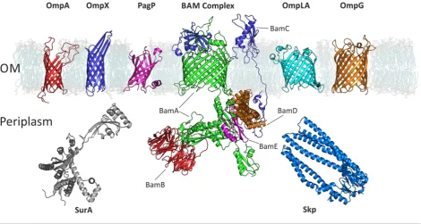
(OMPs) (Figure 1). The assembly of OMPs has received
significant attention in the last few years after the discovery
of an essential protein machinery, the
β
-barrel assembly
machine complex (or BAM complex), which is required
for the assembly of OMPs into the OM of Gram-negative
bacteria (Figure 2) [3–6]. The OM provides a fundamentally
different folding environment compared with the inner
membrane: the bilayer is asymmetric as it is enriched in
lipopolysaccharide in the outer leaflet, it is densely packed
with OMPs, and diffusion is restricted [7,8].
Key words:β-barrel, biogenesis, biophysical techniques, outer membrane protein, protein folding.
Abbreviations: BAM, β-barrel assembly machine; DDPC, didecanoylphosphatidylcholine; DLPC, dilauroylphosphatidylcholine; DMPC, dimyristoylphosphatidylcholine; DOPC, dioleoylphos-phatidylcholine; KTSE, kinetics of tertiary structure formation by electrophoresis; LUV, large unilamellar vesicle; OM, outer membrane; OMP, outer membrane protein; SMT, single-molecule tracking; SUV, small unilamellar vesicles.
1 To whom correspondence should be addressed (email [email protected]).
Early studies of OMP folding focused on obtaining
an understanding of folding/unfolding rates and equilibria
and the conditions that alter them for a small set of
OMPs [9–14]. Most recently, however, application of
modern biophysical techniques is allowing more challenging
mechanistic questions about OMP folding to be tackled, as
highlighted below.
‘Classic’ methods of interrogating folding
applied to
β
-barrel membrane proteins
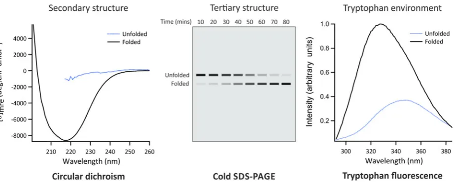
Gel assays
Cold SDS-PAGE exploits the observation that many OMPs
are resistant to SDS denaturation and so the unfolded and
folded states migrate differently on a gel when loaded without
boiling (Figure 3,
top centre
) [9,15–17]. This approach can be
used to determine the fraction of OMPs folded at certain
time points (e.g. following initiation of folding from a
urea-denatured state) and thereby to extract rate constants of
folding [18]. Gel assays were used to examine the effect of
membrane thickness on folding yields into large unilamellar
vesicles (LUVs) formed from didecanoylphosphatidylcholine
(DDPC,
di
C
10PC) to dioleoylphosphatidylcholine (DOPC,
di
C
18:1PC). For most OMPs decreasing lipid chain length
increases the folding yield, while folding into DOPC LUVs
is almost completely abrogated [17,19]. Comparative studies
with OmpA showed a close correlation between the results
obtained using gel assays and tryptophan fluorescence [20].
Circular dichroism (CD)
Figure 1 Examples of OMPs and chaperones mentioned in this review
BAM complex: BamA – green, BamB – red, BamC – blue, BamD – orange, BamE – magenta. PDB ID of structures: OmpA (1G90); OmpX (1QJ8); PagP (1THQ); BamABCDE ([21]); OmpLA (1QD6); OmpG (2IWW); SurA (1M5Y, missing regions built using MODELLER); Skp (1U2M, missing regions built using PyMol). DMPC membrane from O’Neil et al. [21].
lipid phase on folding and insertion of the OmpA
β
-barrel
into LUVs composed of dimyristoylphosphatidylcholine
(DMPC,
di
C
14PC) or
di
C
13PC was tested by following
the change in CD at 216 nm. The authors found that
the folding rates increase when the bilayer is at its
transition temperature (
T
m) [22]. Studies of OmpG folding
into octyl glucoside (OG) showed a ‘burst-phase’ of
β
-sheet formation followed by a second phase in which a
native-like content of secondary structure forms with a
t
1/2of minutes [23]. Gel assays, however, showed that
formation of the native state occurs on a timescale of hours.
Together this indicated that hydrophobic collapse and/or
adsorption to micelles and formation of secondary structure
represent intermediate steps preceding formation of the
native state and closing of the tertiary
β
-barrel of OmpG.
Conversely, a more concerted picture of coupled secondary
and tertiary structure formation was observed using similar
methods to follow the folding of OmpA in DMPC
or 95:5 DMPC/dimyristoylphosphatidylglycerol (DMPG)
small unilamellar vesicles (SUVs) [24], as well as DDPC,
diundecanoylphosphatidylcholine (DUPC,
di
C
11PC),
di-lauroylphosphatidylcholine (DLPC,
di
C
12:0PC) or DMPC
LUVs [19]. Early formation of secondary structure elements
may reflect the formation of misfolded or off-pathway
intermediates as a consequence of the rapid collapse of these
membrane proteins in the aqueous phase prior to membrane
insertion [24,25]. Further experiments will be needed using
different OMPs and different folding conditions to determine
whether this is the case or not for different proteins and
different lipid environments.
Tryptophan fluorescence
All OMPs characterized to date contain aromatic residues
(commonly tryptophan) that form a girdle at the
bilayer:aqueous interface and are thought to be important
in stabilizing OMPs within the membrane. The fluorescence
signal of these Trp residues provides a useful probe of
the folding status of OMPs (Figure 3,
top right
) [26]. By
following fluorescence intensity compared with time, Trp
fluorescence has been used to measure the kinetics of OMP
folding/unfolding and to derive their folding/unfolding free
energies (Figure 3,
lower left
) [27]. Studies following the
change in the centre of spectral mass for Trp emission of
OmpA folding into vesicles of different sizes at different
concentrations of urea, found an influence of stored
membrane curvature elastic stresses on the transition state
of folding [28].
Quenching of tryptophan fluorescence
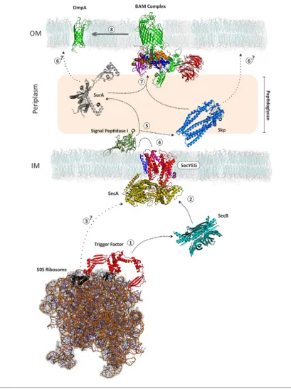
Figure 2 Biogenesis of OMPs
Figure 3 ‘Classic’ methods of interrogating protein folding
Different techniques provide independent, and complementary, information about the kinetics, thermodynamics and mechanism of folding. These approaches have been used for analysis of water soluble and OMP folding (see text).CD
reports on the difference in absorbance of left and right circularly polarized light by peptide bonds in an asymmetric environment. In this case, the asymmetric environment refers to the protein’s secondary structure, with e.g. β-sheet,
α-helix and disorder each giving rise to characteristic spectra. CD in the far UV can be used for the analysis of both water soluble proteins and OMPs. [θ]mre; mean residue ellipticity.Cold SDS-PAGEis useful for the analysis of the formation of
[30]. OmpA contains five tryptophan residues, one on
the periplasmic side of the barrel and the other four on
the extracellular side, with one in each
β
-hairpin loop.
Using this methodology the authors were able to trap a
number of intermediates during the folding of OmpA into
DOPC SUVs [29]. Using site-directed mutagenesis to create
single-Trp variants the authors found that all four hairpins
cross the bilayer concurrently, suggesting a model in which
insertion is directly coupled to folding of the
β
-barrel domain
[31]. More detail on the mechanism and order of insertion and
β
-strand association was obtained through
intra
molecular
quenching of single-Trp variants of OmpA in the same
in vitro
system by a nitroxyl spin-label (which quenches Trp
fluores-cence at distances
<
10–20 A
˚ [32,33]) conjugated to a mutant
cysteine in the neighbouring strand. The results suggested
that association of the N- and C-terminal strands may occur
in tandem with membrane adsorption (i.e. early, not late in the
folding process) and that residues in the extracellular regions
of pairs of
β
-strands in the
β
-barrel associate before those in
the periplasmic ends of the
β
-strands [34].
Chevron plots
Plots of the natural logarithm of observed rate constants
for folding/unfolding against denaturant concentration are
commonly used to analyse the folding pathways of water
soluble proteins (Figure 3) [35]. This approach was used to
interrogate the folding mechanism of PagP (Figure 1) into
DLPC LUVs [36]. The results indicated a reversible two-state
folding mechanism for PagP under the concentration range
of denaturant used involving a transition state that is 50 %
as compact as the native protein [36]. By contrast, studies
of OmpA folding in guanidinium chloride showed that at
low denaturant concentrations the linear folding phase of the
chevron plot becomes non-linear – a phenomenon termed
‘roll-over’ (Figure 3, lower right) which suggests a three-state
folding mechanism [37].
ϕ
-value analysis
ϕ
-value analysis is a powerful technique for acquiring
information on the structure and stability of non-native states
formed during protein folding. Originally developed for
soluble proteins [38,39], this approach has been applied only
recently to membrane proteins [36,40]. It involves making
mutations in specific residues of a protein, measuring the
change in activation energy for unfolding (
G
◦N−trans
, the
free energy required to overcome the transition state barrier)
and the equilibrium free energy of unfolding (
G
◦U), and
comparing the ratio between the two to derive a
ϕ
-value. A
value of 1 implies that native structure has already formed
in the transition state
at that particular residue
, and a value
of 0 that native structure has not yet formed. Analysis of
the transition state for PagP folding/unfolding showed that
the
β
-barrel is largely formed at this stage, that insertion may
occur via a ‘tilted’ orientation and that the N-terminal
α
-helix
(Figure 1) assembles late in the folding pathway [36].
Augmenting our understanding of folding
with advanced biophysical techniques
Structural methods for the analysis of
membrane proteins
Knowing the native structure of a protein is vital in order
to interpret information about its folding mechanism. Large
and dynamic proteins which require a lipid membrane
for solubility present challenges for structural studies but
new methodologies are emerging for acquiring structural
information.
Mass spectrometry (MS)
Until recently, mass spectrometry was limited to analysis of
peptides or water soluble proteins, but recent developments
have enabled previously intractable membrane protein
complexes to be analysed from detergent micelles or
nanodiscs using MS [41,42]. Other membrane mimetics
(such as amphipols) have also been developed [43–45],
and functional and structural studies on OMPs using MS
are beginning to be reported [46]. Full-length OmpA has
been studied in depth using native MS and ion-mobility
spectrometry–MS (IMS–MS). The results revealed a dimer
interface between the periplasmic domains [47] and, along
with cross-linking experiments [48], have been used to build
models of the structure of full length OmpA for the first time.
NMR
NMR is a powerful tool for elucidating protein folding
mechanisms [49]. For water soluble proteins, NMR has
revealed insights into the nature of unfolded and intrinsically
disordered states of proteins, as well as partially folded
intermediates and even rare (1 %) populated partially
structured states [50–52].
1H–
1H NOEs along with a number
of complementary NMR experiments have been used to show
that in 8 M urea OmpX is globally denatured, but contains
locally structured regions [53]. These locally structured
regions are formed in an area of hydrophobic clustering
around a tryptophan residue which may be relevant for
early intermediate stages of folding involving membrane
adsorption [31,54]. NMR studies of
chaperone:unfolded-OMP complexes have proved particularly fruitful in
elucidating the mechanism of chaperoning in the periplasm
and the conformation of OMPs in their chaperone-bound
states [55]. Studies have shown that the chaperone Skp
binds unfolded OMPs in a compact unfolded state via
hydrophobic, low affinity, high avidity interactions within
the internal cavity of Skp (Figure 1) [56,57]. This may indicate
a requirement for folding to proceed from a high entropy, low
enthalpy unfolded state.
Computational methods
MD
and improvements in force fields are opening up new
avenues for research, with reported simulations now passing
the millisecond time-scale [58,59]. Models of DLPC and
simulated OM bilayers have been constructed and the
in
silico
behaviour of OmpLA in these membranes assessed
[60,61]. This system has been used to show that OmpLA
causes local thinning of the bilayer due to hydrophobic
mismatch, and that lipopolysaccharide in the outer leaflet
stabilizes OmpLA’s extracellular loops [60,61].
Course-grained models of membranes have been used to study the
insertion
of ‘pre-folded’ OmpA, revealing that the
β
-barrel
perturbs the bilayer structure and inserts at a 45
◦angle
before equilibrating to an orientation parallel to the bilayer
normal [62]. This tilted-insertion mechanism is consistent
with
in vitro
experiments on PagP [36], although more
examples need to be gathered to determine whether this is
a general mechanism for OMP folding.
Bioinformatics
One open question regarding OMP biogenesis is how the
different folding chaperones and catalysts in the periplasm are
able to recognize OMPs compared with soluble periplasmic
proteins and assist OMP delivery to the BAM complex
(Figure 2). The current consensus is that a C-terminal
aromatic-rich sequence (
β
-signal) found in all OMPs is
key for this process [63–65]. Bioinformatic analysis of the
entire predicted OMP proteome from 437 bacterial strains
showed that the
β
-signal motifs are highly conserved, with
notable variants in
Helicobacter
and
Neisseria
spp. [66].
This emphasizes the significance of the
β
-signal of OMPs
for their biogenesis. Future comparative analysis of the
co-evolution of outlier species’ OMPs with their periplasmic
folding machinery may allow us to identify the regions of
chaperones and their assembly machinery that are tailored to
recognize this signal.
Single molecule methods
Force microscopy
Single molecule force spectroscopy has been used to analyse
the unfolding of proteins by application of a stretching force
and measurement of the protein’s resistance to deformation
[67]. Such studies have shown that OmpG unfolds via
a series of intermediates, corresponding to two
β
-strands
(a
β
-hairpin) unfolding at a time [68]. The interaction
strengths stabilizing
β
-strands in OMPs (150–250 pN) were
found to be around 1.5 times greater than those of
α
-helical
membrane proteins (100–150 pN) [68]. A recent elegant study
analysed the influence of the periplasmic ATP-independent
chaperones, Skp and SurA, on the refolding of the 22-stranded
OMP, FhuA (Figure 1) [69]. The authors found that in
the absence of chaperones, FhuA misfolded or remained
unfolded in 93 % of the experiments. Addition of Skp resulted
in FhuA being trapped in an unfolded state. By contrast,
SurA reduced misfolding, but also increased the probability
of successful folding events (to 40 %). The results give insight
into how OMP biogenesis may proceed
in vivo
and promotes
the idea of the membrane as a free energy sink into which
unfolded OMPs insert and fold [70].
Single-molecule tracking (SMT)
Single-molecule tracking (SMT) microscopy can follow the
fate of individual proteins
in vivo
and
in vitro
and is beginning
to emerge in the study of OMPs [7]. SMT total internal
reflection fluorescence microscopy (SMT-TIRFM) was used
to show that OMPs cluster in highly dense and diffusionally
restricted ‘islands’
in vivo
that co-localize with the BAM
complex [71].
In vitro
it was also shown that even OMPs
at ‘uncrowded’ lipid:protein ratios (100,000:1) self-associate
over time such that their diffusion within a membrane
slows [71]. These data add to an increasingly large literature
suggesting the membrane environment into which OMPs
must fold
in vivo
is crowded and diffusionally restricted [7].
F ¨orster energy transfer (FRET)
FRET occurs when there is radiationless transfer of energy
from a fluorescent donor to an acceptor which may itself
fluoresce, or quench the fluorescence of the donor [72,73].
FRET can be used as a sensitive probe of inter- and/or
intra-molecular distances. Changes to the oligomerization state of
OmpLA were studied
in vitro
by monitoring the association
of two populations of donor and acceptor labelled protein
at putative dimerization sites upon introduction of a calcium
ion [74]. The folding of the OmpA
β
-barrel has also been
probed by FRET using a single-tryptophan mutant as donor
and 1,5-IAEDANS covalently attached to a cysteine residue
as acceptor [75]. This study showed the feasibility of using
FRET to study OMP folding, suggesting that there is early
formation of a pore-like structure prior to traversal of the
membrane, and that later steps may involve extension of
β
-strands and opening of the pore [75].
Conclusions
Acknowledgements
We thank Bob Schiffrin for reading a draft and providing useful suggestions as well as the use of his kinetics and CD data, and models of Skp and SurA; Julia Humes for the use of her chevron plot and all members of the Radford and Brockwell OMP group for their insightful discussions.
Funding
This work was supported by the Biotechnology and Biological Sciences Research Council (BBSRC) White Rose DTP [grant number BB/M011151/1 (to J.E.H.)].
References
1 Anfinsen, C.B. (1973) Principles that govern folding of protein chains. Science181, 223–230CrossRef PubMed
2 Dill, K.A. and MacCallum, J.L. (2012) The protein-folding problem, 50 years on. Science338, 1042–1046CrossRef PubMed
3 Doerrler, W.T. and Raetz, C.R.H. (2005) Loss of outer membrane proteins without inhibition of lipid export in anEscherichia coliYaeT mutant. J. Biol. Chem.280, 27679–27687CrossRef PubMed
4 Werner, J. and Misra, R. (2005) YaeT (Omp85) affects the assembly of lipid-dependent and lipid-independent outer membrane proteins of
Escherichia coli. Mol. Microbiol.57, 1450–1459CrossRef PubMed 5 Wu, T., Malinverni, J., Ruiz, N., Kim, S., Silhavy, T.J. and Kahne, D. (2005)
Identification of a multicomponent complex required for outer membrane biogenesis inEscherichia coli. Cell121, 235–245 CrossRef PubMed
6 Voulhoux, R., Bos, M.P., Geurtsen, J., Mols, M. and Tommassen, J. (2003) Role of a highly conserved bacterial protein in outer membrane protein assembly. Science299, 262–265CrossRef PubMed
7 Kleanthous, C., Rassam, P. and Baumann, C.G. (2015) Protein-protein interactions and the spatiotemporal dynamics of bacterial outer membrane proteins. Curr. Opin. Struct. Biol.35, 109–115 CrossRef PubMed
8 Funahara, Y. and Nikaido, H. (1980) Asymmetric localization of lipopolysaccharides on the outer-membrane ofSalmonella typhimurium. J. Bacteriol.141, 1463–1465 PubMed
9 Schweizer, M., Hindennach, I., Garten, W. and Henning, U. (1978) Major proteins ofEscherichia coliouter cell envelope membrane. Interaction of protein II* with lipopolysaccharide. Eur. J. Biochem.82, 211–217 CrossRef PubMed
10 Dornmair, K., Kiefer, H. and Jahnig, F. (1990) Refolding of an integral membrane protein. OmpA ofEscherichia coli. J. Biol. Chem.265, 18907–18911 PubMed
11 Dekker, N., Merck, K., Tommassen, J. and Verheij, H.M. (1995)In vitro
folding ofEscherichia coliouter membrane phospholipase A. Eur. J. Biochem.232, 214–219CrossRef PubMed
12 Fleming, K.G. (2015) A combined kinetic push and thermodynamic pull as driving forces for outer membrane protein sorting and folding in bacteria. Philos. Trans. R. Soc. B370, 20150026CrossRef
13 Gessmann, D., Chung, Y.H., Danoff, E.J., Plummer, A.M., Sandlin, C.W., Zaccai, N.R. and Fleming, K.G. (2014) Outer membrane beta-barrel protein folding is physically controlled by periplasmic lipid head groups and BamA. Proc. Natl. Acad. Sci. U.S.A.111, 5878–5883CrossRef PubMed 14 Patel, G.J. and Kleinschmidt, J.H. (2013) The lipid bilayer-inserted
membrane protein BamA ofEscherichia colifacilitates insertion and folding of outer membrane protein A from its complex with Skp. Biochemistry52, 3974–3986CrossRef PubMed
15 Heller, K.B. (1978) Apparent molecular weights of a heat-modifiable protein from outer membrane ofEscherichia coliin gels with different acrylamide concentrations. J. Bacteriol.134, 1181–1183 PubMed 16 Locher, K.P. and Rosenbusch, J.P. (1997) Oligomeric states and
siderophore binding of the ligand-gated FhuA protein that forms channels acrossEscherichia coliouter membranes. Eur. J. Biochem.247, 770–775CrossRef PubMed
17 Burgess, N.K., Dao, T.P., Stanley, A.M. and Fleming, K.G. (2008) beta-barrel proteins that reside in theEscherichia coliouter membrane
in vivodemonstrate varied folding behaviorin vitro. J. Biol. Chem.283, 26748–26758CrossRef PubMed
18 Kleinschmidt, J.H. (2015) Folding of beta-barrel membrane proteins in lipid bilayers - Unassisted and assisted folding and insertion. Biochim. Biophys. Acta1848, 1927–1943CrossRef PubMed
19 Kleinschmidt, J.H. and Tamm, L.K. (2002) Secondary and tertiary structure formation of the beta-barrel membrane protein OmpA is synchronized and depends on membrane thickness. J. Mol. Biol.324, 319–330CrossRef PubMed
20 Hong, H.D. and Tamm, L.K. (2004) Elastic coupling of integral membrane protein stability to lipid bilayer forces. Proc. Natl. Acad. Sci. U.S.A.101, 4065–4070CrossRef PubMed
21 O’Neil, P.K., Rollauer, S.E., Noinaj, N. and Buchanan, S.K. (2015) Fitting the pieces of the beta-barrel assembly machinery complex. Biochemistry54, 6303–6311CrossRef PubMed
22 Danoff, E.J. and Fleming, K.G. (2015) Membrane defects accelerate outer membrane beta-barrel protein folding. Biochemistry54, 97–99 CrossRef PubMed
23 Conlan, S. and Bayley, H. (2003) Folding of a monomeric porin, OmpG, in detergent solution. Biochemistry42, 9453–9465CrossRef PubMed 24 Surrey, T. and Jahnig, F. (1995) Kinetics of folding and membrane
insertion of a beta-barrel membrane protein. J. Biol. Chem.270, 28199–28203CrossRef PubMed
25 Danoff, E.J. and Fleming, K.G. (2015) Aqueous, unfolded OmpA forms amyloid-like fibrils upon self-association. PLoS One10, e0132301 CrossRef PubMed
26 Moon, C.P. and Fleming, K.G. (2011) Using tryptophan fluorescence to measure the stability of membrane proteins folded in liposomes. In Methods in Enzymology: Biothermodynamics Part D (Johnson, M.L., Holt, J.M. and Ackers, G.K., eds), pp. 189–211, Academic Press, London, vol. 492CrossRef
27 Fleming, K.G. (2014) Energetics of membrane protein folding. Annu. Rev. Biophys.43, 233–255CrossRef PubMed
28 Pocanschi, C.L., Patel, G.J., Marsh, D. and Kleinschmidt, J.H. (2006) Curvature elasticity and refolding of OmpA in large unilamellar vesicles. Biophys. J.91, L75–L77CrossRef PubMed
29 Kleinschmidt, J.H. and Tamm, L.K. (1999) Time-resolved distance determination by tryptophan fluorescence quenching: probing intermediates in membrane protein folding. Biochemistry38, 4996–5005CrossRef PubMed
30 Bolen, E.J. and Holloway, P.W. (1990) Quenching of tryptophan fluorescence by brominated phospholipid. Biochemistry29, 9638–9643 CrossRef PubMed
31 Kleinschmidt, J.H., den Blaauwen, T., Driessen, A.J.M. and Tamm, L.K. (1999) Outer membrane protein A of Escherichia coli inserts and folds into lipid bilayers by a concerted mechanism. Biochemistry38, 5006–5016CrossRef PubMed
32 London, E. and Feigenson, G.W. (1981) Fluorescence quenching in model membranes. 1. Characterization of quenching caused by a spin-labeled phospholipid. Biochemistry20, 1932–1938CrossRef PubMed 33 Matko, J., Ohki, K. and Edidin, M. (1992) Luminescence quenching by
nitroxide spin labels in aqeuous solution: Studies on the mechanism of quenching. Biochemistry31, 703–711CrossRef PubMed
34 Kleinschmidt, J.H., Bulieris, P.V., Qu, J., Dogterom, M. and den Blaauwen, T. (2011) Association of neighboringβ-strands of outer membrane protein A in lipid bilayers revealed by site-directed fluorescence quenching. J. Mol. Biol.407, 316–332CrossRef PubMed 35 Chan, H.S. and Dill, K.A. (1998) Protein folding in the landscape
perspective: Chevron plots and non-Arrhenius kinetics. Proteins30, 2–33 CrossRef PubMed
36 Huysmans, G.H.M., Baldwin, S.A., Brockwell, D.J. and Radford, S.E. (2010) The transition state for folding of an outer membrane protein. Proc. Natl. Acad. Sci. U.S.A.107, 4099–4104CrossRef PubMed
37 Andersen, K.K., Wang, H. and Otzen, D.E. (2012) A kinetic analysis of the folding and unfolding of OmpA in urea and guanidinium chloride: single and parallel pathways. Biochemistry51, 8371–8383CrossRef PubMed 38 Fersht, A.R., Matouschek, A. and Serrano, L. (1992) The folding of an
enzyme. I. The theory of protein engineering analysis of stability and pathway of protein folding. J. Mol. Biol.224, 771–782CrossRef PubMed 39 Fersht, A.R. and Sato, S. (2004) Phi-value analysis and the nature of
40 Curnow, P. and Booth, P.J. (2009) The transition state for integral membrane protein folding. Proc. Natl. Acad. Sci. U.S.A.106, 773–778 CrossRef PubMed
41 Barrera, N.P. and Robinson, C.V. (2011) Advances in the mass spectrometry of membrane proteins: from individual proteins to intact complexes. Annu. Rev. Biochem.80, 247–271CrossRef PubMed 42 Laganowsky, A., Reading, E., Hopper, J.T.S. and Robinson, C.V. (2013)
Mass spectrometry of intact membrane protein complexes. Nat. Protoc. 8, 639–651CrossRef PubMed
43 Watkinson, T.G., Calabrese, A.N., Giusti, F., Zoonens, M., Radford, S.E. and Ashcroft, A.E. (2015) Systematic analysis of the use of amphipathic polymers for studies of outer membrane proteins using mass spectrometry. Int. J. Mass. Spectrom.391, 54–61CrossRef PubMed 44 Borysik, A.J., Hewitt, D.J. and Robinson, C.V. (2013) Detergent release
prolongs the lifetime of native-like membrane protein conformations in the gas-phase. JACS135, 6078–6083CrossRef
45 Leney, A.C., McMorran, L.M., Radford, S.E. and Ashcroft, A.E. (2012) Amphipathic polymers enable the study of functional membrane proteins in the gas phase. Anal. Chem.84, 9841–9847 CrossRef PubMed
46 Housden, N.G., Hopper, J.T.S., Lukoyanova, N., Rodriguez-Larrea, D., Wojdyla, J.A., Klein, A., Kaminska, R., Bayley, H., Saibil, H.R., Robinson, C.V. and Kleanthous, C. (2013) Intrinsically disordered protein threads through the bacterial outer-membrane porin OmpF. Science340, 1570–1574CrossRef PubMed
47 Marcoux, J., Politis, A., Rinehart, D., Marshall, D.P., Wallace, M.I., Tamm, L.K. and Robinson, C.V. (2014) Mass spectrometry defines the C-terminal dimerization domain and enables modeling of the structure of full-length OmpA. Structure22, 781–790CrossRef PubMed
48 Zheng, C., Yang, L., Hoopmann, M.R., Eng, J.K., Tang, X., Weisbrod, C.R. and Bruce, J.E. (2011) Cross-linking measurements ofin vivoprotein complex topologies. Mol. Cell. Proteomics10, M110.006841
PubMed
49 Wuthrich, K. (1994) NMR assignments a basis for structural characterization of denatured states of globular proteins. Curr. Opin. Struct. Biol.4, 93–99CrossRef
50 Mittermaier, A.K. and Kay, L.E. (2009) Observing biological dynamics at atomic resolution using NMR. Trends Biochem. Sci.34, 601–611 CrossRef PubMed
51 Dyson, H.J. and Wright, P.E. (2004) Unfolded proteins and protein folding studied by NMR. Chem. Rev.104, 3607–3622CrossRef PubMed 52 Baldwin, A.J. and Kay, L.E. (2009) NMR spectroscopy brings invisible
protein states into focus. Nat. Chem. Biol.5, 808–814 CrossRef PubMed
53 Tafer, H., Hiller, S., Hilty, C., Fernandez, C. and Wuthrich, K. (2004) Nonrandom structure in the urea-unfoldedEscherichia coliouter membrane protein X (OmpX). Biochemistry43, 860–869 CrossRef PubMed
54 Kleinschmidt, J.H. and Tamm, L.K. (1996) Folding intermediates of a beta-barrel membrane protein. Kinetic evidence for a multi-step membrane insertion mechanism. Biochemistry35, 12993–13000 CrossRef PubMed
55 Burmann, B.M. and Hiller, S. (2015) Chaperones and chaperone-substrate complexes: dynamic playgrounds for NMR spectroscopists. Prog. Nucl. Magn. Reson. Spectrosc.86–87, 41–64CrossRef
56 Walton, T.A., Sandoval, C.M., Fowler, C.A., Pardi, A. and Sousa, M.C. (2009) The cavity-chaperone Skp protects its substrate from aggregation but allows independent folding of substrate domains. Proc. Natl. Acad. Sci. U.S.A.106, 1772–1777CrossRef PubMed
57 Burmann, B.M., Wang, C. and Hiller, S. (2013) Conformation and dynamics of the periplasmic membrane-protein- chaperone complexes OmpX-Skp and tOmpA-Skp. Nat. Struct. Mol. Biol.20, 1265–1272 CrossRef PubMed
58 Piana, S., Klepeis, J.L. and Shaw, D.E. (2014) Assessing the accuracy of physical models used in protein-folding simulations: quantitative evidence from long molecular dynamics simulations. Curr. Opin. Struct. Biol.24, 98–105CrossRef PubMed
59 Lindorff-Larsen, K., Piana, S., Dror, R.O. and Shaw, D.E. (2011) How fast-folding proteins fold. Science334, 517–520CrossRef PubMed
60 Fleming, P.J., Freites, J.A., Moon, C.P., Tobias, D.J. and Fleming, K.G. (2012) Outer membrane phospholipase A in phospholipid bilayers: a model system for concerted computational and experimental investigations of amino acid side chain partitioning into lipid bilayers. Biochim. Biophys. Acta1818, 126–134CrossRef PubMed
61 Wu, E.L., Fleming, P.J., Yeom, M.S., Widmalm, G., Klauda, J.B., Fleming, K.G. and Im, W. (2014)E. coliouter membrane and interactions with OmpLA. Biophys. J.106, 2493–2502CrossRef PubMed
62 Bond, P.J. and Sansom, M.S.P. (2006) Insertion and assembly of membrane proteins via simulation. JACS128, 2697–2704CrossRef 63 deCock, H., Struyve, M., Kleerebezem, M., vanderKrift, T. and
Tommassen, J. (1997) Role of the carboxy-terminal phenylalanine in the biogenesis of outer membrane protein PhoE of Escherichia coli K-12. J. Mol. Biol.269, 473–478CrossRef PubMed
64 Struyve, M., Moons, M. and Tommassen, J. (1991) Carboxy-terminal phenylalanine is essential for the correct assembly of a bacterial outer-membrane protein. J. Mol. Biol.218, 141–148
CrossRef PubMed
65 Robert, V., Volokhina, E.B., Senf, F., Bos, M.P., Van Gelder, P. and Tommassen, J. (2006) Assembly factor Omp85 recognizes its outer membrane protein substrates by a species-specific C-terminal motif. PLoS Biol.4, 1984–1995CrossRef
66 Paramasivam, N., Habeck, M. and Linke, D. (2012) Is the C-terminal insertional signal in Gram-negative bacterial outer membrane proteins species-specific or not? BMC Genomics13, 510
CrossRef PubMed
67 Bornschloegl, T. and Rief, M. (2011) Single-molecule protein unfolding and refolding using atomic force microscopy. In Single Molecule Analysis: Methods and Protocols (Peterman, E.J.G. and Wuite, G.J.L., eds), pp. 233–250, Humana Press, New York, vol.783CrossRef
68 Sapra, K.T., Damaghi, M., Koester, S., Yildiz, O., Kuehlbrandt, W. and Muller, D.J. (2009) One beta hairpin after the other: exploring mechanical unfolding pathways of the transmembrane beta-barrel protein OmpG. Angew. Chem. Int. Ed.48, 8306–8308CrossRef 69 Thoma, J., Burmann, B.M., Hiller, S. and Mueller, D.J. (2015) Impact of
holdase chaperones Skp and SurA on the folding of beta-barrel outer-membrane proteins. Nat. Struct. Mol. Biol.22, 795–802 CrossRef PubMed
70 Moon, C.P., Zaccai, N.R., Fleming, P.J., Gessmann, D. and Fleming, K.G. (2013) Membrane protein thermodynamic stability may serve as the energy sink for sorting in the periplasm. Proc. Natl. Acad. Sci. U.S.A.110, 4285–4290CrossRef PubMed
71 Rassam, P., Copeland, N.A., Birkholz, O., Toth, C., Chavent, M., Duncan, A.L., Cross, S.J., Housden, N.G., Kaminska, R., Seger, U. et al. (2015) Supramolecular assemblies underpin turnover of outer membrane proteins in bacteria. Nature523, 333–336CrossRef PubMed
72 Jares-Erijman, E.A. and Jovin, T.M. (2003) FRET imaging. Nat. Biotechnol. 21, 1387–1395CrossRef PubMed
73 Roy, R., Hohng, S. and Ha, T. (2008) A practical guide to single-molecule FRET. Nat. Methods5, 507–516CrossRef PubMed
74 Ubarretxena-Belandia, I., Hozeman, L., van der Brink-van der Laan, E., Pap, E.H.M., Egmond, M.R., Verheij, H.M. and Dekker, N. (1999) Outer membrane phospholipase A is dimeric in phospholipid bilayers: a cross-linking and fluorescence resonance energy transfer study. Biochemistry38, 7398–7405CrossRef PubMed
75 Kang, G., Lopez-Pena, I., Oklejas, V., Gary, C.S., Cao, W. and Kim, J.E. (2012) Forster resonance energy transfer as a probe of membrane protein folding. Biochim. Biophys. Acta1818, 154–161
CrossRef PubMed
76 Popot, J.L. (2010) Amphipols, nanodiscs, and fluorinated surfactants: three nonconventional approaches to studying membrane proteins in aqueous solutions. Annu. Rev. Biochem.79, 737–775CrossRef PubMed 77 Postis, V., Rawson, S., Mitchell, J.K., Lee, S.C., Parslow, R.A., Dafforn, T.R.,
Baldwin, S.A. and Muench, S.P. (2015) The use of SMALPs as a novel membrane protein scaffold for structure study by negative stain electron microscopy. Biochim. Biophys. Acta1848, 496–501CrossRef PubMed