RESEARCH NOTE
Method and key points for isolation
of human amniotic epithelial cells with high
yield, viability and purity
Hossein Motedayyen
1, Nafiseh Esmaeil
1, Nader Tajik
2, Fahimeh Khadem
1, Somayeh Ghotloo
3, Behnaz Khani
4and Abbas Rezaei
1*Abstract
Objective: Human amniotic epithelial cells (hAECs) which are isolated from the amniotic membrane have stem cell-like properties and immunomodulatory effects. Several protocols have been proposed for isolation of hAECs, nevertheless, there is no report concerning isolation of highly viable hAECs, with desirable yield, and without signifi-cant purity reduction. In the current study, a detailed protocol with some modification of previous ones is presented in which the amendments led to isolation of hAECs with high purity, yield and viability. Moreover, isolated hAECs were subjected to immuno-phenotyping and their physiological status was assessed using a proliferation assay.
Results: The average yield of obtained hAECs using the new modified method was 190 × 106 cells with a mean viability of 87%, with less than 1% contamination with mesenchymal stem cells (MSCs). The isolated cells were > 95% positive for the epithelial cell markers. The lowest initial plating efficiency of the cells was 80%. Freshly isolated hAECs had the ability to proliferate for 5–6 passages in a standard culture medium.
Keywords: hAECs, Placenta, Amnion, Epithelial cells, Isolation
© The Author(s) 2017. This article is distributed under the terms of the Creative Commons Attribution 4.0 International License (http://creativecommons.org/licenses/by/4.0/), which permits unrestricted use, distribution, and reproduction in any medium, provided you give appropriate credit to the original author(s) and the source, provide a link to the Creative Commons license, and indicate if changes were made. The Creative Commons Public Domain Dedication waiver (http://creativecommons.org/ publicdomain/zero/1.0/) applies to the data made available in this article, unless otherwise stated.
Introduction
Human placenta consists of three layers including
amnion, chorion and deciduas [1]. The amnion layer,
which is derived from the embryo, is the closest layer to the fetus and consists of both cubical and columnar epi-thelial cells [2–4]. These human amniotic epithelial cells (hAECs) have unique properties that distinguish them from other human cell types including immunomodu-latory effects and stem cell-like features which provide capability to differentiate into different cell types origi-nating from three germ layers without any of the ethical concerns related to human stem cells [1, 5–8]. In addi-tion, no tumorigenicity and transplant rejection were observed upon hAECs transplantation [1, 9]. Therefore, hAECs could be safely employed in regenerative medicine and treatment of diseases with immune pathophysiology.
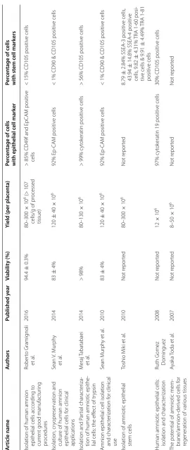
Until now, several protocols were reported by which hAECs were isolated with the average yield ranging from 8 × 10⁶ to 120 × 106 cells and a viability of 83–98%. The isolated cells were positive for the epithelial cell mark-ers in range of 83–98%, while contamination with MSCs was among 1–80% [3, 10–15], nevertheless, as mentioned in Table 1, no report is available concerning isolation of highly viable hAECs with desirable yield and without any significant purity reduction. In this study, a detailed protocol with some modification of previous ones is presented in which by using the new modified method hAECs were isolated with high yield, viability and purity with the minimum contamination with other cell popula-tions. In addition, the isolated hAECs were subjected to immuno-phenotyping, and their physiological status was assessed using a proliferation assay.
Open Access
*Correspondence: [email protected]; [email protected]
1 Department of Immunology, Faculty of Medicine, Isfahan University
of Medical Sciences, Hezar-Jereeb Ave, Isfahan, Iran
Table
1
V
iabilit
y, yield and purit
y of isola
ted hAECs using diff
er en t pr ot oc ols A rticle name A uthors Published y ear Viabilit y (%)
Yield (per plac
en ta) Per cen tage of c ells with epithelial c ell mark er Per cen tage of c ells with st em c ell mark ers
Isolation of human amnion epithelial cells accor
ding t
o
cur
rent good manufac
tur ing pr ocedur es Rober to Gramig noli et al . 2016 94.4 ± 0.3% 80–300 × 10
6 (>
107
cells/g of pr
ocessed
tissue)
>
85% CD49f and EpCA
M positiv
e
cells
<
15% CD105 positiv
e cells Isolation, cr yopr eser vation and cultur
e of human amnion
epithelial cells f
or clinical applications Sean V . M ur ph y et al . 2014 83 ± 4% 120 ± 40 × 10 6 92% Ep -CA M positiv e cells <
1% CD90 & CD105 positiv
e cells
Isolation and P
ar
tial charac
ter
iza
-tion of human amniotic epithe
-lial cells: the eff
ec
t of tr
ypsin M eraj T abatabaei et al . 2014 > 98% 80–130 × 10 6 > 99% c yt ok eratin positiv e cells >
56% CD105 positiv
e cells
Amnion epithelial cell isolation and charac
ter ization f or clinical use Sean M ur ph y et al . 2010 83 ± 4% 120 ± 40 × 10 6 92% Ep -CA M positiv e cells <
1% CD90 & CD105 positiv
e cells
Isolation of amniotic epithelial st
em cells Toshio M ik i et al . 2010 Not r epor ted 80–300 × 10 6 Not r epor ted 8.79 ±
2.84% SSEA-3 positiv
e cells
,
43.94
±
14.8% SSEA-4 positiv
e
cells
, 9.82
±
4.31%
TRA 1-60 posi
-tiv
e cells & 9.91
±
4.49%
TRA 1-81
positiv
e cells
Human amniotic epithelial cells: isolation and charac
ter ization Ruth G omez D ominguez 2008 Not r epor ted 12 × 10 6 97% c yt ok
eratin 19 positiv
e cells
30% CD105 positiv
e cells
The pot
ential of amniotic mem
-
brane/amnion-der
iv
ed cells f
or
regeneration of var
[image:2.595.162.433.82.724.2]Main text
Note
All deliveries which were positive for infectious agents including HBV, HCV and HIV and those with pre-diag-nosed genetic abnormalities were excluded from this study. Full-term human placentas were obtained from six healthy women with a normal singleton pregnancy undergoing uncomplicated elective cesarean section. All the materials and equipment used in this procedure were sterile.
Methods
Preparation of reagents and solutions
Standard culture medium DMEM/F12 (Gibco, Thermo Fisher Scientific, USA) supplemented with 10% FBS, 100 U/ml penicillin, 100 μg/ml streptomycin (1X, Sigma Aldrich, USA) and 10 ng/ml EGF (Thermo Fisher Scien-tific, USA) was used as standard culture medium.
Pre‑digestion buffer To prepare 1000 ml of 1X Pre-diges-tion buffer, all the components menPre-diges-tioned in Table 2 were added to 900 ml of tissue culture-grade water (20–25 °C) and the pH was adjusted to 7.2 (It is because pH of the buffer rises up to 0.1–0.3 units during filtration). Thereaf-ter, tissue culture-grade water was added to the solution and the final volume adjusted to 1000 ml. The buffer was immediately sterilized by filtration using a 0.22 µm filter (Thermo Fisher Scientific, USA) and stored at 4 °C for fur-ther usage.
Isolation of hAECs from placenta
Isolation of the amniotic membrane
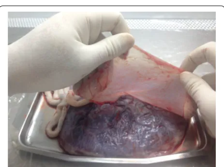
The placenta was transferred to the laboratory in a ster-ile container which was filled with ice-cold Hanks’ bal-anced salt solution (calcium- and magnesium-free HBSS) or RPMI/1640 (Gibco, Thermo Fisher Scientific, USA) containing 1% penicillin/streptomycin solution (Sigma Aldrich, USA) to cover up the placenta surface. The pla-centa was placed in a sterile container (under a horizon-tal laminar flow hood) while the amniotic membrane was
faced up (Fig. 1). The amnion membrane was manually
stripped from the chorion layer, starting from the outer edge of the amniotic membrane and continuing towards
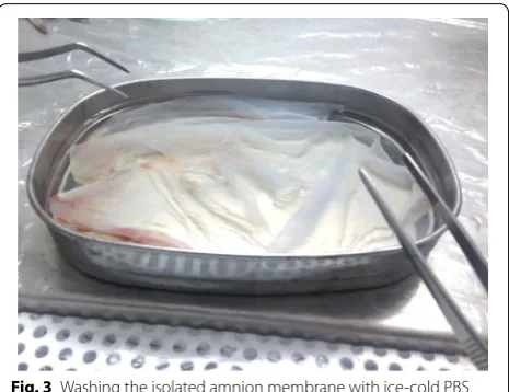
the umbilical cord (Fig. 2). The membrane was washed
several times with ice-clod PBS (pH 7.2) to remove blood clots, torn pieces and cellular debris (Fig. 3).
Digestion of the amniotic membrane
Blood-free amnion was cut into pieces around 7 cm long and transferred with forceps to two new 50 ml falcon conical tubes. To each tube 20 ml pre-digestion buffer was added, and then incubated at 37 °C for 15 min with gentle shaking (30–60 RPM). Afterwards, the amnion pieces were transferred into three new 50 ml falcon coni-cal tubes containing 20 ml of pre-warm 0.05% trypsin/
Table 2 Pre-digestion buffer ingredients
gr/litter
Sodium chloride (NaCl) 8
Potassium chloride (KCL) 0.4
Sodium bicarbonate (NaHCO3) 0.35
D-glucose (Dextrose) 1
Potassium diHydrogen phosphate (KH2PO4) 0.06
Sodium EDTA (Na2-EDTA) 0.2
Sodium hydrogen phosphate, anhydrous (Na2HPO4) 0.047
Fig. 1 The position of placing placenta in a sterile container in which the fetal surface was faced up
[image:3.595.308.538.304.472.2] [image:3.595.306.538.525.697.2] [image:3.595.57.293.616.723.2]EDTA (Thermo Fisher Scientific, USA) and incubated at 37 °C for 10 min with gentle shaking (first digestion). The obtained cells at this step were discarded to exclude blood clots and cellular debris and the membrane pieces were transferred into new tubes. The enzymatic diges-tion was followed by addidiges-tion of 20 ml pre-warm 0.05% trypsin/EDTA and incubation at 37 °C for 30 min with gentle shaking (second digestion). This step was repeated, then the second and the third digestions were neutral-ized for trypsin activity by adding 30 ml HBSS, and finally centrifuged at 200×g for 5 min at 4 °C. The cell pellets were re-suspended in 10 ml standard culture medium, mixed together and filtered through a 100 μm cell strainer (Thermo Fisher Scientific, USA). The filtrate cell number was counted using a haemocytometer and cell viability was determined using trypan blue dye exclusion.
Key points
1. It is possible that the amnion layer is peeled off from the chorion in the operating room, which has its advantages including: using less transportation medium, minimize possibility of non-sterile sam-ples, and decreasing the amnion contamination with blood clots.
2. It is recommended that before hAECs isolation, a piece of the amniotic membrane be observed under
an optical microscope (40 × magnification) in order
to check the status of the cells. An amniotic mem-brane with epithelial cells, which are notably vacu-olated in their cytoplasm, is not suitable for cell
isola-tion (Fig. 4A, B). Do not process to the next steps.
3. It is suggested that the status of the remained cells on the amniotic membrane after each digestion step be checked under an optical microscope and the membrane with highly vacuolated cells is discarded.
Checking a piece of the amniotic membrane under a microscope after each digestion step also helps to realize whether the next-step of enzymatic diges-tion is required or not. Accordingly, the number of required digestion steps to separate majority of the cells from the membrane may be increased or decreased.
4. It is suggested that at the end of each digestion, the membrane pieces be gently shacked using a forceps in 50 ml tubes containing the trypsin digest to sepa-rate all the epithelial cells in case still be (loosely) attached to the membrane.
hAECs immuno‑phenotyping using flow cytometry
The purity and phenotypic characteristics of freshly iso-lated hAECs were determined using flow cytometry. The cells (4–8 × 10⁵) were stained with different antibod-ies (Table 3) or matched-isotype control IgG at 4 °C for 25 min. Matched isotype control antibodies were used as negative controls and MSCs were employed as posi-tive control for anti-CD90 and anti-CD105. Afterwards, the cells were washed three times using cell staining buffer (Biolegend, USA) and centrifugation at 200×g for 5 min at 4 °C. Intracellular staining with FITC-conju-gated–anti-cytokeratin (Biolegend, USA) was performed after fixation and cells permeabilization according to the manufacturer’s instructions (eBioscience, USA). The data was acquired using a FACSCalibur system (Becton–Dick-inson, CA) and analyzed using CellQuest software (Bec-ton–Dickinson, CA).
hAECs proliferation assay
Isolated hAECs were cultured in 75 cm2 tissue culture flasks at a density of 2.5 × 10⁵ cells/cm2 using standard culture medium, and then incubated at 37 °C with 5% CO2. The initial plating efficiency of the cells was deter-mined after 2 days of incubation. The hAECs were dis-sociated using 0.05% trypsin–EDTA solution, whenever they were approximately 80% confluent. The cells were sub-cultured in 1:4 ratios in standard culture medium.
Results
The yield and viability of hAECs
The average yield of obtained hAECs by this method was 190 × 106 cells with a typical range of 90–280 mil-lion cells (Fig. 5a). Noticeably, for this yield the average obtained viability was 87% (ranging from 83 to 89%) (Fig. 5b).
Immuno‑phenotyping of hAECs
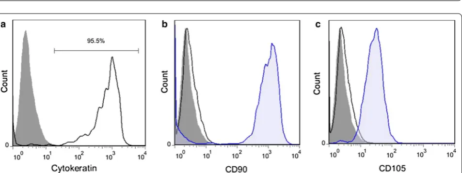
The hAECs purity achieved by this protocol was at least 95.42%, as confirmed by cytokeratin analysis, an epithe-lial cell marker (Fig. 6a). Less than 1% of the isolated cells
[image:4.595.58.291.87.266.2]Fig. 4 Epithelial cells of the amniotic membrane under an optical microscope. A, B An amniotic membrane with epithelial cells which are fully vacuolated in their cytoplasm. C, D An amniotic membrane with normal epithelial cells which is suitable for hAECs isolation
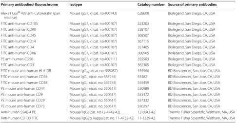
Table 3 Used antibodies to determine phenotypic characterictics of hAECs by flow cytometry
Primary antibodies/ fluorochrome Isotype Catalog number Source of primary antibodies
Alexa Fluor® 488 anti-Cytokeratin (pan
reactive) Mouse IgG1, κ (cat. no:400143) 628608 Biolegend, San Diego, CA, USA FITC anti-human CD105 Mouse IgG1, κ (cat. no:400107) 323203 Biolegend, San Diego, CA, USA FITC anti-human CD90 Mouse IgG1, κ (cat. no:400107) 328107 Biolegend, San Diego, CA, USA FITC anti-human CD45 Mouse IgG1, κ (cat. no:400107) 368507 Biolegend, San Diego, CA, USA FITC anti-human CD14 Mouse IgG1, κ (cat. no:400107) 367115 Biolegend, San Diego, CA, USA FITC anti-human CD4 Mouse IgG1, κ (cat. no:400107) 357405 Biolegend, San Diego, CA, USA FITC anti-human CD8a Mouse IgG1, κ (cat. no:400107) 300905 Biolegend, San Diego, CA, USA PE anti-human CD56 Mouse IgG1, κ (cat. no:400111) 355503 Biolegend, San Diego, CA, USA FITC anti-human CD3 Mouse IgG1, κ (cat. no:400107) 362305 Biolegend, San Diego, CA, USA FITC mouse anti-human HLA-DR Mouse IgG2a, κ(cat. no: 555057) 555560 BD Biosciences, San Jose, CA, USA
FITC mouse anti-human CD34 Mouse IgG1, κ(cat. no: 555748) 555821 BD Biosciences, San Jose, CA, USA FITC mouse anti-human CD38 Mouse IgG1, κ(cat. no: 555748) 555459 BD Biosciences, San Jose, CA, USA
PE mouse anti-human CD44 Mouse IgG1, κ(cat. no: 550617) 550989 BD Biosciences, San Jose, CA, USA
PE mouse anti-human CD9 Mouse IgG1, κ(cat. no: 550617) 555372 BD Biosciences, San Jose, CA, USA
PE mouse anti-human CD29 Mouse IgG1, κ(cat. no: 550617) 557332 BD Biosciences, San Jose, CA, USA
PE mouse anti-human CD73 Mouse IgG1, κ(cat. no: 550617) 550257 BD Biosciences, San Jose, CA, USA
[image:5.595.56.541.88.414.2] [image:5.595.55.542.475.727.2]were positive for MSC markers (CD90, CD105) (Fig. 6b, c). In addition, hAECs immuno-phenotyping from differ-ent donors showed that the isolated cells were almost a homogenous population (Table 4).
The proliferation capability of hAECs
After 48 h, the initial plating efficiency of the isolated
hAECs on culture dishes was at least 80% (Fig. 5b).
Freshly isolated hAECs had the ability to proliferate for 5–6 passages in standard culture medium. In contrast with some reports [5, 16], hAECs which were cultured without EGF were not able to proliferate at all.
Discussion
hAECs are a type of stem cells isolated from the amni-otic membrane of the placenta. In addition to stem cell-like properties, which proposed hAECs as a potential candidate for regenerative medicine, it has been shown that they can be used as an immunomodulatory agent in treatment of diseases with immune pathophysiology.
hAECs isolation from the amniotic membrane is per-formed with enzymatic digestion of the amniotic mem-brane, since amnion epithelium is a single monolayer with a weak cell–cell adhesion at the lateral sides and trypsin is able to separate hAECs from the amniotic
Fig. 5 Yield and viability of isolated hAECs. a The average yield of isolated hAECs was 190 × 106 cells with a typical range of 90–280 million cells.
b An average viability obtained using this protocol was 87% (ranging from 83 to 89%). The depicted results are representative of six independent experiments
[image:6.595.65.539.87.261.2] [image:6.595.62.539.300.479.2]basement membrane [11]. There is a notable variability in the purity, yield and viability of hAECs isolation using previous protocols [3, 10–15]. These variations might be related to several factors including mother related parameters (e.g. age, gestational week and delivery type), size and quality of the placenta (which is affected by duration of the time between the delivery and hAECs isolation), transportation condition (such as cold chain), residual blood on the tissue (in spite of extensive wash-ing), type and concentration of the enzyme solution, and enzymatic digestion time [3, 10–14]. A disadvantage of these methods for cell isolation is that achieving a desir-able yield come at the expense of losing the cell viability and purity. The presented protocol at this study resulted in hAECs isolation with high yield (to our knowledge, the highest yield reported yet), viability and purity.
Using the proposed modified protocol, an average yield of 190 × 106 hAECs (in range of 90–280 million cells) was achieved compared to the average yield of 8 × 10⁶– 120 × 106 cells obtained using previous protocols [3, 10–12, 14, 15]. Regarding the fact that a consistent pro-tocol was followed for hAECs isolation from all donors in the current study, the observed extensive variability in cell yield might be related to quality of the placenta obtained from different donors. The mean viability of iso-lated hAECs in this study was 87%, compared to previ-ously reports with a range from 83 to 99% [14]. Previous methods reported that high yield of the viable cells was
accompanied contamination with other cell populations [10, 11, 14]. However, the current study reported a highly viable hAECs at high yield and minimum contamination with MSCs. These findings could be mainly attributed to two factors, a shorter incubation time compared to previ-ous protocols and discarding the first digestion. Regard-ing the facts that a part of blood clots and cellular debris are not removed after extensive washing of the amniotic membrane with PBS, consequently these parts are sepa-rated from the membrane in the first step of digestion.
The initial plating efficiency of the cells in the current study was at least 80% after 48 h, moreover the cells con-tinued to proliferate for 5–6 passages in standard culture medium. It is found that in addition to the initial viability of the isolated cells which is an essential factor for deter-mining long-term survival of hAECs and their prolifera-tion, the initial plating efficiency is critical as well.
According to the results, isolated hAECs from differ-ent placdiffer-entas were highly homogeneous based on the expression of epithelial (cytokeratin), MSC (CD105, CD73, CD90), embryonic stem cell (SSEA-4), hemat-opoietic stem cell (CD34), and immunologic (HLA-DR, CD56, CD3, CD4, CD8) markers and adhesion molecules (CD29). However, there were differences in the level of CD9, CD14 and CD44 between different donors.
In summary, using the new modified protocol a high hAECs yield with high viability and purity was obtained, and the cells kept their proliferation ability until passage 5–6. Table 4 Determination of markers of hAECs by flow cytometry
–, Not determined; ∓, very low expression (< 10%); +, low expression (10–30%); ++, intermediate expression (30–60%); +++, high expression (60–90%); ++++, very high expression (> 90%)
Marker Donor1 Donor2 Donor3 Donor4 Donor5 Donor6
Pan cytokeratin ++++ ++++ ++++ ++++ ++++ ++++
CD73 +++ +++ +++ +++ +++ +++
SSEA-4 +++ +++ +++ +++ +++ +++
CD133 - - - - -
-CD90 - - - - -
-CD105 - - - - -
-CD34 - - - - -
-CD45 - - - - -
-CD9 ++ - + + - +
CD38 - - - - -
-HLA-DR - - - - -
-Integrin-β2 (CD29) + + + + ++ +
Hyaluronic acid receptor (CD44) - + + + ++ +
CD56 -/+ -/+ -/+ -/+ -/+ -/+
CD14 + ++ - + ++
-CD3 - - - - -
-CD4 - - - - -
[image:7.595.57.544.101.342.2]-Limitation
A limitation of this technique is that increasing the cells yield may result in losing the viability, purity and initial plating efficiency.
Abbreviations
DMEM: Dulbecco’s modified eagle’s medium; RPMI: Roswell park memo-rial institute; PBS: phosphate-buffered saline; FBS: fetal bovine serum; EGF: epidermal growth factor; hAECs: human amniotic epithelial cells; HBSS: Hanks’ balanced salt solution; HBV: hepatitis B virus; HCV: hepatitis C virus; HIV: human immunodeficiency virus.
Authors’ contributions
HM, SG carried out the experiments and preformed statistical analysis. NE participated in the design of the experiments and obtained funding for the work. NT participated in the study design. FK, BK provided the placenta tissue and participated in the design of the experiments. AR drafted the manuscript and participated in the study design. All authors read and approved the final manuscript.
Author details
1 Department of Immunology, Faculty of Medicine, Isfahan University of
Medi-cal Sciences, Hezar-Jereeb Ave, Isfahan, Iran. 2 Immunology Research Center
(IRC), Iran University of Medical Sciences, Tehran, Iran. 3 Department of
Labora-tory Medicine, Kashan University of Medical Sciences, Kashan, Iran. 4
Depart-ment of Gynecology and Obstetrics, Al-Zahra Hospital, Isfahan University of Medical Sciences, Isfahan, Iran.
Acknowledgements
The authors thank all individuals who participated in this study.
Competing interests
The authors declare that they have no competing interests.
Availability of data and materials
All data generated or analyzed during this study are included in this published article.
Consent for publication
Not applicable.
Ethics approval and consent to participate
This study was approved by the Ethics Committee of Isfahan University of Medical Science. All contributors signed informed consent before entering the study.
Funding
This study was financially supported by Kashan University of Medical Sciences (KAUMS).
Publisher’s Note
Springer Nature remains neutral with regard to jurisdictional claims in pub-lished maps and institutional affiliations.
Received: 9 June 2017 Accepted: 26 October 2017
References
1. Miki T, Lehmann T, Cai H, Stolz DB, Strom SC. Stem cell characteristics of amniotic epithelial cells. Stem cells. 2005;23:1549–59.
2. van Herendael B, Oberti C, Brosens I. Microanatomy of the human amni-otic membranes: a light microscopic, transmission, and scanning electron microscopic study. Am J Obstet Gynecol. 1978;131:872–80.
3. Miki T, Marongiu F, Dorko K, Ellis E, Strom SC. Isolation of amniotic epithelial stem cells. Curr Protoc Stem Cell Biol. 2010. https://doi. org/10.1002/9780470151808.sc01e03s12.
4. Kakishita K, Elwan MA, Nakao N, Itakura T, Sakuragawa N. Human amni-otic epithelial cells produce dopamine and survive after implantation into the striatum of a rat model of Parkinson’s disease: a potential source of donor for transplantation therapy. Exp Neurol. 2000;165:27–34. 5. Liu YH, Vaghjiani V, Tee JY, To K, Cui P, Oh DY, et al. Amniotic epithelial cells
from the human placenta potently suppress a mouse model of multiple sclerosis. PLoS ONE. 2012;7:e35758.
6. McDonald CA, Payne NL, Sun G, Moussa L, Siatskas C, Lim R, et al. Immu-nosuppressive potential of human amnion epithelial cells in the treat-ment of experitreat-mental autoimmune encephalomyelitis. J Neuroinflamm. 2015;12:112.
7. Ilancheran S, Michalska A, Peh G, Wallace EM, Pera M, Manuelpillai U. Stem cells derived from human fetal membranes display multilineage differentiation potential. Biol Reprod. 2007;77:577–88.
8. Niknejad H, Peirovi H, Jorjani M, Ahmadiani A, Ghanavi J, Seifalian AM. Properties of the amniotic membrane for potential use in tissue engi-neering. Eur Cells Mater. 2008;15:88–99.
9. Akle C, Welsh K, Adinolfi M, Leibowitz S, McColl I. Immunogenicity of human amniotic epithelial cells after transplantation into volunteers. Lancet. 1981;318:1003–5.
10. Gomez Dominguez R. Human amniotic epithelial cells: isolation and characterisation. Giessen: Laufersweiler Publishers; 2008.
11. Gramignoli R, Srinivasan RC, Kannisto K, Strom SC. Isolation of human amnion epithelial cells according to current good manufacturing proce-dures. Curr Protoc Stem Cell Biol. 2016. https://doi.org/10.1002/cpsc.2. 12. Murphy S, Rosli S, Acharya R, Mathias L, Lim R, Wallace E, et al. Amnion epithelial cell isolation and characterization for clinical use. Curr Protoc Stem Cell Biol. 2010. https://doi.org/10.1002/9780470151808.sc01e06s13. 13. Tabatabaei M, Mosaffa N, Nikoo S, Bozorgmehr M, Ghods R, Kazemnejad
S, et al. Isolation and partial characterization of human amniotic epithelial cells: the effect of trypsin. Avicenna J Med Biotechnol. 2014;6:10. 14. Murphy SV, Kidyoor A, Reid T, Atala A, Wallace EM, Lim R. Isolation,
cryopreservation and culture of human amnion epithelial cells for clinical applications. J Vis Exp. 2014. https://doi.org/10.3791/52085.
15. Toda A, Okabe M, Yoshida T, Nikaido T. The potential of amniotic membrane/amnion-derived cells for regeneration of various tissues. J Pharmacol Sci. 2007;105:215–28.