JOURNAL OFVIROLOGY, Mar. 1983,p.1195-1199
0022-538X/83/031195-05$02.00/0
Copyright C 1983,AmericanSocietyforMicrobiology
Vol.45, No.3
Cloning and
Analysis of Reverse Transcript
P160
Genomes
of
Abelson Murine Leukemia Virus
SAMUELA. LATT,t STEPHEN P.GOFF,1 CLIFFORD J. TABIN, MICHAEL PASKIND, JEANY.-J. WANG, ANDDAVIDBALTIMORE*
Centerfor Cancer Research and Departmentof Biology, Massachusetts Institute of Technology, and Whitehead Institute for Biomedical Research, Cambridge, Massachusetts 02139
Received 27September1982/Accepted22November 1982
Circular duplexreversetranscripts of thegenomeofastrain of Abelson murine
leukemia virus that encodes a 160,000-molecular-weight protein were isolated,
cleaved with Hindlllrestrictionendonuclease,andcloned into the unique HindIII
site of lambda phage Charon 21A. Recombinant phage clones, some of which
were infectious in transfection assays, were found to contain a 789-base-pair
region specific for Abelson murine leukemia virus; this region isnotfoundin other
strains ofthis virus. Theextrasequence waslocalized byrestriction endonuclease
and electron microscopic heteroduplex analysis. Sequence analysis showed no
homology atthe ends of theextra sequence, implying that it was deletedby an
eventthatdidnotutilizesequencehomology. Thesequenceof this unique region
has anopenreadingframethrough its entirety.
Abelson murineleukemia virus(A-MuLV)isa
replication-defectiveretroviruscapableof trans-formingmouselymphoid (1, 13)and fibroblastic
(15) cells. Thegenome of this virus isa hybrid
RNAmoleculecontaining portions ofthe
paren-tal Moloney MuLV at its ends and a large substitution, termed v-abl, in the center (16).
This central substitution is homologous to a
cellulargene, termedc-abl, which isapparently
interruptedbyseveralinterveningsequences not
presentin the viralgenome(7).
Several variantsofA-MuLVhave been
isolat-edwithalterations in the central portion of the
genome. These variants encode related mem-brane-associated proteins ranging in size from
160,000 to 85,000 daltons (14). The largest
genome,thatofA-MuLV(P160),is
approximate-ly 6.2kilobases(kb) longandis knowntoencode
the largest of these polypeptides, termed P160.
Other genomes,
including
thoseencoding
pro-teins P120, P100, and P90, are smaller and
apparentlyshareacommondeletionof 0.7to0.8
kbwith thatencodingP160(7,9). A
transforma-tion-defective genome (21) encoding a protein termed P92td has suffered yetanother deletion
(9).
To isolate DNA clones representing the
genome ofA-MuLV(P160), virus was isolated
t Present address: Genetics Division, Childrens Hospital MedicalCenter, Boston, MA 02115.
tPresentaddress:DepartmentofBiochemistry, Collegeof
Physiciansand Surgeons, ColumbiaUniversity, New York, NY 10027.
from the A-MuLV(P160)-transformed NIH 3T3
fibroblast cell line AN-P160-54 (9), which had
been coinfected with the clone 3A strain of
Moloney MuLV. The virus was used to infect
sensitive NIH 3T3 cellsfromwhich supercoiled
circularduplexA-MuLV(P160) DNAwas
isolat-ed (7). This DNA was cleaved with HindIII
endonuclease and cloned into the unique
HindIlI site of lambda phage Charon 21A (20).
A total of10 independent isolates that
hybrid-ized (4) to an abl-specific probe (plasmid
pAB3SUB3) (7) weregrown intophage stocks,
the DNA from which was characterized for
restriction endonucleasecleavage sites.
Prelimi-nary restriction maps of these 10 clones were
prepared by using theabl-specific probeto
iden-tify (19) majorrestrictionfragments. Five of the
clones appeared to contain full-length
A-MuLV(P160) genomes with one long terminal repeat(LTR)sequence (6.2kbtotal), and three
had apparently full-length genomes with two
tandem LTR sequences (6.7 kb total). The
re-mainingtwoclonescontained large internal de-letions (1.0 and 0.2 kb) andwere not
character-izedfurther(Table 1).
Comparison of the restriction enzyme cleav-age maps ofthese clones with those ofother
strainsofA-MuLV DNA(9)suggests that the
A-MuLV(P160) genome contains an extra 0.7 to
0.8 kb when compared with the P120, P100, or
P90strains andthat a newSacd siteispresentin
this extra DNA. This unique sequence was
localized near the center ofthe abl region. In
addition, theLTRregion ofthe P160cloneswas
1195
on November 10, 2019 by guest
http://jvi.asm.org/
_
p160~~~16
A.~~~~~~~~~~4
!b
----
v--
ns
Ok2t
V
X,ly~~~~~~s_~, S dj e C t ^ F'_ _^\
_ ~~> ^
sC'"'-"4
r¼
4*re
*
~~~~~~t
(4 it~ a ttiW^b*'- ^ XD s, v * .t * ';As ' b , a
4,1
~~p6
MuLV(P92td)p9
~~W'"~~W~~~ ~, '0.* A..
.V
V
thtfth thrA-MuVgnmseaie. [eie fro A-uV(9) (7) rv alecm
rA~~ ~ ~ ~ ~ ~~~~~~.t
~~.
K..'
\t§
-AW. ~ ~ A"
FIG. 1. (A) Heteroduplex (diagrammed in inset) between cloned inserts from A-MuLV(P160) and
A-MuLV(P90). (B) Heteroduplex (diagrammed in inset) between cloned inserts from A-MuLV(P160) and
A-MuLV(P92td.
approximately 80 base pairs (bp) shorter than heteroduplexes(6)betweenXAB160-1and XAB3 that of the other A-MuLV genomesexamined. [derived from A-MuLV(P90)] (7), revealed
com-A prototypeclone withoneLTR
(XAB160-1)
pletehomology,
exceptforasingle
loop,
0.73kbwaschosenfor further
analysis.
Examination of insize,
mapping
1.7 kb from one end of the1196 NOTES J. VIROL.
on November 10, 2019 by guest
http://jvi.asm.org/
[image:2.489.53.447.63.575.2]NOTES 1197
TABLE 1. Properties of A-MuLV(P160) clones
Clone No. of LTRs
Transforma-tiona
AAB160-1 1
XAB160-2 1 +
XAB160-3 1 +
XAB160-5 2 +
XAB160-6 1
XAB1609b 1
XAB160-12 2 +d
XAB160-13 2
XAB160-15 1 +d
XAB16016c 1
aEach DNA preparation was cleavedwith HindIII endonuclease and religated to produce some DNA fragments containing a nonpermuted copyof the pro-virus. These DNA samples were then cotransfected as described in the text with a 10-fold molar excess of DNA of a biologically active proviral clone of Moloney MuLV termed pZAP (18). The cells were replated 1 day later, and foci were scored after 15 to 20 days in culture.
[image:3.489.48.241.82.205.2]bDeletion of 1.0 kb in 2.75-kb Sacl-Saclfragment (Fig. 3).
[image:3.489.254.452.490.587.2]cDeletionof 0.2 kb in 2.75-kb SacI-SacIfragment
(Fig. 3).
dTransformedcellsproduced P160.
inserts (Fig. 1A). Thus, the extra DNA
con-tained in the A-MuLV(P160)genomeapparently
lies in a single contiguous stretch.
Heterodu-plexes between XAB160-1 and XAB2 [derived
from A-MuLV(P92td)] (7) showed twoseparate
deletionloops 0.7 and 0.6 kb in size, separated
by 0.3 kb of duplex DNA(Fig. 1B). These data
(summarized inFig. 2) show that thetwo
dele-tionspresentinA-MuLV(P92td)donotoverlap.
To determine the sequence of the unique
information in A-MuLV(P160), a 0.94-kb PstI
fragment of XAB160-3, which appeared to
con-taintheentireunique region,wassubcloned into thePstI siteofplasmid pBR322 (5). Comparison
of thesequence(12) of thisfragment with that of
an A-MuLV(P90) clone (Lee etal., manuscript
in preparation) shows that the uniqueregion is
789bp long and hasasingleopenreading frame
through its entirety. The sequence itself will be
published as part of the complete A-MuLV
sequence (manuscript in preparation). Certain
restriction enzymecleavage sites are shown in
Fig. 3. DNA sequence andrestriction fragment
analysis placed the 3' boundary of the
A-MuLV(P160)-specific insert 1.75 kb interior to
the 3' end of the total A-MuLV(P160) genome,
as compared with 1.69 ± 0.05 kb estimated by
heteroduplex analysis. Agreement between
these twoestimates, and betweentheinsert size
asdeterminedbyDNAsequencing(789bp)and
by electronmicroscopy (725 ± 48and734 ± 62
bp) (Fig. 2), is reasonably good.
L3
A)
L2 X/I LI p160P90FRACTIONAL LENGTH (n-14) Li 0.312±0.005
L2 0.688±0.005
L3 0.136±0.005 734±62 BP (L1 +L2=5.42KB)
B)
r XL5 L3
T7K7
L2' ' LI
FRACTIONAL LENGTH (n=26) Li 0.356 ±0.003
L2' 0.644 ±0.003
L3 0.151±0.006 725±49 BP)
L4 0.064±0.001 314 ±56 BP (L1+L2' +L5-5.42KB)
L5 0.129 ±0.005 629 ±82 BP)
FIG. 2. Analysis of heteroduplex data involving
cloned A-MuLV molecules. Heteroduplex figures
were identified on 8 by 10 in (20 by 25 cm)
photograph-ic prints. Relative segment lengthswere determined from duplicate scans with an X-Y digitizer kindly
made available by Jonathan King. (A)
A-MuLV(P160)-A-MuLV(P90) heteroduplex. (B)
A-MuLV(P160)-A-MuLV(P92td) heteroduplex.The seg-mentsidentifiedin thediagrambased on the number of pictures (n)indicated in parentheses arerepresented
first as average fractionallengths (± standarderrorof themean), from which thesizes oftheinserted seg-ments areestimated.
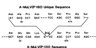
Animportantaspectof the uniquesequencein
XAB160-3is that there isno homology between
the end points ofthe region (Fig. 4), implying
that if (as seems most likely) A-MuLV(P160)
suffers a deletion toproducetheA-MuLV(P120) genome, that deletion involves nohomology. In
addition, the sequence at the deletion is not
compatiblewith the ideathat an aberrant
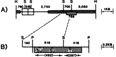
splic-H S S
A)
II°r
2.750
S S H
17001 2.050 1
- I--, -yiro
I ~
~~I/
I- I
I
p s p
B)
1182
5sa
418<0 (492)----
--4297)P-VKA
FIG. 3. Summary map of A-MuLV(P160) genomes derived fromelectrophoresis and heteroduplexdata. (A) Insert with two LTRS. (B) Detail of(A). ,
v-ablsequences. Scale is indicatedattheright. Enzyme cleavage sitesareindicated as follows:H,HindIll;S,
Sacl; P,PstI;T, TaqI;SU,Sau96I.Thesize of the
A-MuLV(P160)-specific insert (mm. is basedon DNA sequence data. The data in(B)arederivedlargelyfrom
analysis of the subclonedPstIfragment.
- mmmrnm==9
VOL. 45,1983
I
on November 10, 2019 by guest
http://jvi.asm.org/
1198 NOTES
A-MuLV(P160) Unique Sequence
Asp Ala Pro Asp Ser Ser Pro Gly Ser -AT GCG CCT GAC---TCC AGC CCT GGC
T-\ 30 810 /
Ala Glu GIn Lys [Al Ser Pro Pro Ser ---GCT GAG CAG AAA G\C0 AGC CCT CCC
AGC---20 820
A-MuLV(P120)Sequence
FIG. 4. DNA sequences at the junctions of the A-MuLV(P160)-specific DNA insert. Base pairs are numbered starting with the 5' terminus of the PstI
fragment. The boundaries of the surrounding A-MuLV(P120) sequence are also indicated. Written above each triplet is the corresponding amino acid, and in the caseof the A-MuLV(P120) sequence, the amino acid (in brackets) replaced in A-MuLV(P160) because the insert occurred within a codon.
ing eventcauses the removal of the 789 bp; no
homology tothe consensus sequences at splice
joints (2) was detected. One mechanism that
couldgenerate thedeletion isanunequal,
illegit-imate recombination between A-MuLV(P160)
genomes, such as that postulated by Goldfarb
and Weinberg (10) to occur between deleted
Harvey sarcoma virus and Moloney MuLV
ge-nomes.
The cloned permuted A-MuLV(P160)
ge-nomes were tested for biological activity in a
transfectionassay(8)asmodifiedby Andersson
etal. (3) fromthemethodof Graham andvander
Eb (11), with NIH 3T3 cells used as indicator
cells. Of eight independent clones lacking a
deletion in the abl sequence,five were capable
oftransformingmouse NIH 3T3 cells(Table1).
Preliminary data (Rotter, unpublished) are
con-sistent with the production of the P160 protein
bymost,ifnotall, of these transfectants. Three
of the infective clones contained one LTR; the
other two contained two LTRs. The LTRs of
these A-MuLV(P160) clones were shorter by
approximately 80bpthan the LTRsof other A-MuLV strains. The exact position of this
dele-tion has notyet been determined, but it might
well correspond to loss ofoneofthe tandemly
repeated 75-bp sequences starting 117 and 192
bp fromthe 5' end of theLTR in M-MuLV(17).
Thebiological significance of this deletion isnot
yet clear. Since itwaspresent in all 10ofthe
A-MuLV(P160) clones examined, it presumnably
preexisted in the A-MuLV(P160) RNA and
hence was compatible with biological
transfor-mation.
Theacquisition and initial characterizationof
A-MuLV(P160) clones should permit more
de-tailed study oftheablsequencesegmentsinthe mousegenome andofstructure-activity
relation-ships within the family of transformation-associ-ated A-MuLV proteins.
We thank Chris Riser for advice on the use of the electron microscope.
This work was supported by Public Health Service grants CA-26717 (to D.B.), GM-21121 (to S.A.L.), and CA-14501 (core grant to S. E. Luria) from the National Institutes of Health. S.P.G. was a postdoctoral fellow of the Jane Coffin Childs Memorial Fund for Medical Research, as is J.Y.-J.W. D.B. is anAmerican Cancer Society Research Professor.
LITERATURECITED
1. Abelson, H. T., and L. S. Rabstein. 1970.
Lymphosarco-ma: virus-induced thymic-independent disease in mice. Cancer Res. 30:2213-2222.
2. Abelson, J. 1979. RNA processing and the intervening sequenceproblem.Annu. Rev.Biochem. 48:1035-1069. 3. Andersson, P., M.P.Goldfarb,and R. A.Weinberg. 1979.
Adefined subgenomic fragment ofin vitrosynthesized Moloneysarcomavirus DNA thatcaninduce cell
trans-formation upon transfection. Cell 16:63-75.
4. Benton, W. D., and R. W. Davis. 1977. Screening gt recombinant clones by in situ hybridization to single plaques.Science 196:180-181.
5. Bolivar,F., R. L. Rodriquez,P.J. Green,M.C. Betlach,
H. L.Heyneker,H.W.Boyer, J.H.Crosa, and S. Falkow. 1977. Construction and characterization ofnewcloning vehicles.II.Amultipurpose cloning system. Gene 2:95-111.
6. Ferguson, J.,and R. W.Davis. 1978.Quantitativeelectron microscopy of nucleic acids, p. 123-171. In J. K. Koehler (ed.), Advancedtechniquesinbiologicalelectron micros-copy.Springer-Verlag KG,Berlin.
7. Goff,S.P.,E.Gilboa,0.N.Witte,andD. Baltimore.1980. Structure of the Abelson murine leukemia virus genome and the homologous cellular gene: studies with cloned
viralDNA.Cell 22:777-785.
8. Goff, S. P., C.J. Tabin, J. Y.-J. Wang,R. A.Weinberg,
andD. Baltimore. 1982.Transformation of fibroblastsby
cloned Abelsonmurineleukemia virus DNA and recovery of transmissible virusbyrecombination withhelpervirus. J.Virol. 41:271-285.
9. Goff,S.P.,0.N.Witte,E.Gilboa,N.Rosenberg,and D. Baltimore. 1981. Genome structure of Abelson murine
leukemia virus variants: proviruses in fibroblasts and
lymphoidcells. J. Virol. 38:460-468.
10. Goldfarb,M.P.,and R. A.Weinberg. 1981. Generation of
novel, biologically active Harvey sarcoma viruses via apparentillegitimaterecombination. J. Virol.38:136-150.
11. Graham, F. L., and A. J. van der Eb. 1973. A new
technique for the assay ofinfectivityof human adenovirus DNA.Virology52:456-471.
12. Maxam,A.M.,and W.Gilbert. 1977.A newmethod for
sequencingDNA. Proc.Natl. Acad. Sci. U.S.A.
74:560-564.
13. Rosenberg, N.,and D. Baltimore.1980.Abelsonvirus,p. 7187-7203. In G. Klein (ed.), Viral oncology. Raven Press,NewYork.
14. Rosenberg, N., and 0.N. Witte. 1980. Abelson murine leukemia virus mutants with alterations in the
virus-specificP120molecules. J. Virol.33:340-348.
15. Scher,C.D.,and R.Siegler. 1975. Direct transformation of 3T3 cellsby Abelson murine leukemia virus. Nature
(London)253:729-731.
16. Shields, A., S. P. Goff, M. Paskind, G. Otto, and D. Baltimore. 1979.Structure oftheAbelsonmurine leuke-mia virus genome. Cell 18:955-962.
17. Shinnick,T.M.,R. A.Lerner,andJ. G.Sutcliff. 1981. Nucleotide sequence ofMoloney mufine leukaemia virus.
Nature(London) 293:543-548.
18. Shoemaker,C., J. Hoffmann,S. P.Goff,and D.Baltimore.
1981. Intramolecularintergration withinMoloney leuke-mia virusDNA.J. Virol.40:164-172.
J.VIROL.
on November 10, 2019 by guest
http://jvi.asm.org/
[image:4.489.47.243.64.162.2]VOL. 45, 1983 NOTES 1199
19.Southern, E. M. 1975. Detection ofspecific sequences among DNAfragmentsseparatedby gel electrophoresis. J. Mol.Biol. 98:503-517.
20. Williams,B.G., and F. R. Blattner. 1980.Bacteriophage
lambdavectors for DNA cloning, p. 201-281. InJ.Setlow
andA.Hollaender (ed.),Genetic engineering: principles
and methods, vol. 2. Plenum Publishing Corp., New York.
21. Witte, 0. N., S. P.Goff, N. Rosenberg, and D. Baltimore. 1980. Atransformation-defective mutant of Abelson mu-rine leukemia virus lacks protein kinase activity. Proc. Natl. Acad. Sci. U.S.A.77:4993-4997.
on November 10, 2019 by guest
http://jvi.asm.org/