2007
Functional divergence after gene duplication and
sequence-structure relationship: a case-study of
G-protein alpha subunits
Ying Zheng
Iowa State University
Follow this and additional works at:https://lib.dr.iastate.edu/rtd
Part of theBioinformatics Commons
This Thesis is brought to you for free and open access by the Iowa State University Capstones, Theses and Dissertations at Iowa State University Digital Repository. It has been accepted for inclusion in Retrospective Theses and Dissertations by an authorized administrator of Iowa State University Digital Repository. For more information, please [email protected].
Recommended Citation
Zheng, Ying, "Functional divergence after gene duplication and sequence-structure relationship: a case-study of G-protein alpha subunits" (2007).Retrospective Theses and Dissertations. 15063.
by
Ying Zheng
A thesis submitted to the graduate faculty
in partial fulfillment of the requirements for the degree of
MASTER OF SCIENCE
Major: Bioinformatics and Computational Biology
Program of Study Committee: Xun Gu, Co-Major Professor Karin Dorman, Co-Major Professor
Xiaoqiu Huang
Iowa State University
Ames, Iowa
2007
1446062 2007
UMI Microform Copyright
All rights reserved. This microform edition is protected against unauthorized copying under Title 17, United States Code.
ProQuest Information and Learning Company 300 North Zeeb Road
P.O. Box 1346
Ann Arbor, MI 48106-1346
TABLE OF CONTENTS
ABSTRACT iii
CHAPTER I. GENERAL INTRODUCTION 1
CHAPTER II. DATA AND METHODS 7
CHAPTER III. RESULTS AND DISSCUSSION 11
CHAPTER IV. CONCLUSIONS 29
REFERENCES CITED 30
ABSTRACT
In this study, I use animal G-protein alpha subunit family as an example to illustrate a
comprehensive analytical pipeline for detecting different types of functional divergence of
protein families, which is phylogeny-dependent, combined with ancestral sequence
inference and available protein structure information. In particular, I focus on (i) Type-I
functional divergence, or site-specific rate shift, as typically exemplified by amino acid
residue highly conserved in a subset of homologous genes but highly variable in a
different subset of homologous genes, and (ii) Type-II functional divergence, or the shift
of cluster-specific amino acid property, as exemplified by a radical shift of amino acid
property between duplicate genes, which is otherwise evolutionally conserved. We
utilized the software DIVERGE2 to carry out these analyses. In the case of G-protein
alpha subunit gene family, I have tested the significance of functional divergence between
subfamily Gq and Gs, and predicted the candidate amino acid residues related to either
Type-I or Type-II functional divergence. Then, the inferred ancestral sequences and
current amino acid configuration of these candidate sites were combined with
phylogenies to explore the trends of functional divergence. Finally, these predicted
residues are mapped to the protein structures to test whether these residues may have
CHAPTER I. GENERAL INTRODUCTION
1.1 Gene duplication and gene families
A gene family is a set of genes evolved from a common ancestral gene. They generally
share similarity whereas with divergence in sequence, structure/function. Gene
duplications and domain duplication/shuffling, have been considered to provide the major
source of the innovation and complexity of gene families (Ohno, 1970; Doolittel, 1995;
Sidow, 1996; Henikoff et al., 1997; Li et al, 2001).
1.2 Functional divergence of gene families
Understanding functional divergence after gene duplication is one of the major goals in
functional genomics (Henikoff et al. 1997; Bork and Koonin 1998). Under the framework
of phylogenomic annotation of gene function (Golding and Dean 1998; Eisen and Fraser
2003), the importance of gene ‘function’ can be measured quantitatively in terms of the
functional constraints of the protein sequence (Kimura 1983). As an amino acid residue is
said to be functionally important if it is evolutionarily conserved, it has been recognized
recently that change of the evolutionary conservation at a particular residue may indicate
its involvement of functional divergence (Lichtarge et al. 1996; Gu 1999). Following this
idea, many research groups including ours have developed statistical methods for testing
and predicting functional divergence after gene duplication (e.g., Lichtarge et al. 1996;
Gu 1999, 2001, 2006; Landgraf et al. 2001; Knudsen and Miyamoto 2001; Lopez et al.
methods, many case studies have shown the association between sequence and
function/structure divergence (e.g., Gaucher et al. 2002a; Landgraf et al. 2001; Wang and
Gu 2001; Jordan et al. 2001; Gribaldo et al. 2003; Gao et al. 2005; Rastogi and Liberles
2005; Zhou et al. 2006).
1.2.1 Types of functional divergence in protein sequence evolution
From the view of molecular evolution, an amino acid residue is said to be functionally or
structurally important if it is evolutionarily conserved (Kimura, 1983). Therefore, change
of the evolutionary conservation at a particular residue may indicate the involvement of
functional divergence during the evolution of a gene family (Gu, 1999). Furthermore, Gu
(2001) made a distinction between Type-I and Type-II functional divergences. Note that
these two types of functional divergence may have other names. For instance, the basic
Evolutionary Trace approach (Lichtarge et al. 1996; Madabushi et al. 2004) mainly
focused on cluster-specific residues related to Type-II functional divergence. Gribaldo et
al. (2003) also looked at Type-II functional divergence as called ‘constant-but-different’.
Meanwhile, the weighted Evolutionary Trace approach proposed by Landgraf et al. (2001)
was similar to Type-I functional divergence (Gu 1999).
Type-I functional divergence (Site-specific rate shift)
This type of functional divergence refers to the evolutionary process, resulting in
site-specific rate shifts after gene duplication (Gu 1999; Gaucher et al. 2002b; Landgraf et
al. 2001; Knudsen and Miyamoto 2001; Lopez et al. 1999). Typically, an amino acid
(1999) has developed a statistical method to test the significance of Type-I functional
divergence between duplicate genes. Briefly, the two-state model proposed by Gu (1999)
assumed that an amino acid residue (site) is in either one of two states: related to
functional divergence if its evolutionary rate is shifted (up or down) after gene
duplication; or unrelated to functional divergence for otherwise. The coefficient of
(Type-I) functional divergence between duplicate genes, denoted by θI, is defined as the
probability of being related to functional divergence. Clearly, a large value of θI indicates
a high level of Type-I functional divergence, and vice versa. In a typical case when two
gene clusters are generated by a gene duplication event, the coefficient of (Type-I)
functional divergence between them can be estimated (Gu 1999; Gu and Vander Velden
2002). Rejection of the null hypothesis θI =0 means that the evolutionary rate has become
different between the duplicate genes at some sites. Using this method, many case studies
has demonstrated the functional-structural basis, e.g., the Caspase family (Wang and Gu
2001), and the Jak protein kinase family (Gu et al. 2002). Moreover, a site-specific profile
based on the empirical posterior analysis is useful to predict amino acid residues that are
crucial for functional divergence.
Type-II functional divergence (site-specific property shift)
As opposed to site-specific shift of evolutionary rate (Type-I functional divergence),
Type-II functional divergence results in site-specific property shift. A typical case is that
at a homologous residue (one column in the multiple alignment of the gene family), a
radical shift of amino acid property, e.g., positively versus negatively charged, has
within each of orthologous genes. Gu (2006) proposed a statistical method for the Type-II
functional divergence inference by extending the two-state model (Gu 1999, 2001) to
Type-II (cluster-specific) functional divergence: (i) In the early (E) stage after gene
duplication, an amino acid residue can be in either of two states: Type-II unrelated and
Type-II related. The probability of a residue being under the Type-II related status is
denoted by θII, as called the coefficient of Type-II functional divergence. (ii) In the late (L)
stage, any amino acid residue has no further Type-II functional divergence, so amino acid
substitutions in this stage are mainly under purifying selection. Under the functional
divergence unrelated status, the substitution model largely reflects the conserved
evolution of protein sequences, which can be empirically determined by the Dayhoff
model (Dayhoff et al. 1978), or the JTT model (Jones, Taylor, and Thornton 1992). In
contrast, under the functional divergence-related status, radical amino acid substitutions
may occur more frequently (Lichtarge et al. 1996). To avoid over-parameterization, Gu
(2006) proposed a simple model that can distinguish between the radical and conserved
amino acid substitutions. First, we tentatively classify twenty amino acids into four
groups: charge positive (K, R, H), charge negative (D, E), hydrophilic (S, T, N, Q, C, G, P),
and hydrophobic (A, I, L, M, F, W, V, Y). An amino acid substitution is called radical if it
changes from one group to another; otherwise it is called conserved. In a typical case
when two gene clusters generated by a gene duplication event, the coefficient of (Type-II)
functional divergence between them can be estimated. Moreover, a site-specific profile
based on the empirical posterior analysis is useful to predict amino acid residues that are
1.2.2 Evidence for the association between functional divergence and changes in
function after gene duplication
Using statistical methods in software DIVERGE (Gu and Vander 2002), our group has
conducted a large-scale analysis showing that site-specific rate shift (type-I functional
divergence) is a general evolutionary pattern. Moreover, we found evidence that the level
of site-specific rate shift of member genes could be related to protein structure differences
(Wang and Gu, 2001; Gaucher et al. 2002a), the severity of knockout phenotypes, and
tissue-specificity.
1.3 G-protein alpha subunit as an example
G proteins, short for guanine nucleotide binding proteins, are a family of proteins
involved in second messenger cascades. These proteins are activated by G
protein-coupled receptors and are made up of alpha, beta and gamma subunits. There are
over 16 G-protein alpha subunits in animals, which can be further divided into four major
classes: Gs, Gio, Gq, and G12, respectively (Simon et al. 1991; Neer 1995; Downes et al
1999; Cabrera-Vera et al. 2003), depending on their actions upon the effectors. In the
project, we have predicted amino acid residues that are related to either Type-I or Type-II
functional divergence between the Gq and Gs subfamilies. The inferred ancestral
sequences for these sites are helpful to explore the trends of functional divergence. Finally,
these predicted residues are mapped to the protein structures to test whether these
how to identify amino acid residues that are crucial for different types of functional
divergence between duplicate genes, infer the trend of evolutionary changes at these
CHAPTER II. DATA AND METHODS
2.1 Animal G-protein alpha subunits family data set preparation
Protein sequence data
The starting set of vertebrate G-protein alpha subunits sequences were downloaded from
the Homologous Vertebrate Genes Database
(http://pbil.univ-lyon1.fr/databases/hovergen.html). A standard BLAST search against
NCBI non-redundant protein sequence database added more vertebrate sequences and the
invertebrate out group to the data set. Finally we got 81 amino acid sequences of animal
G-protein alpha subunits. The multiple alignments were made using the program ClustalX.
The phylogenetic tree of the whole family was inferred by the neighbor-joining (NJ)
method (Saitou and Nei 1987). The parsimony (PAUP4.0) and likelihood (PHYLIP)
methods give virtually the same topology.
Protein structure data
We downloaded the 3D structures of Gs and Gq from the RCSB Protein database
(http://www.rcsb.org/), and the MMDB
(http://www.ncbi.nlm.nih.gov/entrez/query.fcgi?db=Structure) with respective data
formats. In our study, we utilized the structure of Gs_BOVIN, the chain C of the PDB
2.2 DIVERGE2: Analytical pipeline for comprehensive functional divergence
analysis
The software DIVERGE (Gu and Vander Velden 2002) is a software system to study
functional divergence between member genes of a protein family based on (site-specific)
shifted evolutionary rates (Type-I) after gene duplication.
Posterior analysis results in a site-specific profile for predicting important amino acid
residues that are responsible for this type of functional divergence. Moreover, when the
3D protein structure is available, these predicted amino acid residues can be mapped to
the 3D structure viewer to explore their structural basis.
The updated version, named DIVERGE2, has provided more options (e.g., Type-II
functional divergence) to explore functional evolution of protein family sequences. One
can use the site-specific profiles to detect amino acid residues that are crucial for this type
(I or II) of functional divergence. In practice, one may use the site (k)-specific score QI (k),
or QII (k), the posterior probability that site k is related to Type-I or Type-II functional
divergence. Another commonly used measure is based on the posterior ratio; in our case,
it is given by RI (k) = QI (k)/[1-QI (k)], or RII (k) = QII(k)/[1- QII (k)]. When a cutoff is
given, important residues for two types of functional divergence are predicted.
2.3 Ancestral sequence inference
Ancestral sequence inference under a given phylogeny is becoming an important
1998). This is partly because evolution has selected proteins for function over hundred or
even thousand millions of years, keeping those that carried out critical functions, and
eliminating deleterious mutations. We have recognized that ancestral sequence
reconstruction is a powerful technique for linking sequence to function. An important
development of DIVERGE2 is to provide an analytical pipeline for combining functional
divergence and ancestral sequence inference, which can be used to infer the trends of
functional divergence. Currently, DIVERGE2 adopts the Bayesian algorithm of Zhang et
al. (1997) to infer the ancestral sequences under a known phylogeny of gene family. It is a
simplified version of Yang et al. (1995) in which the branch lengths of the phylogenetic
tree are estimated using a least squared method rather than the maximum-likelihood
method. Each site in the inferred ancestral sequence receives the assignment of amino
acid with the highest posterior probability. Using this approach one may determine
whether an amino acid residue that was highly conserved in the ancestral protein
sequence now becomes highly variable, or vice versa.
2.4 Mapping predicted amino acid residues to protein structure
Important features of the physical environment of a residue such as secondary structure,
and whether the site is on the surface or in the interior of a protein, can be extracted from
the solved 3D structure. We take the criteria of accessible surface area (ASA) to define
the surface area of a bimolecule accessible to a solvent (Lee and Richards 1971).
Residues are considered to be solvent exposed (on the surface, o for short) or be buried
(Mizuguchi et al. 1998) was used to compute the relative ASA and assign all the residues
to “solvent inaccessible” or “solvent accessible” with the default cutoff of 7% relative
accessibility. As an alternative method for computing residue accessibility, NACCESS
CHAPTER III. RESULTS AND DISSCUSSION
Consistent to previous results, the four major classes of G-protein alpha subunit family
are monophyletic. As an example, we choose Gs and Gq, the two major classes of
G-protein alpha subunits, to demonstrate the ancestral-based analysis of functional
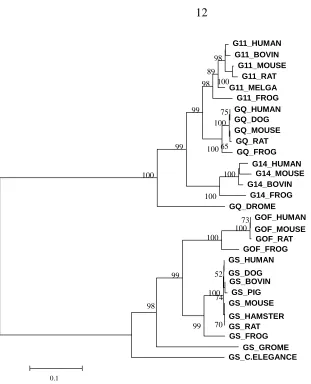
divergence. Fig.1 is the phylogenetic tree of Gs and Gq classes. The Gs class, which
consists of Gs and Golf subtypes, is involved in hormonal stimulation of adenylate
cyclase and opening of Ca2+ channels. While the Gs subtype is expressed in almost all
tissue types, the Golf subtype is expressed exclusively in olfactory cells and is thought to
be involved specifically in odorant signal transduction (Kaziro et al. 1991). On the other
hand, the class Gq has the function of simulating phospholipase C (PLC), which has four
subtypes: Gq, G11, G14, and G15. In spite of the fact that Gq and G11 are widely
distributed and often found in the same cell types, they may have different receptors and
effectors or act in different developmental stages. G14 and G15 are tissue-specific, which
may interact with different members of the phospholipase family. In this demonstration,
G15 subtype is not included in the Gq class. It should be noted that the alignment of all
the 81 protein sequences is the input of our software; whereas Gs and Gq(without G15)
Fig. 1. The NJ tree of Gq and Gs based on the multiple sequence alignment with Poisson distance. Bootstrap score >50% are presented. The accession
numbers for protein sequences are P50148(GQ_HUMAN), Q28294(GQ_DOG),
P21279(GQ_MOUSE), P82471(GQ_RAT), P38410(GQ_FROG), P29992(G11_HUMAN), P38409(G11_BOVIN), P21278(G11_MOUSE), Q9JID2(G11_RAT), P45645(G11_MELGA), P43444(G11_FROG), O95837(G14_HUMAN), P38408(G14_BOVIN), P30677(G14_MOUSE), O73819(G14_FROG), JN0115(GQ_DROME), P04895(GS_HUMAN), CAA78161(GS_DOG), P04894(GS_MOUSE), AAA40827(GS_RAT), CAA35516GS_HAMSTER), P04896(GS_BOVIN), P29797(GS_PIG), CAA39571(GS_FROG), Q8CGK7(GOF_MOUSE), P38406(GOF_RAT), P38405(GOF_HUMAN), CAC82735(GOF_FROG), NP_477506(GS_DROME),
3.1 Functional divergence between Gs and Gq proteins
Type-I functional divergence
We first tested the site-specific shift of evolutionary rate (Type-I functional divergence)
after the gene duplication event leading to Gs and Gq subtypes. The coefficient of Type-I
functional divergence between Gs and Gq is θI = 0.53 ± 0.08, which is significantly larger
than 0. Hence, site-specific rate difference mayoccur at some amino acid residues after
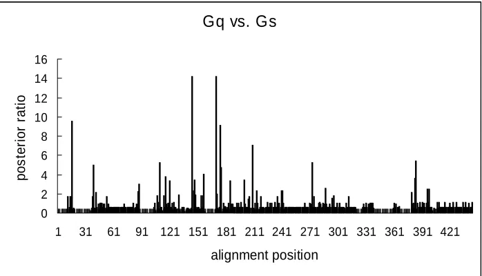
the gene duplication. Fig.2 shows the site-specific profile of posterior ratio, RI (k); notably,
most sites are unlikely to be involved in the Type-I functional divergence. We used the
cutoff RI >2 (the posterior probability QI (k)>0.67) to identify the (Type-I) functional
divergence-related residues between Gs and Gq, and obtained twenty-five amino acid
residues (Fig.3). This cutoff value is empirical. Generally, the cutoff value in terms of
posterior ratio is large than 1(large than 0.5 for Q, the posterior probability); for large θ
values, we should choose a large cutoff value to avoid too much false positive results.
These sites clearly show a typical pattern of Type-I functional divergence, i.e., conserved
amino acid in one cluster, and diverse amino acids in the other one. Moreover, these
predicted sites can be divided into two groups. Group A in Fig. 3 includes 15 sites that
conserved in Gq but not conserved in Gs, while the group B includes 10 sites that
conserved in Gs but not conserved in Gq. Consequently, Gq proteins become more
Gq vs. Gs
0 2 4 6 8 10 12 14 16
1 31 61 91 121 151 181 211 241 271 301 331 361 391 421
alignment position
p
o
s
te
ri
o
r ra
ti
[image:19.595.149.498.98.298.2]o
(A) (B)
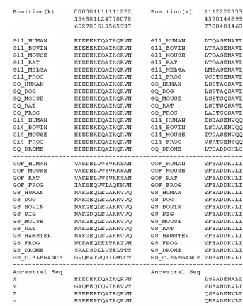
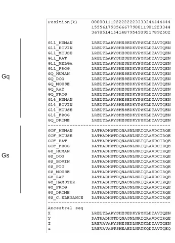
Fig. 3. Type-I functional divergence related amino acid sites candidates and ancestral sequence inference of these sites. (A): Category I: amino acids conserved in Gq cluster but variable in Gs cluster. (B): Category II: amino acids conserved in Gs cluster but variable in Gq cluster. X: The ancestral amino acids for the Gq cluster at the candidate sites. Y: The ancestral amino acids for the Gs cluster at the candidate sites. Z: The ancestral amino acids for the common ancestor of Gs and Gio cluster at the candidate sites. z: The ancestral amino acids for the common ancestor of the Gq and G12 cluster at the candidate sites.
Gq
Type-II functional divergence
Based on the same multiple alignment of protein sequences, we obtained the estimate of
the coefficient of Type-II functional divergence, θII = 0.325±0.055, between the Gs and
Gq alpha proteins, which is significantly larger than 0. It suggests that, after the gene
duplication, some amino acid residues that are evolutionarily conserved in both Gs and
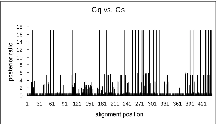
Gq proteins may have radical changes in their amino acid properties. Fig.4 shows the
site-specific profile based on the posterior ratio, RII (k), for Type-II functional divergence
between Gs and Gq proteins. Notably, most residues receive very low scores, indicating
that only a small portion of amino acid residues that have involved in this type of
functional divergence. 29 amino acid residues with the highest scores (the posterior ratio
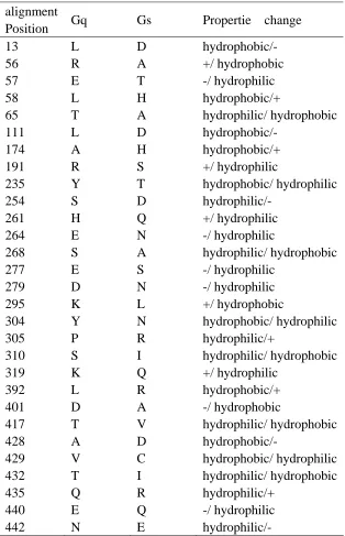
RII >17) show a typical shift of amino acid properties at conserved residues (Fig.5), as
been demonstrated in Table 1.
Gq vs. Gs
0 2 4 6 8 10 12 14 16 18
1 31 61 91 121 151 181 211 241 271 301 331 361 391 421
[image:21.595.93.445.472.673.2]alignment position p o s te ri o r ra ti o
Fig. 5. Type-II functional divergence related amino acid sites candidates and ancestral sequence inference of these sites. X, Y, Z, and z are the same definitions as in the Fig. 3.
Gq
Table 1. Overview of the amino acid changes in the predicted 29 sites in Type-II functional divergence.
alignment
Position Gq Gs Propertie change
13 L D hydrophobic/-
56 R A +/ hydrophobic
57 E T -/ hydrophilic
58 L H hydrophobic/+
65 T A hydrophilic/ hydrophobic
111 L D hydrophobic/- 174 A H hydrophobic/+
191 R S +/ hydrophilic
235 Y T hydrophobic/ hydrophilic
254 S D hydrophilic/-
261 H Q +/ hydrophilic
264 E N -/ hydrophilic
268 S A hydrophilic/ hydrophobic
277 E S -/ hydrophilic
279 D N -/ hydrophilic
295 K L +/ hydrophobic
304 Y N hydrophobic/ hydrophilic
305 P R hydrophilic/+
310 S I hydrophilic/ hydrophobic
319 K Q +/ hydrophilic
392 L R hydrophobic/+
401 D A -/ hydrophobic
417 T V hydrophilic/ hydrophobic
428 A D hydrophobic/-
429 V C hydrophobic/ hydrophilic
432 T I hydrophilic/ hydrophobic
435 Q R hydrophilic/+
440 E Q -/ hydrophilic
3.2 Evolutionary trends of functional divergence - Ancestral inference analysis
Using the Bayesian ancestral sequence inference implemented in the software
DIVERGE2, we inferred the ancestral sequences of all internal nodes on the phylogeny of
G-protein alpha subunits, which provides further information about the evolutionary
trends of functional divergence. Since our study is focused on the Gq and Gs clusters, we
are interested in the ancestor node for the Gs cluster, Gq cluster, the Gs and Gio clusters,
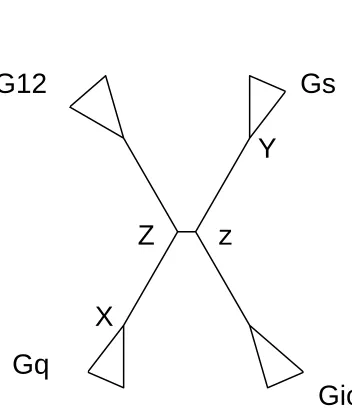
and the Gq and G12 clusters. All these four major internal nodes are represented in Fig. 6,
where the “X” stands for the ancestor for the Gq cluster, the “Y” stands for the ancestor
for the Gs cluster, the “Z” stands for the common ancestor for the Gs and Gio clusters,
and the “z” stands for the common ancestor for the Gq and G12 clusters. The whole
sequences for these four ancestors are concerned. In particular, the inferred ancestral
amino acid residues related to Type-I or Type-II functional divergence sites are presented
in Fig. 3 and Fig. 5, respectively, where X is the ancestral residues for the Gq cluster, Y is
the ancestral residues for the Gs cluster, Z is the common ancestral residues for the Gs
Fig. 6. The ancestral inference points for the G-protein alpha subunit family.
X: The ancestor for the Gq cluster. Y: The ancestor for the Gs cluster. Z: The common ancestor of Gs and Gio clusters. z: The common ancestor of the Gq and G12 clusters.
As shown in Fig. 3, there are two groups of Type-I functional divergence. Among fifteen
residues in Group A, i.e., conserved in Gq but variable in Gs, five residues (e.g., position
088) show the conserved Gq-type amino acid at all four major internal nodes (X, Y, Z,
and z), while six residues (e.g., position 016) show the conserved Gq-type amino acid in
three internal nodes but not at the Y (common ancestor of Gs). Putting these two ancestral
patterns together, it appears that the ancestral states of these eleven residues are all
conserved Gq-type, whereas the variable Gs-type residues are the derived characters that
are only specific to Gs-proteins. The only difference between these two ancestral patterns
is that Type-I functional divergence at the first five residues likely occurred after the
common ancestor of Gs (Y), while those at the second six residues occurred before the
z
Z
Y
X
Gio
Gq
node Y. On the other hand, among ten residues in Group B, i.e., conserved in Gs but
variable in Gq, two residues (positions 200 and 396) show the conserved Gs-type amino
acid at all four major internal nodes, while another two residues (positions 170 and 384)
show such pattern except for the common ancestor of Gq (X). Interestingly, for the two
residues at positions 214 and 398, the conserved Gs-type is recently derived, which is
specific to the Gs cluster. For the rest of residues, one can not determine the trend of
functional divergence, due to the statistical uncertainty of phylogenetic inference or
ancestral sequence inference.
In the same manner, we examined the ancestral amino acid residues for Type-II
functional divergence (Fig.5). Among twenty-nine predicted Type-II divergence related
residues, seventeen residues (e.g., position 013) show an ancestral patterns indicating
these amino acid property-shifts at conserved residues may occur in the evolutionary
trend that can be simply represented as from the internal nodes z (ancestral type) to Y
(Gs-type). In contrast, the ancestral pattern of four residues (e.g., position 065) indicates
the evolutionary trend of Type-II functional divergence from the internal nodes Z
(ancestral type) to X (Gq-type).
3.3 Protein structure mapping
Comparative study of molecular sequences and protein structures has provided many
insights into protein folding, stability and evolution (Golding and Dean, 1998). The
could provide a deep insight into the evolution trends. The crystal structures of G-proteins,
Gs and Gq, are both determined (Tesmer et al. 1997; Tesmer et al. 2005; Wall et al. 1995;
Lambright et al. 1996; Cabrera-Vera et al. 2003), which provide the structural basis to
investigate the functional interpretations of these Type-I and Type-II predicted residues.
The software DIVERGE2 we have developed is capable of mapping a subset of amino
acid residues onto the protein structure. It should be noted that some residues in either N
or C termini are not available or simply disordered in the solved protein structures
(Cabrera-Vera et al. 2003). Consequently, predicted Type-I and Type-II residues (sites) in
these regions have to be excluded in the sequence-structure study.
A G-protein alpha subunit contains two domains: a GTPase domain involved in the
binding and hydrolysis of GTP, and a helical domain connected to the GTPase domain by
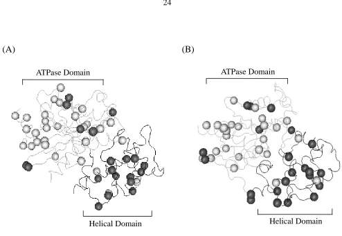
two linker regions (Cabrera-Vera et al. 2003). Fig. 7 shows the location of predicted
Type-I and Type-II functional divergence related sites on the protein 3D structures of
Gs_BOVIN and Gq_MOUSE. The mapping result suggests that the Type-I and Type-II
sites have different patterns of location distribution. Among twenty-five Type-I sites
predicted at the cutoff posterior ratio RI >2, there are thirteen sites in the helical domain
(in the range of PDB sites 86 - 202 in Gs, or PDB sites 70-185 in Gq), while the rest,
around half of the total, are located in the GTPase domain. It should be noted that some
predicted sites in N-terminal can not be mapped due to lacking of structure information as
mentioned in previous paragraph, therefore Fig. 7 only shows part of Type-I sites in the
predicted at the posterior ratio cutoff 17 (or posterior probability 0.94), there are only
three sites located in the helical domain, while twenty-six sites are in the GTPasedomain
(p<0.001, binomial test). Fig.7 shows the locations of twenty-eight sites out of the
predicted twenty-nine Type-II sites and reveals that most of the predicted Type-II sites
are located in the ATPase domain. Cabrera-Vera et al. (2003) mentioned that the helical
domain is the most divergentdomain among the G-protein alpha subunits, whereas the
GTPase domain is much more conserved. Our result indicates that the Type-II sites may
be involved in more conserved domain, implying the relevance of these sites to the
(A) (B)
Fig. 7. The mapping of the predicted Type-I and Type-II sites onto protein 3D structures
(A): The mapping of the predicted Type-I and Type-II sites onto the Gs-BOVIN protein structure (Chain C in the structure 1AZS). (B): The mapping of Type-I and Type-II sites onto the Gq-MOUSE structure (Chain Q in the structure 2BCJ). The dark ball represents the predicted Type-I functional divergence-related residues; the light ball represents the predicted Type-II functional divergence-related residues. The gray domain is ATPase domain of the G protein alpha subunit; the black domain is helical domain of the G-protein alpha subunit.
ATPase Domain
Helical Domain Helical Domain
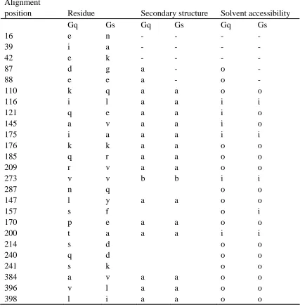
Moreover, we examined the distribution of Type-I and Type-II functional divergence
related sites in the secondary structure elements. Table 2 (A, and B) shows that most of
these sites are within the typical repeat elements of secondary structure such as alpha
helices, beta strands and 310 helices.
Besides the secondary structure distribution of these functional divergence-related sites,
we are concerned about whether they are located on the surface or in the interior of the
protein. To address this issue, we take the criteria of accessible surface area (ASA) to
define the surface area of a bimolecule accessible to a solvent (Lee and Richards 1971).
Residues are considered to be solvent exposed (on the surface, o for short) or be buried
(inside, i for short) according to the relative ASA in the protein. Overall, we found that
the Type-I and the Type-II sites may have the tendency to be located in the surface area
(Table 2 C), though the differences are not statistically significant (p-value >0.05).
Moreover, the accessibility of most Type-I sites, except for three sites(positions 121, 145
and 157), has no changes between Gq and Gs, while in the Type-II sites, eight sites are
found to have accessibility changes between the Gq and Gs proteins. Interestingly, there
Table 2. The structure features of the Type-I and Type-II functional divergence related residues in Gq and Gs: the secondary structure conformation and the solvent accessibility.
A: Type-I functional divergence related sites and their structure features.
Alignment
position Residue Secondary structure Solvent accessibility
Gq Gs Gq Gs Gq Gs
16 e n - - - -
39 i a - - - -
42 e k - - - -
87 d g a - o -
88 e e a - o -
110 k q a a o o
116 i l a a i i
121 q e a a i o
145 a v a a i o
175 i a a a i i
176 k k a a o o
185 q r a a o o
209 r v a a o o
273 v v b b i i
287 n q o o
147 l y a a o o
157 s f o i
170 p e a a o o
200 t a a a i i
214 s d o o
240 q d o o
241 s k o o
384 a v a a o o
396 v l a a o o
B: Type-II functional divergence related sites and their structure features.
In this table, the letter “a” stands for alpha helices, “b” stands for beta strands and “3” stands for 310 helices.
“i ”means solvent inaccessible and “o” means solvent accessible. “- ” means there is no residue structure information available.
Alignment
position Residue Second structure Solvent accessibility
Gq Gs Gq Gs Gq Gs
13 L D - - - -
56 R A o o
57 E T b b o o
58 L H b b o i
65 T A i o
111 L D a a o o
174 A H a a i o
191 R S 3 o o
235 Y T b b o o
254 S D 3 o o
261 H Q 3 3 o o
264 E N o o
268 S A b b i i
277 E S 3 3 i i
279 D N 3 3 i o
295 K L a a o o
304 Y N i o
305 P R 3 i o
310 S I i o
319 K Q a a o o
392 L R a a o o
401 D A o o
417 T V i i
428 A D a a o o
429 V C a a i i
432 T I a a o i
435 Q R a a o o
440 E Q - a - o
C: the comparison of the solvent accessibility of Type-I, Type-II functional divergence related residues to all the residues in the sequence.
Gs Gq
TypeI TypeII
Whole
sequence TypeI TypeII
Whole sequence
#sites 20 28 339 22 26 317
#Accessible sites 15 22 238 16 16 198
Accessible /total 0.75 0.79 0.70 0.73 0.62 0.62
The table only considered the residues with available structure information, i.e., some N
and C termini are not included. The p-value for testing the non-difference of the
accessibility between the functional divergence related (Type-I or Type-II) sites and the
functional divergence unrelated sites is large than 0.1 and reveals no significant difference
CHAPTER IV. CONCLUSIONS
Providing substantial genomic data, plus powerful computational tools, it is now desirable
to develop a comprehensive analytical pipeline to perform functional divergence analysis
of protein families. In this project, we use animal G-protein alpha subunit family as an
example to illustrate such an analytical pipeline. Advanced to our previous works, which
can only detect type-I functional divergence between duplication genes (or subfamilies) in
protein families and identify residues responsible for the functional divergence, this
approach includes detecting different types, say, type-I and type-II, of functional
divergence, as well as identifying the functional residues, and can be further combined
with ancestral sequence inference and available residue protein structure information.
By this approach, we are more than able to exam two types of functional divergences
between duplication genes. With the combination of functional divergence analysis and
the ancestral sequence inference, we are able to trace the evolutionary trend of two types
of functional divergence of amino acid residues after the gene duplication. With the
sequence-3D structure mapping we can get the structure features of the particular
functional divergence of amino acid residues, and explore the sequence-structure
relationship during the evolution. Clearly, these pieces of evolutionary information are
useful for making testable hypothesis about functional divergence between subtypes of
REFERENCES CITED
Berman HM, Westbrook J, Feng Z, Gilliland G, Bhat TN, Weissig H, Shindyalov IN, Bourne PE. 2000. The Protein Data Bank. Nucleic Acids Res 28(1):235-242.
Bork P, Koonin EV. 1998. Predicting functions from protein sequences--where are the bottlenecks? Nat Genet 18(4):313-318.
Cabrera-Vera TM, Vanhauwe J, Thomas TO, Medkova M, Preininger A, Mazzoni MR, Hamm HE. 2003. Insights into G protein structure, function, and regulation. Endocr Rev 24(6):765-781.
Dayhoff MO, Schwartz RM, Orcutt BC. 1978. A model of evolutionary change in proteins. In: Dayhoff, M. O. (ed.) Atlas of Protein Sequence and Structure, vol. 5, (suppl. 3),345-352. National Biomedical Research Foundation, Washington DC.
Doolittle RF. 1995. The origins and evolution of eukaryotic proteins. Philos Trans R Soc Lond B Biol Sci 349(1329):235-240.
Downes GB, Gautam N. 1999. The G protein subunit gene families. Genomics 62(3):544-552.
Eisen JA, Fraser CM. 2003. Phylogenomics: intersection of evolution and genomics. Science 300(5626):1706-1707.
Gao X, Vander Velden KA, Voytas DF, Gu X. 2005. SplitTester: software to identify domains responsible for functional divergence in protein family. BMC Bioinformatics 6:137.
Gaucher EA, Das UK, Miyamoto MM, Benner SA. 2002a. The crystal structure of eEF1A refines the functional predictions of an evolutionary analysis of rate changes among elongation factors. Mol Biol Evol 19(4):569-573.
Gaucher EA, Gu X, Miyamoto MM, Benner SA. 2002b. Predicting functional divergence in protein evolution by site-specific rate shifts. Trends Biochem Sci 27(6):315-321.
Golding GB, Dean AM. 1998. The structural basis of molecular adaptation. Mol Biol Evol 15(4):355-369.
Gu J, Wang Y, Gu X. 2002. Evolutionary analysis for functional divergence of Jak protein kinase domains and tissue-specific genes. J Mol Evol 54(6):725-733.
Gu X. 1999. Statistical methods for testing functional divergence after gene duplication. Mol Biol Evol 16(12):1664-1674.
Gu X. 2001. Maximum-likelihood approach for gene family evolution under functional divergence. Mol Biol Evol 18(4):453-464.
Gu X. 2006. A simple statistical method for estimating type-II (cluster-specific) functional divergence of protein sequences. Mol Biol Evol 23(10):1937-1945.
Gu X, Vander Velden K. 2002. DIVERGE: phylogeny-based analysis for functional-structural divergence of a protein family. Bioinformatics 18(3):500-501.
Henikoff S, Greene EA, Pietrokovski S, Bork P, Attwood TK, Hood L. 1997. Gene families: the taxonomy of protein paralogs and chimeras. Science 278(5338):609-614.
Hubbard, SJ, Thornton, JM. 1993. 'NACCESS', Computer Program, Department of Biochemistry and Molecular Biology, University College London.
Jones DT, Taylor WR, Thornton JM. 1992. A new approach to protein fold recognition. Nature 358(6381):86-89.
Jordan IK, Bishop GR, Gonzalez DS. 2001. Sequence and structural aspects of functional diversification in class I alpha-mannosidase evolution. Bioinformatics 17(10):965-976.
Kaziro Y, Itoh H, Kozasa T, Nakafuku M, Satoh T. 1991. Structure and function of signal-transducing GTP-binding proteins. Annu Rev Biochem 60:349-400.
Kimura, M. 1983. The Neutral Theory of Molecular Evolution. Cambridge University Press, Cambridge.
Knudsen B, Miyamoto MM. 2001. A likelihood ratio test for evolutionary rate shifts and functional divergence among proteins. Proc Natl Acad Sci U S A 98(25):14512-14517.
Lambright DG, Sondek J, Bohm A, Skiba NP, Hamm HE, Sigler PB. 1996. The 2.0 A crystal structure of a heterotrimeric G protein. Nature 379(6563):311-319.
Lee B, Richards FM. 1971. The interpretation of protein structures: estimation of static accessibility. J Mol Biol 55(3):379-400.
Li WH, Gu Z, Wang H, Nekrutenko A. 2001. Evolutionary analyses of the human genome. Nature 409(6822):847-849.
Lichtarge O, Bourne HR, Cohen FE. 1996. An evolutionary trace method defines binding surfaces common to protein families. J Mol Biol 257(2):342-358.
Lopez P, Forterre P, Philippe H. 1999. The root of the tree of life in the light of the covarion model. J Mol Evol 49(4):496-508.
Madabushi S, Gross AK, Philippi A, Meng EC, Wensel TG, Lichtarge O. 2004.
Evolutionary trace of G protein-coupled receptors reveals clusters of residues that determine global and class-specific functions. J Biol Chem 279(9):8126-8132.
Mizuguchi K, Deane CM, Blundell TL, Johnson MS, Overington JP. 1998. JOY: protein sequence-structure representation and analysis. Bioinformatics 14(7):617-623.
Neer EJ. 1995. Heterotrimeric G proteins: organizers of transmembrane signals. Cell 80(2):249-257.
Nei M. 1987. Molecular evolutionary genetics.Columbia University Press, New York. Ohno S. 1970. Evolution by gene duplication Springer-Verlag, Berlin.
Rastogi S, Liberles DA. 2005. Subfunctionalization of duplicated genes as a transition state to neofunctionalization. BMC Evol Biol 5(1):28.
Saitou N, Nei M. 1987. The neighbor-joining method: a new method for reconstructing phylogenetic trees. Mol Biol Evol 4(4):406-425.
Sidow A. 1996. Gen(om)e duplications in the evolution of early vertebrates. Curr Opin Genet Dev 6(6):715-722.
Simon MI, Strathmann MP, Gautam N. 1991. Diversity of G proteins in signal transduction. Science 252(5007):802-808.
Tesmer JJ, Sunahara RK, Gilman AG, Sprang SR. 1997. Crystal structure of the catalytic domains of adenylyl cyclase in a complex with Gsalpha.GTPgammaS. Science 278(5345):1907-1916.
activated G proteins at the membrane: the Galphaq-GRK2-Gbetagamma complex. Science 310(5754):1686-1690.
Wall MA, Coleman DE, Lee E, Iniguez-Lluhi JA, Posner BA, Gilman AG, Sprang SR. 1995. The structure of the G protein heterotrimer Gi alpha 1 beta 1 gamma 2. Cell 83(6):1047-1058.
Wang Y, Gu X. 2001. Functional divergence in the caspase gene family and altered functional constraints: statistical analysis and prediction. Genetics 158(3):1311-1320.
Yang Z, Kumar S, Nei M. 1995. A new method of inference of ancestral nucleotide and amino acid sequences. Genetics 141(4):1641-1650.
Zhang J, Nei M. 1997. Accuracies of ancestral amino acid sequences inferred by the parsimony, likelihood, and distance methods. J Mol Evol 44 Suppl 1:S139-146.
ACKNOWLEDGEMENTS
I would first like to thank my major professor, Dr. Xun Gu, for being my mentor,
supporting me, offering me great guidance on my research projects, and always giving me
encouragements during my graduate studies.
I would also like to thank my co-major professor, Dr. Karin Dorman, for kindly
discussing with me and providing great suggestions on both my research projects and
graduated studies. I must thank Dr. Xiaoqiu Huang, my committee professor, your
guidance and help was very much appreciated. Thank you all for being on my committee.
Next, I would give special thanks to Dongping Xu for developing the DIVEGE2 software
and giving me valuable suggestions on the usage of software and analysis of the data.
Special thanks to Dr. Jianying Gu for introducing me into the research area of gene
duplication functional divergence.
Then, I would like to thank all my former and current labmates, Dr. Zhongqi Zhang, Dr.
Shiquan Wu, Dr. Zhixi Su and Yong Huang. I will forever enjoy our deep friendship and
appreciate your help during my studies.
Last, I would like to thank my dear parents and husband. It is with their supports and