Open Access
Vol 11 No 6Research article
A selective eradication of human nonhereditary breast cancer
cells by phenanthridine-derived polyADP-ribose polymerase
inhibitors
Dana Inbar-Rozensal
1, Asher Castiel
2, Leonid Visochek
1, David Castel
1, Françoise Dantzer
3,
Shai Izraeli
2and Malka Cohen-Armon
11The Neufeld Cardiac Research Institute and Dept. of Physiology and Pharmacology, Sackler School of Medicine, Tel-Aviv University, Tel-Aviv 69978, Israel
2Cancer Research Institute, Sheba Medical Center, Tel-Hashomer, Ramat-Gan 52621, Israel
3Laboratory of Molecular and Structural Biology, Ecole Superieure de Biotechnologie de Strasbourg, F-67400, Illkrich-Graffenstaden, France
Corresponding author: Malka Cohen-Armon, [email protected]
Received: 19 Feb 2009 Revisions requested: 6 Apr 2009 Revisions received: 28 Jul 2009 Published: 9 Nov 2009
Breast Cancer Research 2009, 11:R78 (doi:10.1186/bcr2445)
This article is online at: http://breast-cancer-research.com/content/11/6/R78 © 2009 Inbar-Rozensal; licensee BioMed Central Ltd.
This is an open access article distributed under the terms of the Creative Commons Attribution License (http://creativecommons.org/licenses/by/2.0), which permits unrestricted use, distribution, and reproduction in any medium provided the original work is properly cited.
Abstract
Introduction PARP-1 (polyADP-ribose polymerase-1) is known to be activated in response to DNA damage, and activated PARP-1 promotes DNA repair. However, a recently disclosed alternative mechanism of PARP-1 activation by phosphorylated externally regulated kinase (ERK) implicates PARP-1 in a vast number of signal-transduction networks in the cell. Here, PARP-1 activation was examined for its possible effects on cell proliferation in both normal and malignant cells.
Methods In vitro (cell cultures) and in vivo (xenotransplants) experiments were performed.
Results Phenanthridine-derived PARP inhibitors interfered with cell proliferation by causing G2/M arrest in both normal (human
epithelial cells MCF10A and mouse embryonic fibroblasts) and human breast cancer cells MCF-7 and MDA231. However, whereas the normal cells were only transiently arrested, G2/M
arrest in the malignant breast cancer cells was permanent and was accompanied by a massive cell death. In accordance, treatment with a phenanthridine-derived PARP inhibitor prevented the development of MCF-7 and MDA231 xenotransplants in female nude mice. Quiescent cells (neurons and cardiomyocytes) are not impaired by these PARP inhibitors.
Conclusions These results outline a new therapeutic approach for a selective eradication of abundant nonhereditary human breast cancers.
Introduction
PolyADP-ribose polymerases (PARPs) catalyze a posttransla-tional and energy-consuming modification of proteins by poly-ribosylation. This enzymatic reaction is initiated by ADP-ribose transferase activity, which proceeds with polymerization of ADP-riboses into long and branched polymers [1]. In the chromatin, polyADP-ribosylation apparently regulates the interaction of PARPs and their substrates with protein part-ners and DNA. PARP-1 is a highly conserved DNA-binding protein and the most abundant nuclear PARP. The enzyme is known to be activated in response to DNA single-strand breaks [1], and its activation induces chromatin remodeling,
rendering the DNA more accessible to transcription factors and repair enzymes [1,2].
Our recent findings in quiescent cells and in cell-free systems disclosed an alternative mechanism, inducing an intense and long-lasting activation of PARP-1 in the absence of DNA dam-age [3,4]. In this process, PARP-1 interaction with phosphor-ylated ERK2 (externally regulated kinase) resulted in PARP-1 activation and polyADP-ribosylation in a positive-feedback mechanism that kept PARP-1 polyADP-ribosylated as long as ERK2 was phosphorylated [3]. In addition, polyADP-ribo-sylated PARP-1 highly augmented the activity of
phosphorylated ERK, enhancing phosphorylation of ERK-tar-geted transcription factors, core histone acetylation, and the expression of ERK target genes, some of which are onco-genes [3-6]. Because ERK activity in the nucleus is a key mod-ulator for inducing proliferation versus differentiation in a variety of cancer cells [7,8], these findings suggest that PARP-1 activation might be a possible target for mechanisms induc-ing cell proliferation.
PARP inhibitors were designed to prevent PARP-1 activation in response to nicked DNA, in an attempt to suppress PARP mediated DNA repair [9-12]. Several generations of PARP inhibitors were designed to prevent PARP-1 activity by block-ing the bindblock-ing of the nicotinamide moiety of NAD+ in the
cat-alytic site of the enzyme. PARP inhibitors differ in their chemical structure, their potency, stability, solubility in water, and apparently even in their therapeutic potential [9-12]. Sev-eral groups of PARP inhibitors (including phenanthridine deriv-atives) were designed to protect cells under stress conditions from cell death induced by a massive activation of PARP-1 (for example, stroke, inflammation; [10,12]), or to cause cell death in malignant cells by preventing polyADP-ribosylation-depend-ent DNA repair [9,11]. In accordance with this concept, PARP inhibitors were tested for their therapeutic potential in malig-nant cells with impaired DNA-repair machinery [13,14] (bear-ing mutations in the tumor-suppressor genes BRCA1 and
BRCA2 that cause an impaired DNA repair [15]) or in combi-nation with DNA-damaging treatments [11]. However, in view of findings indicating that activated PARP-1 highly augments the activity of ERK in the nucleus even in the absence of DNA damage [3,4], a different therapeutic potential of PARP inhib-itors is examined in breast cancer cells lacking BRCA mutations.
Materials and methods
Human breast cancer cell line MCF-7 and MDA231 and human epithelial cells MCF-10A were supplied by ATCC Co. (American Type Culture Collection, P.O. Box 1549, Manas-sas, VA 20108, USA. The dealer in Israel is Almog Diagnostic & Medical equipment Ltd.). Mouse embryonic fibroblasts were prepared in the laboratory of Dr. Dantzer (Strasbourg, France).
MCF-7 and MDA231 cells were cultured in six-well multidish plates (Nunc, Thermo Fisher scientific, Reskilde, Denmark). MCF-7 and MDA231 cells were maintained in a medium con-taining DMEM (cat. 01055-1A), 10% Fetal bovine serum (FBS; cat. 04-124-1A), 1% L-glutamine (cat. 03-020-1B), and 1% Pen-Strep Ampho (cat. 03-033-1B) (Gibco, purchased from Rhenium, Rehovot, Israel).
MCF-10A human epithelial cells were cultured in DMEM/F12 (Gibco) with FBS (Gibco) 6%, EGF (100 μg/ml, Cytolab, Rehovot, Israel) 0.02%, hydrocortisone (50 μM, Sigma) 2.8%, insulin (10 mg/ml, Sigma) 0.1%, and Pen/Strep (Gibco) 1%.
Mouse embryonic fibroblasts were cultured in a medium con-taining DMEM (cat. 01055-1A), 10% FBS (cat. 04-121-1A), 1% L-glutamine (cat. 03-020-1B), and 1% Pen-Strep Ampho (cat. 03-033-1B) (Gibco, purchased from Rhenium, Rehovot, Israel).
Phenanthridine-derived PARP inhibitors
We examined the effect of phenanthridine-derived com-pounds that act as potent PARP inhibitors. These included PJ-34, (N-(6-oxo-5,6-dihydrophenanthridin-2-yl)-N, N -dimethyl-acetamide), Tiq-A, (4H-thieno [2,3-c]isoquinolin-5-one) and Phen, (6(5H) phenanthridinone). PJ-34 and Phen were pur-chased from Alexis Biochemicals (Alexis Corporation, Lausanne, Switzerland). Tiq-A was purchased from Sigma, Israel. Their chemical structure is presented in Figure 1.
The effect of PJ-34 on the development of MCF-7 and MDA231 xenotransplants
Xenotransplants were developed in female CD-1 nu/nu mice 5 to 6 weeks old (Charles River Labs, Sulzfeld, Germany) pur-chased in Israel from Harlan Labs, Jerusalem. MCF-7 and MDA231 cells were injected subcutaneously, about 107
MCF-7 or MDA231 cells in 150 μl of PBS and 150 μl of Matrigel Basement Membrane Matrix (Becton Dickinson, Bedford, MA, USA; In Israel, Bactolab Diagnostics). In mice treated with PJ-34, Injection was adjacent to subcutaneous osmotic pumps dripping PJ-34 by a slow release. The mice were maintained under specific pathogen-free conditions with access to mouse chow and water ad libitum. PJ-34 (2 mM dissolved in 100 μl PBS) was inserted in subcutaneously implanted Alzet osmotic pumps, designed to release PJ-34 continuously (at about 0.6 nmol/h) for 14 days. For comparison, in the in vitro experi-ments, the amount of PJ-34 per dish was approximately 20 nmol. Subcutaneous implantation of these pumps was per-formed before injection by a veterinarian (Dr. Kastel David). All the experiments with nude mice conform with the Guide for the Care and Use of Laboratory Animals published by the NIH (publication No. 85-23, revised 1996). Approval was granted by the Israeli Ministry of Health ethics review board in the Tel-Aviv University (M08033).
Figure 1
The chemical structures of the PARP inhibitors PJ-34, Tiq-A, and Phen
Cell survival in cells treated with PJ-34
MCF-7 or MDA231 cells (seeded about 500,000 cells/3-mm well in six-well plates) were treated with PJ-34 applied only once, 24 hours after seeding. In some experiments, cells were reseeded in PJ-34-free medium in 10-cm plates for colony for-mation. In these experiments, after 2 weeks of incubation with-out application of PJ-34, cells were fixed (methanol/acetic acid, 3:1), stained with crystal violet, and counted to determine cell survival.
Flow cytometry was used to monitor changes in the ploidy level of malignant and normal cells labeled with propidium Iodide (PI) staining. Counting the cells with flow cytometry in selected time periods for several hours, indicated changes in their cell cycle caused by treatment with PJ-34. Flow cytome-try was performed by using a Becton Dickenson machine and the FlowJo software (Tree Star, Ashland, OR, USA). Untreated cells were used as controls for each cell type.
Results
Phenanthridine-derived PARP inhibitors efficiently eradicated MCF-7 and MDA231 breast cancer cells without impairing human epithelial MCF-10A or mouse embryonic fibroblasts
We examined the effect of PARP inhibitors on MCF-7 and MDA231 human breast cancer cells. Cells were treated with the potent PARP inhibitors, PJ-34, Tiq-A, and Phen (Figure 1) applied only once, 24 hours after seeding. MCF-7 cells did not survive after 48 to 72 hours of incubation with 10 μM PJ-34 (Figure 2a), nor after incubation with Tiq-A (100 μM) or Phen (50 μM) (Figure 2a). At these concentrations, PJ-34, Tiq-A, and Phen inhibit the activity of PARP-1 [3,10,16,17]. Signifi-cant cell death was observed even at lower concentrations of PJ-34 (Figure 2b). More than 99% of MCF-7 cells were erad-icated after 48 hours of incubation with 10 μM PJ-34 (Figure 2b). The damage was irreversible. No recovery was observed in MCF-7 cells treated with PJ-34 for 48 to 72 hours and then reseeded in PJ-34-free medium and incubated for 2 additional weeks in the absence of PJ-34 (Figure 2c). Massive cell death also was observed in MDA231 incubated for 72 to 96 hours with PJ-34 applied only once, 24 hours after seeding. These cells were completely eradicated by incubation with 20-30 μM PJ-34 (Figure 2d). No recovery was observed in MDA231 cells incubated with 30 μM PJ-34 for 72 hours, after reseeding in PJ-34-free medium and incubation for 2 additional weeks.
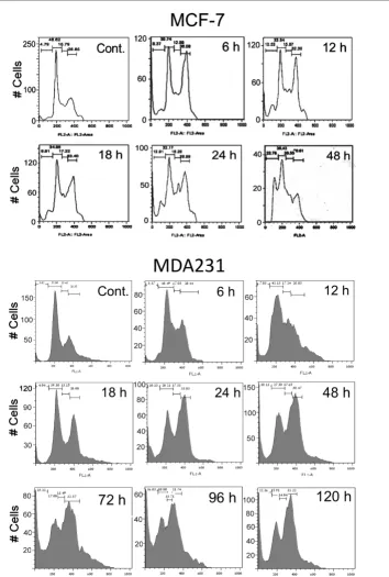
Flow cytometry disclosed G2/M arrest and cell death in both MCF-7 and MDA231 cells. G2/M arrest was already observed
in both cell types 6 hours after treatment with 10 μM PJ-34. It was not relieved within 120 hours of the experiment and was accompanied by massive cell death (Figure 3).
Normal dividing cells, human epithelial cells MCF-10A (Figure 4a), were similarly arrested at G2/M (Figure 4b). Their arrest
also was detected 6 hours after application of PJ-34 (10 μM).
However, unlike the malignant cells, MCF10A cells were only temporarily arrested (no arrest observed after 18 hours of incubation with PJ-34), and this transient arrest was not accompanied by cell death (Figure 4a and 4b). MCF-10A cells overcame the cell-cycle arrest, and continued to proliferate as normal cells, even when incubated with the same concentra-tions of PJ-34 and for the same duraconcentra-tions used to eradicate MDA231 cells (compare Figures 2d and 4a). Also, prolifera-tion of MCF-10A cells was not significantly reduced, even after a long incubation of 14 days with 10 μM PJ-34 (Figure 4a).
G2/M cell-cycle arrest also was detected in mouse embryonic fibroblasts (Figure 5a) after 6 hours of incubation with PJ-34 (10 μM) (Figure 5b). These cells also overcame the cell-cycle arrest, and the arrest in cell cycle was not accompanied by cell death (Figure 5b).
Thus, treatment with PJ-34 induced a transient G2/M arrest in these normal proliferating cells, which was not accompanied by cell death (Figures 4 and 5), whereas the cell cycle of malig-nant cells MCF-7 and MDA231 was permanently arrested, and these cells were eradicated by incubation with PJ-34 applied only once 24 hours after seeding (Figures 2 and 3). An efficient eradication of MCF-7 cells was observed after 48 hours of incubation with 10 μM PJ-34, whereas MDA231 cells were massively eradicated only after 72 hours of incubation with PJ-34, 20-30 μM. Quiescent cells, brain cortical neurons, and cardiomyocytes were not impaired by incubation with the examined phenanthridine-derived PARP inhibitors (10 to 20
μM PJ-34, 100 μM Tiq-A, and 50 μM Phen [3,10,16,17]).
PJ-34 prevented the development of MCF-7 and MDA231 xenotransplants in nude female mice
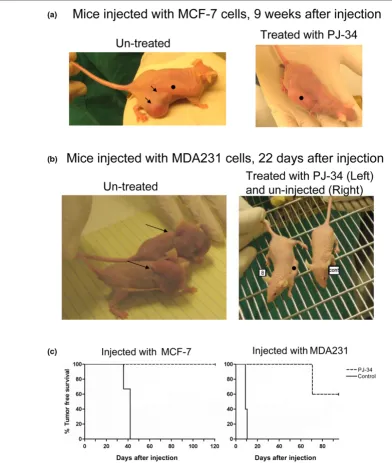
In vivo experiments were carried out in nude female mice (nu/ nu) injected subcutaneously with MCF-7 or MDA231 cells (Figure 6 and Additional data file 1). To test the effect of PJ-34 on the development of xenotransplants in the injected mice, PJ-34 (2 mM dissolved in 100 μl PBS) was inserted into sub-cutaneously implanted osmotic pumps (Alzet) that enable its constant slow release for 14 days (Methods). In the control nude mice, pumps contained only PBS, or pumps were not implanted. On the next day, 24 hours after pump implantation, each mouse was injected subcutaneously with approximately 107 MCF-7 or MDA231 cells dispersed in Matrigel (Methods).
Figure 2
Eradication of MCF-7 and MDA231 human breast cancer cells by phenanthridine-derived PARP inhibitors
Figure 3
G2/M arrest and cell death in MCF-7 and MDA231 treated with PJ-34
Figure 4
PJ-34 did not eradicate normal human epithelial cells MCF-10A
Figure 5
PJ-34 did not eradicate mouse embryonic fibroblasts
Figure 6
Treatment with PJ-34 prevented the development of MCF-7 and MDA231 xenotransplants
After 10 weeks, we detected tumors in two of the five mice that were injected with MDA231 cells and treated with PJ-34. These tumors were of human origin, as indicated by histo-chemistry (labeling with mouse human mitochondria anti-body (Millipore/Biotest) applied after blocking ("mouse-on-mouse"; Vector Labs/Zotal)).
Tumor-free survival curves [18] for mice injected with MCF-7 cells and for mice injected with MDA231 cells are presented in Figure 6c. The effect of treatment with PJ-34 on tumor-free survival is indicated, and significance was calculated with the log-rank significance test [18].
Discussion
Findings indicating that polyADP-ribosylation is required for spindle formation [19] are in accordance with the observed G2/M arrest in both normal and malignant dividing cells treated
with the potent PARP inhibitor PJ-34 (Figures 3, 4b, 5b). It is puzzling how normal cells overcome the G2 arrest while
malig-nant cells die.
G2 arrest could be induced by DNA damage in proliferating
cells [20]. Because of their genomic instability and fast prolif-eration, malignant cells might be more susceptible to inhibition of PARP mediated DNA repair than are normal proliferating cells [13,14]. However, eradication of MCF-7 and MDA231 cells was not shared by other non- phenanthridine-derived PARP inhibitors, and the examined cell lines MCF-7 and MDA231 were neither BRCA-deficient cells [21] nor PTEN
(phosphatase and tensin homologue)-deficient cells [22], in which DNA-repair mechanisms are impaired [15,23].
Other possible mechanisms underlying G2/M arrest implicate
signal-transduction pathways involving p53, p21/WAF1, cdc25c, cdc2, and suppressed cyclin-B gene expression [20,24-26]. Stabilization of p21 could promote G2 arrest and induce cell death [25]. ERK activation also is implicated in G2/ M transition control. Nuclear translocation of cyclin B/Cdc2 complex, which is required for G2/M transition, could be medi-ated by ERK activation [20]. Other mechanisms involving ERK activation in G2/M transition control are not fully resolved. They
include autostabilization of the p21cip/cyclin D1 complex leading to cell-cycle arrest, and mechanisms promoting degra-dation of cyclin-dependent inhibitors such as p27kip1 [26-31]. Various impairments in some of these regulatory mecha-nisms were detected in human cancer cell types.
Numerous primary human tumors and derived cell lines display a constitutive activation of ERK [8,31-34]. Some of them har-bor mutations of the Ras protein that render it and the Raf-MEK-ERK cascade constitutively active [35-37]. Constitutively activated Raf has been found in many human cancers [35-37]. ERK targets are constitutively activated in B-Raf melanomas, colon cancer, PC-3 prostate cancer, and pancreatic cancer cells [37-39]. Growth and proliferation of the human breast
cancer MCF-7 and MDA231 cells are also highly dependent on ERK activity [40-42].
Blocking the activity of ERK by blocking the Ras/Raf/MEK/ ERK pathway is one of the main targets for human cancer treatment. However, previous clinical studies showed an insuf-ficient antitumor activity of MEK inhibitors [39], suggesting that the MEK/ERK phosphorylation pathway is resilient to dim-inution of the activity of MEK and can adapt rapidly to maintain nearly normal ERK activation [39].
The recently disclosed augmentation of ERK activity in the nucleus by polyADP-ribosylated PARP-1 [3], and the fact that PARP-1 silencing with targeted siRNA downregulated ERK phosphorylation in nuclei of both MCF-7 and MDA231 cells (not shown), urged us to examine PARP inhibitors for their possible effect on cell proliferation in the absence of DNA-damaging factors. However, whereas phenanthridine-derived PARP inhibitors efficiently arrested the cell proliferation of MCF-7 and MDA231 cancer cells, other potent PARP inhibi-tors did not cause a similar effect. PARP inhibitor, ABT-888 (benzimidazole-4-carboxamide) hardly affected MCF-7 and MDA231 cells (not shown), and potent PARP inhibitors effi-ciently eradicating BRCA mutated breast cancer cells did not similarly eradicate MCF-7 cells [13,14] and cancer cells not carrying BRCA mutations [43]. Hence, additional features of the phenanthridine-derived PARP inhibitors apparently under-lie their very promising potency to arrest proliferation and cause cell death in cancer cells lacking mutations that impair DNA repair, without impairing normal proliferating or quiescent cells.
Conclusions
Phenanthridine-derived PARP inhibitors possess a promising potency to arrest proliferation (G2/M arrest) and cause cell
death in MCF-7 and MDA231 human breast cancer cells, without impairing normal proliferating human epithelial cells, fibroblasts, or quiescent cells (neurons, cardiomyocytes).
Competing interests
The authors declare that they have no competing interests. This study was not supported by any company or commercial fund.
MC-A is the inventor of patent WO 2009/0477052, submit-ted by Tel-Aviv University.
Authors' contributions
Additional files
Acknowledgements
This work was supported by The Israeli Science Foundation and the Israeli Ministry of Health, grants of Prof. M. Cohen-Armon.
References
1. Schreiber V, Dantzer FJ-C, Ame de Murcia G: PolyADP-ribose: novel functions for an old molecule. Nat Rev Mol Cell Biol
2006, 7:517-528.
2. Kraus WL: Transcriptional control by PARP-1:chromatin modu-lation, enhancer-binding, coregumodu-lation, and insulation. Curr Opin Cell Biol 2008, 20:294-302.
3. Cohen-Armon M, Visochek L, Rozensal D, Kalal A, Geistrikh I, Klein R, Bendetz-Nezer S, Yao Z, Seger R: DNA-independent PARP-1 activation by phosphorylated ERK2increases Elk1 activity: a link to histone acetylation. Mol Cell 2007, 25:297-308. 4. Cohen-Armon M: PARP-1 activation in the ERK signaling
pathway. Trends Pharmacol Sci 2007, 28:556-560.
5. Cohen-Armon M: PARP-1 activation mediates the expression of immediate early genes implicated in long-term memory formation. FEBS J 2008, 275(Suppl 1):92.
6. Carbone M, Rossi MN, Cavaldesi M, Notari A, Amati P, Maione R: Poly(ADP-ribosyl)ation is implicated in the G0-G1 transition of resting cells. Oncogene 2008, 27:6083-6092.
7. Buchwalter G, Gross C, Wasylyk B: Ets ternary complex tran-scription factors. Gene 2004, 324:1-14.
8. Hoshino R, Chantani Y, Yamori T, Tsuruo T, Oka H, Yoshida O, Shimada Y, Ari-i S, Wada H, Fujimoto J, Kohno M: Constitutive activation of the 41/42-kDa mitogen-activated protein kinase signaling pathway in human tumors. Oncogene 1999, 18:813-822.
9. Ratnam K, Low JA: Current development of clinical inhibitors of polyADP-ribose polymerase in oncology. Clin Cancer Res
2007, 13:1383-1388.
10. Chiarugi A, Meli E, Calvani M, Picca R, Picca R, Baronti R, Camaioni E, Costantino G, Marinozzi M, Pellegrini-Giampietro DE, Pellicciari R, Moroni F: Novel isoquinolinone-derived inhibitors of polyADP-ribose polymerase-1: pharmacological characteri-zation and neuroprotective effects in an in vitro model of cer-ebral ischemia. J Pharmacol Exp Ther 2003, 305:943-949. 11. Peralta-Leal A, Rodríguez-Vargas JM, Aguilar-Quesada R,
Rodrígu-ez MI, Linares JL, de Almodóvar MR, Oliver JF: PARP inhibitors: new partners in the therapy of cancer and inflammatory diseases. Free Radic Biol Med 2009, 47:13-26.
12. Jagtap P, Szabó C: Poly(ADP-ribose) polymerase and the ther-apeutic effects of its inhibitors. Nat Rev Drug Disc 2005, 4:421-440.
13. Bryant HE, Schultz N, Thomas HD, Parker KM, Flower D, Lopez E, Kyle S, Meuth M, Curtin NJ, Helleday T: Specific killing of BRCA2-deficient tumours with inhibitors of poly(ADP-ribose) polymerase. Nature 2005, 434:913-917.
14. Farmer H, McCabe N, Lord CJ, Tutt ANJ, Johnson DA, Richardson TB, Santarosa M, Dillon KJ, Hickson I, Knights C, Martin NM, Jack-son SP, Smith GC, Ashworth A: Targeting the DNA repair defect in BRCA mutant cells as a therapeutic strategy. Nature 2005, 434:917-921.
15. Grant SG, Das R, Cerceo CM, Rubinstein WS, Latimer JJ: Ele-vated levels of somatic mutation in a manifesting BRCA1 mutation carrier. Pathol Oncol Res 2007, 13:276-283. 16. Cohen-Armon M, Visochek L, Katzoff A, Levitan D, Susswein AJ,
Klein R, Valbrun M, Schwartz JH: Long-term memory requires polyADP-ribosylation. Science 2004, 304:1820-1823. 17. Visochek L, Vulih I, Steingart RA, Klein R, Priel E, Gozes I,
Cohen-Armon M: PolyADP-ribosylation is involved in neurotrophic activity. J Neurosci 2005, 25:7420-7428.
18. Harrington D: Linear rank tests in survival analysis. In Encyclo-pedia of biostatistics New York: Wiley Interscience; 2005. DOI: 10.1002/0470011815.b2a11047
19. Chang P, Jacobson MK, Mitchison TJ: Poly(ADP-ribose)is required for spindle assembly and structure. Nature 2004, 432:645-649.
20. Stark GR, Taylor WR: Control of the G2/M transition. Mol Biotechnol. 2006, 32:227-248.
21. Fustier P, Le Corre L, Chalabi N, Vissac-Sabatier C, Communal Y, Bignon Y-J, Bernard-Gallon DJ: Resveratrol increases BRCA1 and BRCA2 mRNA expression in breast tumour cell lines. Br J Cancer 2003, 89:168-172.
22. Yu K, Toral-Barza L, Discafani C, Zhang W-G, Skotnicki J, Frost P, Gibbons JJ: mTOR, a novel target in breast cancer: the effect of CCI-779, an mTOR inhibitor, in preclinical models of breast cancer. Endocr Relat Cancer 2001, 8:249-258.
23. Mendes-Pereira AM, Martin SA, Brough R, McCarthy A, Taylor JR, Kim J-S, Waldman T, Lord CJ, Ashworth A: Synthetic lethal tar-geting of PTEN mutant cells with PARP inhibitors. EMBO Mol Med. 2009, 1:315-322.
24. Yoshida M, Matsui Y, Iizuka A, Ikarashi Y: G2-phase arrest through p21(WAF1/Cip1) induction and cdc2 repression by gnidimacrin in human hepatoma HLE cells. Anticancer Res
2009, 29:1349-1354.
25. Abbas T, Dutta A: p21 in cancer: intricate networks and multiple Activities. Nat Rev Cancer 2009, 9:400-414.
26. Nigam N, Prasad S, George J, Shukla Y: Lupeol induces p53 and cyclin-B-mediated G2/M arrest and targets apoptosis through activation of caspase in mouse skin. Biochem Biophys Res Commun 2009, 381:253-258.
27. Aaltonen K, Amini R-M, Heikkilä P, Aittomäki K, Tamminen A, Nevanlinna H, Blomqvist C: High cyclin B1 expression is asso-ciated with poor survival in breast cancer. Br J Cancer 2009, 100:1055-1060.
28. Chambard J-C, Lefloch R, Pouyssegur J, Lenormand P: ERK impli-cation in cell cycle regulation. Biochim Biophys Acta 2007, 1773:1299-1310.
29. Whitmarsh AJ, Davis RJ: Transcription factor AP-1 regulation by mitogen-activated protein kinase signal transduction pathways. J Mol Med 1996, 74:589-607.
30. Shaulian E, Karin M: AP-1 in cell proliferation and survival.
Oncogene 2001, 20:2390-2400.
31. Milde-Langosch K: The Fos family of transcription factors and their role in tumourigenesis. Eur J Cancer 2005, 41:2449-2461.
32. Brunet A, Roux D, Lenomand P, Dowd S, Keyse S, Pouyssegur J: Nuclear translocation of p42/p44 mitogen-activated protein kinase is required for growth factor-induced gene expression and cell cycle entry. EMBO J 1999, 18:664-674.
33. Chalmers CJ, Gilly R, March HN, Balmanno K, Cook SJ: The dura-tion of ERK1/2 activity determines the activadura-tion of c-Fos and Fra-1 and the composition and quantitative transcriptional out-put of AP-1. Cell Signal. 2007, 19:695-704.
34. Balmanno K, Cook SJ: Sustained MAP kinase activation is required for the expression of cyclin D1, p21 and a subset of AP-1 proteins in CCL39 cells. Oncogene 1999, 18:3085-3097. 35. Davies H, Bignell GR, Cox C, Stephens P, Edkins S, Clegg S, Teague J, Woffendin H, Garnett MJ, Bottomley W, Davis N, Dicks E, Ewing R, Floyd Y, Gray K, Hall S, Hawes R, Hughes J, Kosmidou V, Menzies A, Mould C, Parker A, Stevens C, Watt S, Hooper S, Wilson R, Jayatilake H, Gusterson BA, Cooper C, Shipley J, et al.: Mutations of the B-RAF gene in human cancer. Nature 2002, 417:949-954.
36. Karasarides M, Chiloeches A, Hayward R, Niculescu-Duvaz D, Scanlon I, Friedlos F, Ogilvie L, Hedley D, Martin J, Marshall CJ, Springer CJ, Marais R: B-RAF is a therapeutic target in melanoma. Oncogene 2004, 23:6292-6298.
The following Additional files are available online:
Additional file 1
The following additional data are available with the online version of this article: A documented experiment, testing PJ-34 treatment in nude mice injected with MDA231 breast cancer cells (Additional data file 1).
37. Mercer KE, Pritchard CA: Raf proteins and cancer: B-Raf is identified as a mutational target. Biochim Biophys Acta 2003, 1653:25-40.
38. Tan X, Egami H, Abe M, Nazawa F, Hirota M, Ogawa M: Involve-ment of MMP-7 in invasion of pancreatic cancer cells through activation of the EGFR mediated MEK-ERK signal transduction pathway. J Clin Pathol 2005, 58:1242-1248.
39. Rinehart J, Adjei AA, LoRusso PMD, Hecht RJ, Natale RB, Hamid O, Varterasian M, Asbury P, Kaldjian EP, Gulyas S, Mitchell DY, Herrera R, Sebolt-Leopold JS, Meyer MB: Multicenter phase II study of the oral MEK inhibitor, CI-1040, in patients with advanced non-small-cell lung, breast, colon, and pancreatic cancer. J Clin Oncol 2004, 22:4456-4462.
40. Duan R, Xie W, Burghardt RC, Safe S: Estrogen receptor-medi-ated activation of the serum response element in MCF-7 cells through MAPK-dependent phosphorylation of Elk1. J Biol Chem 2001, 276:11590-11598.
41. Lu C, Shen Q, DuPre E, Kim H, Hilsenbeck S, Brown PH: cFos is critical for MCF-7 breast cancer cell growth. Oncogene 2005, 24:6516-6524.
42. Camirand A, Zakikhani M, Young F, Pollak M: Inhibition of insulin-like growth factor-1 receptor signaling enhances growth-inhibitory and proapoptotic effects of gefitinib (Iressa) in human breast cancer cells. Breast Cancer Res 2005, 7:R570-R579.