STUDY OF PREVALENCE OF THYROID
DYSFUNCTION IN SEROPOSITIVE HIV PATIENTS
Dissertation submitted for
DOCTOR OF MEDICINE
BRANCH - I (GENERAL MEDICINE)
APRIL 2015
THE TAMILNADU
BONAFIDE CERTIFICATE
This is to certify that the dissertation titled “A STUDY ON
PREVALANCE OF THYROID DYSFUNCTION IN SEROPOSITIVE
HIV PATIENTS” submitted by Dr. A. PRABHU to the Tamil Nadu Dr.
M.G.R. Medical University, Chennai in partial fulfilment of the requirement for
the award of M.D Degree Branch I (General Medicine) is a bonafide research
work and it was carried out by him under my direct supervision & guidance.
Dr. S. VADIVEL MURUGAN M.D., Dr. J. SANGUMANI M.D.,
Professor and HOD, Unit Chief,
Dept. of General Medicine, Dept. of General Medicine,
Government Rajaji Hospital, Government Rajaji Hospital,
Madurai Medical College, Madurai Medical College,
Madurai. Madurai.
CERTIFICATE FROM THE DEAN
This is to certify that this dissertation titled “A STUDY ON
PREVALANCE OF THYROID DYSFUNCTION IN SEROPOSITIVE
HIV PATIENTS” is the bonafide work of DR A. PRABHU, in partial
fulfilment of the university regulations of the Tamil Nadu Dr. M.G.R. Medical
University, Chennai, for M.D General Medicine Branch I examination to be
held in April 2015.
Captain.Dr.B.SANTHAKUMAR, M.Sc(F.Sc),
M.D(F.M). PGDMLE., Dip.N.B (F.M).,
The Dean,
Madurai Medical College and
Government Rajaji Hospital,
DECLARATION
I, Dr. A. PRABHU solemnly declare that, I carried out this dissertation
titled, “A STUDY ON PREVALANCE OF THYROID DYSFUNCTION IN
SEROPOSITIVE HIV PATIENTS” at the Department of Medicine, Govt.
Rajaji Hospital during the period of JUNE 2014 to SEPTEMBER 2014. I also
declare that this bonafide work or a part of this work was not submitted by me
or any others for any award, degree, and diploma to any other University, Board
either in India or abroad.
This is submitted to The Tamilnadu Dr. M.G.R. Medical University,
Chennai, in partial fulfilment of the rules and regulations for the M.D degree
examination in General Medicine.
Place : Madurai Dr. A. PRABHU
ACKNOWLEDGEMENT
Above all I thank God Almighty for His kindness and benevolence. At
the outset, I wish to thank Captain.Dr. B. SANTHAKUMAR , M.Sc(F.Sc),
M.D (F.M)., PGDMLE., Dip.N.B (F.M)., Dean, Madurai Medical College and
Government Rajaji Hospital, for permitting me to utilize the facilities of
Madurai Medical College and Government Rajaji Hospital facilities for this
dissertation.
I wish to express my respect and sincere gratitude to my beloved teacher
and Head of The Department, Prof. Dr. S.VADIVELMURUGAN, M.D., and
Professor of Medicine for his valuable guidance and encouragement during the
study and also throughout my course period.
I would like to express my deep sense of gratitude, respect and thanks to
my beloved Unit Chief and my guide Prof. Dr. J.SANGUMANI, M.D., for his
valuable suggestions, guidance and support throughout the study and also
throughout my course period.
I am greatly indebted to my beloved Professor, Dr. V.T.PREMKUMAR,
M.D., Dr. R.BALAJINATHAN, M.D., Dr. M.NATARAJAN, M.D.,
Dr.G.BAGYALAKSHMI, M.D., Dr. DHARMARAJ, M.D., and
Dr.R.PRABAKARAN, M.D., for their valuable suggestions throughout the
I am extremely thankful to the Assistant Professors of Medicine of my
Unit, Dr.S.MURUGESAN, M.D., and Dr.R.SUNDARAM, M.D., for their
valid comments, suggestions and encouragement. I am extremely grateful
to the Nodal Officer of ART centre, Government Rajaji Hospital,
Prof. Dr.T.PREMKUMAR. M.D., and Senior ART medical officer,
Dr.SELVARAJ MANOHARAN without whose constant support, guidance,
cooperation and encouragement this study would not have been possible.
I sincerely thank HOD and Chief (in charge) Prof. Dr. J.SANGUMANI,
M.D., Dept of Endocrinology, Assistant Professor of Endocrinology,
Dr.SOMASUNDARAM M.D., Dr. SRIDHAR, M.D., D.M., for their
guidance and suggestions in my dissertation work.
I sincerely thank all the staffs of Department Of Medicine and
Department of Endocrinology, Regional ART Centre for their timely help
rendered to me, whenever needed.
I express my thanks to my family and friends who have stood by me
during my times of need. Their help and support have been invaluable to the
study and throughout my course period.
I extend my thanks to all my batch mates, senior and junior colleagues
who have supported me throughout my study and course period.
Finally, I thank all my patients, who form the backbone of my study, for
their patience and co-operation. I pray for their well-being and their speedy
CONTENTS
S.NO CONTENTS PAGE.NO.
1. INTRODUCTION 1
2. AIM OF STUDY 3
3. REVIEW OF LITERATURE 4
4. MATERIALS AND METHODS 81
5. RESULTS AND INTERPRETATION 85
6. DISCUSSION 98
7. CONCLUSION 100
8. SUMMARY 101
ANNEXURES
1. BIBLIOGRAPHY
2. PRO FORMA
3. ABBREVIATIONS
4. MASTER CHART
5. ETHICAL COMMITTEE APPROVAL LETTER
ABSTRACT
INTRODUCTION:
Several endocrinopathies have been reported to be associated with HIV
infection when the CD4 count is low. So before the advent of HAART, subclinical
and clinical thyroid, adrenal and gonadal disturbances have been identified1.
An abnormal thyroid function test results are seen among human
immunodeficiency virus (HIV) infected patients and is caused by various
mechanisms such as infiltration of the gland by opportunistic infections or a
systemic manifestation of the infection itself. This causes subclinical
Hypothyroidism4 which is a precursor to overt hypothyroidism.
Subclinical hypothyroidism is associated with only mild clinical
abnormalities and is characterised by normal T4 values but with elevation in
thyroid-stimulating hormone (TSH).Specific thyroid function test abnormalities are
identified among HIV-infected patients. But the incidence of overt thyroid illness
is same as that of the general populationAmong the HIV patients:
Overt thyroid disease 1-2%
Subtle abnormalities in thyroid function test 5, 6, and 7 35%
AIMS AND OBJECTIVES OF THE STUDY:
To evaluate patients with human immunodeficiency virus (HIV)
infection for the prevalence of thyroid dysfunction and to correlate the results with
the CD4 count.
MATERIALS AND METHODS:
STUDY POPULATION:
This study is to be conducted among 50 patients with Seropositive HIV,
attending the Department of Medicine & ART Centre in Govt. Rajaji Hospital,
Madurai.
STUDY PROTOCOL:
In Patients of HIV Positive on ART, both sex, T3, T4, TSH, CD4 Count
are done .Results are then analysed
RESULTS:
People with CD4 count between 100- 200 were having a mean TSH value
of 8.45+/-0.32 and people with CD4 less than <100 were having TSH values
9.75+/-0.39 (p= <.001) and there is no significant correlation between thyroid
CONCLUSIONS:
Thyroid dysfunction is found in significant association with HIV infection
and a hypothyroid state occurs in HIV infection as the disease progresses. Males
and females suffering from HIV show equal incidence of thyroid dysfunction.
All individuals with CD4 count less than 200 should be screened for
hypothyroidism. In individuals with a low CD4 count, a lower BMI is observed as
compared to other patients with CD4 counts higher than them. There is no
significant correlation between thyroid dysfunction and patients treated with
HAART.
KEY WORDS: Thyroid dysfunction, Thyroid function test, HIV, HAART, CD4
1
INTRODUCTION
Several endocrinopathies have been reported to be associated with HIV
infection when the CD4 count is low. So before the advent of HAART,
subclinical and clinical thyroid, adrenal and gonadal disturbances have been
identified1.
An abnormal thyroid function test results are seen among human
immunodeficiency virus (HIV) infected patients and is caused by various
mechanisms such as infiltration of the gland by opportunistic infections or a
systemic manifestation of the infection itself. This causes subclinical
Hypothyroidism4 which is a precursor to overt hypothyroidism.
Subclinical hypothyroidism is associated with only mild clinical
abnormalities and is characterised by normal T4 values but with elevation in
thyroid-stimulating hormone (TSH).
Euthyroid sick syndrome (i.e. non thyroidal illness) is more common in
HIV patients with advanced disease. Specific thyroid function test
abnormalities are identified among HIV-infected patients. But the incidence of
overt thyroid illness is same as that of the general population. During HAART
therapy, many patients screened had an elevated thyroid-stimulating hormone
2
In immune reconstitution syndrome the exact opposite occurs. It is
characterised by Graves’ disease with low TSH and increased thyroxin levels.
Routine thyroid screening of asymptomatic HIV infected individuals is not
recommended. But testing of symptomatic patients should begin with
measurement of the TSH level.
This review highlights the current evidence regarding the optimal
evaluation of thyroid function test and discusses the controversies of routine
screening.
Among the HIV patients:
Overt thyroid disease 1-2%
Subtle abnormalities in thyroid function test 5, 6, and 7 35%
The physician must interpret abnormal thyroid function test with the
above results in mind. The diagnosis, treatment and interpretation of thyroid
function tests in HIV-infected patients are reviewed and the indications for
screening are formulated. Current concepts in thyroid dysfunction in HIV
patients, in contrast to the general population with a broader context for
3
AIMS AND OBJECTIVES OF THE STUDY
To evaluate patients with human immunodeficiency virus (HIV)
infection for the prevalence of thyroid dysfunction.
4
REVIEW OF LITERATURE
HISTORY:
Hypothyroidism is a clinical syndrome which was described for the first
time in London in 1870. It was named myxedema. In 1888 it was accepted
widely that cretinism myxedema and post thyroidectomy changes were a result
of loss of function of thyroid body. Kendall isolated thyroxin for the first time
in 1914. Harrington synthesized it for the first time in 1926.However;
synthesis of thyroxin was done in large scale in 1949. Later it became a
universally accepted therapy for hypothyroidism8
THYROID GLAND:
Anatomy of Thyroid:
Thyroid gland comprises of:
A midline isthmus lying horizontally just below the cricoid cartilage
Right and left, two lateral lobes that extend superiorly together, in
front of neck giving the appearance of a butterfly shape.
The gland is fully enclosed by pre tracheal fascia, under the strap
5
Histology of thyroid:
Thyroid gland is divided by Thin fibrous septa into pseuolobules
These pseudolobules are composed of vesicles otherwise called follicles
or acini, are densely surrounded by a capillary network.
Follicular walls are surrounded by cuboidal epithelium
Proteinaceous colloidal material is filled within the lumen of follicles
which contains the unique protein called thyroglobulin. The peptide
sequences of T4 and T3 are stored and synthesized as a component of
thyroglobulin.
Development of thyroid:
Develops from the ectoderm of the floor of the pharynx with some
contribution from the lateral pharyngeal pouches.
The thyroglossal duct, which extends from the foramen caecum near the
base of the tongue to the isthmus of the thyroid arise from descent of
the midline thyroid anlagen.
The posterior aspect of the thyroid gland becomes associated with the
parathyroid gland and the para follicular C cells, during the
development, which are derived from ultimo-bronchial body, which
become incorporated in to its substance25.
While they undergo malignant transformation, the C cells are the source
6
At about 10-12 weeks of intra uterine life, the foetal thyroid begins to
concentrate and organify iodine.
Maternal TSH and T4 do not cross the placenta, but the maternal TRH
crosses the placenta.
The major source of thyroid hormone in the foetal life is T4 from the
foetal thyroid.
The functional unit is foetal pituitary- thyroid axis which is distinct
from that of mother.
PHYSIOLOGY OF THYROID GLAND:
Thyroid secretes three hormones – thyroxin (T4), triiodothyronine (T3)
and calcitonin. Thyroid follicles secrete the first two hormones, have similar
biological activity and the term “thyroid hormones” is pertinent to these 2
hormones only. Calcitonin is chemically and biologically different entirely and
is secreted from parafollicular C cells. It regulates calcium metabolism and it is
considered along with parathormone.
Thyroid hormone contains iodine. Iodine enters the thyroid in the form
of inorganic or organic iodide is oxidized by a peroxidase enzyme at the cell-
colloidal interface. Subsequent reactions results in formation of thyroxin. The
only source of T4 is thyroid gland. Thyroid secretes 20% of T3; extra
glandular tissues produce the remaining amount by the peripheral conversion
7
CHEMISTRY AND SYNTHESIS OF THYROID HORMONE:
Both T4 and T3, which is a condensation product of 2 molecules of
tyrosine are iodine containing derivatives of thyronine.
Thyroxine (T4) - 3, 5, 3’, 5’- tetraiodothyronine
T3 - 3, 5, 3’ – triiodothyronine
Thyroid hormones are synthesized and stored in thyroid follicles as part
of thyroglobulin molecule which is a glycoprotein synthesized in thyroid cells,
contains 10% of sugar, MW 660 KDa. There are 5 steps in synthesis of
thyroid hormones.
1. IODIDE UPTAKE OR IODIDE TRAPPING: Iodine from peripheral
circulation is taken into the follicles by active transport process called
Na + I- symporter or NIS. Iodine content of follicle regulates the iodide
trap. Meager storage activates and large storage inhibits this trap. This
process is mediated by TSH. Percholarate, thiocyanates and nitrates
inhibits this trapping.
2. OXIDATION AND IODINATION :Iodide trapped by follicular cells is
transported by one another transporter across the apical membrane called
as “pendrin” and oxidized by thyroid peroxidase enzyme present in
follicular membrane and forms iodinium ions (I+) or hypoiodous acid
(HOI) or enzyme linked hypoiodate (E-OI) with the help of H2O2.
These various forms of iodine bound avidly with thyroglobulin and
8
3. COUPLING: Pairs of iodinated tyrosine residues forms T3 and T4 by
coupling with one another .Coupling belongs to oxidative reaction and is
catalysed by the same thyroid peroxidase. Oxidation and coupling, both
reactions are regulated by TSH.
4. STORAGE AND RELEASE: Tyrosil residues are stored as thyroid
colloid. These materials is taken back into follicular cells by endocytosis
and undergo lysosomal proteolysis then released as T4 and T3. This
colloidal uptake and proteolysis are mediated by TSH. At rest, follicles
filled with colloid has flat or cuboidal epithelium and TSH stimulated
follicles has columnar cells, colloid emptied.
5. PERIPHERAL CONVERSION OF T4 TO T3: Conversion occurs
predominantly in kidney and liver. One third of T4 undergoes conversion
and most of T3 in plasma is derived from liver. Target organs take up T3
for metabolic functions except brain and pituitary which take up T4 and
converts in to T3 by their own cellular mechanisms.
Relation between T3 and T4:
Normally thyroid secretes more amount of T4 compared to T3. But
this difference is reduced in iodine deficient state.
Normally T4 is the major circulating form because it is avidly bound
with plasma proteins 15 times more.
9
T3 acts very faster than T4.
Peak effect of T3 comes earlier (1-2 days), but peak effect of T4
comes later (6-8 days).
T3 is more tightly bound to the nuclear receptors than T4 and the
T4-receptor complex is not able to activate or depress the gene
transcription.
About one third of T4 is converted in to T3 in peripheral tissues, in
liver and kidney, by D1 type of 5’ Deiodinase (D1type 5’DI) and
released in to circulation. But in addition, T3 is generated within the
target cells like skeletal muscles, brain, pituitary and heart, by
another enzyme type called type 2 deiodinase (D2 type 5’DI). T4 is
converted in to metabolically active T3 or inactive reverse T3 (r T3).
T4 and T3 metabolized in liver by conjugation with glucuronate and
sulfate. Enzyme inducers such as phenobarbitone, carbamazepine and
phenytoin increase the metabolic clearance of the hormones without
decreasing the proportion of free hormones in the circulation.
Finally, T3 is an active form. T4 is a transport form i.e. precursor
of T3.
Normal daily secretion of T3- 10-30 mcgm. T4- 60-90 mcgm.
T3 and T4 bound with 3 plasma proteins – they are
10
ii) Thyroxin binding pre albumin ( Transthyretin )
iii) Albumin
Plasma t ½ of T3 is 1-2 days; of T4 is 6-7 days. The half life
is increased in hypothyroidism and shortened in
hyperthyroidism due to enhanced and blunted metabolism
respectively.
Thyroid is the only source of T4.
Circumstances with altered concentration of TBG25:
INCREASED TBG DECREASED TBG
1. New born 1. Phenytoin
2. OCP / Estrogens/Tamoxifen 2.Acromegaly
3. Biliary cirrhosis 3.Androgens
4. HAV/Chronic active hepatitis 4. Nephrotic syndrome
5. Acute intermittent porphyria 5. Large doses of glucocorticoids
and Cushing’s syndrome
11
REGULATION OF SECRETION:
Thyroid hormone secretion is regulated by
hypothalamo-pituitary-thyroid axis. Thyrotropin releasing hormone (TRH) from hypothalamus
stimulates anterior pituitary to secrete TSH. This in turn stimulates thyroid
gland and thyroxin is released from thyroid follicles. T3 and T4 are then
released into circulation. T3 and T4 by the negative feedback mechanism
directly control both hypothalamus and anterior pituitary.
Thyrotropin releasing hormone:
This is major positive regulator for pituitary TSH synthesis and release.
TRH production starts infetus as early as 30 days of the gestation. It undergoes
rapid degradation in the serum. It reaches pituitary by a pathway consisting of
TRH fibres that enter median eminence and release TRH into portal system.
TRH also reach pituitary by direct diffusion from hypothalamus or through
cerebrospinal fluid and sub arachnoid process.
The anterior pituitary:
Anterior lobe contains multiple hormones cell type including cells that
produce TSH.TSH cells are thought to be part of the lineage that is dependent
on home box transcription factor pit-12. Fetal pituitary TSH synthesis can be
detected by 13 weeks but remains low till 18 weeks after which both serum
and pituitary TSH levels rise dramatically. This is followed by increase in the
12
TSH Action:
TSH regulates thyroid gland function through TSH-R, a
seven-transmembrane G protein–coupled receptor (GPCR). The TSH-R is coupled to
the subunit of stimulatory G protein (G), activates adenylyl cyclase, leading to
increased production of cyclic AMP. TSH also stimulates phosphatidylinositol
turnover by activating phospholipase C. The functional role of TSH-R is
exemplified by consequences of naturally occurring mutations. Recessive
loss-of-function mutations cause congenital hypothyroidism and thyroid
hypoplasia. Dominant gain-of-function mutations cause sporadic or familial
hyperthyroidism that is characterized by thyroid cell hyperplasia, goiter and
autonomous function.
This mimics the changes induced by TSH covalent binding or the
interactions with thyroid-stimulating immunoglobulin’s (TSI) in Graves'
disease. Activating TSH-R mutations occur as somatic events, leading to
clonal selection and expansion of the affected thyroid follicular cell and
autonomously functioning thyroid nodules.
Although TSH is the dominant hormonal regulator of thyroid gland
growth and function, many growth factors, secreted in the thyroid gland
regulates the synthesis of thyroid hormone. They are endothelia, transforming
growth factor (TGF),epidermal growth factor and insulin-like growth factor I
13
The quantitative roles of these factors are not well understood, but they
are important in selected disease states. In Acromegaly, increased levels of
growth hormone and IGF-1 are associated with goiter and predisposition to
multinodular goiter (MNG).Certain interleukins (ILs) and cytokines produced
in association with autoimmune thyroid disease induce thyroid growth,
whereas others lead to apoptosis. Iodine deficiency upregulates the NIS. It
increases blood flow to thyroid and iodine uptake. Transient inhibition of
thyroid iodide organification, by excess iodide itself is called Wolff-Chaikoff
effect. In individuals with normal thyroid, iodide organification resumes and
the gland escapes from this inhibitory effect; the suppressive action of high
iodide may persist in patients with underlying autoimmune thyroid disease10.
CAUSES OF HYPOTHYROIDISM
Primary:
Subtotal or total thyroidectomy, Iatrogenic treatment, external
irradiation of neck for lymphoma.
Type 3 deoiodinase over expression - infantile hemangioma
Congenital hypothyroidism: TSHR mutation, dyshormonogenesis,
absent or ectopic thyroid gland
Infiltrative disorders like sarcoidosis, scleroderma, cystinosis,
14
Autoimmune hypothyroidism: atrophic thyroiditis, Hashimoto's
thyroiditis.
Drugs: sunitinib, iodine excess (including iodine-containing contrast
media and amiodarone),antithyroid drugs, interferon, cytokines,
aminoglutethimide,lithium andp-aminosalicylic acid,
Deficiency of iodine.
Transient:
Withdrawal of thyroxine treatment
Post treatment or subtotal thyroidectomy for Graves' disease
Subacute thyroiditis
Silent thyroiditis, including postpartum thyroiditis.
Secondary:
Hypothalamic disease:Infiltrative disorders, tumors, trauma, idiopathic
Hypopituitarism: tumors, pituitary surgery / irradiation, or infiltrative
disorders
Isolated TSH inactivity or deficiency
Genetic forms of combined pituitary hormone deficiencies
Sheehan's syndrome, trauma
15
CLINICAL PRESENTATION OF HYPOTHYROID DISORDERS:
Symptoms:
Feeling cold
Dry skin
Tiredness, weakness
Hair loss
Signs:
Puffy face, hands, and feet (my edema)
Constipation
Diffuse alopecia
Delayed tendon reflex relaxation
Weight gain with poor appetite
Dry coarse skin; cool peripheral extremities
Serous cavity effusions
Difficulty in concentrating and poor Memory
Dyspnoea
Peripheral edema
Bradycardia
Menorrhagia
Hoarse voice
16
Clinical Examination:
Examination is normal in most of the cases. However some may present
with clinical signs such as typical hypothyroid facies suggestive of overt
hypothyroidism. Skin maybe cold, dry, rough and scaly. Peripheral edema of
feet and hand typically non pitting maybe seen. Nails maybe brittle and
thickened.
Some patients can have loss of hair in the lateral third of the eyebrows
and scalp .Patients may have sinus bradycardia and diastolic hypertension.
Blood pressure maybe normal or low in subclinical hypothyroidism. The
thyroid gland may be rubbery, enlarged and firm. It is not tender and no bruit
is heard. Thyroid maybe normal in size also. Patients can have memory loss
and slow speech. A polyneuropathy or mononeuropathy like carpel tunnel
syndrome with involvement of several peripheral nerves with complaints such
as parasthesia may be seen in some cases.
LABORATORY EVALUATION:
Measurement of Thyroid Hormones:
The TSH levels change dynamically in response to alterations of T4 and
T3. The first approach to thyroid testing is to first find out whether TSH is
normal, suppressed or elevated. With very rare exceptions, a normal TSH level
17
and specificity of TSH assays have greatly improved laboratory assessment of
thyroid function.
Immune chemiilluminometric assays (ICMAs) for TSH are sensitive
enough to discriminate between the suppressed values that occur with
thyrotoxicosis and the lower limit of the reference range. Extremely sensitive
(fourth-generation) assays can detect TSH levels 0.004 mU/L, but for practical
purposes, assays sensitive to 0.1 mU/L are enough. The TRH stimulation test
is now obsolete because of the widespread availability of the TSH ICMA. Also
there is often a failure of TSH to rise after an intravenous bolus of 200–400 g .
The finding of an abnormal TSH level should then be followed by
circulating thyroid hormone levels to correctly diagnose hypothyroidism
(elevated TSH) or hyperthyroidism (suppressed TSH). Radio immunoassays
are widely available for serum totalT4 and total T3. T4 and T3 are highly
protein-bound. Medications, illness, genetic factors etc. can influence protein
binding. So the free or unbound hormone levels, which correspond to the
biologically available hormone pool should be measured next.
This is because total thyroid hormone level is not affected by changes in
serum binding protein affinity. Serum TSH level is the first line investigation
in the diagnosis of primary hypothyroidism and hyperthyroidism. However the
18
Thyroid hormones level in various clinical conditions25:
CONDITION FREE T3 FREE T4 TSH
Subclinical hypothyroidism Increased Normal Normal
Subclinical hyperthyroidism Normal Normal Low
Primary hyperthyroidism Increased increased Undetectable
Primary hypothyroidism Low or normal Low High
Secondary hyperthyroidism
(TSHoma)
Increased Increased Normal /increased
Secondary hypothyroidism Low or normal Low Low or normal
19
Drugs influencing metabolism and thyroid hormone function25:
METABOLIC
PROCESS
INCREASED DECREASED
Binding proteins Heroin, Estrogens,
clofibrate
Androgens,Glucocorticoids,
phenytoin, Carbamazepine
T4synthesis/ release Iodine Lithium, Iodide
T4 / T3 binding in
serum
Frusemide, Amiodarone,
mefenamic acid, beta blockers,
glucocorticoids, Salicylates
T 4 metabolism Rifampicin
Anticonvulsants
TSH secretion Amiodarone Phenytoin, Glucocoticoids,
20
THYROID HORMONE RESISTANCE25:
This is a syndrome characterized by elevated free T3, free T4 but with
normal TSH level. But TSH responsiveness to TRH is normal. Patient may
have thyromegaly, short stature, attention deficit, mental retardation and
learning difficulty and hyperactivity. The differential diagnosis for this is
TSH-secreting pituitary tumour. Treatment is by suppressing TSH with
bromocriptine, D T4, tri iodo-thyroacetic acid and octreotide. Thyroid ablation
by either radioiodine or surgery is tried if refractory to medical management.
SCREENING FOR THYROID DISEASE23:
The following patients should be screened for thyroid illness:
1. Annual TFT for diabetic patients
2. Type 1 DM women in first trimester of pregnancy and post delivery
3. Patients with hyperlipidaemia
4. Monthly assessment in patients on amiodarone and lithium
5. History of post partum thyroiditis
6. Atrial fibrillation patients
7. Annual TFT in Turner’ syndrome, Down’s syndrome, and Addison’s
disease,because of high prevalence of hypothyroidism in those
21
ATYPICAL THYROID FUNCTION TESTS25:
TESTS CAUSE
Detectable TSH , Elevated FT3,FT4 Heterophile antibodies
TSH secreting pituitary tumour
Thyroid hormone resistance
Low FT3, Elevated FT4 Amiodarone
Normal FT4, Suppressed TSH T3 toxicosis
Suppressed TSH , Normal FT3,FT4 Excess thyroxine replacement
Early subclinical thyrotoxicosis
Sick euthyroidism
Recovery from thyrotoxicosis
SICK EUTHYROID SYNDROME/ NON THYROIDAL ILLNESS
SYNDROME:
Low T4 and T3 with normal or low TSH
Low concentration of thyroid hormones in all tissues
Found in - starvation, severe systemic illness, cardiac failure, liver
Failure, infections, malignancy ,adrenal failure
Benefit of thyroxine replacement is controversial
22
ATYPICAL CLINICAL SITUATIONS 25:
Struma ovarii :
Ovarian teratoma with hyperfunctioning thyroid tissue. There is no
thyroid enlargement. Diagnosed by body scan after radioiodine.
Thyrotoxicosis factitia :
Without thyroid enlargement, increased free T4 and lowTSH, depressed
uptake in scintigraphy. To differentiate it from thyroditis, thyroglobulin level is
done which is low.
Choriocarcinoma of testes :
Associated with thyrotoxicosis and gynaecomastia – measure HCG.
Transient hyperthyroidism of Hyperemesis gravidarum:
Increased beta HCG level is the most accepted mechanism. LH, FSH,
TSH and beta HCG are glycoprotein hormones. They contain a specific beta
subunit and a common alpha subunit. TSH level is decreased and serum free
T4 raised. TFT returns to normal after recovery from hyperemesis gravidarum.
Anti thyroid drugs need not be initiated.
Trophoblast tumours :
These tumours secrete HCG. This HCG is structurally similar to TSH
23
AMIODARONE AND THYROID FUNCTION21:
Amiodarone is a benzofuronic derivative with structural similarity with
thyroxin. High concentration of iodine is present in amiodarone (39 % by
weight). Daily optimal intake of iodine is around 150 – 200 micro gram. But
with a dose of amiodarone between 200 – 700 mg per day, 7 – 21 mg of iodine
enters the body. Half life of amiodarone is 52.6 ± 23.7 days.
Thyroid function abnormality occurs in 50 % of patients on chronic
amiodarone therapy.
In areas with high iodine intake amiodarone induced hypothyroidism
(AIH ) develops. Amiodarone induced thyrotoxicosis (AIT ) occurs in areas
with low iodine intake. AIT develops even after several months of
discontinuation of amiodarone due to its long half life. Hypothyroidism is
common in females and in patients with positive thyroid auto antibodies. the
thyroid function test should be done every 6 months in patients on amiodarone
therapy. Thyrotoxicosis due to iodine excess is referred as AIT type I.
24
FEATURE AIT TYPE I AIT TYPE II
Etiology Iodine excess Thyroiditis
Vascularity ( Doppler ) Normal / increased Decreased
Goiter Frequent Infrequent
Thyroid antibodies Positive Negative
Late hypothyroidism No Possible
Thyroid Clinical signs Present Absent
Serum IL 6 Normal Highly elevated
Thyroglobulin Normal / mild elevation Highly elevated
Radioiodine uptake Normal Decreased
SUBCLINICAL HYPOTHYROIDISM:
TSH level is raised but free thyroid hormones are normal. Indication for
treatment of subclinical hypothyroidism is:
1. TSH level > 10
2. Thyroid auto antibodies positive
3. Previous treatment of grave’s disease / radioiodine treatment
4. Pregnancy.
SUBCLINICAL HYPERTHYROIDISM:
Undetectable TSH level and normal levels of T3 and T4. This may be
endogenous when associated with nodular thyroid disease or underlying
25
endogenous type, if patient is older ablative therapy with I131is the best initial
treatment. In exogenous type, dose of thyroxin has to be reduced except in
those with prior thyroid malignancy in whom TSH suppression is mandatory.
And if there is new onset cardiac failure, atrial fibrillation, angina, accelerated
bone loss or borderline high serum T3 level is present, the dose of
levothyroxine must be reduced.
THYROID AND PREGNANCY:
TBG level is increased during pregnancy. Total T3 and T4 levels may be
raised but the free hormone levels are normal. Trimester adjusted reference
values are to be taken as normal values during pregnancy.
The free T4 index (“adjusted T4”) can be taken as a reliable assay
during gestational period. The nonpregnant total T4 level (50–150 nmol/liter)
can be taken by multiplying this range by 1.5-foldin the second and third
trimesters of pregnancy.
SCREENING FOR HYPOTHYROIDISM11:
Various recommendations have been proposed.
1. AmericanThyroid Association:
Men and women >35 years must be screened every 5 years
2. American association of clinical endocrinologist:
Women and older people should be screened
26
Women with autoimmune disease and family history of thyroid disease
screened at 19 years
4. American college of Physicians:
Symptomatic thyroid disease in Women>50 years
5. U.S Preventive services task force:
No proper evidence for or against screening
6. Royal College of Physicians:
Healthy adult population is not justified
7. Indian thyroid society:
Routine screening is not indicated.
SCREENING OF HIGH RISK POPULATION12:
In this subset of patients routine screening for thyroid dysfunction would
be beneficial
1. Menstrual irregularities
2. Infertility
3. Dyslipidemia
4. Unexplained hyponatremia
5. Type 1 diabetes
6. Carpel tunnel syndrome
7. Depression
27
9. Recurrent abortion
10. Down’s syndrome
11. Pregnancy
12. Family history of thyroid disease
13. Exposure to radiation.
DIAGNOSIS13:
TSH estimation is a very effective method to screen thyroid dysfunction.
IF TSH levels are elevated T3/T4 estimation should be done. If T3/T4 levels
are normal possibility of subclinical hypothyroidism should be considered/
Free T3/T4 estimation maybe done to confirm the diagnosis. Anti TPO
antibodies maybe done if Hashimoto thyroiditis is suspected.
TREATMENT14:
Treatment of all cases of subclinical hypothyroidism is not generally
indicated. Follow up with repeat TSH levels may be required in some cases.
There are three principal reasons for starting therapy in subclinical
hypothyroidism
1. To avert the symptoms of overt thyroid failure
2. To reverse the effect of mild thyroid failure on various organ systems
and relieve subtle signs and symptoms caused by mild thyroid failure
3. In specific situations like:
28
b.Dyslipidemia
c. Pregnancy
d. Infertility
e. Goitre
f. Obesity
g. Carpel tunnel syndrome
h. Menstrual irregularities
i. Unexplained hyponatremia
j. Short stature
Whereas dosage of thyroxin of overt hypothyroidism is
1.6-1.8μg/kg/day, an initial dose of thyroxin of up to 50-75μg per day is sufficient
to bring the serum TSH level to normal in patients with subclinical
hypothyroidism15.
A lower dose (12.5-25μg daily)is given to coronary artery disease
patients. After four to six weeks of therapy serum TSH is measured to see the
response to treatment. It is also done after any change in the dose. Once the
levels become stable, follow up TSH measurements are done annually.
Thyroxine requirements increase pregnancy16 as there is a progressive thyroid
failure and hence TSH has to be repeated at regular intervals and dose
29
GOALS OF THERAPY:
Goal should be to keep the patient’s thyroid profile in euthyroid range
with optimum dose of thyroxin supplementation. TSH level should be kept in
the range of 0.5 to 5.0μIU/ml. In the pregnant TSH should be <2.5μIU/ml. The
patient should be followed up regularly till optimal control is achieved.
If the patient had abnormal lipid profile before initiation of the treatment
repeat lipid profile once in 3 months till the biochemical parameters
normalizes.
COMPLICATIONS OF THERAPY:
1. Osteoporosis: Elderly, post-menopausal women are more prone to
develop osteoporosis. This complication can be minimized by giving
calcium supplements and encouraging calcium rich diet.
2. Subclinical hyperthyroidism: This complication can be prevented by
regular follow up and dose titration. Initially TSH should be repeated once
in 6 weeks later once in 3 months. Once TSH levels stabilizes patient can
be followed up once in 6 months and yearly thereafter.
3. Overt manifestation of Ischemic heart disease: In those patients with
established coronary artery disease is advisable to start with the lowest
dose (12.5μ/day) followed by gradual titration according to TSH values. If
high dose of thyroxin is initiated at the start of the therapy patient may
30
HIV
HISTORY:
The origin of AIDS dates back to the late 1960s when scientists had no
clue about this dreadful disease. A virus that affects the chimpanzees of Africa,
known as the SIV (simian immunodeficiency virus) is very similar to the one
causing AIDS in humans. It is believed that this deadly virus has spread from
the remote parts of Africa to the rest of the world. The first case was reported
among gay men known as GRID – gay related immunodeficiency33 in America
in 1981. But later on an investigation of the blood of a bantu tribal man in
Belgian Congo in 1959, who was identified to have a mysterious illness
revealed the same virus. He was then declared as he first reported confirmed
case of AIDS.
Soon a large number of young people were identified with an illness
characterised by a near total destruction of their immune system. Centre for
Disease Control and Prevention (CDC) identified a large number of cases of
pneumocystis carinii pneumonia and Kaposi sarcoma in the same year. This
illness causing immunodeficiency and rare opportunistic infections was termed
as Acquired Immuno Deficiency Syndrome (AIDS). In Pasteur Institute,
France, Luc Montagnier isolated a virus from the lymph node of a patient in
1983 and named it LAV (Lymphadenopathy Associated Virus)34.In 1984, Dr.
Robert Gallo, isolated a virus which he called HTLV- III in the National
31
same. An antibody test to detect the virus called ELISA was developed in
1985. In 1986, the name of the virus was coined as the Human
Immunodeficiency Virus (HIV) and the first drug ‘zidovudine’ was developed
against HIV.
“World AIDS Day” was first observed on December 1st
1988. Deaths
due to AIDS continued to multiply until the late 1990s. Then a newer treatment
regimen was devised called HAART (Highly Active Anti Retroviral Therapy)
and it revolutionised the entire treatment of AIDS. More potent drugs like
Nevirapine and Saquinavir were discovered which decreased the death rate
to a great extent.
GLOBAL AIDS CRISIS:
AIDS continues to be the greatest challenge to modern medicine even
today.
AIDS statistics – WORLD 2013
No. of deaths due to HIV AIDS : 1.7 million (approx)
No. of people living with HIV AIDS : 35.5 million (approx)
32
AIDS statistics – INDIA 2013
No. of deaths due to HIV AIDS : 1.8 lakhs (approx.)
No. of new infections with HIV : 1.04 lakhs (approx.)
No. of people living with HIV AIDS : 24 lakhs (approx)
Prevalence of HIV AIDS : 0.30%
By the HAART regimen, the rate of new infections has fallen by 33% ,
the death rates have improved significantly but the number of PLHA (people
living with HIV and AIDS) is on the rise due to the increased longevity due to
the ART. The rate of spread and death toll continue to rise in sub Saharan
Africa .
The practical problems in reaching out to the AIDS victims are:
Unawareness of their HIV status
Poor access to treatment due to it either lack of resources or lack of
initiative
Low economic status of people of the developing nations
Social stigma25,26.
Because of these inequalities ‘The AIDS pandemic’ has begun to plateau
33
DEFINITION AND CLASSIFICATION24:
DEFINITION:
HIV positive person with T lymphocyte CD4+ count of <200 /µL has
been termed as AIDS irrespective of associated infections or significant
symptoms.
CLASSIFICATION:
The current CDC classification system for HIV patients takes into
account, the CD4+ T lymphocyte count and the associated clinical conditions.
CD4+ T cell
count (per µL)
Category A –
asymptomatic/
acute HIV/ PGL
Category B –
symptomatic
Category C –
AIDS indicators
>500 A1 B1 C1
200 – 400 A2 B2 C2
<200 A3 B3 C3
Once a patient is designated category B as defined by his present
clinical condition and CD4+ T cell count, it cannot be reverted back again to ‘
category A’ regardless of his recovery from the respective condition/
symptoms under category B. Similarly this applies to category C patients
34
CATEGORY A :
Patients with one or more of the following conditions or symptoms, but
conditions enumerated under category B or C must not be present.
These include:
Generalised lymphadenopathy (persistent)
HIV infection– Asymptomatic
Acute symptomatic HIV infection (clinical disease or history of acute
HIV infection)
CATEGORY B:
Patients with one or more of the following conditions, but those under
category C must not be present plus the conditions related to HIV or deficiency
in cell mediated immunity.
These include24:
Oral thrush
Oral hairy leukoplakia
>1 episode/ >1 dermatome – herpes zoster
Listeria infection
Fever/ diarrhoea> 1 month
Bacillary angiomatosis
Idiopathic thrombocytopenic pupura
35
Vulvovaginal candidiasis (non responsive to routine treatment/
recurrence)
Cervical dysplasia/ carcinoma in situ
PID like tubo ovarian mass or abscess
Peripheral neuropathy
CATEGORY C:
Diseases or infections coined together as AIDS defining illness24
Invasive candidiasis (esophagus, trachea, lung or bronchi)
Pulmonary or extrapulmonary TB
Histoplasmosis
Cryptococcosis
Chronic intestinal Cryptosporidiosis / isospora infection
CMV retinitis
Mycobacterium Avium Complex infection
Herpes simplex infections ( bronchitis, pneumonia, esophagitis)
Coccidioidomycosis
Pneumocystis carinii pneumonia
CNS toxoplasmosis
Recurrent pneumonia/ salmonella sepsis
36
Kaposi sarcoma
Primary CNS lymphoma
Burkitt s lymphoma
HIV encephalopathy
AIDS cachexia
37
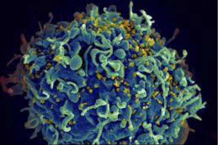
Figure 1 :THE AIDS VIRUS
38
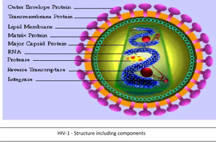
HUMAN IMMUNODEFICIENCY VIRUS - STRUCTURE:
The human immune deficiency virus is classified under the family
retroviridae and the sub family of lentiviruses. There are 2 types of viruses,
HIV-1 and HIV-2. HIV- 1 is responsible for the widely spread global
pandemic. Whereas HIV-2 isolated from western Africa is sporadic and its
distribution is widespread. The virus has an icosahedral structure and has 2
major envelope proteins, a surface protein gp 120 and another transmembrane
protein, gp41.Being a retroviruses, HIV-1 has a single-stranded and a
[image:48.595.101.533.411.696.2]plus-sense RNA.
Figure 2: COMPONENTS OF HIV
39
The reverse transcriptase enzyme also called as RNA-dependent DNA
polymerase, is found within the virion core. This enzymereplicates the
single-stranded RNA of the virus to a double-single-stranded DNA intermediate. This viral
DNA serves as the initiating precursor molecule needed for proviral
integration, This takes place within the host cell genome. The core proteins of
HIV-1 namely the capsid protein p24 and the matrix protein p18forms the
major structure.
A bilayered lipid covering which is a part of the host cell outer limiting
membrane surrounds the viral core protein structures. During replication, it is
from this membrane that the virus buds from the cell surface. The envelope
glycoproteins gp120 and gp41 cover this outer membrane. These glycoproteins
are encoded by virus-specific genes. The outer envelope proteins are
40
41
LIFE CYCLE OF HIV:
The virus by means of its surface protein gp120 begins its replication
cycle by binding to the CD4+ T lymphocyte. It undergoes conformational
change after entering the cell with the help of gp120. Then it attaches with one
of the major HIV co-receptors CCR5 and CXCR4. The binding of HIV to
CD4+ T cell, by binding the gp120 to C type lectin receptor on their surface by
the dendritic cells is known as DC – SIGN. The penetration of the host cell
membrane by the virion occurs with the help of the transmembrane protein
gp41. The pre- intergration complex which comprises of the viral RNA and
viral enzymes is released into the host cytoplasm surrounded by a capsid
protein coat 31.
The target of the preintegration complex is the host cell nucleus. The
RNA to DNA transcription is catalysed by viral reverse transcriptase
enzyme. The resultant pro viral DNA is released from the nuclear capsid to
enter the host nucleus. The enzyme integrase fuses the pro viral DNA with the
host chromosome. The further course is variable. Either the provirus may now
remain dormant or undergo different levels of genetic expression leading to
production of large number of virions in the host.
The host cell on activation initiates transcription of the integrated pro viral
DNA into RNA or mRNA.
The mRNA is modified by :
42
Glycosylation
Phosphorylation
Myristoylation
The assembly of the viral RNA, enzymes and proteins forms the entire
virus particle. Lipid rafts are specialised areas of the host cell lipid membrane
through which the virus buds out. The precursor proteins are cleaved by the
viral enzyme protease to release the mature virion.
Each step is regulated by viral regulatory gene products which can be
targeted for therapeutic interventions.
These include24 :
Virus – target cell binding
Virus – target cell fusion
Reverse transcriptase
Integrase
43
44
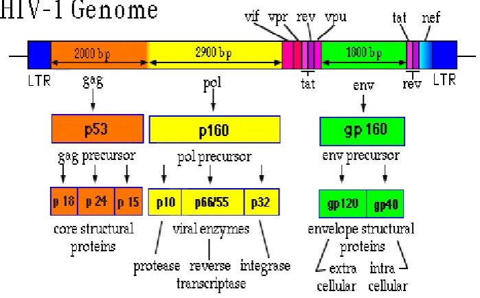
GENOME AND ITS DIVERSITY:
There are numerous genes that code for functional and structural
proteins.
GENE FUNCTION
Env Envelope glycoproteins
pol Protease, reverse transcriptase and integrase
enzymes
tat, rev, nef, vif, vpr,
vpu
Regulates viral gene replication and host cell
modification to enhance viral growth
gag Core of virion (including p24 antigen)
The HIV 2 genome has vpx gene and lacks vpu gene which is present
in HIV 1. The cluster mutations occurring in the surface proteins accounts for
the genomic diversity of HIV. The genomic reverse transcriptase is more or
less conserved. These diversities lead to subclassifications into groups,
subtypes and sub – subtypes .
GROUPS24:
4 groups – group N, group M (major), group P and group O (outlier),
Group M is subclassified into 9 different subtypes : A, B, C, D, F, G,
45
Patients may be infected with more than one subtype which give rise to
CRFs in combination (circulating recombinant forms). Examples include
CRF02_AG and CRF01_AE
Subtype A and F are then sub classified into sub – sub – types like A1,
A2 and F1, F2.
The geographic distribution of these different strains is widely
distributed. Subtype C is the most prevalent strain all over the world.
Other common strains globally are CRF01_AE, CRF01_AG, A, B, C,
D, and G.
There are numerous implications to this genetic diversity such as :
1. Different rates of disease progression
2. Varied response to therapy
3. Development of resistance
4. Continuous viral evolution
46
GEOGRAPHIC DISTRIBUTION OF VARIOUS HIV STRAINS:
1. India Subtype C
2. China Subtypes B, C and
BC recombinant forms
3. Western Europe Subtype B
3. Eastern Europe Subtype A,B and
AB recombinant forms
4. Sub-Saharan Africa Subtype C (most common)
Subtype B and G,
CRFO2_AG
5. Australia Subtype B
6. Southeast Asia CRF01_AE
7. North America and some parts of South
America
Subtype B
NEW EMERGING STRAINS:
CRF35_AD Afghanistan and Iran*
BF recombinant forms South America
CRF14_BG Spain* Portugal*
Thai B. Indian C. southern China*
CRF03_AB Former soviet union
47
MODES OF TRANSMISSION:
Multiple ways of HIV transmission are:
Sexual contact
Vertical
Perinatal
Breast milk.
Blood and blood products
No transmission has been reported through casual contact or insect bite.
SEXUAL TRANSMISSION26,34:
May be homosexual or hetero sexual transmission
Higher rates of transmission with higher HIV RNA load
Increased risk with unprotected anal intercourse (receptive)
Increased risk with genital infections ( as increased number of
inflammatory cells are present)
Higher rate of transmission from male to female than vice versa
Increased risk with genital ulcers caused by Chlamydia, Trichomonas,
Neisseria, Herpes etc. due to exposed mucous membrane
Due to absence of fore skin, male circumcision decreases risk of male
transmission (decreased local concentration of inflammatory cells,
decreased susceptibility to ulcerative infections, micro trauma)
Due to immature genital tract, adolescent girls are more susceptible to
48
TRANSMISSION BY BODY FLUIDS26,27:
Screening of blood and blood products for presence of HIV
antibodies, HIV p24 antigen and HIV RNA is practiced in most
countries
Transfusion related transmission cannot be completely avoided as HIV
RNA levels cannot be detected in the first 10 – 15 days following
infection due to undetectable levels of viremia, despite best technology
HIV transmission can occur through transfusions of whole blood,
plasma, factor concentrates in haemophiliacs, leukocytes and platelet
concentrates
No transmission occurs via hepatitis B Immunoglobulin, Rh
immunoglobulin, hyper immune gamma globulin and plasma derived
hepatitis B vaccine.
TRANSMISSION IN INTRAVENOUS DRUG USERS:
Transmitted in injection drug abusers by sharing needles and drugs27.
Their sexual partners are also affected through homosexual or
heterosexual transmission.
Their children are affected through perinatal transmission of HIV
49
TRANSMISSION IN HEALTH CARE WORKERS26,27,28:
Health care workers and laboratory personnels are at risk of procuring
HIV infection from patients
Risk due to contact of infected blood with mucous membrane or
breached skin by needle stick injuries or cuts
Increase risk of transmission by contact of blood with intact skin has
not been documented
Other potentially infectious body fluids are:
o Pleural
o Peritoneal
o Cerebrospinal
o Pericardial
o Semen
o Vaginal secretions
o Synovial
o Amniotic fluids
No substantial risk of transmission: Saliva, sputum, sweat, feces, nasal
secretions, vomitus, urine and tears.
Transmission increases with long contact, large volume blood contact
and port of entry in debraded mucous membrane or breached skin.
Post exposure prophylaxis is warranted within 24 hours, in case of
accidental exposure.
Universal precautions with specialized disposable kits should be used
50
51
UNIVERSAL PRECAUTIONS:
Safe hand washing with soap and water
Disposable gloves
Protective long gowns
Protective eyewear/ goggles
Mask
Heat inactivation or chemical decontamination of reusable equipments
Blood spills- disinfection/ disposal
Impervious containers- sharps disposal
Caution of workers with raw area, denuded skin, active dermatitis etc.
FETO – MATERNAL TRANSMISSION34:
Maternal Transmission to fetus can occur antenatally, during
labour or during breast feeding.
Higher risk of transmission are associated with :
Low maternal CD4 count
Pre term delivery
High maternal plasma viremia
Long duration between rupture of amniotic membrane &
delivery
Procedures like amniocentesis, amnioscopy, episiotomy etc.
Close match of maternal and fetal human leucocyte antigen
Chorioamnionitis/STD during pregnancy
52
TRANSMISSION BY BREAST FEEDING:
Transmission by breast milk is high during the early months of breast feeding
Risk is increased with :
Mastitis
Low maternal CD4 count
Detectable levels of HIV in breast milk
Maternal vitamin A deficiency
Risk of maternal transmission can be reduced by providing Zidovudine
with or without lamivudine, to the mother in the last few weeks of
gestation and to the fetus during the first week postnatally.
Perinatal transmission risk is reduced by :
Anti retro viral prophylaxis antenatally
Reducing exposure of fetus to maternal blood and
genital secretions (LSCS)
Universal voluntary testing / counselling of all pregnant
women
53
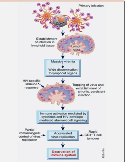
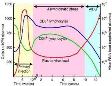
PATHOGENESIS OF HIV:
HIV disease is a chronic infection which eventually leads to a
quantitative and qualitative immunodeficiency because of ongoing immune
destruction. Mechanisms by which this immune dysfunction is achieved are:
Destruction of the immune cells by the replicating virus
Indirect effects like clearance of infected immune cells by the reticulo
endothelial system
Excess immune activation leading to immune exhaustion
Activation induced apoptosis (cell death).
The virus gains entry into the body by dendritic cells on the surface of
mucosa or through microscopic rents in the mucosa. The virus then seeks its
target -CD4+ T lymphocyte. The lymphocyte may be resting or activated. The
activated T cells help in virus replication. It then spreads to the nearby draining
lymph node where CD4 lymphocytes are available in large amounts. This
causes an initial burst of viremia and wide dissemination of virus in primary
infection leading to an early acute HIV syndrome24.
Despite the massive immune response generated against acute viral
infection, the virus is never eliminated from the human system and progresses
to a chronic infection. The replication and immune destruction continues for
almost 10years before the patient begins to deteriorate clinically.
54
PROTEINS THAT PLAY A VITAL ROLE IN PATHOGENESIS:
CD4 Surface protein on T lymphocytes
Gp 41 Transmembrane protein helps to penetrate host membrane and coil upon itself thereby helping in fusion
Gp120 CD4 receptor ligand, numerous spikes over the envelope of
the virion
CCR5 beta chemokine receptor for host cells like lymphocytes, dendritic cells, macrophages and glial cells
CXCR4 Co receptor used by HIV during late stages of infection
FUSION:
Gp120 of the virus gets attached to CD4 molecule which is found in T
helper cells. Gp120 undergoes conformational change. This leads to exposure
of another underlying protein Gp41 present beneath Gp120. It also causes
binding to host cell through co-receptors CCR4/CXCR5. Gp41 penetrates the
host membrane, succeeds in bringing together viral and cellular membrane
resulting in fusion.
FOUNDER VIRUS:
All viruses of infected individual do not transmit disease. The virus
during its replication in various lymphoid tissues acquires extreme genetic
expression and diversity. So there is a high degree of variation in genetic
characteristics and immunological response of existing virus in the plasma and
55
CHARACTERISTICS OF FOUNDER VIRUS:
1. Minimal N-linked Glycosylation
2. Limited genetic diversity
3. Rapid divergence after transmission
4. Presence of effective neutralizing antibodies in TP*
5. Under representation in the plasma viremia of TP*
6. Short V1-V2 loop
*TP-Transmitting partner
IMMUNE SYSTEM EVASION OF HIV24:
The Human Immunodeficiency virus dodges all the defence mechanisms
mounted against it and makes way for ambient survival conditions inside the
host. Thus HIV infection is almost impossible to eradicate from the infected
individual.
The mechanism by which the virus evades the immune system includes:
Wide diversity of mutations and recombinations causing a sustained
level of chronic viremia.
The Nef protein on the virus downregulates the HLA class I antigens on
the virus affected cells and hence escapes immune recognition.
Mutant virus population that helps virus propagation by escaping
56
The neutralising antibodies are directed only against gp120 and gp41 of
HIV because of:
Hypervariable regions in the genetic sequence of the
envelope proteins.
post translational modification like glycosylation.
Conformational masking of antigenic epitopes.
Sequestration of the infected cells preferentially in the immune
previledged sites like central nervous system to avoid detection.
Infection of virus specific CD4 cells selectively leading to elimination of
virus specific immune response, with a profound damage to immune
regulation and control of infection.
VIRAL RESERVOIRS AND LATENCY:
The wide range of infected cells in the dormant state in the body which
act as a potential virus reservoir, is the greatest obstacle to eradication of HIV.
These viral reservoirs exhibit pre – integration or post – intergration latency[24].
Pre – intergration latency is caused when the virus enters an inactive
CD4 cell, and the reverse transcription is incomplete and the pro viral DNA is
unable to immediately intergrate into the host genome . This phase can last for
hours to days. The pro viral DNA degenerates, if no host cell activation occurs
57
Post – intergration latency is caused when the virus enters into an active
host CD4 cell and the pro viral DNA integrates into the host cell genome
following which the host cell remains dormant but can replicate upon receiving
an activation signal from cytokines.
Reservoirs of HIV genome –exist in peripheral blood, lymph nodes,
central nervous system and in other unidentified areas either active or in
dormant form. Once the infection is established there is a progressive rise in
the viral load and a progressive gradual decline in the CD4 cell count. This
asymptomatic period is called clinical latency. It does not include microbial
latency as there is continuous viral replication despite the absence of clinical
58