STEROID RESPONSIVENESS – A SINGLE CENTER
STUDY IN SOUTH INDIA
Dissertation submitted in partial fulfillment of the
Requirement for the award of the degree of
MASTER OF PHARMACY
IN
BRANCH VII (PHARMACY PRACTICE)
Of
THE TAMILNADU Dr.M.G.R.MEDICAL UNIVERSITY
CHENNAI
\ DEPARTMENT OF PHARMACY PRACTICE
K.M.COLLEGE OF PHARMACY
UTHANGUDI, MADURAI-625107
This is a bonafide dissertation work entitled “CHILDHOOD NEPHROTIC SYNDROME AND STEROID RESPONSIVENES – A SINGLE CENTER STUDY
IN SOUTH INDIA” submitted by KUMARAN.N to The Tamilnadu Dr. M.G.R. Medical University, Chennai, in partial fulfillment of the requirement for the award of MASTER OF PHARMACY in PHARMACY PRACTICE at K.M. College of Pharmacy, Madurai – 625107. It is a bonafide work carried out by him under my guidance and supervision during the year 2011-2012. This dissertation partially or fully has not been submitted for any other degree or Diploma of this University.
GUIDE HEAD OF THE DEPARTMENT
Mr.K.Thirupathy, M.Pharm. Prof.M.Nagarajan
Assistant Professor M.Pharm.,DMS(BM)., DMS(IM)., Dept. of Pharmacy Practice, Head, Dept. of Pharmacy Practice, K.M. College of Pharmacy, K.M. College of Pharmacy, Uthangudi, Madurai- 625107. Uthangudi, Madurai-625107.PRINCIPAL
Dr. S. Jayaprakash M.Pharm.,Ph.D., Head of theDept. of Pharmaceutics,
INTRODUCTION
1-14BACKGROUND
Pediatric nephrotic syndrome, also known as nephrosis, is defined by the presence of nephrotic-range proteinuria, edema, hyperlipidemia, and hypoalbuminemia. Nephrotic-range proteinuria in adults is characterized by protein excretion of 3.5 g or more per day. However, because of the great range of body sizes in children, the pediatric definition of nephrotic-range proteinuria is more cumbersome.
Nephrotic-range proteinuria in children is protein excretion of more than 40 mg/m2/h. Because 24-hour urine collections are potentially unreliable and
burdensome, especially in young children, many pediatric nephrologists instead rely on a single, first-morning urine sample to quantify protein excretion by the ratio of protein to creatinine.
The use of a first-morning urine sample eliminates the contribution of potentially nonpathological orthostatic proteinuria, which might otherwise falsely elevate the protein level in a urine sample collected while a patient is active during the day. A urine protein/creatinine value of more than 2-3 mg/mg indicates nephrotic range proteinuria and correlates with results from 24-hour urine collection.
Kidneys affected by nephrotic syndrome have small pores in the podocytes, large enough to permit proteinuria (and subsequently hypoalbuminemia, because some of the protein albumin has gone from the blood to the urine) but not large enough to allow cells through (hence no hematuria). By contrast, in nephrotic syndrome, RBCs pass through the pores, causing hematuria.
CAUSES
PRIMARY
Primary causes of nephrotic syndrome are usually described by the histology, i.e. minimal change disease (MCD) like minimal change nephropathy which is the most common cause of nephrotic syndrome in children, focal segmental glomerulosclerosis (FSGS) and membranous nephropathy (MN) like membranous glomerulonephritis which is the main cause of nephrotic syndrome in adult.
They are considered to be "diagnoses of exclusion", i.e. they are diagnosed only after secondary causes have been excluded.
SECONDARY
Secondary causes of nephrotic syndrome have the same histologic patterns as the primary causes, though may exhibit some differences suggesting a secondary cause, such as inclusion bodies.
They are usually described by the underlying cause.
SECONDARY CAUSES BY HISTOLOGICAL PATTERN
Membranous nephropathy (MN):
• Hepatitis B& Hepatitis C
• Sjögren's syndrome
• Systemic lupus erythematosus(SLE)
• Diabetes mellitus
• Sarcoidosis
• Drugs (such as corticosteroids, gold, intravenous heroin)
• Malignancy (cancer)
• Bacterial infections, e.g. leprosy & syphilis
Focal segmental glomerulosclerosis (FSGS)
• Hypertensive nephrosclerosis
• HIV
• Obesity
• Kidney loss
Minimal change disease (MCD)
• Drugs, especially NSAIDs in the elderly
• Malignancy, especially Hodgkin's lymphoma
• Leukemia
Drugs that can cause secondary nephrotic syndrome include the following:
• Penicillamine
• Gold
• Nonsteroidal anti-inflammatory drugs (NSAIDs)
• Interferon
• Mercury
• Heroin
• Pamidronate
• Lithium
•
EPIDEMIOLOGY
histological pattern and outcome of INS. Moreover, there are some reports indicating the changing face of childhoodINS with time. Recent studies show that the frequency of FSGS in children has dramatically increased over the past two decades in some parts of world.
In India 20 cases per million are affected by nephrotic syndrome. The ISKDC found that 76.6% of children with INS had MCNS on kidney biopsy findings, with 7% of cases associated with FSGS on biopsy findings.
RACE, SEX, AND AGE-RELATED DEMOGRAPHICS
Black and Hispanic children appear to have an increased risk of steroid-resistant nephrotic syndrome and FSGS.An increased incidence of INS is reported in Asian children, (6 times the rate seen in European children). An increased incidence of INS is also seen in Indian, Japanese, and Southwest Asian children.
Primary, SSNS is rare in Africa, where nephrotic syndrome is more likely to be secondary or steroid-resistant. These variations in ethnic and geographic distribution of INS underscore the genetic and environmental influences in the development of PNS.
In children younger than 8 years at onset, the ratio of males to females varies from 2:1 to 3:2 in various studies. In older children, adolescents, and adults, the male-to-female prevalence is approximately equal. ISKDC data indicate that 66% of patients with either MCNS or FSGS are male, whereas 65% of individuals with MPGN are female.
PROGNOSIS
Since the introduction of corticosteroids, the overall mortality of INS has decreased dramatically from over 50% to approximately 2-5%. Despite the improvement in survival, INS is usually a chronic, relapsing disease and most patients experience some degree of morbidity, including the following:
• Hospitalization, in some instances
• Frequent monitoring both by parents and by physician
• Administration of medications associated with significant adverse events
• A high rate of recurrence (relapses in >60% of patients)
• The potential for progression to chronic kidney failure and end-stage kidney failure (ESKD)
Additionally, INS is associated with an increased risk of multiple complications, including edema, infection, thrombosis, hyperlipidemia, acute kidney failure, and possible increased risk of cardiovascular disease. The prognosis varies, depending on whether the nephrotic syndrome is steroid responsive or steroid resistant.
STEROID-RESPONSIVE NEPHROTIC SYNDROME
Patients who remain responsive to steroids with remission of proteinuria, even with frequent relapses, generally have a good prognosis. The ISKDC found that in 93% of children with INS who responded to steroids, kidney biopsy revealed MCNS. In contrast, 75% of patients who did not initially respond to steroids had histology other than MCNS.
Despite the generally favorable prognosis in patients who respond to steroids, the ISKDC reported a 60% rate of subsequent relapses, which can lead to complications, increased morbidity, and decreased quality of life.A longer course of initial steroid treatment (12 wk rather than the original ISKDC protocol of 8 wk) may reduce the rate of subsequent relapse to 36%,which still represents a large number of patients who undergo repeated courses of immunosuppression, with possible hospitalizations, edema, infections, medication side effects, and other comorbidities.
A long-term study of 398 children with INS found that the percentage of children who became free of relapses during the course of their disease rose from 44% one year after diagnosis to 69% at 5 years and 84% 10 years after diagnosis.Although most children with INS who respond to steroids achieve long-term remission, relapses may continue into adulthood.
Older studies suggested that more than 90% of children achieve long-term remission without further relapses by puberty. However, this has recently been challenged by surveys indicating a rate of relapse during adulthood as high as 27-42%.
In a retrospective study, Vivarelli et al reported that the length of time between initiation of steroid treatment and syndrome remission is an early prognostic indicator for children with INS.In study patients who did not suffer relapse or who relapsed infrequently, the median time from treatment onset to remission was less than 7 days. In patients who had frequent relapses or who developed steroid-dependent nephrotic syndrome, the median time to remission was more than 7 days.
STEROID-RESISTANT NEPHROTIC SYNDROME
Most patients who do not achieve remission of proteinuria with steroids have kidney biopsy findings other than MCNS. The most common diagnosis in these patients is FSGS.
More than 60% of patients with nephrotic syndrome and FSGS who fail to achieve remission with any treatment progress to end-stage kidney disease (ESKD). In contrast, only 15% progression to ESKD is observed in patients with FSGS who achieve remission by any treatment.Gipson et al reported a 90% lower risk of progression to ESKD in patients with INS who achieved remission.
Thus, patients with steroid-resistant INS have a good prognosis if remission of proteinuria can be achieved by other medications. Failure to respond to treatment (ie, failure to achieve remission) and kidney insufficiency at presentation are predictors of poor outcome and progression to ESKD.
PATHOPHYSIOLOGY
PROTEINURIA AND HYPOALBUMINEMIA
The hallmark of INS is massive proteinuria, leading to decreased circulating albumin levels. The initiating event that produces proteinuria remains unknown. INS is believed to have an immune pathogenesis. Studies have shown abnormal regulation of T-cell subsets and expression of a circulating glomerular permeability factor.
A circulating factor may play a role in the development of proteinuria in INS. This can be demonstrated by the rapid development of proteinuria in recurrence of nephrotic syndrome after kidney transplantation, the improvement in nephrotic syndrome in such patients after treatment with plasmapheresis, and the experimental induction of proteinuria in animals by plasma from patients with INS.
The nature of this circulating factor is not known. Various cytokines and molecules have been implicated, including the following:
• Interleukin (IL)-2, IL-4, IL-12, IL-13, IL-15, IL-18
• IL-2 receptor
• Interferon-γ
• Tumor growth factor (TGF)-β • Vascular permeability factor
• Nuclear factor (NF)-κB
• Tumor necrosis factor (TNF)-α
The association of allergic responses with nephrotic syndrome also illustrates the role of the immune system in INS. Nephrotic syndrome has been reported to occur after allergic reactions to bee stings, fungi, poison ivy, ragweed, house dust, jellyfish stings and cat fur. Food allergy might play a role in relapses of INS; a reduced-antigenic diet was associated with improved proteinuria and complete remission in one study.
Additionally, INS is 3-4 times more likely in children with human leukocyte antigen (HLA)-DR7. Steroid sensitive INS has also been associated with HLA-B8 and the DQB1 gene of HAL-DQW2. A greater incidence of INS is also observed in children with atopy and HLA-B12.
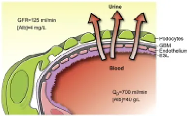
FIGURE NO: 1 SCHEMATIC DRAWING OF THE GLOMERULAR BARRIER
Podo = podocytes; GBM = glomerular basement membrane; Endo = fenestrated endothelial cells; ESL = endothelial cell surface layer (often referred to as the glycocalyx). Primary urine is formed through the filtration of plasma fluid across the glomerular barrier (arrows); in humans, the glomerular filtration rate (GFR) is 125 mL/min. The plasma flow rate (Qp) is close to 700 mL/min, with the filtration fraction being 20%. The concentration of albumin in serum is 40 g/L, while the estimated concentration of albumin in primary urine is 4 mg/L, or 0.1% of its concentration in plasma.
The glomerular filtration barrier consists of the fenestrated capillary endothelium, the extracellular basement membrane, and the intercalated podocyte foot processes, connected by 35-45 nm slit diaphragms. Nephrotic syndrome is associated with the biopsy finding of fusion (effacement) of podocyte foot processes. This effacement of the podocytes long was thought to be a secondary phenomenon of nephrotic syndrome.
However, theories have shifted toward the podocyte as playing a primary role in the development of proteinuria. The understanding of the pathophysiology of proteinuria in renal diseases has greatly expanded with insights into the molecular biology of the podocyte. Various forms of INS have been described with mutations in podocyte genes, such as those associated with the following:
• Glomerular basement membrane -LAMB2
• Mitochondria -COQ2
• Transcription factors -WT1, LMX1B
Nephrin is a transmembrane protein that is a major structural element of the slit diaphragm and is encoded by the NPHS1 gene on chromosome 19. Mutations in the NPHS1 gene are responsible for autosomal recessive, congenital nephrotic syndrome of the Finnish type (FNS).
FNS is characterized by massive proteinuria in the first year of life (usually within the first 3 month) and progression to end-stage kidney disease within the first decade of life, although milder forms of the disease have been described. Mutations in NPHS1 are usually associated with congenital nephrotic syndrome, but Philipe and colleagues have described NPHS1 mutations in children aged 6 months to 8 years with later-onset SRNS. Santin has described NPHS1 mutations in patients with later childhood onset as well as adult-onset SRNS.
Podocin is another podocyte protein that interacts with nephrin and CD2AP and is integral to the assembly of the slit diaphragm. Podocin is encoded by the NPHS2 gene on chromosome 1. Mutations in the NPHS2 gene were originally described in patients with autosomal recessive, steroid-resistant INS with FSGS on biopsy. Podocin mutations account for approximately 45-55% of familial and 8-20% of sporadic cases of SRNS.
α-Actinin-4, encoded by the gene ACTN4 on chromosome 19, cross-links actin filaments of the podocyte cytoskeleton and anchors them to the glomerular basement membrane. The TRPC6 gene on chromosome 11 encodes for a calcium channel associated with the slit diaphragm. Disruptions in either ACTN4 or TRPC6 are associated with autosomal dominant forms of FSGS.
nephrotic syndrome and FSGS. One report describes a single patient with a homozygous mutation in CD2AP and early onset of nephrotic syndrome with FSGS and diffuse mesangial sclerosis.
Nonmuscle myosin heavy chain 9 (MYH9) is a podocyte protein that binds to the podocyte actin cytoskeleton to perform intracellular motor functions. MYH9 polymorphisms are estimated to account for more than 40% of end-stage kidney disease cases among blacks and has been associated with FSGS, HIV-associated nephropathy, and focal global glomerulosclerosis (which had been previously attributed to hypertensive nephrosclerosis in many patients). The E1 haplotype of MYH9 is present in 60% of blacks compared with 4% of individuals with European ancestry.
Other genetic forms of nephrotic syndrome continue to shed light on the pathogenesis of INS. Mutations in the developmental regulatory gene WT1 are associated with forms of congenital nephrotic syndrome associated with male pseudohermaphroditism, Wilms tumor (Denys-Drash syndrome), and gonadoblastoma (Frasier syndrome).
Mutations in phospholipase C epsilon 1 (PLCE1), a cytoplasmic enzyme required for podocyte maturation, have been associated with as many as 28% of cases of congenital nephrotic syndrome due to isolated (nonsyndromic) diffuse mesangial sclerosis. Nail-patella syndrome, a disorder characterized by skeletal and nail dysplasia as well as nephrotic syndrome, is caused by mutations in the LMX1B gene, which regulates expression of type IV collagen and the podocyte proteins nephrin, podocin, and CD2AP.
The role of alterations in the slit diaphragm in MCNS has not been elucidated. Podocin appears to be expressed normally in MCNS but decreased in FSGS. Mutations in nephrin and podocin do not at this time appear to play a role in steroid-sensitive nephrotic syndrome. However, acquired alterations in slit diaphragm architecture might play a role in INS apart from actual mutations in the genes encoding podocyte proteins. Various authors have reported changes in expression and distribution of nephrin in MCNS.
Coward et al demonstrated that nephrotic plasma induces translocation of the slit diaphragm proteins nephrin, podocin, and CD2AP away from the plasma membrane into the cytoplasm of the podocyte. These authors also demonstrated the normal plasma might contain factors that maintain the integrity of slit diaphragm architecture and that the lack of certain factors (rather than the presence of an abnormal circulating factor) might be responsible for alterations in the podocyte architecture and development of INS.
Apart from the podocyte and slit diaphragm, alterations in the glomerular basement membrane also likely play a role in the proteinuria of nephrotic syndrome. In INS, the glomerular capillary permeability to albumin is selectively increased, and this increase in filtered load overcomes the modest ability of the tubules to reabsorb protein.
EDEMA
The classical explanation for edema formation is a decrease in plasma oncotic pressure, as a consequence of low serum albumin levels, causing an extravasation of plasma water into the interstitial space. The resulting contraction in plasma volume (PV) leads to stimulation of the renin-angiotensin-aldosterone axis and antidiuretic hormone. The resultant retention of sodium and water by the renal tubules contributes to the extension and maintenance of edema.
While the classical model of edema (also known as the "underfill hypothesis") seems logical, certain clinical and experimental observations do not completely support this traditional concept. First, the PV has not always been found to be decreased and, in fact, in most adults, measurements of PV have shown it to be increased. Only in young children with MCNS have most (but not all) studies demonstrated a reduced PV.
Additionally, most studies have failed to document elevated levels of renin, angiotensin, or aldosterone, even during times of avid sodium retention. Active sodium reabsorption also continues despite actions that should suppress renin effects (eg, as albumin infusion or angiotensin-converting enzyme [ACE] inhibitor administration).
Coupled with these discrepancies is the fact that, in the steroid-responsive nephrotic, diuresis usually begins before plasma albumin has significantly increased and before plasma oncotic pressure has changed. Some investigators have demonstrated a blunted responsiveness to atrial natriuretic peptide (ANP) despite higher-than-normal circulating plasma levels of ANP.
ANP might play a role is this mechanism; studies have shown an impaired response to ANP in nephrotic syndrome. This ANP resistance, in part, might be caused by overactive efferent sympathetic nervous activity, as well as enhanced tubular breakdown of cyclic guanosine monophosphate.
Other mechanisms that contribute to a primary increase in renal sodium retention include overactivity of the Na+ -K+ -ATPase and renal epithelial sodium
channel (RENaC) in the cortical collecting duct and shift of the Na+/H+ exchanger NHE3 from the inactive to active pools in the proximal tubule.
A more recent theory of edema formation posits that massive proteinuria leads to tubulointerstitial inflammation and release of local vasoconstrictors and inhibition of vasodilation. This leads to a reduction in single-nephron glomerular filtration rate and sodium and water retention.
Thus, the precise cause of the edema and its persistence is uncertain. A complex interplay of various physiologic factors, such as the following, probably contributes:
• Decreased oncotic pressure
• Increased activity of aldosterone and vasopressin
• Diminished atrial natriuretic hormone
• Activities of various cytokines and physical factors within the vasa recti
HYPERLIPIDEMIA
INS is accompanied by disordered lipid metabolism. Apolipoprotein (apo)-B– containing lipoproteins are elevated, including very-low-density lipoprotein (VLDL), intermediate-density lipoprotein (IDL), low-density lipoproteins (LDL), and lipoprotein(a), with resultant increases in total cholesterol and LDL-cholesterol. The level of high-density lipoprotein (HDL) cholesterol is normal or low. Elevations in triglyceride levels occur with severe hypoalbuminemia.
hypoalbuminemia. However, serum cholesterol levels have been shown to be independent of albumin synthesis rates.
Decreased plasma oncotic pressure may play a role in increased hepatic lipoprotein synthesis, as demonstrated by the reduction of hyperlipidemia in patients with INS receiving either albumin or dextran infusions. Also contributing to the dyslipidemia of INS are abnormalities in regulatory enzymes, such as lecithin-cholesterol acyltransferase, lipoprotein lipase, and lecithin-cholesterol ester transfer protein.
THROMBOSIS
Patients with nephrotic syndrome are at increased risk for thrombosis. The incidence rate of thromboembolic complications (TEC) is about 1.8-5% but may be underestimated. One study found the subclinical rate of pulmonary embolism to be 28% using scintigraphic pulmonary ventilation and perfusion studies.Incidence is higher in adults and children with secondary nephrotic syndrome. The incidence is especially high in membranous nephrotic syndrome.
Renal vein thrombosis, deep vein thrombosis, and pulmonary embolism (PE) are the most frequently encountered TEC in children. Other venous sites of thrombosis include the superior sagittal sinus, other cerebral venous sites, and the inferior vena cava.
Arterial thrombosis, although less common than venous TEC, can occur and has been reported at the axillary, subclavian, femoral, coronary, and mesenteric arteries.
Various factors play a role in the increased incidence of thrombosis. Abnormalities described in INS include the following:
• Increased platelet activation and aggregation
• Elevation in levels of factors V, VII, VIII, and XIII and fibrinogen
• Increased activities of tissue plasminogen activator and plasminogen activator inhibitor-1
These abnormalities in hemostatic factors, combined with potential hypovolemia, immobility, and increased incidence of infection, lead to a hypercoagulable state in INS.
INFECTION
Patients with INS are at increased risk of infection. Peritonitis and sepsis are the most common and serious infections. Peritonitis occurs at a rate of approximately 2-6% and may be accompanied by sepsis or bacteremia. The predominant bacterial causes are Streptococcus pneumoniae and Gram-negative enteric organisms such as Escherichia coli.
Various infections can also occur, including meningitis, cellulitis, viral infections, and others. Varicella is a particular concern in immunosuppressed patients and can be lethal. Prompt recognition and treatment with acyclovir (or postexposure prophylaxis with varicella-zoster immune globulin [VZIG]) is essential. Routine childhood varicella immunization has alleviated some of the concern regarding this complication.
Infection, viral or bacterial, can trigger relapse of INS and further complicate the course of the condition.
Risk of infection may be increased in INS because of low immunoglobulin (Ig)G levels, which do not appear to be the result of urinary losses. Instead, low IgG levels seem to be the result of impaired synthesis, again pointing to a primary disorder in lymphocyte regulation in INS.
function may also be present in INS, which contributes to the susceptibility to infection. Finally, the medications used to treat INS, such as corticosteroids and alkylating agents, further suppress the immune system and increase the risk of infection.
ACUTE KIDNEY FAILURE
Acute kidney failure (AKF) is a rare complication of INS, occurring in about 0.8% of cases.Causes include the following:
• Rapid progression of underlying disease (nephrotic syndrome other than MCNS, secondary nephrotic syndrome)
• Bilateral renal vein thrombosis
• Acute interstitial nephritis (AIN) due to drug therapy (eg, antibiotics, nonsteroidal anti-inflammatory agents [NSAIDs], diuretics)
• Acute tubular necrosis (ATN) due to hypovolemia or sepsis
Use of ACE inhibitors or angiotensin II receptor blockers (ARBs) in conjunction with volume depletion can also precipitate AKF.
PHYSICAL EXAMINATION
The most common clinical finding is edema. The edema is pitting and is typically found in the lower extremities, face and periorbital regions, scrotum or labia, and abdomen (ascites). In those children with marked ascites, mechanical restriction to breathing may be present, and the child may manifest compensatory tachypnea. Pulmonary edema and effusions can also cause respiratory distress. Hypertension can be present and is more common in children with FSGS and MPGN rather than MCNS.
DIAGNOSTIC CONSIDERATIONS
Acute kidney failure (AKF) is a rare complication of idiopathic nephrotic syndrome. Fever, rash, arthralgia and eosinophilia with a "bland" urinalysis (minimal cellular elements) in the presence of AKF are typical for acute interstitial nephritis. However, obvious clinical symptoms may be absent except for the AKF and unremarkable urinalysis. Gross hematuria, flank pain, and thrombocytopenia may be signs of renal vein thrombosis. Hemoconcentration in the patient with anasarca might indicate intravascular volume depletion.
APPROACH CONSIDERATIONS
The first step in evaluating the child with edema is to establish whether nephrotic syndrome is present, because hypoalbuminemia can occur in the absence of proteinuria (such as from protein-losing enteropathy), and edema can occur in the absence of hypoalbuminemia (eg, in angioedema, capillary leak, venous insufficiency, congestive heart failure).
In order to establish the presence of nephrotic syndrome, laboratory tests should confirm (1) nephrotic-range proteinuria, (2) hypoalbuminemia, and (3) hyperlipidemia. Therefore, initial laboratory testing should include the following:
• Urinalysis
• Urine protein quantification (by first-morning urine protein/creatinine or 24-hour urine protein)
• Serum albumin
• Lipid panel
• Metabolic panel (Serum electrolytes, BUN and creatinine, calcium, phosphorus, and ionized calcium levels)
• Testing for HIV, hepatitis B and C
• Complement studies (C3, C4)
• Antinuclear antibody (ANA), anti–double-stranded DNA antibody (in selected patients)
Patients with INS lose vitamin D–binding protein, which can result in low vitamin D levels, and thyroid binding globulin, which can result in low thyroid hormone levels. Consideration should be given, especially in the child with frequently relapsing or steroid-resistant nephrotic syndrome, to testing for 25-OH-vitamin D; 1,25-di(OH)-vitamin D; free T4; and thyroid-stimulating hormone (TSH).
Other tests and procedures in selected patients may include the following:
• Genetic studies
• Kidney ultrasonography
• Chest radiography
• Mantoux test
• Kidney biopsy
Age plays an important role in the diagnostic evaluation of nephrotic syndrome. Children presenting with nephrotic syndrome younger than 1 year of age should be evaluated for congenital/infantile nephrotic syndrome. In addition to the above tests, infants should have the following tests:
• Congenital infection (syphilis, rubella, toxoplasmosis, cytomegalovirus, HIV)
• Kidney biopsy
• Genetic tests for NPHS1, NPHS2,WT1, and LAMB2 mutations as guided by biopsy findings and clinical presentation
• No routine surgical care is indicated for this condition.
the peritonitis is Streptococcus pneumoniae; however, enteric bacteria may also cause peritonitis. Treatment is medical rather than surgical.
URINE STUDIES
Microscopic hematuria is present in 20% of cases of INS and cannot be used to distinguish between minimal change nephrotic syndrome (MCNS) and other forms of glomerular disease.
RBC casts, if present, are suggestive of acute glomerulonephritis, such as postinfectious nephritis, or a nephritic presentation of chronic glomerulonephritis, such as membranoproliferative glomerulonephritis (MPGN). Granular casts may be present and are non-specific to etiology.
The presence of macroscopic (gross) hematuria is unusual in MCNS and suggests another cause, such as MPGN, or a complication of idiopathic nephrotic syndrome (INS), such as renal vein thrombosis.
URINE PROTEIN QUANTIFICATION
First morning urine protein/creatinine is more easily obtained than 24-hour urine studies, is possibly more reliable, and excludes orthostatic proteinuria. A urine protein/creatinine ratio of more than 2-3 mg/mg is consistent with nephrotic-range proteinuria.
A 24-hour urine protein level of more than 40 mg/m2/h also defines nephrotic-range proteinuria.
BLOOD STUDIES
• Elevated total cholesterol, low-density lipoprotein (LDL)-cholesterol
• Elevated triglycerides with severe hypoalbuminemia
• High-density lipoprotein (HDL)-cholesterol (normal or low)
The patient with INS, even MCNS, can present with acute kidney failure due to intravascular volume depletion and/or bilateral renal vein thrombosis. In the absence of the above, elevated BUN and creatinine levels and signs of chronic kidney failure (such as poor growth, anemia, acidosis, hyperkalemia, hyperphosphatemia, elevated parathyroid hormone) suggest a chronic glomerular disease other than MCNS, such as one of the following:
• Focal segmental glomerulosclerosis (FSGS)
• Membranous nephropathy (MN)
• MPGN
• Immunoglobulin (Ig)A nephropathy
Serum sodium levels are low in patients with INS because of hyperlipidemia (pseudohyponatremia), as well as dilution due to water retention. Total calcium levels are low because of hypoalbuminemia, but ionized calcium levels are normal.
On the CBC, an increased hemoglobin and hematocrit indicate hemoconcentration and intravascular volume depletion. The platelet count is often increased.
HIV infection, hepatitis B, and hepatitis C are important secondary causes of nephrotic syndrome. Consequently, screening for these viruses should be performed in all patients presenting with nephrotic syndrome. Consider checking liver enzymes, such as alanine aminotransferase (ALT) and aspartate aminotransferase (AST), when screening for liver disease.
Low complement levels (C3, C4) are found in postinfectious nephritis, MPGN, and lupus nephritis.
loss, joint pain) or any patient with nephrotic syndrome presenting in later school-age or adolescent years when lupus has a higher incidence.
GENETIC TESTING
WT1 testing is indicated for patients with pseudohermaphroditism, Wilms tumor, gonadoblastoma, and diffuse mesangial sclerosis on biopsy. NPHS1 testing is indicated in patients with biopsy and clinical findings consistent with Finnish-type nephrotic syndrome Because congenital NS has been reported with mutations in NPHS2, analysis of this gene should also be considered. Infants with nephrotic syndrome and neurological or visual disturbances should be considered candidates for LAMB2 testing (Pierson syndrome).
In patients initially or subsequently unresponsive to steroid treatment, in addition to kidney biopsy, consideration should be given to testing for mutations in podocin (NPHS2), ACTN4 and TRPC6.
KIDNEY ULTRASONOGRAPHY
Kidney ultrasonography might help to distinguish between MCNS and other chronic kidney disease, but findings are usually nonspecific. In all cases of nephrotic syndrome, the kidneys are usually enlarged due to tissue edema. Increased echogenicity is usually indicative of chronic kidney disease other than MCNS, in which echogenicity is usually normal. A finding of small kidneys indicates chronic kidney disease other than MCNS and is usually accompanied by elevated serum creatinine levels.
CHEST RADIOGRAPHY
Chest radiography also should be considered prior to steroid therapy to rule out tuberculosis (TB) infection, especially in the child with positive or previously positive Mantoux test or prior treatment for TB.
MANTOUX TEST
Mantoux test (purified protein derivative [PPD]) should be performed prior to steroid treatment to rule out TB infection.
Mantoux testing can be performed concurrent to starting steroid treatment, as treatment with steroids for 48 hours prior to reading the PPD does not mask a positive result and the risk associated with 2 days of steroids is minimal (if tests results are positive, steroids should be immediately stopped).
In children with a positive PPD, previously positive PPD, or prior treatment for TB, chest radiography should be performed.
KIDNEY BIOPSY
A kidney biopsy is not indicated for first presentation of PNS in the child 1-8 years of age unless the history, physical findings, or laboratory results indicate the possibility of secondary nephrotic syndrome or primary nephrotic syndrome other than MCNS. Kidney biopsy is indicated in patients younger than 1 year when genetic forms of congenital nephrotic syndrome are more common, and in patients older than 8 years, when chronic glomerular diseases such as FSGS have a higher incidence.
In select preadolescent patients older than 8 years, empirical steroid treatment can be considered prior to kidney biopsy, but this should occur only under the care of a pediatric nephrologist experienced with nephrotic syndrome. Some authors have recommended performing a kidney biopsy in patients older than 12 years.
• Symptoms of systemic disease (eg, fever, rash, joint pain)
• Laboratory findings indicative of secondary nephrotic syndrome (eg, positive ANA findings, positive anti–double-stranded DNA antibody findings, low complement levels)
• Elevated creatinine levels unresponsive to correction of intravascular volume depletion
• A relevant family history of kidney disease
Finally, in patients who are initially or subsequently unresponsive to steroid treatment, kidney biopsy should be performed, because steroid unresponsiveness has a high correlation with prognostically unfavorable histology findings such as FSGS or MGN.
HISTOLOGIC FINDINGS
If a kidney biopsy is performed, various histologic findings can be present, depending on the etiology of the nephrotic syndrome. Briefly, the most common histological types of INS are as follows.
MINIMAL CHANGE NEPHROTIC SYNDROME
MCNS indicates glomerular morphology that on light microscopic examination is little different from normal. Minimal mesangial hypercellularity may be present. Immunofluorescent microscopy (IF) usually reveals no presence of immune deposits.
DIFFUSE MESANGIAL PROLIFERATION
Diffuse mesangial proliferation (DMP) refers to increased mesangial matrix and increased mesangial hypercellularity. IF findings are negative and EM reveals the typical foot process effacement of MCNS. Patients with DMP have an increased incidence of steroid resistance, although whether these patients are at increased risk for progression to kidney failure is unclear.
FOCAL SEGMENTAL GLOMERULOSCLEROSIS
FSGS describes a lesion in which, as seen on LM, discrete segments of the glomerular tuft reveal sclerosis (segmental); some glomeruli are involved, and others are spared (focal).
Adhesion of the glomerular tuft to Bowman capsule (synechiae) is observed. Glomerular hypertrophy is common. Interstitial fibrosis and tubular atrophy are often present and correlate with the severity of disease.
IF reveals IgM and C3 trapped in the sclerotic areas. As in MCNS, EM reveals effacement of the podocyte foot processes. Additionally, EM reveals obliteration of capillary lumens by fine granular and lipid deposits.
A subtype of FSGS, in which the glomerular tufts demonstrate collapse of capillaries (collapsing glomerulopathy) on LM, has a poorer prognosis and high rate of progression to end-stage kidney failure (ESKD).
FSGS is not a specific disease but a histopathological finding that can be associated with INS but can also be found in a wide variety of other conditions, including HIV nephropathy, heroin nephropathy, reflux nephropathy, obstructive uropathy, renal hypoplasia, hypertension, obesity, and Alport syndrome.
MEMBRANOPROLIFERATIVE GLOMERULONEPHRITIS
MPGN is also known as mesangiocapillary glomerulonephritis. Glomeruli are typically lobulated in appearance on LM findings and demonstrate mesangial proliferation. Silver stain may reveal characteristic duplication of the glomerular basement membrane ("tram-track" appearance). IF findings reveal characteristic capillary deposition of C3.
Three types of MPGN are recognized and can be distinguished by electron microscopy findings according to the location of immune deposits. Type 1 is subendothelial; type 2 has ribbonlike, dense intramembranous deposits; and type 3 is subendothelial and subepithelial. Some controversy surrounds the existence of type 3 MPGN as a distinct entity or a variant of type 1.
MEMBRANOUS NEPHROPATHY
MN is a rare finding in INS of childhood, comprising only approximately 1% of biopsies, whereas in adult INS, MN can be found in 25-40% of cases. Light microscopy typically reveals thickening of the glomerular basement membrane. Silver stain may reveal characteristic "spikes," resulting from protrusion of basement membrane around immune deposits. IF reveals fine granular IgG and complement staining along the periphery of the glomerular capillary wall. EM reveals subepithelial electron-dense deposits.
TREATMENT
The various causes in nephrotic syndrome can be treated as follows:
HYPERLIPIDEMIA
Lipid abnormalities generally resolve when nephrotic syndrome is in remission. Dietary modification does not appear to be effective in limiting hyperlipidemia during active nephrotic syndrome.
Chronic hyperlipidemia has been linked to increased risk of atherosclerosis and coronary artery disease. Chronic hyperlipidemia has also been associated with progression of renal disease. However, the small studies to date of lipid-lowering agents in pediatric INS have not shown an improvement in proteinuria or progression of renal disease.
Dyslipidemias in adults with nephrotic syndrome have been successfully treated with the following:
• Statins (simvastatin, lovastatin)
• Fibrates (gemfibrozil)
• Bile-acid binding resins (cholestyramine)
• Probucol
Children with INS have been effectively treated with probucol, but this agent has been associated with prolonged QT interval and is not available in the United States. Gemfibrozil has also been shown to be effective in childhood nephrotic syndrome in small studies.
In order to monitor for treatment-associated rhabdomyolysis, children treated with statins should have creatine kinase measured prior to initiating therapy and every 6-12 weeks during treatment. Patients and families should be instructed to report muscle soreness, tenderness, or pain. Aspartate aminotransferase (AST) and alanine aminotransferase (ALT) levels should be measured before initiating treatment and about every 3 months thereafter to monitor for liver toxicity.
Long-term safety studies regarding statins in pediatrics are lacking, and routine use of statins were not recommended by an expert panel in 2004. The only drugs recommended at this time by the panel were bile acid sequestrants.
THROMBOEMBOLISM
Initial treatment of thromboembolic complications includes thrombolysis with anticoagulants (such as heparin) and/or fibrinolytic agents (ie, tissue plasminogen activator, streptokinase, urokinase). For secondary prevention, warfarin is often prescribed for a period of as long as 6 months.
Empiric prophylactic anticoagulation is not routinely indicated in INS. Some practitioners advocate the use of long-term, low-dose aspirin in patients with chronic nephrotic syndrome (eg, frequently relapsing nephrotic syndrome, SDNS, SRNS). However, adequate controlled trials examining the use of aspirin have not been performed.
ACUTE KIDNEY FAILURE
CORTICOSTEROID THERAPY
The original International Study of Kidney Disease in Children (ISKDC) protocol recommended induction therapy with oral prednisone or prednisolone at 60 mg/m2/d (2 mg/kg/d), with a maximum of 60 mg, daily for 4 weeks. Traditionally, the total daily dose was split into two doses. However, a single daily dose of steroids has efficacy equal to split dosing and fewer side effects.
Subsequent studies have shown that a longer period of initial steroid treatment (6 weeks rather than 4 weeks) reduces the subsequent rate of relapse. Thus, the general consensus now is to prescribe the initial daily steroids for 6 weeks.
Original guidelines recommended that induction therapy be followed with maintenance therapy with oral prednisone or prednisolone at 40 mg/m2 (or 1.5 mg/kg), with a maximum of 40 mg, given as a single dose on alternate days for 4 weeks. Subsequent studies demonstrated that a longer alternate-day maintenance period of 6 weeks resulted in a lower rate of relapse.
Thus, the general consensus now is daily induction steroid treatment for 6 weeks, followed by alternate-day maintenance therapy for another 6 weeks. After 6 weeks of alternate day treatment, steroids may be stopped or slowly tapered over a variable length of time.
TREATMENT OF RELAPSES
For infrequent relapses, steroids are resumed, although for a shorter duration than treatment during initial presentation. Prednisone, 2 mg/kg/d (60 mg/m2/d), is given as a single morning dose until the patient has been free of proteinuria for at least 3 days.
Following remission of proteinuria, prednisone is reduced to 1.5 mg/kg (40 mg/m2) given as a single dose on alternate days for 4 weeks. Steroids may then be stopped or gradually tapered.
DIURETIC THERAPY
Diuretic therapy may be beneficial, particularly in children with symptomatic edema. Loop diuretics, such as furosemide (starting at 1-2 mg/kg/d) may improve edema; their administration, however, should be handled with care because plasma volume contraction may already be present, and hypovolemic shock has been observed with overly aggressive therapy.
Metolazone may be beneficial in combination with furosemide for resistant edema. Patients must be monitored carefully on this regimen. If the child is sent home on diuretic therapy, the family must have clear guidelines about discontinuing therapy when edema is no longer present and careful communication with the family should continue.
When a patient presents with anasarca and signs of intravascular volume depletion (such as a high hematocrit, indicative of hemoconcentration), consideration should be given to administration of 25% albumin, although this is controversial. Rapid administration of albumin can result in pulmonary edema.
ANTIHYPERTENSIVE THERAPY
Antihypertensive therapy should be given when hypertension is present and particularly if it persists, but caution should be exercised. In some patients, the hypertension will respond to diuretics. Angiotensin-converting enzyme (ACE) inhibitors or angiotensin II receptor blockers (ARBs) may also contribute to reducing proteinuria but should be used cautiously in the presence of acute kidney failure or volume depletion because they can worsen kidney function in these settings.
ACE inhibitors and ARBs can cause birth defects, so adolescent women who are taking these agents must be counseled regarding use of birth control, and pregnancy testing should be considered before starting these agents.
Calcium channel blockers and beta-blockers may also be used as first-line agents for hypertension.
FREQUENTLY RELAPSING AND STEROID-DEPENDENT DISEASE
Frequently-relapsing nephrotic syndrome (FRNS) is defined as steroid-sensitive nephrotic syndrome (SSNS) with 2 or more relapses within 6 months or more than 3 relapses within a 12-month period. Steroid-dependent nephrotic syndrome (SDNS) is defined as SSNS with 2 or more consecutive relapses during tapering or within 14 days of stopping steroids.
ALKYLATING AGENTS
Alkylating agents (eg, cyclophosphamide [CYP], chlorambucil, nitrogen mustard) offer the benefit of possible sustained remission after a defined course of treatment, although with the possible risk of infertility and other side effects (see Side Effects of Drug Therapy).
An increased risk of seizures is noted with chlorambucil. Additionally, a higher incidence of infections and leukopenia may be seen with chlorambucil compared with CYP. Because of these risks, and the need to give nitrogen mustard intravenously, CYP has generally been the preferred alkylating agent.
Steroids (prednisone 2 mg/kg/d in single dose, max 60 mg) are usually started prior to administration of CYP in order to induce remission of proteinuria before starting CYP. After initiation of CYP, if proteinuria remains in remission, prednisone is reduced to 1.5 mg/kg on alternate days for 4 weeks, then slowly tapered over 4 weeks.
CYP (2 mg/kg daily) is given orally for 8-12 weeks. An influential study found that a 12-week course was more effective than an 8-week course in producing sustained remission of nephrotic syndrome. However, a subsequent randomized trial did not reach this same conclusion, and the optimal duration of CYP treatment is still unclear at this time.
CALCINEURIN INHIBITORS
Calcineurin inhibitors (eg, cyclosporine A [CSA], tacrolimus [TAC]) are useful steroid-sparing agents. These agents can also be used in those children who fail to respond to, or subsequently relapse after, treatment with CYP, or in children whose families object to the use of CYP.
Calcineurin inhibitors have disadvantages: prolonged courses of treatment are needed, nephrotic syndrome tends to recur when treatment is stopped, and nephrotoxic injury may occur. Consideration should be given to kidney biopsy after prolonged treatment to monitor for calcineurin inhibitor–induced nephrotoxicity and fibrosis.
Limited studies are available regarding the effectiveness of TAC compared with CSA. A single-center study by Choudhry et al in 41 patients with idiopathic SRNS found that TAC (0.1-0.2 mg/kg/d) or CSA (5-6 mg/kg/d) have similar efficacy in inducing remission in patients with idiopathic SRNS at 6 months and 1 year when combined with alternate-day low-dose corticosteroids and enalapril.
Relapse was significantly greater with CSA than with TAC; in addition, TAC decreased blood cholesterol levels to a greater extent and resulted in fewer incidents of nephrotoxicity that necessitated discontinuance than CSA. Cosmetic adverse effects (eg, hypertrichosis, gum hypertrophy) were significantly more frequent with CSA. Thus, TAC therapy is a promising alternative to CSA because of the lower relapse risk and lack of cosmetic adverse effects.
CYCLOSPORINE A
Kidney function and drug levels must be carefully monitored due to the risk of CSA-induced nephrotoxicity.
Low-dose steroids are continued for a variable length of time. As many as 40% of patients may need to remain on steroids during CSA treatment to maintain remission.
TACROLIMUS
TAC is started at a dose of 0.1 mg/kg daily divided every 12 hours and adjusted to keep trough level about 5-10 ng/mL.Our practice is to use the lowest possible dose that sustains remission and to aim for a trough level of around 3-5 ng/mL. TAC trough levels correspond better to AUC than CSA, allowing better determination of dosing and exposure with TAC than with CSA.As with CSA, continuing low-dose steroids is often necessary to maintain remission, although some patients may eventually be able to discontinue steroid treatment.
LEVAMISOLE
Levamisole is an anthelmintic drug that has immune-modulating effects and can be effective in reducing the relapse rate in FRNS. However, it is unavailable in the United States. Side effects include leukopenia, hepatic dysfunction, agranulocytosis, vasculitis, and encephalopathy. Levamisole is prescribed at a dose of 2.5 mg/kg given on alternate days.
MYCOPHENOLATE MOFETIL
Thus, MMF might be a useful steroid-sparing agent in stable patients (without excessive edema, need for hospitalizations and without other serious complications) whose families wish to avoid the possible side effects of CYP, CSA, or TAC. However, response to MMF varies and is less reliable than other treatments.
MMF is started at a dose of 600 mg/m2 twice daily. Complete blood counts should be monitored for bone marrow suppression, and liver function tests should occasionally be performed to monitor for hepatic toxicity.
STEROID-RESISTANT DISEASE AND FOCAL SEGMENTAL GS
Adequate randomized controlled trials have not yet been reported to give sufficient evidence to guide treatment of steroid-resistant nephrotic syndrome (SRNS).A current National Institutes of Health (NIH)-sponsored, multicenter, randomized clinical trial has recently concluded enrollment and will compare CSA versus MMF for treatment of focal segmental glomerulosclerosis (FSGS).
The most frequently recommended treatment for FSGS and SRNS is CSA. Approximately 36% of children with SRNS may achieve remission with this agent.CSA is dosed as for FRNS and SDNS. However, higher doses and trough levels may be required to achieve remission in SRNS and FSGS.TAC may be effective as well, although studies are limited at this time.Most studies to date have shown no clear benefit to the use of alkylating agents in FSGS and SRNS.
The histology in these patients consisted of MCNS, FSGS, MPGN, crescentic glomerulonephritis, and membranous nephropathy). Further studies are needed on the use of MMF in SRNS and FSGS.
A controversial treatment involves high-dose, intravenous methylprednisolone tapered over 78 weeks, in combination with alternate-day oral prednisone; CYP or chlorambucil is added if remission is not achieved in the first 10 weeks. The authors reported a 52% remission rate in SRNS.However, subsequent studies using this protocol have not duplicated the initial success. The risk of steroid toxicity and infection, as well as lack of sufficient evidence for the effectiveness of this protocol, have dampened enthusiasm for this treatment.
The use of an ACE inhibitor (eg, enalapril), either alone or in combination with an ARB (eg, losartan), has been shown to reduce proteinuria in FSGS/SDNS and should be considered in all patients, even in the absence of hypertension. Accordingly, ACE inhibitors and ARB should be considered as preferred agents in patients with hypertension. ACE inhibitor and ARB treatment may also have a renoprotective effect and slow progression of renal disease by inhibiting pathways of fibrosis.
DIET AND ACTIVITY
Reduce sodium intake to 1000–2000 mg daily. Foods high in sodium include salt used in cooking and at the table, seasoning blends (garlic salt, Adobo, season salt, etc.) canned soups, canned vegetables containing salt, luncheon meats including turkey, ham, bologna, and salami, prepared foods, fast foods, soy sauce, ketchup, and salad dressings. On food labels, compare milligrams of sodium to calories per serving. Sodium should be less than or equal to calories per serving.
Avoid saturated fats such as butter, cheese, fried foods, fatty cuts of red meat, egg yolks, and poultry skin. Increase unsaturated fat intake, including olive oil, canola oil, peanut butter, avocadoes, fish and nuts. Eat low-fat desserts.
Increase intake of fruits and vegetables. No potassium or phosphorus restriction necessary.
Monitor fluid intake, which includes all fluids and foods that are liquid at room temperature. Fluid management in nephrotic syndrome is tenuous, especially during an acute flare.
A normal activity plan is recommended. Because viral respiratory illnesses are often associated with relapses of nephrotic syndrome, keeping children with INS away from those who have obvious respiratory tract infections may be beneficial. However, children should not be kept out of school and should have as normal a routine as possible.
VACCINATION
Yearly influenza vaccination is recommended to prevent serious illness in the immunocompromised patient, as well as to prevent this possible trigger of relapse.
Pneumococcal vaccination should be administered to all patients with INS upon presentation. Vaccination should be repeated every 5 years while the patient continues to have relapses.
Because of the high risk of varicella infection in the immunocompromised patient, postexposure prophylaxis with varicella-zoster immune globulin is recommended in the nonimmune patient. Patient with varicella-zoster infection should be treated with acyclovir and carefully monitored. Varicella immunization is safe and effective in patients with INS who are in remission and off steroid treatment (with the usual precautions for administering live viral vaccines to patients who have received steroids).
Routine nonlive viral vaccines should be administered according to their recommended schedules. Despite the former belief that routine immunization can trigger relapse of nephrotic syndrome, no solid evidence supports this, and the risk of these preventable childhood illnesses exceeds the theoretical, unproven risk for triggering relapses.
CONSULTATIONS AND LONG-TERM MONITORING
Because of the complexity of care of INS in all but the simplest of cases, the lack of strong clinical evidence supporting treatment options, and the great deal of experience required in successfully managing these patients, care of the patient with INS should always be performed in consultation with a pediatric nephrologist.
In cases that have initially been managed by the primary care specialist, referral to a pediatric nephrologist is mandatory in cases of FRNS, SDNS, SRNS, secondary nephrotic syndrome, and situations in which a kidney biopsy is necessary.
Referral to a pediatric nephrologist is mandatory for all children with nephrotic syndrome whose symptoms fail to respond to initial therapy (complete remission of proteinuria); in most of these patients, a percutaneous renal biopsy is indicated, and an alternative treatment plan may be desirable.
LONG-TERM MONITORING
Ambulatory monitoring of the child's condition and response to treatment is a very important aspect of the overall management of nephrotic syndrome.
Home monitoring of urine protein and fluid status is an important aspect of management. Parents and/or caregivers should be trained to monitor first morning urine proteins at home with urine dipstick. Weight should be checked every morning as well and a home logbook should be kept recording the patient’s daily weight, urine protein, and steroid dose if the child is receiving steroids.
Families and patients are instructed to call for any edema, weight gain, or urine testing 2+ or more for protein for more than 2 days. Rapid detection of relapse of proteinuria by home testing of urine can allow early initiation of steroid treatment before edema and other complications develop. Urine testing at home is also useful in monitoring response (or nonresponse) to steroid treatment.
PATIENT EDUCATION
Soon after nephrotic syndrome is diagnosed, the patient and family should be educated about the disease, its management, and its expected course. The family should participate in therapeutic decisions and should be encouraged to adhere to the medical regimen.
As with all chronic illnesses, many psychosocial issues may need to be addressed, including (but not limited to) the following:
• Behavior
• Adherence to medication
• Adequate parental/caretaker supervision
• Medical insurance
• Missed work and school due to hospitalizations and outpatient visits
B IU C M S D m pr in P D M in a w by BRAND NAM UPAC NAM CHEMICAL MOLECULA TRUCTUR DESCRIPTI Predn metabolized i rednisolone ntermediate rednisolone DOSAGE FO MECHANIS Gluco nflammation molecular with high aff
y modifyin
ME: WYSO
ME:(11β) –
L FORMUL
AR MASS:3
RAL FORM
ION
nisolone is th in the liver is roughly between h was first ap
ORMS: Tab
SM OF ACT
ocorticoids a n and immun level, unbou finity to spec ng transcrip
PRED
OLONE, NU
11, 17, 21 –
LA:C21H28O
360.44 gm/m
MULA
he most com to its active y 4 times a hydrocortison
proved by th
blet, Injection TION are naturally ne responses und glucoco cific cytopla ption and,
DNISOLON
UCORT, DEL trihydroxyp 5 mol mmonly-presc e form, prednas potent as ne and dex he FDA in 1
n. y occurring when admin orticoids rea asmic recept ultimately
NE
15-17 LTACORTIpregna – 1,4
cribed oral c nisolone. Re s a glucoco xamethasone
955.
hormones nistered at p adily cross c tors. This bin
protein sy
IL, HOSTAC
– diene – 3,
corticosteroid elative to hy orticoid. Pre e in duratio
that prevent pharmacolog cell membra nding induc ynthesis to CORTIN-H.
20 – dione.
d. The drug ydrocortison ednisolone on of action
at the site of inflammation, interference in the function of mediators of inflammatory response, and suppression of humoral immune responses. Some of the net effects include reduction in edema or scar tissue, as well as a general suppression in immune response. The degree of clinical effect is normally related to the dose administered. The anti-inflammatory actions of corticosteroids are thought to involve phospholipase A2 inhibitory proteins, collectively called lipocortins. Lipocortins, in turn, control the biosynthesis of potent mediators of inflammation such as prostaglandins and leukotrienes by inhibiting the release of the precursor molecule arachidonic acid. Anti-inflammatory and immunosuppressive actions:
• Inhibition of gene transcription for COX-2, cytokines, cell adhesion molecules, and inducible NO synthetase
• Blockage of Vit D3-mediated induction of osteocalcin gene in osteoblasts
• Modification of collegenase gene transcription
• Increase synthesis annexin-1, important in negative feedback on hypothalamus and anterior pituitary gland
• Anti-inflammatory action, it is presumed.
CLINICAL PHARMACOLOGY
PHARMACODYNAMICS
PHARMACOKINETICS
Absorption
Prednisolone is rapidly absorbed across the GI membrane following oral administration. Peak effects can be observed after 1—2 hours. The circulating drug binds extensively to the plasma proteins albumin and transcortin, with only the unbound portion of a dose active.
Distribution
Systemic prednisolone is quickly distributed into the kidneys, intestines, skin, liver and muscle. Corticosteroids distribute into the breastmilk and cross the placenta.
Plasma Protein Binding: 90%
Metabolism
Extensively metabolized in the liver.
Elimination
The inactive metabolites, as well as a small portion of unchanged drug, are excreted in the urine.
Half-life: 18 to 36 hours.
Paediatric Dose for Nephrotic Syndrome
Maintenance dose for frequent relapses: 0.5 to 1 mg/kg/ dose given every other day for 3 to 6 months.
OVERDOSAGE
Long-term/overdose use of high prednisolone doses can lead to symptoms such as thinning skin, easy bruising, changes in the shape or location of body fat (especially in your face, neck, back, and waist), increased acne or facial hair, menstrual problems, impotence, or loss of interest in sex.
ROUTE OF ADMINISTRATION: Oral, intramuscular, intraarticular.
DOSAGE FORM/ STRENGTH: 5, 10, 20, 40 mg tablets.
INDICATIONS AND CLINICAL USE
• acute lymphocytic leukemia
• acute respiratory distress syndrome • Addison's disease
• adrenal hyperplasia
• adrenocortical insufficiency • allergic conjunctivitis • amyloidosis
• angioedema
• ankylosing spondylitis
• anterior segment inflammation • asthma
• atopic dermatitis • autoimmune hepatitis • Behcet's syndrome • berylliosis
• bone pain • bursitis
• iritis
• juvenile rheumatoid arthritis • keratitis
• kidney transplant rejection • Loeffler's syndrome • lupus nephritis
• mixed connective tissue disease
• carpal tunnel syndrome • chorioretinitis
• chronic lymphocytic leukemia • corneal ulcer
• Crohn's disease • dermatitis • dermatomyositis • endophthalmitis • epicondylitis • erythroblastopenia • gout
• gouty arthritis
• graft-versus-host disease • headache
• hemolyticanemia • Hodgkin's disease • hypercalcemia • hypoplasticanemia
• idiopathic thrombocytopenic purpura
• pneumonitis • polyarteritisnodosa • polychondritis • polymyositis • psoriasis
• rheumatic carditis • rheumatoid arthritis • sarcoidosis
• severe pain
• Stevens-Johnson syndrome • systemic lupus erythematosus • temporal arteritis
• tenosynovitis • thrombocytopenia • thyroiditis
• tuberculosis • ulcerative colitis
• urticaria
• Wegener's granulomatosis
CONTRAINDICATIONS
• abrupt discontinuation • breast-feeding
• cataracts • children • coagulopathy • Cushing's syndrome • diabetes mellitus • diverticulitis • fungal infection • GI disease • glaucoma
• inflammatory bowel disease • measles
• myasthenia gravis • myocardial infarction • osteoporosis
• peptic ulcer disease • psychosis
• renal disease • seizure disorder • surgery
• hepatic disease • herpes infection • hypertension • hypothyroidism • infection
• ulcerative colitis • vaccination • varicella • viral infection • visual disturbance
PRECAUTIONS
Drug-induced secondary adrenocortical insufficiency may be minimized by gradual reduction of dosage. This type of relative insufficiency may persist for months after discontinuation of therapy; therefore, in any situation of stress occurring during that period, hormone therapy should be reinstituted. Since mineralocorticoid secretion may be impaired, salt and/or a mineralocorticoid should be administered concurrently.
• There is an enhanced effect of corticosteroids on patients with hypothyroidism and in those with cirrhosis.
• Corticosteroids should be used cautiously in patients with ocular herpes simplex because of possible corneal perforation.
• The lowest possible dose of corticosteroid should be used to control the condition under treatment, and when reduction in dosage is possible, the reduction should be gradual.
• Psychic derangements may appear when corticosteroids are used, ranging from euphoria, insomnia, mood swings, personality changes, and severe depression, to frank psychotic manifestations. Also, existing emotional instability or psychotic tendencies may be aggravated by corticosteroids.
• Aspirin should be used cautiously in conjunction with corticosteroids in hypoprothrombinemia.
• Steroids should be used with caution in nonspecific ulcerative colitis, if there is a probability of impending perforation, abscess or other pyogenic infection; diverticulitis; fresh intestinal anastomoses; active or latent peptic ulcer; renal insufficiency; hypertension; osteoporosis and myasthenia gravis.
ADVERSE EFFECTS
• A lengthy course of prednisolone can cause bloody or black tarry stools from bleeding into the stomach (this requires urgent medical attention); filling or rounding out of the face; muscle cramps or pain; muscle weakness; nausea; pain in back, hips, ribs, arms, shoulders, or legs; reddish-purple stretch marks on arms, face, legs, trunk, or groin; thin and shiny skin; unusual bruising; urinating at night; rapid weight gain; and wounds that will not heal.
• Prolonged use of prednisolone can lead to the development of osteoporosis which makes bones more fragile and susceptible to fractures. One way to help alleviate this side effect is through the use of calcium and vitamin D supplements.
• Swelling of the pancreas has also been reported.
• Prednisolone can cause increased blood sugar levels for diabetics.
• Other effects include decreased or blurred vision, increased eye pressure, increased thirst, cataract formation, confusion, rare cases of dementia in otherwise-healthy elderly patients, and nervousness.
DRUG-DRUG INTERACTIONS
Many drugs can interact with prednisolone. Below is a list of drugs which shows interaction with prednisolone.
• aspirin (taken on a daily basis or at high doses);
• a diuretic (water pill);
• a blood thinner such as warfarin (Coumadin, Jantoven);
• cyclosporine (Gengraf, Neoral, Sandimmune);
• insulin or diabetes medications you take by mouth;
• ketoconazole (Nizoral);
• rifampin (Rifadin, Rifater, Rifamate, Rimactane); or
• seizure medications such as phenytoin (Dilantin) or phenobarbital (Luminal, Solfoton).
B IU C M P S D m to so D M m P an ir ch BRANDNAM UPAC NAM CHEMICAL MOLECULA PHARMACO TRUCTUR DESCRIPTI Cyclo monohydrate o the nitrog oluble in wa
DOSAGE FO
MECHANIS
The m mustard. Thi
hosphorami nd within (i rreversible a hemotherapy
C
ME:ENDOXA
ME:N, N-bis L FORMUL
AR MASS:2 OLOGICA RAL FORM ION ophosphamid . Cyclophos en mustards ater, saline, o
ORMS: Tab
SM OF ACT
main effect o is metabolite de mustard ntrastrandcr and leads to y toxicity a
CYCLOP
AN,CYCLOX s (2-chloroet LA:C17H15Cl 261.086 gm/ L CLASSIF MULAde is a steri phamide is a s. Cyclopho or ethanol.
blet, Injection
TION
of cyclophos e is only fo forms DNA rosslinkages) o cell death. as ALDHs a
HOSPHA
XAN,NEOSA thyl)-1,3,2 o l2N2O2P /mol FICATIONile white po a synthetic a osphamide is
n.
sphamide is ormed in ce A crosslink ) DNA stran . Cyclophos are present
AMIDE
15-17 AR, PROCYT oxazaphosph :Alkylating owder conta antineoplasti s a white cdue to its m ells that hav s between ( nds at guani sphamide ha in relatively 7 TOX, REVIM inan-2-amin agent, nitrog aining cyclo ic drug chem crystalline po
metabolite ph ve low leve
(interstrandc ine N-7 posi as relatively y large conc
MMUNE. ne-2-oxide. gen mustard ophosphamid mically relate owder and hosphoramid ls of ALDH crosslinkage itions. This y little typic centrations i
proliferating tissues against toxic effects phosphoramide mustard and acrolein by converting aldophosphamide to carboxyphosphamide that does not give rise to the toxic metabolites (phosphoramide mustard and acrolein)
CLINICAL PHARMACOLOGY
PHARMACODYNAMICS
Cyclophosphamide is converted by mixed function oxidase enzymes in the liver to active metabolites. The main active metabolite is 4-hydroxycyclophosphamide, which exists in equilibrium with its tautomer, aldophosphamide. Most of the aldophosphamide is oxidised by the enzyme aldehyde dehydrogenase (ALDH) to make carboxyphosphamide. A small proportion of aldophosphamide is converted into phosphoramide mustard and acrolein. Acrolein is toxic to the bladder epithelium and can lead to hemorrhagic cystitis. This can be prevented through the use of aggressive hydration and/or mesna.
PHARMACOKINETICS
Absorption
Cyclophosphamide is rapidly absorbed across the GI membrane following oral administration. Peak effects can be observed after 1—2 hours. The drug gets transformed into active metabolite in the liver and then binds to the acting site.
Distribution
Systemic cyclophosphamide is quickly distributed into the kidneys, intestines, skin, liver and muscle. They have a bioavailability of 75%.
Metabolism
Extensively metabolized in the liver.
Elimination
The inactive metabolites, as well as a small portion of unchanged drug, are excreted in the urine.
Half-life: 3 to 12 hours.
Paediatric Dose for Nephrotic Syndrome
2-3 mg/kg/day for up to 12 weeks when corticosteroids unsuccessful
OVERDOSAGE
Long-term/overdose use of high cyclophosphamide doses can lead to symptoms such as thinning skin, easy bruising, changes in the shape or location of body fat (especially in your face, neck, back, and waist), increased acne or facial hair, menstrual problems, impotence, or loss of interest in sex.
ROUTE OF ADMINISTRATION: Oral, intramuscular, intravenous.
DOSAGE FORM/ STRENGTH
Powder for injection: 100 mg, 200 mg, 500 mg, 1 g, 2 g Tablets: 25 mg, 50 mg
INDICATIONS AND CLINICAL USE
CONTRAINDICATIONS
• Hypersensitivity to drug
• Severe bone marrow depression