Copyright © 1998, American Society for Microbiology. All Rights Reserved.
The PK Domain of the Large Subunit of Herpes Simplex Virus
Type 2 Ribonucleotide Reductase (ICP10) Is Required for
Immediate-Early Gene Expression and Virus Growth
C. C. SMITH,
1T. PENG,
1† M. KULKA,
1ANDL. AURELIAN
1,2*
Virology/Immunology Laboratories
1and Departments of Pharmacology and Experimental Therapeutics and
Microbiology,
2University of Maryland School of Medicine, Baltimore, Maryland 21201
Received 30 March 1998/Accepted 7 July 1998
The large subunit of herpes simplex virus (HSV) ribonucleotide reductase (RR), RR1, contains a unique
amino-terminal domain which has serine/threonine protein kinase (PK) activity. To examine the role of the PK
activity in virus replication, we studied an HSV type 2 (HSV-2) mutant with a deletion in the RR1 PK domain
(ICP10
D
PK). ICP10
D
PK expressed a 95-kDa RR1 protein (p95) which was PK negative but retained the ability
to complex with the small RR subunit, RR2. Its RR activity was similar to that of HSV-2. In dividing cells, onset
of virus growth was delayed, with replication initiating at 10 to 15 h postinfection, depending on the multiplicity
of infection. In addition to the delayed growth onset, virus replication was significantly impaired (1,000-fold
lower titers) in nondividing cells, and plaque-forming ability was severely compromised. The RR1 protein
expressed by a revertant virus [HSV-2(R)] was structurally and functionally similar to the wild-type protein,
and the virus had wild-type growth and plaque-forming properties. The growth of the ICP10
D
PK virus and its
plaque-forming potential were restored to wild-type levels in cells that constitutively express ICP10.
Immedi-ate-early (IE) genes for ICP4, ICP27, and ICP22 were not expressed in Vero cells infected with ICP10
D
PK early
in infection or in the presence of cycloheximide, and the levels of ICP0 and p95 were significantly (three- to
sevenfold) lower than those in HSV-2- or HSV-2(R)-infected cells. IE gene expression was similar to that of the
wild-type virus in cells that constitutively express ICP10. The data indicate that ICP10 PK is required for early
expression of the viral regulatory IE genes and, consequently, for timely initiation of the protein cascade and
HSV-2 growth in cultured cells.
Herpes simplex virus (HSV) expresses a distinct
ribonucle-otide reductase (RR) that consists of two heterologous protein
subunits. The small subunit (RR2) is a 38-kDa protein
en-coded by UL40; the large subunit (RR1), designated ICP6 and
ICP10 for HSV type 1 (HSV-1) and HSV-2, respectively, is a
140-kDa protein encoded by UL39 (3, 6, 24, 45). The two RR
subunits have different expression kinetics and can function
independently. Thus, RR2 is regulated with characteristic
b
(also known as delayed-early)-class kinetics. Its expression
peaks at 6 to 8 h postinfection (p.i.), and it requires functional
ICP4 (73). It imparts
b
-class kinetics to RR activity (13, 36,
73). By contrast, RR1 expression is regulated with
a
(also
known as immediate-early [IE])-class kinetics, as evidenced by
the onset of synthesis at 2 h p.i. and RR1 production in the
presence of cycloheximide (3, 29, 69, 75). The RR1 promoter
has an octamer/TAATGARAT sequence that responds to the
VP16/oct1 complex (18, 70, 77, 78). Basal expression from the
RR1 promoter requires AP-1 factors, but not functional ICP4.
RR1 is expressed in cells infected with ICP4- or ICP0-defective
mutants (17, 43–45, 59). Its expression is enhanced by ICP0,
involving the interaction of ICP0 with AP-1 factors (18, 70, 77,
78, 81).
RR1 is a multifunctional protein. It consists of an intrinsic
serine/threonine-specific protein kinase (PK) localized at the
amino terminus and RR1 localized at the carboxy terminus
(10, 11, 14, 16, 41, 42, 46, 50). Sequences homologous to ICP10
PK DNA were cloned from human tissue, suggesting that the
PK domain may have evolved from a cellular gene (62). This
implies that by participating in the viral life cycle, the cellular
gene provided a functional advantage which justified its
con-servation. Studies of HSV-2 (63) and HSV-1 (25, 26) RR1
mutants led to the conclusion that RR1 is required for virus
growth in nondividing cells in culture. Furthermore, HSV-1
RR1 mutants are less neurovirulent (7, 31) and less likely to
reactivate from latency (33, 58). Inasmuch as RR activity in
infected cells is regulated with
b
-class kinetics, like the RR2
protein, it seems reasonable to conclude that the IE
compo-nent of RR1 regulation is required for the role of PK activity
early in infection. Indeed, the RR and PK activities of the RR1
proteins can be dissociated by various means, including cellular
proteolysis (10, 32, 37, 42). However, PK activity is not
re-quired for ribonucleotide reduction (15), and its role in virus
growth is still unknown.
Here we describe the results of our studies with an HSV-2
mutant (ICP10
D
PK) with a deletion in the PK domain of
ICP10. The data indicate that ICP10 PK activity is required for
virus growth in exponential-phase and growth-restricted cells
in culture, involving optimal expression of IE genes.
MATERIALS AND METHODS
Cells.Vero (African green monkey kidney) cells were grown in Eagle’s
min-imal essential medium (EMEM) supplemented with 10% fetal calf serum (FCS) and antibiotics. JHLa1 cells (which constitutively express ICP10) were previously described (30, 41, 64). They were cultured in EMEM with 10% FCS, 1 mM Na pyruvate (GIBCO-BRL, Gaithersburg, Md.), 13 nonessential amino acids (GIBCO-BRL), and antibiotics. Vero-ICP10 cells were derived by transfection of Vero cells with an ICP10 expression vector that has an SV2-neo cassette
(pJW17N) (41). For serum starvation, cells grown to confluency in EMEM
* Corresponding author. Mailing address: Virology/Immunology
Laboratories, University of Maryland School of Medicine, 10 S. Pine
St., Baltimore, MD 21201. Phone: (410) 706-3895. Fax: (410) 706-2513.
E-mail: [email protected].
† Present address: Department of Microbiology, School of
Medi-cine. University of Pennsylvania, Philadelphia, Pa.
9131
on November 9, 2019 by guest
http://jvi.asm.org/
containing 10% FCS were washed with phosphate-buffered saline (PBS) at pH 7.0 and grown for 2 days in medium containing 1 or 0.5% FCS.
Construction of ICP10 mutant viruses.The construction of the ICP10DPK
virus was described previously (50). Briefly, wild-type sequences in a plasmid (TP101) that contains the HSV-2 BamHI E and T fragments were replaced with the 1.8-kb SalI/BglII fragment from pJHL9 (ICP10 mutant with a deletion in the PK catalytic domain [41]). The resulting plasmid, TP9, contains sequences which code for ICP10 with a deletion in the PK catalytic domain flanked by 4- and 2.8-kb HSV-2 DNA sequences at the 59and 39ends, respectively. The 10-kb
HindIII/EcoRI fragment from TP9 was introduced by marker transfer into
ICP10DRR, in which the RR domain of ICP10 had been replaced with the lacZ gene. The resulting recombinant virus, designated ICP10DPK, was obtained by selecting white plaques on a background of blue plaques after staining with 5-bromo-4-chloro-3-indolyl-b-D-galactopyranoside (X-Gal). A few white plaques were picked, purified, and grown in Vero cells in EMEM with 10% FCS.
For construction of revertant virus, designated HSV-2(R), Vero-ICP10 cells were cotransfected with 1mg of infectious viral DNA from ICP10DPK, and a 10-fold molar excess of the wild-type BamHI E/T fragment and progeny virus were titrated on serum-starved Vero cells (EMEM–1% FCS). ICP10DPK plaque formation is significantly impaired in serum-starved cells (ratio of plaquing efficiencies [expressed as PFU per milliliter] of recombinant virus/wild-type virus, 0.0001), and the plaques are morphologically distinct. Therefore, recombinants with wild-type plaque morphology were easily detected. A few such plaques were picked, purified, and grown in Vero cells. The identity of the revertant virus was confirmed by restored plaquing efficiency (ratio, 0.9) and the ability to express the 140-kDa ICP10 protein. Revertants were not obtained by cotransfection of ICP10DPK DNA with a BamHI E/T fragment that had a deletion in the domain which codes for ICP10 PK due to MscI/StuI digestion, nor with a BglII I fragment which contains the VP16 coding sequences.
Plaque assay.Virus titers were determined by plaque assay as previously
described (5). Vero-ICP10 cells were used under an overlay consisting of EMEM supplemented with 10 or 0.5% FCS and 0.3% pooled human serum immuno-globulin G (IgG).
Antibodies.The production and specificity of the anti-LA-1 antibody against
ICP10 amino acids 13 to 26 were previously described (4, 12). Capsid antibody was prepared in rabbits by using HSV-2 nucleocapsids purified as previously described (67). It recognized primarily the 155-kDa major capsid protein (VP5) in an immunoblotting assay (data not shown). The antibody was adsorbed with Vero cells fixed with paraformaldehyde (PFA) and permeabilized with Triton X-100 (30) and used at the highest dilution that gave no signal with uninfected cells. ICP4 and ICP0 monoclonal antibodies were purchased from Advanced Biotechnologies, Columbia, Md.
Southern blot hybridization with an oligonucleotide probe.Viral DNA was
isolated from cytoplasmic virions as previously described (52, 63). Briefly, Vero cells were infected at a multiplicity of infection (MOI) of 5. At 48 h p.i., cells were resuspended (23107/ml) in a buffer consisting of 10 mM Tris-HCl (pH 7.9), 10
mM EDTA, and 0.25% Triton. Following incubation on ice (15 min), NaCl was added at a final concentration of 0.2 M and the nuclei were pelleted by centrif-ugation at 1,0003g (10 min, 4°C). The supernatant containing cytoplasmic
virions was incubated in 200-mg/ml proteinase K and 0.2% sodium dodecyl sulfate (SDS) (4 h at 37°C), mixed with saturated NaI (final concentration, 1.525 g/ml) and ethidium bromide (final concentration, 3mg/ml), and centrifuged at 100,0003g for 16 h.
Viral DNA (5mg) was digested with BamHI, and the fragments were sepa-rated by 1% agarose gel electrophoresis in Tris-acetate-EDTA buffer (40 mM Tris-acetate, 1 mM EDTA) and transferred to GeneScreen membranes (New England Nuclear Corp., Boston, Mass.). The membranes were incubated at 42°C for 2 h in a prehybridization solution containing 53SSC (750 mM NaCl, 75 mM sodium citrate, pH 7.0), 2% casein, 0.1% N-laurylsarcosine, and 0.02% SDS. The hybridization probes were oligonucleotides AU25 (CAAATGGGATTCATGG ACACGTTA) and AU26 (CCCCTTCATCATGTTTAAGGA), which represent sequences in the ICP10 promoter and RR coding regions, respectively. They were 39tailed with digoxigenin (DIG)-dUTP by terminal transferase (Boehringer Mannheim, Indianapolis, Ind.) in a 20-ml volume with 13reaction buffer (5 mM CoCl2, 0.05 mM DIG-dUTP, 5-nmol/ml AU25 or AU26, 0.5 mM dATP, and
2.5-U/ml terminal transferase) at 37°C for 15 min and diluted to a final concen-tration of 5 pmol/ml in prehybridization solution. Hybridization was done at 42°C for 3 h. Membranes were washed once (room temperature) in a solution con-taining 23SSC–0.1% SDS for 5 min and twice in 0.53SSC–0.1% SDS for 15 min each time. For detection of the hybridized DNA fragments, the membranes were rinsed in buffer 1 (100 mM Tris-HCl [pH 7.5], 150 mM NaCl), incubated in buffer 2 (2% [wt/vol] casein in buffer 1) for 40 min and in buffer 2 containing 33
1024-U/ml alkaline phosphatase-conjugated anti-DIG antibody (Boehringer
Mannheim) for 30 min. After washing with buffer 1 (twice) and soaking in buffer 3 (100 mM Tris-HCl [pH 9.5], 100 mM NaCl, 50 mM MgCl2) for 2 min, the
membranes were exposed to the chemiluminescent substrate Lumi-Phos 530 (Boehringer Mannheim) and the reaction was developed on X-ray film.
Metabolic labeling and immunoprecipitation.Cells were mock infected with
PBS (pH 7.4) or infected with 200-PFU/cell HSV-2, ICP10DPK, or HSV-2(R). They were labeled with [35S]methionine (100mCi/ml) (specific activity, 1,120
Ci/mmol; Dupont, NEN) in methionine-free EMEM with 10% dialyzed FCS (64). In some experiments, infection was done in the presence of cycloheximide
(50mg/ml) for 6 h. At that time, the cycloheximide was removed and the cells were washed extensively with PBS and incubated (3 h) in the presence of 10-mg/ml actinomycin D and 100-mCi/ml [35S]methionine (69). For
immunopre-cipitation, cell lysates were incubated in cold radioimmunoprecipitation assay buffer (0.01 M Tris-HCl [pH 8.0], 0.1% SDS, 1% Nonidet P-40, 1% deoxy-cholate, 0.15 M NaCl) with 1 mM phenylmethylsulfonyl fluoride and aprotinin at 100 kallikrein U/ml (Sigma, St. Louis, Mo.) for 15 min on ice and cleared of cell debris by centrifugation for 30 min at 20,0003g. They were incubated (1 h, 4°C)
with 15 to 20ml of antibody (30 min, 4°C) and with 100ml of protein A-Sepharose CL4B beads (10 mg; Sigma) in a buffer consisting of 0.1 M Tris-HCl (pH 8.0), 0.15 M NaCl, and 0.5% Nonidet P-40. Beads were washed extensively with ice-cold radioimmunoprecipitation assay buffer, and bound proteins were eluted by boiling (5 min) in 100ml of denaturing solution (150 mM Tris-HCl [pH 7.0], 5.7% SDS, 14% 2-mercaptoethanol, 17% sucrose, 0.04% bromothymol blue). Proteins were resolved by SDS-polyacrylamide gel electrophoresis (PAGE) on 7 or 8.5% polyacrylamide gels and visualized by autoradiography as previously described (10, 42, 64). In some experiments, cells were resuspended directly in denaturing solution, boiled for 5 min, and analyzed by SDS-PAGE.
Immunocomplex PK assay.Immunoprecipitates of cell extracts normalized for
protein concentration by the bicinchoninic acid protein assay kit (Pierce, Rock-ford, Ill.) were washed with TS buffer containing 20 mM Tris-HCl (pH 7.4) and 0.15 M NaCl, suspended in 50ml of kinase reaction buffer consisting of 20 mM Tris-HCl (pH 7.4), 5 mM MgCl2, 2 mM MnCl2and 10mCi of [g-32P]ATP (3,000
Ci/mmol; Dupont, NEN), and incubated at 30°C for 15 min. The beads were washed once with 1 ml of TS buffer, resuspended in 100ml of denaturing solution, and boiled for 5 min. The proteins were resolved by SDS-PAGE on 7% polyacrylamide gels as previously described (10, 11, 41, 50, 64).
Western blot assay.Cell extracts or immunoprecipitates were subjected to
SDS-PAGE on 7% polyacrylamide gels. Proteins were electrotransferred onto nitrocellulose membranes, and immunoblotting was performed by incubation for 1 h each at room temperature with the respective antibodies, followed by protein A-peroxidase (Sigma). Detection was done with ECL reagents (Amersham, Chicago, Ill.) as previously described (64).
Immunofluorescence staining.Vero cells were grown on coverslips for 1 to 2
days, until they reached 70% confluency, and infected with HSV-2 or ICP10DPK at an MOI of 100 PFU/cell. They were fixed with 3% PFA for 20 min, and then the remaining fixative was quenched with 50 mM NH4Cl for 10 min and the cells
were permeabilized with 0.1% Triton X-100 for 4 min (30, 66). Cells were washed with PBS (pH 7.4), exposed to 10% normal goat serum for 30 min, and stained with the primary antibody in 10% goat serum for 20 min. The coverslips were rinsed three times (5 min each time) and incubated with a fluorescein-conjugated secondary antibody in 10% goat serum for 30 min. After extensive washing in PBS and one short wash in water, the coverslips were mounted in Mowiol containing 2.5% (wt/vol) 1,4-diazabicyclo-[2.2.2]octane on glass slides and exam-ined with a Zeiss fluorescence microscope (30, 66).
RR assay.RR activity was assayed as previously described (12, 63). Extracts
from 12-h-infected cells or mock-infected cells were resuspended in HD buffer (100 mM HEPES buffer [pH 7.6], 2 mM dithiothreitol) at 23107cell
equiva-lents/ml, incubated on ice for 15 min, disrupted by sonication (30 to 60 s at the maximum setting of an Ultrasonics 220F Sonifier), and clarified of cell debris by centrifugation (100,0003g; 1 h, 4°C). The HSV RR activity was precipitated
with crystalline ammonium sulfate at 45% saturation (0.258 g/ml). Following dialysis and centrifugation (16,0003g, 30 min), the partially purified enzyme
preparations were incubated (37°C, 10 min) with equal volumes of a 23standard reaction mixture containing 400 mM HEPES buffer (pH 8.0), 20 mM dithiothre-itol, and 0.2 mM [3H]CDP (17.8 Ci/mmol; Amersham). The reaction was
termi-nated by addition of 100 mM hydroxyurea with 10 mM EDTA (pH 8.0) and boiling for 3 min. Crotalus atrox venom (Sigma) was added (0.5 mg/ml in 12 mM Tris-HCl [pH 9.0]–4 mM MgCl2–1 mM deoxycytidine), and the mixture was
incubated for 30 min at 37°C, boiled for 3 min, and applied to a 0.5-ml Dowex-1 borate column (Sigma). The column was washed with 2.5 ml of H2O, and 0.5-ml
eluate fractions were mixed with Biofluor (NEN) for scintillation counting. RR activity is expressed as units per milligram, where 1 U represents the conversion of 1 nmol of [3H]CDP to dCDP/h/mg of protein.
Northern blot hybridization.The guanidinium isothiocyanate-cesium chloride
gradient method was used to isolate and purify RNA from Vero cells infected with HSV-2, ICP10DPK, or HSV-2(R) (MOI, 200 PFU/cell). Northern blot hybridization was done as previously described (22). Hybridization was for 16 h at 42°C with a32P-labeled ICP4 or ICP0 DNA probe in a solution containing
50% formamide, 53SSPE (13SSPE is 0.18 M NaCl, 10 mM NaH2PO4, and 1
mM EDTA [pH 7.7]), 23Denhardt’s solution, 0.1% SDS, and 250-mg/ml salmon sperm DNA. The ICP4 probe was a 1.9-kb BamHI DNA fragment derived from pXhoI-C (22). The ICP0 probe was a 1.7-kb NruI fragment derived from pGH15 (47). The human GAPDH probe was a 40-mer oligonucleotide purchased from Oncogene Science (catalog no. ON407). Probes were [a-32P]dCTP labeled by the
random priming method using an oligonucleotide kit (Pharmacia, Uppsala, Swe-den) in accordance with the manufacturer’s instructions. Blots were washed twice in 23SSC–0.1% SDS and twice in 0.13SSC–0.01% SDS for 10 min each time at ambient temperature and then washed once in 0.13SSC–0.1% at 50°C and visualized by autoradiography.
on November 9, 2019 by guest
http://jvi.asm.org/
RESULTS
Characterization of the ICP10
D
PK and HSV-2(R) viruses.
The AU25 and AU26 probes, which recognize sequences
within the ICP10 promoter and its RR coding region,
respec-tively (Fig. 1), were used to confirm the construction of the
ICP10
D
PK mutant and its revertant [HSV-2(R)]. DNA (5
m
g)
from HSV-2, ICP10
D
PK, or HSV-2(R) was digested with
BamHI, separated on a 1% agarose gel, and used for
hybrid-ization. Bands of 7.6 kb (representing the BamHI E fragment)
were observed for DNAs from HSV-2 (Fig. 2, lanes 2 and 5)
and HSV-2(R) (Fig. 2, lanes 3 and 6) hybridized with AU26
and AU25, respectively. The AU26-hybridizing band seen for
ICP10
D
PK DNA was 2.2 kb (Fig. 2, lane 1), and the
AU25-hybridizing band was 4.4 kb (Fig. 2, lane 4), as predicted from
deletion of the PK coding region.
Expression of the ICP10 protein with a deletion in the PK
domain (p95).
We have previously shown (41) that the size of
the ICP10 protein with a deletion in its PK domain is 95 kDa
(p95). To determine whether the ICP10
D
PK virus expresses
p95, Vero cells were infected with 100 PFU/cell and labeled
with [
35S]methionine (100
m
Ci/ml) from 6 to 16 h p.i. Cells
similarly infected with HSV-2 or HSV-2(R) served as controls.
Cell extracts were precipitated with ICP10 antibody, and the
proteins were resolved by SDS-PAGE on 7% polyacrylamide
gels. A 140-kDa protein was precipitated from cells infected
with HSV-2 (Fig. 3A, lane 1) or HSV-2(R) (Fig. 3A, lane 3),
while a 95-kDa protein (p95) was precipitated from cells
in-fected with ICP10
D
PK (Fig. 3A, lane 2). A 38-kDa protein,
consistent with RR2, was equally coprecipitated from cells
infected with all three viruses, indicating that p95 can complex
with RR2. Preimmune serum was negative (Fig. 3A, lane 4).
p95 lacks kinase activity.
Studies of RR and PK expression
vectors have shown that the PK activity is associated with the
57- to 60-kDa amino-terminal domain of RR1 and is
indepen-dent of its 90- to 95-kDa carboxy-terminal domain (10, 41).
To determine whether this is also true within the context of
the virus, extracts of cells infected for 16 h with HSV-2,
ICP10
D
PK, or HSV-2(R) (MOI, 200 PFU/cell) were
precipitated with ICP10 antibody and subjected to
immuno-complex PK assays. The resolved proteins were transferred to
a nitrocellulose membrane and immunoblotted with ICP10
[image:3.612.53.287.67.241.2]antibody to determine the protein levels in the
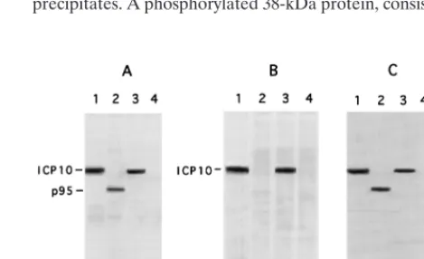
immunopre-cipitates. A phosphorylated 140-kDa protein consistent with
ICP10 was observed in HSV-2 (Fig. 3B, lane 1)- or HSV-2(R)
(Fig. 3B, lane 3)-infected cells, but p95 was no phosphorylated
(Fig. 3B, lane 2). This is not due to low levels of protein in the
precipitates, since the levels of p95 detected by
immunoblot-ting of the precipitates from ICP10
D
PK-infected cells with
ICP10 antibody (Fig. 3C, lane 2) were similar to those of ICP10
in HSV-2 (Fig. 3C, lane 1) and HSV-2(R) (Fig. 3C, lane 3)
precipitates. A phosphorylated 38-kDa protein, consistent with
FIG. 1. Schematic representation of ICP10DPK DNA. Oligonucleotide probe AU26 recognizes the 7.6-kb BamHI E fragment from HSV-2 or HSV-2(R) DNA and a 2.2-kb BamHI fragment from ICP10DPK DNA. Oligonucleotide probe AU25 recognizes the 7.6-kb BamHI E fragment from HSV-2 or HSV-2(R) DNA and a 4.4-kb BamHI fragment from ICP10DPK DNA.
[image:3.612.331.522.67.322.2]FIG. 2. Southern blot hybridization of BamHI-digested DNA from ICP10DPK (lanes 1 and 4), HSV-2 (lanes 2 and 5), or HSV-2(R) (lanes 3 and 6) with a DIG-labeled AU26 (lanes 1 to 3) or AU25 (lanes 4 to 6) oligonucleotide probe. Size markers are shown in the right margin.
FIG. 3. Expression and PK activity of the p95 protein from ICP10D PK-infected cells. (A) Vero cells were PK-infected with HSV-2 (lane 1), ICP10DPK (lanes 2 and 4), or HSV-2(R) (lane 3) and labeled with [35S]methionine from 6
to 16 h p.i. Cell extracts obtained at this time were immunoprecipitated with ICP10 antibody (recognizes amino acids 13 to 26) (lanes 1 to 3) or preimmune serum (lane 4). (B) Immunocomplex PK assays with ICP10 antibody (lanes 1 to 3) or preimmune serum (lane 4) of extracts from Vero cells infected for 16 h with HSV-2 (lane 1), ICP10DPK (lanes 2 and 4), or HSV-2(R) (lane 3). (C) Immu-noprecipitates from panel B immunoblotted with ICP10 antibody.
on November 9, 2019 by guest
http://jvi.asm.org/
[image:3.612.309.544.509.652.2]RR2, was also seen in the ICP10 precipitates from HSV-2 (Fig.
3B, lane 1) and HSV-2(R) (Fig. 3B, lane 3)-infected cells, but
it was not seen in those from ICP10
D
PK-infected cells (Fig. 3B,
lane 2). Preimmune serum was negative (Fig. 3B, C, and lanes
4). These data are consistent with previous reports that ICP10
PK phosphorylates both viral and cellular substrates (2, 6, 10,
47, 50) and indicate that the PK coding region is required for
kinase activity, also within the context of virus infection.
The ICP10
D
PK virus has RR activity.
Although p95
copre-cipitates with RR2 (Fig. 3A, lane 2), the question arises of
whether the loss of the ICP10 PK domain affects RR activity.
To address this question, RR assays were performed on
ex-tracts from cells infected with ICP10
D
PK, 2, or
HSV-2(R) (MOI, 20 PFU/cell) for 12 h as previously described (12,
63). As shown in Table 1, the RR activity of the ICP10
D
PK
virus (8.4 U) was similar to those of HSV-2 and HSV-2(R)
(10.2 and 8.8 U, respectively), supporting the conclusion that
the PK and RR activities can be functionally dissociated (10,
32, 37, 42).
The ICP10
D
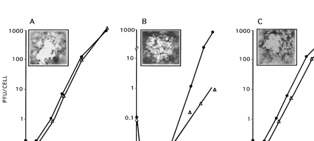
PK virus is defective for growth in culture.
In a
first series of experiments, we examined the growth of the
ICP10
D
PK virus in dividing (10% serum) and nondividing
(0.5% serum) Vero cells infected with 2 PFU/cell. Adsorption
was for 2 h (0 h on the growth curve), and virus titers were
determined at 2 to 36 h after adsorption. Cells similarly
in-fected with HSV-2 or HSV-2(R) served as controls. As shown
in Fig. 4A, HSV-2 grew equally well in dividing and
nondivid-ing cells. Virus replication began at 2 h after adsorption. The
burst size was 1,000 PFU/cell at 36 h after adsorption. A similar
growth pattern was evidenced by HSV-2(R) (Fig. 4C). By
con-trast, onset of ICP10
D
PK replication was not seen until 15 h
after adsorption in both exponentially growing and
serum-starved cells (Fig. 4B). In dividing cells, the rate of virus growth
between 15 and 36 h and the virus titers at 36 h after
adsorp-tion were similar to those seen for HSV-2 (burst size, 1,000
PFU/cell). However, virus titers were significantly (1,000-fold)
lower in serum-starved cells (burst size, 1 PFU/cell at 36 h).
In a second series of experiments, we considered the
possi-bility that growth defects may be MOI dependent and repeated
the growth curve measurement with cells infected at a
signifi-cantly higher MOI (200 PFU/cell). The growth of the ICP10
D
PK
virus in dividing cells was somewhat improved by infection
under these conditions in that virus replication began at 10 to
12 h after adsorption, compared to 15 h at a low MOI.
How-ever, virus titers in serum-starved cells were still approximately
1,000-fold lower than in dividing cells (burst sizes, 1 and 980
PFU/cell, respectively). The growth defect does not appear to
be cell type determined, as similar results were obtained with
HeLa, L, BHK (data not shown), and 293 (Fig. 5A) cells.
[image:4.612.50.290.81.139.2]Single step growth kinetics indicate that ICP10
D
PK grows as
well as HSV-2 and HSV-2(R) in cells which supply ICP10 PK
activity in trans, such as JHLa1 (41, 64). 293 and JHLa1 cells
were infected with the ICP10
D
PK virus at 200 PFU/cell in
EMEM containing 1% FCS, a condition which does not
sup-port efficient virus growth. In JHLa1 cells, replication was first
seen at 2 h after adsorption, and the burst size at 20 h was 2,500
PFU/cell. This compares to growth onset at 10 h after
adsorp-tion in 293 cells and a burst size of 8 PFU/cell at 20 h after
adsorption (Fig. 5A). By contrast, HSV-2 grew equally well in
JHLa1 and 293 cells. Replication began at 2 h, and the burst
sizes at 20 h after adsorption were 2,800 and 2,750 PFU/cell in
FIG. 4. Virus growth in dividing and nondividing cells. Vero cells were grown and infected (MOI, 2 PFU/cell) with HSV-2 (A), ICP10DPK (B), or HSV-2(R) (C) in 10% (F) or 0.5% (‚) FCS. Adsorption was for 2 h at 37°C (0 h of the growth curve). Virus titers were determined at 2 to 36 h after adsorption, and results are
[image:4.612.51.552.461.687.2]expressed as PFU per cell (burst size). The insets in each panel show the morphologies of the respective virus plaques in Vero cells overlaid with EMEM–10% FCS and 0.3% IgG and stained with Giemsa at 48 h. ICP10DPK plaques in Vero-ICP10 cells were identical to those of HSV-2 in Vero cells.
TABLE 1. RR activity of ICP10
D
PK virus
Virus Radioactivity (cpm)a RR sp act (U)b
HSV-2
11,534
10.2
HSV-2(R)
10,037
8.8
ICP10
D
PK
9,540
8.4
Mock infected
3,060
2.7
aIn cpm/270 mg of protein.
bOne RR unit equals conversion of 1 nmol of CDP to dCDP/h/mg of protein.
on November 9, 2019 by guest
http://jvi.asm.org/
JHLa1 and 293 cells, respectively (Fig. 5B). Similar results
were obtained for HSV-2(R), with replication beginning at 2 h
after adsorption and burst sizes of 2,570 and 2,610 PFU/cell in
JHLa1 and 293 cells, respectively (Fig. 5C). We interpret these
findings to indicate that ICP10
D
PK evidences two growth
de-fects: (i) delayed growth onset, which is seen in both dividing
and nondividing cells, and (ii) impaired replication, which is
seen only in nondividing cells. An induced or activated cellular
function(s) compensates for the missing viral protein in
divid-ing cells but not in serum-starved cells.
ICP10
D
PK has altered plaque morphology and
compro-mised plaquing ability.
To analyze the plaque-forming ability
of the ICP10
D
PK virus, we used Vero and Vero-ICP10 cells
grown in 10 or 0.5% serum. ICP10
D
PK plaque-forming ability
was severely compromised in serum-starved Vero cells but not
in dividing cells, in which it was similar to that of HSV-2.
Plaque-forming ability was also normal in Vero-ICP10 cells,
which supply ICP10 PK activity (Table 2). The size of the
ICP10
D
PK plaques was similar to that of HSV-2 or HSV-2(R)
plaques. However, in both dividing and nondividing Vero cells,
the ICP10
D
PK plaques differed from those of 2 or
HSV-2(R) in that they were hazy, apparently reflecting incomplete
cell lysis (Fig. 4B, inset). The extent of cell lysis differed
some-what from one experiment to the next, but it was never as
complete as that seen for HSV-2 (Fig. 4A, inset) or HSV-2(R)
(Fig. 4C, inset). The morphology of the ICP10
D
PK plaques in
Vero-ICP10 cells was similar to that of HSV-2 and HSV-2(R)
plaques (data not shown).
ICP10
D
PK has wild-type adsorption-and-penetration
kinet-ics and is not defective in capsid transport to the nucleus.
One
possible interpretation for the growth and plaquing patterns
evidenced by the ICP10
D
PK virus is that it is defective in the
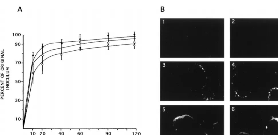
ability to adsorb to and penetrate target cells. To address this
question, we exposed Vero cells in six-well plates to 5 or 200
PFU of HSV-2, ICP10
D
PK, or HSV-2(R) for 0, 10, 30, 60, 90,
and 120 min at 4°C. At this time, the plates were extensively
washed with PBS and overlaid with EMEM–10% FCS and
0.3% IgG. The plates were reincubated at 37°C for 48 h and
then scored for the number of plaques. Under these
condi-tions, each plaque represents the progeny of 1 adsorbed PFU.
As shown in Fig. 6A, the number of HSV-2 plaques increased
as a function of exposure time, reaching maximal levels at 20 to
30 min and plateauing thereafter. Similar patterns were seen
for ICP10
D
PK and HSV-2(R) and at both MOIs, suggesting
that ICP10
D
PK is not defective for adsorption and
penetra-tion.
Another interpretation for the growth and plaquing defect
evidenced by ICP10
D
PK is that PK activity is required for the
transport of capsids from the cell periphery to the nucleus. To
address this possibility, Vero cells were exposed to HSV-2 or
ICP10
D
PK at 100 PFU/cell and virus adsorption was allowed
to occur at 4°C for 2 h. At this time (0 h), the cultures were
transferred to 37°C. They were stained with capsid antibody at
0, 2, 3, and 4 h thereafter. For both HSV-2 and ICP10
D
PK,
staining was not seen at 0 h, indicating that capsids present in
surface-bound intact viruses are not recognized by the
body (Fig. 6B, panels 1 and 2). Presumably, this reflects
anti-gen inaccessibility due to epitope masking in the intact virus
particles by envelope and/or tegument components. By 2 h
after adsorption, isolated labeled spots were seen at the
nu-clear membrane, with a similar distribution in HSV-2 (Fig. 6B,
panel 3)- and ICP10
D
PK (Fig. 6B, panel 4)-infected cells.
Their number appeared to increase with time, such that by 4 h
they had coalesced into rings localizing around the nuclear
membrane. The distribution and intensity of the labeled spots,
and the number of staining cells, were similar in HSV-2 (Fig.
6B, panel 5)- and ICP10
D
PK (Fig. 6B, panel 6)-infected cells.
Similar results were obtained for HSV-2(R) (data not shown).
While we do not exclude the possibility that the number of
capsids represented by a fluorescent spot differs for ICP10
D
PK
and HSV-2, the data suggest that capsid transport to the
nu-cleus is not significantly different for the two viruses. The
transport of tegument proteins was not studied.
p95 expression in ICP10
D
PK-infected cells and its
intracel-lular localization.
These studies sought to examine whether
the kinetics of p95 expression in ICP10
D
PK-infected cells and
its intracellular localization are similar to those of ICP10. Vero
cells were infected with HSV-2, HSV-2(R), or ICP10
D
PK for
6, 8, 12, or 18 h (in 10% serum) and stained by an indirect
immunofluorescence assay with ICP10 antibody. The results
are shown in Fig. 7. Approximately 70% of the HSV-2- and
HSV-2(R)-infected cells stained with the ICP10 antibody at 6 h
p.i., and the proportion reached 100% at 8 h p.i., as previously
reported for HSV-2-infected cells (10). Staining was localized
in the cytoplasm and the perinuclear region and had a diffuse
distribution pattern. By contrast, cells infected with ICP10
D
PK
for 6 h did not stain with the ICP10 antibody. Staining was first
seen at 8 h p.i. and in only 5 to 10% of the infected cells. It was
in the perinuclear space and in restricted cytoplasmic granules.
FIG. 5. Virus growth in cells that constitutively express ICP10. JHLa1 cells, which constitutively express ICP10 (‚), and 293 cells, which were used to
estab-lish JHLa1 (Œ), were infected with ICP10DPK (A), HSV-2 (B), or HSV-2(R) (C)
[image:5.612.52.289.68.208.2]at an MOI of 200 PFU/cell and overlaid with medium containing 1% FCS. Adsorption was for 2 h (0 h of the growth curve), and virus titers were assayed at 2 to 20 h after adsorption. Results are expressed as PFU per cell (burst size). Onset of ICP10DPK replication in 293 cells infected in 10% FCS is similarly delayed.
TABLE 2. Plaquing efficiency of ICP10
D
PK virus in dividing and
serum-starved cells
Virus (serum concn [%])Cells a Virus titer b (wild-type/mutant ratio)
HSV-2
Vero (10)
5.0
3
10
7ICP10
D
PK
Vero (10)
2.8
3
10
7(1.8)
HSV-2
Vero (0.5)
4.7
3
10
7ICP10
D
PK
Vero (0.5)
3.5
3
10
4(1.3
3
10
3)
HSV-2
Vero-ICP10 (10)
4.9
3
10
7ICP10
D
PK
Vero-ICP10 (10)
2.2
3
10
7(2.2)
HSV-2
Vero-ICP10 (0.5)
4.6
3
10
7ICP10
D
PK
Vero-ICP10 (0.5)
2.0
3
10
7(2.3)
aPlaque assays were done in medium containing 10 or 0.5% serum. bIn PFU per milliliter.on November 9, 2019 by guest
http://jvi.asm.org/
Diffuse cytoplasmic staining was not observed at this time (Fig.
7). The proportion of staining cells increased with time p.i.,
reaching levels of 15 to 20% and 85 to 100% at 12 and 18 h p.i.,
respectively. These findings indicate that the expression of p95
is delayed relative to that of ICP10 and, at least during the first
12 h p.i., appears to be partially sequestered within granular
structures in the cytoplasm. The delay in p95 expression and its
sequestration in granular structures were also seen in cells
infected with ICP10
D
PK in the presence of 1% FCS (data not
shown).
Onset of protein synthesis is delayed in ICP10
D
PK-infected
cells.
Delayed onset of p95 expression and ICP10
D
PK
replica-tion may reflect the failure to initiate the protein synthesis
cascade. To examine the validity of this interpretation, Vero
cells were mock infected (with PBS) or infected with HSV-2,
ICP10
D
PK, or HSV-2(R) (MOI, 200 PFU/cell) in medium
containing 10% FCS. At 2, 7, or 11 h p.i., the cultures were
pulse labeled with [
35S]methionine for 60 min. Proteins in the
cell extracts obtained at that time were resolved by
SDS-PAGE. As previously described (51, 68, 76), protein profiles in
cells infected with HSV-2 for 3 h included IE species ICP4,
ICP0, ICP22, and ICP27, as well as ICP10 (Fig. 8A, lane 2).
Host protein synthesis was significantly decreased relative to
that of mock-infected cells (Fig. 8A, lane 1), as exemplified by
host cell protein H (Fig. 8A, lane 2). Additional viral proteins
were seen in cells infected with HSV-2 for 8 h (Fig. 8A, lane 3)
or 12 h (Fig. 8B, lane 4). Similar protein profiles were seen in
cells infected with HSV-2(R), as shown in Fig. 8A (lane 8) for
3-h-infected cells.
By contrast, the protein profile in cells infected with
ICP10
D
PK for 3 h (Fig. 8A, lane 5) was not significantly
dif-ferent from that in mock-infected cells (Fig. 8A, lane 1). ICP4
(identity confirmed by immunoblotting [Fig. 8C, lane 3]),
ICP22, and ICP27 were not seen in ICP10
D
PK-infected cells,
and the levels of ICP0 (identity confirmed by immunoblotting
[Fig. 8C, lane 1]) were fourfold lower than in cells infected with
HSV-2 or HSV-2(R) [3,130, 3,099 and 782 densitometric
inte-gration U for HSV-2, HSV-2(R), and ICP10
D
PK,
respective-ly]. The levels of p95 (identity confirmed by immunoblotting
[Fig. 8C, lane 2]) were also lower (sevenfold) than the ICP10
levels in HSV-2- or HSV-2(R)-infected cells [3,567, 3,630, and
480 densitometric integration U for HSV-2, HSV-2(R), and
ICP10
D
PK, respectively]. At 8 h p.i., with ICP10
D
PK, the
lev-els of ICP0 and p95 were higher, and bands consistent with
ICP4, ICP22, and ICP27 were also seen (Fig. 8A, lane 6).
Protein profiles in cells infected with ICP10
D
PK for 12 h (Fig.
8A, lane 7) were similar to those seen in HSV-2-infected cells
at 8 h p.i. (Fig. 8A, lane 3). This is consistent with the growth
kinetics in cells infected at a high MOI in that virus replication
under these conditions begins at 10 to 12 h after adsorption
both in 10 and 1% FCS (Fig. 4 and 5). In JHLa1 cells,
expres-sion of the major IE genes (those for ICP4, ICP22, ICP27, and
ICP0) was seen as early as 3 h p.i. with ICP10
D
PK (Fig. 8B,
lane 1), and their levels were comparable to those seen in
HSV-2-infected cells (Fig. 8B, lane 2).
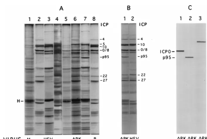
[image:6.612.59.545.64.301.2]ICP10 PK is required for expression of ICP4, ICP27, and
ICP22.
To further examine the synthesis of IE proteins in
ICP10
D
PK-infected cells, we used a cycloheximide block, a
condition which allows expression of IE but not other viral
FIG. 6. Virus adsorption and penetration of cells and capsid transport to the nucleus. (A) Six-well plates of Vero cells were exposed to 200 PFU of HSV-2 (F),
ICP10DPK (E), or HSV-2(R) ({) at 4°C for 0, 10, 30, 60, 90, and 120 min. At this time, the cells were overlaid with EMEM–10% FCS and 0.3% IgG and reincubated
at 37°C. They were scored for plaque numbers at 48 h. (B) Vero cells were infected at an MOI of 100 PFU/cell with HSV-2 (panels 1, 3, and 5) or ICP10DPK (panels 2, 4, and 6), and adsorption was allowed to occur for 2 h at 4°C. The cells were fixed in 3% PFA, permeabilized with 0.1% Triton X-100, and stained by immunofluorescence with capsid antibody at 0 (panels 1 and 2), 2 (panels 3 and 4), or 4 (panels 5 and 6) h after adsorption.
FIG. 7. p95 synthesis and intracellular localization. Vero cells were infected with HSV-2 for 6 h or with ICP10DPK for 6 or 8 h and stained with ICP10 antibody.
on November 9, 2019 by guest
http://jvi.asm.org/
genes (29, 69). Cells were infected with ICP10
D
PK, HSV-2, or
HSV-2(R) at 200 PFU/cell in the presence of 50-
m
g/ml
cyclo-heximide (6 h) and labeled with [
35S]methionine for 3 h in
medium containing 10-
m
g/ml actinomycin D. Proteins
consis-tent with ICP4, ICP10, ICP0, ICP22, and ICP27 were seen in
cells infected with HSV-2 (Fig. 9A, lane 2) or HSV-2(R) (Fig.
9A, lane 3) under these conditions. ICP4, ICP22, and ICP27
were not seen in cells similarly infected with ICP10
D
PK (Fig.
9A, lane 4). A 110-kDa protein which is recognized by
anti-ICP0 antibody (Fig. 9B, lane 1) was seen in the ICP10
D
PK-infected cells (Fig. 9A, lane 4), but its levels were twofold lower
than in HSV-2 (Fig. 9A, lane 2)- or HSV-2(R) (Fig. 9A, lane
3)-infected cells (1,760 and 3,520 densitometric integration U
for ICP10
D
PK- and HSV-2-infected cells, respectively). p95
was also seen in ICP10
D
PK-infected cells (Fig. 9A, lane 4, and
B, lane 2), but its levels were fivefold lower than those of ICP10
in HSV-2-infected cells (413 and 2,200 densitometric
integra-tion U for p95 and ICP10, respectively). The data support the
conclusion that ICP10 PK is required for optimal IE gene
expression. Significantly, host protein synthesis was not shut off
in ICP10
D
PK-infected cells (Fig. 9A, lane 4), although the
infection was at the same MOI as for HSV-2 or HSV-2(R).
This may reflect the role of ICP10 PK in the phosphorylation
of vhs (55), which is responsible for host shutoff (54).
IE gene transcription is delayed in ICP10
D
PK-infected
cells.
Northern hybridization was used to examine whether the
defect in IE gene expression in ICP10
D
PK-infected cells is at
the level of transcription. RNA was obtained from Vero cells
infected with HSV-2, ICP10
D
PK, or HSV-2(R) (in 10%
se-rum), and ICP4 and ICP0 DNAs were used as probes. GAPDH
served as a control transcript. The relative abundance of ICP4
and ICP0 mRNAs was estimated by first normalizing to the
value of GAPDH mRNA in each sample. The kinetics of ICP4
expression in HSV-2-infected cells were similar to those
pre-viously described for HSV-1-infected cells (27). Optimal levels
were seen at 3 h p.i. (Fig. 10B, lane 2), and the transcript was
no longer detectable at 8 h p.i. (Fig. 10B, lane 4). By contrast,
in cells infected with ICP10
D
PK, ICP4 mRNA was not seen at
3 and 4 h p.i. (Fig. 10A, lanes 2 and 3). It was first seen at 8 h
p.i. (Fig. 10A, lane 4), at which time its levels were similar to
those seen in HSV-2-infected cells at 3 h p.i. (Fig. 10B, lane 2).
The ICP4 transcript was still seen at 12 h p.i. (Fig. 10A, lane 5),
indicating that the eventual expression of ICP4 correlates with
the rise of virus growth late in infection. The transcript was no
longer detected at 20 h p.i. with ICP10
D
PK (Fig. 10A, lane 7).
Similar results were obtained for ICP27 (data not shown).
ICP0 mRNA was seen at 3 h p.i. with ICP10
D
PK (Fig. 10C,
lane 2), but its relative abundance (expressed as ICP0/GAPDH
mRNA) was threefold lower than in cells similarly infected
with HSV-2 (Fig. 10C, lane 1) or HSV-2(R) (Fig. 10C, lane 3)
(ICP0/GAPDH ratios of 0.32, 1.0, and 0.95, respectively). We
interpret these data to indicate that ICP10 PK is required for
early transcription of the IE genes.
DISCUSSION
[image:7.612.124.477.66.303.2]Studies of deletion and temperature-sensitive mutants
showed that RR1 is required for virus growth in nondividing
cells in culture (25, 26, 63), as well as for optimal
neuroviru-lence (7, 31) and latency reactivation (33, 58) in infected
ani-mals. However, these studies did not differentiate between the
respective contributions of the PK and RR domains of the
multifunctional RR1 proteins. In nondividing cells, the RR
domain presumably functions to supply the RR activity which
is necessary for virus growth (25, 26, 63). The PK activity is not
required for ribonucleotide reduction (15), and it can be
dis-sociated from the RR activity (10, 15, 32, 37, 42). Because the
PK domain likely evolved from a cellular gene and was
con-served through many evolutionary cycles (62), it seems
reason-able to conclude that the PK activity is critical for virus growth.
The studies described in this report were designed to test this
FIG. 8. Protein profiles of HSV-2- and ICP10DPK-infected cells. (A) Vero cells were mock infected (lane 1) or infected with HSV-2 (lanes 2 to 4), ICP10DPK (lanes 5 to 7), or HSV-2(R) (lane 8) in EMEM containing 10% FCS. They were labeled with [35S]methionine from 2 to 3 h p.i. (lanes 1, 2, 5, 8), 7 to 8 h p.i. (lanes 3 and
6), or 11 to 12 h p.i. (lanes 4 and 7), and proteins were resolved by SDS–8.5% PAGE. H, host protein. (B) JHLa1 cells, which express ICP10 constitutively, were infected with ICP10DPK (lane 1) or HSV-2 (lane 2) for 2 h and labeled with [35S]methionine from 2 to 3 h p.i. in EMEM containing 1% FCS. Proteins were resolved by
SDS–8.5% PAGE. (C) Duplicate samples of the extracts from cells infected with ICP10DPK for 3 h (lanes 1 and 2) or 8 h (lane 3) were immunoblotted with antibody to ICP0 (lane 1), ICP10 (lane 2), or ICP4 (lane 3). Protein profiles in Vero-ICP10 cells were similar to those in JHLa1 cells; profiles in 293 cells were similar to those in Vero cells.
on November 9, 2019 by guest
http://jvi.asm.org/
hypothesis. The following comments seem pertinent with
re-spect to our findings.
The virus used in these studies (ICP10
D
PK) is an HSV-2
mutant with a deletion in the RR1 PK domain. It expresses a
95-kDa protein (p95) that lacks PK activity but retains the
ability to complex with RR2, giving rise to an RR activity
similar to that of the wild-type virus. This is consistent with our
previous finding that ICP10 residues which complex with RR2
are at the C terminus (amino acids 1096 to 1144) (12).
ICP10
D
PK is unlikely to have defects other than a PK-deficient
ICP10, since a revertant virus [HSV-2(R)] was generated by
recombination of ICP10
D
PK DNA with the wild-type BamHI
E/T fragment that encompasses the ICP10 coding sequences,
but not with the BamHI E/T fragment with the sequences
which code for ICP10 PK deleted. Because VP16 activates the
expression of IE genes (8, 53) as well as that of RR1 (18, 70, 77,
78), we considered the possibility that growth defects
evi-denced by ICP10
D
PK may be due to a mutated VP16 gene.
However, this is unlikely, since the wild-type phenotype was
not restored in a rescue experiment with VP16-encoding DNA,
and the levels of VP16 were similar in ICP10
D
PK- and
HSV-2-infected cells (data not shown). Nonetheless, we do not
ex-clude the possibility that VP16 is involved in the timely
expres-sion of IE genes in cells infected with ICP10
D
PK, because
VP16 is phosphorylated on serine residues (48) and it may be
a substrate for ICP10 PK.
Single-step growth curve analyses indicated that ICP10
D
PK
has two, apparently distinct, growth defects. First, onset of
virus replication was significantly delayed (10 to 15 h) in both
dividing and nondividing cells. Second, in nondividing cells,
virus titers were approximately 1,000-fold lower than in
divid-ing cells, and plaqudivid-ing ability was severely compromised.
Sim-ilar defects were observed in various cell lines, indicating that
they are not determined by the cell type. They were not
evi-denced by the restored virus [HSV-2(R)] or in cells that
con-stitutively express ICP10 (JHLa1 or Vero-ICP10), indicating
that they are due to the lack of a functional ICP10 PK.
[image:8.612.78.259.66.313.2]The delayed onset of virus growth is consistent with previous
conclusions that the IE component of RR1 regulation is
re-quired for early expression of PK activity (18, 70, 77, 78, 81).
Presumably, growth onset at 10 to 15 h p.i. reflects
compensa-tion for the missing ICP10 PK by a cellular funccompensa-tion which may
FIG. 9. IE protein synthesis in HSV-2- and ICP10DPK-infected cells. (A) Vero cells were mock infected (lane 1) or infected with 2 (lane 2), HSV-2(R) (lane 3), or ICP10DPK (lane 4) in the presence of 50-mg/ml cycloheximide (6 h) and labeled with [35S]methionine for 3 h in medium containing 10-mg/ml
actinomycin D. Proteins were resolved by SDS–8.5% PAGE. (B) Duplicate samples of extracts from cells infected with ICP10DPK were immunoblotted with ICP0 antibody (lane 1) or ICP10 antibody (lane 2).
FIG. 10. ICP4 and ICP0 RNA synthesis in ICP10DPK-infected cells. (A) RNA was isolated from cells infected (in 10% FCS) with ICP10DPK at 0 to 20 h p.i. (lanes 1 to 7) and from cells infected with HSV-2(R) at 3 h p.i. (lane 8). It was hybridized with a32P-labeled ICP4 DNA probe or GAPDH oligonucleotide (bottom). Molecular
size marker positions are indicated in the margin. (B) RNA was isolated from cells infected with HSV-2 at 1 to 8 h p.i. (lanes 1 to 4) and hybridized with a32P-labeled
ICP4 DNA probe or GAPDH oligonucleotide (bottom). Molecular size marker positions are indicated in the margin. (C) RNA was isolated from cells infected with HSV-2 (lane 1), ICP10DPK (lane 2), or HSV-2(R) (lane 3) at 3 h p.i. and hybridized with a32P-labeled ICP0 DNA probe or GAPDH oligonucleotide (bottom).
Molecular size marker positions are indicated in the margin.
on November 9, 2019 by guest
http://jvi.asm.org/
[image:8.612.124.479.450.675.2]be induced or activated by virion structural proteins. Such an
interpretation is consistent with the finding that replication
begins 3 to 5 h earlier, when the cells are infected at a high (200
PFU/cell), rather than a low (2 PFU/cell), MOI. Virion
pro-teins that could induce or activate such a cellular function
include VP16, which was previously shown to activate cellular
promoters such as beta interferon (38) and Gal4 (79), and the
promoter-independent promiscuous transactivator ICP0 (20,
39), which was recently shown to be a virion protein (80).
Inasmuch as ICP10
D
PK growth began at the same time in
dividing and nondividing cells, the putative compensatory
function presumably does not require de novo protein
synthe-sis and may involve the activation of a PK cascade. It is
note-worthy that a mutant defective in both the PK and RR
activ-ities did not replicate, even in dividing cells, suggesting that the
RR domain may be involved in induction or activation of the
PK compensatory function (data not shown). Implicit in such
an interpretation is the conclusion that, in dividing cells,
suf-ficient levels of RR activity are produced early in infection with
ICP10
D
PK to induce or activate the PK compensatory
func-tion. Ongoing studies were designed to examine the validity of
this interpretation.
What is the function of ICP10 PK in the early onset of virus
growth? Our data suggest that ICP10
D
PK is not defective in
adsorption and penetration or in the transport of incoming
capsids to the nucleus. However, IE gene expression is
selec-tively delayed. Thus, ICP4, which is required for synthesis of
early and late viral proteins (17, 19, 47), was first seen in cells
infected with ICP10
D
PK at 8 h p.i., compared to 3 h p.i. for
HSV-2 and HSV-2(R). ICP4 mRNA was not seen until 8 h p.i.,
and neither ICP4 mRNA nor protein was seen with a
cyclo-heximide block, a condition that allowed IE gene expression in
cells infected with HSV-2 or HSV-2(R). Cells infected with
ICP10
D
PK for less than 8 h or in the presence of cycloheximide
were also negative for ICP27, which often functions together
with ICP4 to initiate early gene expression (60), and ICP22,
which is required for late gene expression (40, 57, 61). ICP0
was expressed early after infection with ICP10
D
PK (3 h) and in
the presence of cycloheximide, but its levels were significantly
lower than those in HSV-2- or HSV-2(R)-infected cells. p95
was also expressed early after infection with ICP10
D
PK and in
the presence of cycloheximide, but is levels were lower than
those of ICP10 in cells similarly infected with 2 or
HSV-2(R), suggesting that the PK domain is involved in ICP10
self-regulation. p95 expression in the absence of ICP4 is
con-sistent with previous findings that basal expression from the
RR1 promoter requires AP-1 transcription factors and is
in-dependent of ICP4 (18, 77, 78, 81).
Shutoff of host protein synthesis was delayed in ICP10
D
PK-infected cells, also in the presence of cycloheximide, and this is
consistent with the incomplete lysis of the infected cells and the
hazy appearance of the ICP10
D
PK plaques. Because vhs
phos-phorylation affects its ability to induce mRNA degradation
(55), impaired host shutoff may reflect the role of ICP10 PK in
vhs phosphorylation. Indeed, vhs is not phosphorylated by
an-other HSV PK species (UL13) (49), and a phosphorylated
57-to 59-kDa species consistent with vhs was not seen in
ICP10
D
PK-infected cells (data not shown). If vhs is
phosphor-ylated by ICP10 PK, ICP10
D
PK grown in JHLa1 cells may
contain a vhs-encoded protein which is relatively more
acti-vated (has higher mRNA degradation activity) than the
vhs-encoded protein of ICP10
D
PK propagated in Vero cells.
On-going studies were designed to test this interpretation.
The exact role of ICP10 PK in IE gene expression is
un-known. It is unlikely that it functions as a transactivator of IE
gene expression, because transactivating activity was not
ob-served in transient transfection assays with pICP4-cat
con-structs (unpublished data). Because ICP10 is located in the
virion tegument (65), its PK domain could be involved in the
transport of incoming VP16 to the nucleus, for example, by
maintaining tegument integrity. In addition, ICP10 PK could
phosphorylate and consequently activate VP16, thereby
deter-mining early onset of IE gene expression. Implicit in the
inter-pretation that ICP10 PK functions at the level of VP16 is the
conclusion that VP16 is not required for early expression of
ICP0, which is seen as early as 3 h p.i. with ICP10
D
PK.
How-ever, previous studies showed that VP16 is required for
expres-sion of ICP0 but not ICP4 (1). Also, inasmuch as the kinetics
of synthesis of the IE proteins and their levels were restored to
wild-type patterns in ICP10
D
PK-infected cells, which provide
ICP10 PK activity in trans, it is unlikely that ICP10 PK is
required for transport of tegument proteins to the nucleus.
Nonetheless, ongoing studies were designed to test this
hy-pothesis.
Delayed onset of ICP10
D
PK growth could be due to the
inhibition of IE gene expression by PK-defective ICP10. This
implies that in the wild-type virus, ICP10 PK downregulates a
protein which inhibits early onset of IE gene expression. A
function that represses accumulation of ICP4 transcripts was
recently described in mouse neurons latently infected with
HSV-1, but it was attributed to the latency-associated
tran-script (LAT) locus (9). It may also be that the PK domain is not
involved in IE gene expression but, rather, that in its absence,
the RR domain causes transdominant inhibition of IE gene
expression and virus growth. If this were the case, a virus with
deletions in both the PK and RR domains should grow as well
as HSV-2 in 10% FCS. However, a mutant with a deletion in
ICP10 failed to replicate under these conditions (data not
shown), suggesting that the RR domain does not have
trans-dominant downregulatory activity. Finally, ICP10 PK could
phosphorylate, and thereby activate, one or more factors
in-volved in IE gene transcription. For example, carboxy-terminal
domain (CTD) kinase(s), a component(s) of transcription
fac-tor IIH, is activated by phosphorylation (23, 28) and, in turn,
phosphorylates the CTD of the large subunit of polymerase II.
If their phosphorylation is altered by the direct or indirect
contribution of virion-associated PKs, polymerase II might be
redirected from cellular to viral IE promoters (56). This
pos-sibility has been excluded for HSV-1 virions. They contain only
one trans-phosphorylating kinase activity (UL13), and it does
not phosphorylate CTD kinases (57). However, ICP10 PK is
structurally and functionally different from ICP6 PK (14, 16,
46). ICP10 is located within the tegument fractions of HSV-2
virions and has trans-phosphorylating activity (65). It is
there-fore in a position to be involved in alterations of the
phosphor-ylation of CTD kinases. Implicit in this interpretation is the
conclusion that ICP10 PK functions in the nucleus. While the
available data indicate that ICP10 is localized in the cytoplasm
and on the surfaces of cells infected with HSV-2 for at least 6 h
(10), we recently found nuclear staining with monoclonal
an-tibodies to epitopes within the ICP10 PK (but not RR) domain
in cells infected with HSV-2 for 1 to 3 h, i.e., before the onset
of viral DNA synthesis (4a).
The finding that p95 is localized primarily within restricted
cytoplasmic compartments in cells infected with ICP10
D
PK for
8 to 12 h suggests that, in addition to its role in the early onset
of virus growth, the PK domain is involved in RR1 intracellular
localization. RR1 sequestration may render it unavailable for
complexation with RR2 and the generation of appropriate
levels of RR activity, thereby explaining the reduced virus
titers in nondividing cells.
What is the role of ICP10 PK in virus pathogenesis? In vivo,
on November 9, 2019 by guest
http://jvi.asm.org/
ICP10 PK might be required for virus replication at the site of
infection and, thereby, efficient latency establishment, and/or
for reactivation from latency. It has been proposed that in
addition to IE genes, early genes involved in viral DNA
syn-thesis must be turned on by the reactivating stimuli resulting in
limited DNA replication. This, in turn, stimulates a viral
func-tion that upregulates IE gene expression, leading to the lytic
cascade and the production of infectious virus (35). However,
reactivating stimuli upregulate AP-1 transcription factors (21,
34, 72), and RR1 is the only viral promoter that contains AP-1
cis response elements (77, 78, 81). Because the ICP10
pro-moter responds to AP-1 with basal expression which is
inde-pendent of VP16, ICP0, or ICP4 (18, 77, 78, 81), we propose
that ICP10 is an early response to latency-reactivating stimuli.
An AP-1 amplification loop is further provided by the ability of
ICP10 PK to activate the ras signaling pathway (30, 64). RR1
is uniquely compatible with a virus-reactivating function
be-cause it provides both the PK activity which is necessary for IE
gene expression and the RR activity which further upregulates
IE gene expression by inducing viral DNA synthesis in
nondi-viding neuronal cells (25, 26). ICP0, the expression of which is
thus increased, cooperates with AP-1 to further activate
ex-pression from the ICP10 promoter (18, 77, 78, 81). It also
upregulates other HSV IE genes. The outcome is initiation of
the lytic cascade and the production of infectious virus. Indeed,
recent studies indicate that the HSV-2 LATs, generally
as-sumed to be the only viral transcripts involved in latency
tivation, are inefficient and weak determinants of HSV-2
reac-tivation, at least as reflected by the quantity of the major LATs
in the ganglia (74). Furthermore, studies of the mouse
trigem-inal model indicate that during reactivation, early viral
scripts, notably, RR1 and TK, are detected before IE
tran-scripts (71). Consistent with these interpretations, HSV-2 was
not reactivated from latently infected ganglia explanted in the
presence of an antisense oligonucleotide that inhibits ICP10
expression (65a). Ongoing studies were designed to examine
the role of ICP10 PK in latency reactivation.
REFERENCES
1. Ace, C. I., T. A. McKee, M. Ryan, J. M. Cameron, and C. M. Preston. 1989. Construction and characterization of a herpes simplex virus type 1 mutant unable to transinduce immediate-early gene expression. J. Virol. 63:2260– 2269.
2. Ali, M. A., S. S. Prakash, and R. J. Jariwalla. 1992. Localization of the antigenic sites and intrinsic protein kinase domain within a 300 amino acid segment of the ribonucleotide reductase large subunit from herpes simplex virus type 2. Virology 187:360–367.
3. Anderson, K. P., R. J. Frink, G. B. Devi, B. H. Gaylord, and E. K. Wagner. 1981. Detailed characterization of the mRNA mapping in the HindIII frag-ment K region of the herpes simplex virus type 1 genome. J. Virol. 37:1011– 1027.
4. Aurelian, L., P. Terzano, C. C. Smith, T. D. Chung, A. Shamsuddin, S. Costa,
and C. Orlandi.1989. Amino-terminal epitope of herpes simplex virus type
2 ICP10 protein as a molecular diagnostic marker for cervical intraepithelial neoplasia. Cancer Cells 7:187–191.
4a.Aurelian, L., et al. Unpublished data.
5. Aurelian, L. 1992. Herpes simplex viruses, p. 473–494. In S. Specter, and G. Lancz (ed.), Clinical virology manual, 2nd ed. Elsevier Science Publishers, New York, N.Y.
6. Bacchetti, S., M. J. Evelegh, B. Muirhead, C. S. Sartari, and D. Huszar. 1984. Immunological characterization of herpes simplex virus type 1 and 2 polypeptide(s) involved in viral ribonucleotide reductase activity. J. Gen. Virol. 49:591–593.
7. Cameron, J. M., I. McDougall, H. W. Marsden, V. G. Preston, D. M. Ryan,
and S. H. Subak-Sharpe.1988. Ribonucleotide reductase encoded by herpes
simplex virus is a determinant of the pathogenicity of the virus in mice and a valid antiviral target. J. Virol. 69:2607–2612.
8. Campbell, M. E. M., J. W. Palfreyman, and C. M. Preston. 1984. Identifi-cation of herpes simplex virus DNA sequences which encode a trans-acting polypeptide responsible for stimulation of immediate early transcription. J. Mol. Biol. 180:1–19.
9. Chen, S. H., M. F. Kramer, P. A. Schaffer, and D. M. Coen. 1997. A viral function represses accumulation of transcripts from productive-cycle genes
in mouse ganglia latently infected with herpes simplex virus. J. Virol. 71: 5878–5884.
10. Chung, T. D., J. P. Wymer, C. C. Smith, M. Kulka, and L. Aurelian. 1989. Protein kinase activity associated with the large subunit of the herpes simplex virus type 2 ribonucleotide reductase (ICP10). J. Virol. 63:3389–3398. 11. Chung, T. D., J. P. Wymer, M. Kulka, C. C. Smith, and L. Aurelian. 1990.
Myristylation and polylysine-mediated activation of the protein kinase do-main of the large subunit of herpes simplex virus type 2 ribonucleotide reductase (ICP10). Virology 179:168–178.
12. Chung, T. D., J. H. Luo, J. P. Wymer, C. C. Smith, and L. Aurelian. 1991. Leucine repeats in the large subunit of herpes simplex virus type 2 ribonu-cleotide reductase (RR; ICP10) are involved in RR activity and subunit complex formation. J. Gen. Virol. 72:1139–1144.
13. Cohen, G. H. 1972. Ribonucleotide reductase activity of synchronized KB cells infected with herpes simplex virus. J. Virol. 9:408–418.
14. Conner, J., J. Cooper, J. Furlong, and J. B. Clements. 1992. An autophos-phorylating but not transphosautophos-phorylating activity is associated with the unique N terminus of the herpes simplex virus type 1 ribonucleotide reduc-tase large subunit. J. Virol. 66:7511–7516.
15. Conner, J., J. Macfarlene, H. Lankinen, and H. Marsden. 1992. The unique N-terminus of the herpes simplex virus type 1 large subunit is not required for ribonucleotide reductase activity. J. Gen. Virol. 73:103–112.
16. Cooper, J., J. Conner, and J. B. Clements. 1995. Characterization of the novel protein kinase activity present in the R1 subunit of herpes simplex virus ribonucleotide reductase. J. Virol. 69:4979–4985.
17. DeLuca, N. A., and P. A. Schaffer. 1985. Activation of immediate-early, early, and late promoters by temperature-sensitive and wild-type forms of herpes simplex virus type 1 protein ICP4. Mol. Cell. Biol. 5:1997–2008.
18. Desai, P., R. Ramakrishnan, Z. W. Lin, B. Osak, J. C. Glorioso, and M.
Levine.1993. The RR1 gene of herpes simplex virus type 1 is uniquely trans
activated by ICP0 during infection. J. Virol. 67:6125–6135.
19. Dixon, R. A. F., and P. A. Schaffer. 1980. Fine-structure mapping and func-tional analysis of temperature-sensitive mutants in the gene encoding the herpes simplex virus type 1 immediate early protein VP175. J. Virol. 36:189–203. 20. Everett, R. E., C. M. Preston, and N. W. Stow. 1991. Functional and genetic
analysis of the role of Vmw110 in herpes simplex virus replication, p. 49–76.
In E. K. Wagner (ed.), Herpesvirus transcription. CRC Press, Inc., Boca
Raton, Fla.
21. Fawl, R. L., and B. Roizman. 1993. Induction of reactivation of herpes simplex virus in murine sensory ganglia in vivo by cadmium. J. Virol. 67: 7025–7031.
22. Feng, C. P., M. Kulka, C. C. Smith, and L. Aurelian. 1996. Herpes simplex virus-mediated activation of human immunodeficiency virus is inhibited by oligonucleotide methylphosphonates that target immediate-early mRNAs 1 and 3. Antisense Nucleic Acid Drug Dev. 6:25–35.
23. Fisher, R. P., P. Jin, H. M. Chamberlin, and D. O. Morgan. 1995. Alternative mechanisms of CAK assembly require an assembly factor or an activating kinase. Cell 83:47–57.
24. Frame, M. C., H. S. Marsden, and B. M. Dutia. 1985. The ribonucleotide reductase induced by herpes simplex virus type 1 involves minimally a com-plex of two polypeptides (136K and 38K). J. Gen. Virol. 66:1581–1587. 25. Goldstein, D. J., and S. K. Weller. 1988. Herpes simplex virus type 1-induced
ribonucleotide reductase activity is dispensable for virus growth and DNA synthesis: isolation and characterization of an ICP6 lacZ insertion mutant. J. Virol. 62:196–205.
26. Goldstein, D. J., and S. K. Weller. 1988. Factor(s) present in the herpes simplex virus type-1 infected cells can compensate for the loss of the large subunit of the viral ribonucleotide reductase: characterization of an ICP6 deletion mutant. Virology 166:41–51.
27. Harris-Hamilton, E., and S. L. Bachenheimer. 1985. Accumulation of herpes simplex virus type 1 RNAs of different kinetic classes in the cytoplasm of infected cells. J. Virol. 53:144–151.
28. Hermann, C. H., M. O. Gold, and A. P. Rice. 1995. Viral transactivators specifically target distinct cellular protein kinases that phosphorylate the RNA polymerase II C-terminal domain. Nucleic Acids Res. 24:501–504. 29. Honess, R. W., and B. Roizman. 1974. Regulation of herpesvirus
macromo-lecular synthesis. I. Cascade regulation of the synthesis of three groups of viral proteins. J. Virol. 14:8–19.
30. Hunter, J. C. R., C. C. Smith, D. Bose, M. Kulka, R. Broderick, and L.
Aurelian.1995. Intracellular internalization and signaling pathways triggered
by the large subunit of HSV-2 ribonucleotide reductase (ICP10). Virology
210:345–360.
31. Idowu, A. D., E. B. Fraser-Smith, K. L. Paffenberger, and R. C. Herman. 1992. Deletion of herpes simplex virus type 1 ribonucleotide reductase gene alters virulence and latency in vitro. Antiviral Res. 17:145–156.
32. Ingemarson, R., and H. Lankinen. 1987. The herpes simplex virus type 1 ribonucleotide reductase is a tight complex of the type alpha 2 and beta 2 composed of 40K and 140K proteins, of which the latter shows multiple forms due to proteolysis. Virology 156:417–422.
33. Jacobson, J. G., D. A. Leib, D. J. Goldstein, C. L. Bogard, P. A. Schaffer,
S. K. Weller, and D. M. Coen.1989. A herpes simplex virus ribonucleotide
reductase deletion mutant is defective for productive acute and reactivatable