0022-538X/95/$04.0010
Copyrightq1995, American Society for Microbiology
Equal Levels of gp120 Retention and Neutralization Resistance
of Phenotypically Distinct Primary Human Immunodeficiency
Virus Type 1 Variants upon Soluble CD4 Treatment
MARTIJN GROENINK,
1JOHN P. MOORE,
2SILVIA BROERSEN,
1AND
HANNEKE SCHUITEMAKER
1*
Department of Clinical Viro-Immunology, Central Laboratory of the Netherlands Red Cross Blood Transfusion Service,
and Laboratory for Experimental and Clinical Immunology of the University of Amsterdam, Amsterdam,
The Netherlands,
1and Aaron Diamond AIDS Research Center, New York University School of Medicine,
New York, New York 10016
2Received 23 May 1994/Accepted 29 September 1994
Human immunodeficiency virus type 1 (HIV-1) variants passaged in T-cell lines, often called laboratory
isolates, are potently neutralized by soluble CD4 (sCD4), whereas primary HIV-1 variants are highly resistant
to sCD4 neutralization. Previously, it was demonstrated that the domain from V1 to V3 of the HIV-1 gp120
molecule contains one of the major determinants of sCD4 neutralization sensitivity, and the same region has
also been implicated as influencing syncytium-inducing (SI) capacity and T-cell-line tropism. To determine
possible differences in sCD4 neutralization sensitivity between phenotypically distinct primary HIV-1 variants,
a panel of non-syncytium-inducing (NSI) and SI HIV-1 variants was studied. Primary NSI and SI HIV-1
variants appeared to be equally resistant to sCD4 neutralization. Consistent with this observation, sCD4 did
not induce gp120 shedding from either primary NSI or SI HIV-1 variants at 37
&
C. Thus, it is not the potential
of certain primary HIV-1 variants to infect T-cell lines but rather their adaptation to T-cell lines that is
reflected in specific properties of the viral envelope which influence sCD4 neutralization sensitivity.
The biological variability of human immunodeficiency virus
type 1 (HIV-1) plays a role in the pathogenesis of AIDS (5, 7,
13, 26, 45, 46). In the asymptomatic stage of HIV-1 infection,
non-syncytium-inducing (NSI), preferentially
macrophage-tropic HIV-1 variants predominate (40), and these variants
remain present throughout the infection (7, 41). In contrast,
syncytium-inducing (SI) HIV-1 variants, which can replicate
efficiently in T-cell lines (27), emerge during the course of
HIV-1 infection in 50 to 60% of infected individuals (12, 26,
46). The emergence of more cytopathic, SI HIV-1 variants
precedes an accelerated CD4 T-cell depletion and progression
to AIDS (4, 7, 26).
The CD4 molecule serves as the principal receptor for
HIV-1 (9, 24, 28, 29). The interaction of the HIV-1 external
envelope glycoprotein gp120 with the CD4 molecule ultimately
mediates a pH-independent fusion of virus and cell membrane
that is required for HIV-1 entry (44). Consequently, the
gp120-CD4 interaction has provided a target for therapeutic
intervention in HIV-1 infection (33). One experimental
ap-proach was to compete for binding of HIV-1 to the CD4
receptor on the cell with soluble forms of the CD4 molecule. In
vitro experiments demonstrated that soluble CD4 (sCD4)
potently neutralizes HIV-1 variants that have been passaged
extensively in T-cell lines (so-called laboratory HIV-1 variants)
(10, 14, 20, 43, 49). Studies with such laboratory HIV-1 variants
revealed that the binding of sCD4 to gp120 on virions and
HIV-1-infected cells (19, 32, 37), as well as sCD4 binding to
gp120 on cells stably expressing HIV-1 envelope glycoproteins
(15, 32), resulted in the dissociation of gp120 from its complex
with gp41. The dissociation of gp120 may contribute to the
sCD4-induced neutralization. However, upon therapeutic
ap-plication of sCD4, no significant antiviral effect could be
detected, as assessed by p24 antigen levels in serum (23, 39)
and infectious HIV-1 titers in blood (8). The clinical failure of
sCD4, although some short-term beneficial effects of high-dose
sCD4 treatment have been reported (38), could be explained
by the observation that, compared with laboratory HIV-1
variants, primary HIV-1 variants which had been passaged only
in peripheral blood mononuclear cells (PBMC) were highly
resistant to neutralization by sCD4 in vitro (8, 23, 36, 39). This
relative resistance of primary HIV-1 variants correlated with
reduced sCD4 binding to virions and increased virion gp120
retention (31).
Studies with recombinant viruses generated from laboratory
HIV-1 variants and primary NSI, macrophage-tropic HIV-1
variants revealed that the region from the V1 domain to the V3
domain of the gp120 molecule contains a major determinant of
sCD4 neutralization sensitivity of HIV-1 (22, 25, 34). The same
region of gp120 has also been delineated as important for
differences in SI capacity and cytotropism (2, 6, 11, 17, 25, 35,
42). Thus, the available data suggest that sensitivity to sCD4
neutralization segregates with SI capacity.
To explore possible differences between primary NSI and
primary SI HIV-1 variants in their interactions with sCD4, a
panel of four primary NSI and two primary SI HIV-1 variants
was chosen (Table 1) (16, 18). All HIV-1 variants were
recovered from cryopreserved PBMC from participants of the
Amsterdam cohort studies. PBMC from the infected
individ-uals were cocultivated with 3-day-old
phytohemagglutinin-prestimulated PBMC from seronegative blood donors (45).
For comparison, the SI HIV-1 variant ACH-320.3.1, which was
molecularly cloned after a single passage through the H9 cell
line, and two HIV-1 variants extensively passaged in T-cell
lines (IIIB and the recombinant NL4-3 [1] that contains the 39
half of IIIB) were used.
To determine the sCD4 neutralization sensitivity of each
HIV-1 variant, an inoculum of fifty 50% tissue culture infective
* Corresponding author. Mailing address: Department of Clinical Viro-Immunology, Central Laboratory of the Netherlands Red Cross Blood Transfusion, Plesmanlaan 125, 1066 CX Amsterdam, The Netherlands. Phone: 31-20-5123317. Fax: 31-20-5123310.
523
on November 9, 2019 by guest
http://jvi.asm.org/
dose per ml was incubated for 2 h with 0, 0.12, 0.24, 0.49, 0.98,
1.95, 3.91, 7.81, 15.63, 31.25, 62.50, 125, or 250
m
g of
recom-binant sCD4 (Genentech Inc.) per ml. For use in the
experi-ments, all viral stocks had been grown and their titers had been
determined on 3-day-old phytohemagglutinin-prestimulated
PBMC. Since it was previously shown that, in contrast to
laboratory-adapted HIV-1 variants, primary viruses shed
gp120 to a greater extent after sCD4 treatment at 4
8
C than at
37
8
C (30, 31), the sCD4 neutralization sensitivity was
deter-mined at preincubation temperatures of both 4 and 37
8
C
before the addition of the virus-sCD4 mixture to the target
cells at 378C. These were 1.5
3
10
53-day
phytohemagglutinin-prestimulated PBMC from seronegative blood donors. The
cells were cultured in the presence of interleukin 2 (10 U/ml),
and on days 7 and 14 the culture supernatants were analyzed
for virus production as reflected by the presence of HIV-1 p24
(47). Each datum point represents quadruplicate cultures
(Table 1).
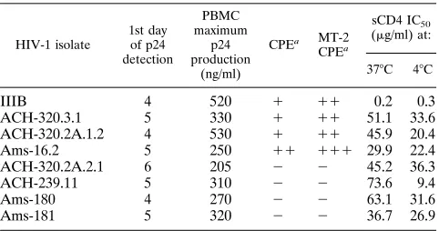
The sCD4 concentrations that reduced the virus titer by 50%
(50% inhibitory concentrations [IC
50s]) at 4 and 37
8
C are
shown in Table 1. The sCD4 IC
50s did not vary between days
7 and 14 (data not shown). As was shown previously (10, 14, 20,
43, 49), laboratory HIV-1 variant HIV-1 IIIB was highly
sensitive to neutralization by sCD4 at 37
8
C (IC
50of 0.2
m
g/ml).
Despite its single earlier passage in H9 cells, the PBMC-grown
molecular clone ACH-320.3.1 was highly resistant to sCD4
neutralization. In comparison with HIV-1 IIIB, the primary
HIV-1 variants required a 150- to 368-fold (IC
50of 29.9 to 73.6
m
g/ml) larger amount of sCD4 for 50% neutralization at 37
8
C.
However, no profound distinction between the sCD4
neutral-ization sensitivities of the NSI and SI primary HIV-1 variants
was observed. The sCD4 IC
50s at 37
8
C of the primary HIV-1
variants were somewhat lower than those at 4
8
C. However, as
found at 37
8
C, no major difference between the sCD4
neutral-ization sensitivities of NSI and SI primary HIV-1 variants was
observed when the virus-sCD4 preincubation temperature was
reduced to 4
8
C. Furthermore, PBMC-grown HIV-1 IIIB
re-mained highly sensitive to neutralization by sCD4 at 4
8
C (IC
50of 0.3
m
g/ml), whereas the H9-passaged PBMC-grown HIV-1
variant ACH-320.3.1 was as resistant to sCD4 neutralization as
the primary HIV-1 variants. In comparison with HIV-1 IIIB,
the primary HIV-1 variants required a 31- to 121-fold (IC50
of
9.4 to 36.3
mg/ml) larger amount of sCD4 for 50%
neutraliza-tion at 48C.
To investigate the effect of sCD4 on gp120 retention of
primary HIV-1 variants, 700-
m
l aliquots of HIV-1-containing
culture supernatants of the NSI primary HIV-1 variants
ACH-320.2A.2.1 and ACH-239.11 and the SI primary HIV-1 variants
ACH-320.2A.1.2 and Ams-16.2 were incubated with or without
10
mg of sCD4 per ml at 4 and 378C. For comparison, the single
H9-passaged PBMC-grown HIV-1 variant ACH-320.3.1 and
the frequently T-cell-line-passaged HIV-1 recombinant NL4-3
were used. One hundred-microliter samples were assayed for
gp120 retention at 0, 0.5, 1, 2, 4, and 8 h. The fractionation of
untreated controls and the HIV-1-sCD4 mixtures was
per-formed as described before, by a combination of gel filtration
on Sephacryl S-1000 and enzyme-linked immunosorbent assay
(31, 32).
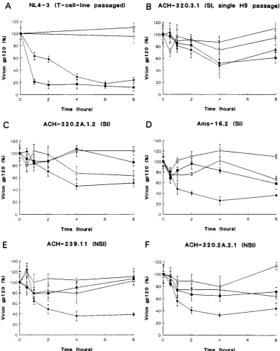
In the absence of sCD4, SI primary HIV-1 variant
ACH-320.2A.1.2 showed some spontaneous gp120 dissociation after
4 h at 4
8
C (Fig. 1C), whereas none of the other HIV-1 variants
lost significant amounts of gp120 within 8 h at 4 or 37
8
C (Fig.
1B and D to F). In the presence of sCD4, the primary HIV-1
variants and the single-H9-passaged HIV-1 variant
ACH-320.3.1 did not differ in their gp120 loss at 37
8
C. Thus, sCD4
did not cause significant gp120 shedding from either the NSI or
the SI primary HIV-1 variants within 8 h at 37
8
C (Fig. 1B to F).
The observation that sCD4 induced gp120 shedding from
NL4-3 (Fig. 1A) at both 4 and 37
8
C is consistent with results
obtained previously with other laboratory HIV-1 variants (19,
31, 32).
In contrast to the results obtained at 37
8
C, the primary
HIV-1 variants demonstrated a clear gp120 loss within 2 h in
the presence of sCD4 at 4
8
C (Fig. 1B to E). No distinction
between gp120 losses of NSI and SI primary HIV-1 variants
was observed. The rate of sCD4-induced gp120 loss from the
H9-passaged HIV-1 variant ACH-320.3.1 at 4
8
C was identical
to that observed at 378C (Fig. 1B).
Previously, it was demonstrated that a segment of gp120
spanning the V1 domain up to the V3 domain harbors major
determinants of sCD4 neutralization sensitivity (22, 25, 34), SI
capacity (2, 11, 17), and cytotropism (6, 21, 25, 35, 42). We
show here that the SI phenotype, with the ability to enter a
T-cell line, does not segregate with sCD4 sensitivity at 37
8
C.
Dissociation of the gp120 molecule from its complex with the
gp41 molecule may not be required for syncytium formation
and HIV-1 entry in T cells (48), but our data do not
unequiv-ocally resolve this issue. This is in agreement with the finding
that envelope glycoprotein mutants of the laboratory HIV-1
variant HXBc2, which exhibited marked decreases in gp120
loss in the presence of sCD4, could still efficiently form syncytia
(48). Moreover, it has been demonstrated with the laboratory
HIV-1 variant BH10 that syncytium formation can occur under
conditions in which sCD4 does not induce gp120 dissociation
(15). These findings imply that upon passage in T-cell lines a
selection for additional mutations in the region from the V2 to
the V3 domain or elsewhere in the envelope of certain viruses
of primary SI, T-cell-line-tropic HIV-1 variants may occur.
These additional mutations may increase the capacity of HIV-1
to replicate in T-cell lines, which may be concurrent with the
acquisition of sCD4 sensitivity at 37
8
C (50, 51). The HIV-1
variant ACH-320.3.1, which was obtained after only a single
passage in H9 cells, did not differ for sCD4-induced gp120
shedding and was as resistant to sCD4 neutralization as the
primary HIV-1 variants. The sCD4 neutralization sensitivity
and gp120 shedding observed for NL4-3 suggest that multiple
passages in T-cell lines, which will lead to an accumulation of
mutations, are required to create an HIV-1 variant fully
adapted to the T-cell line (laboratory HIV-1 variant) (3, 50,
51).
[image:2.612.58.299.91.218.2]In conclusion, we have demonstrated that primary SI,
po-tentially T-cell-line-tropic, and NSI HIV-1 variants can be
equally resistant to neutralization by sCD4 and do not differ in
TABLE 1. sCD4 IC50s for neutralization of HIV-1 variants with
distinct biological phenotypes
HIV-1 isolate 1st day of p24 detection PBMC maximum p24 production (ng/ml)
CPEa MT-2 CPEa
sCD4 IC50
(mg/ml) at:
378C 48C
IIIB 4 520 1 11 0.2 0.3
ACH-320.3.1 5 330 1 11 51.1 33.6
ACH-320.2A.1.2 4 530 1 11 45.9 20.4
Ams-16.2 5 250 11 111 29.9 22.4
ACH-320.2A.2.1 6 205 2 2 45.2 36.3
ACH-239.11 5 310 2 2 73.6 9.4
Ams-180 4 270 2 2 63.1 31.6
Ams-181 5 320 2 2 36.7 26.9
aCPE, cytopathic effect (i.e., SI capacity).2, no syncytia;1, 1 to 10 syncytia; 11, 11 to 50 syncytia;111,.50 syncytia per 1003field.
on November 9, 2019 by guest
http://jvi.asm.org/
their gp120 retention in the presence of sCD4 at 4 and 37
8
C.
Thus, although SI HIV-1 variants have the potential to
repli-cate in T-cell lines, the properties of the viral envelope that are
involved in sCD4 sensitivity are gained only after viral
adap-tation to T-cell lines. Hence, we emphasize that primary SI
variants are not the equivalent of HIV-1 variants extensively
passaged in T-cell lines.
This study was supported in part by grant 92-025 from RGO/WVC (Ministry of Public Health), by NIH grant AI 36082-01, and by the
Aaron Diamond Foundation. This work was performed as part of the Amsterdam cohort studies on AIDS.
We thank Shu-Wing Poon for technical assistance and Ron A. M. Fouchier for a critical reading of the manuscript. We are grateful to Genentech Inc. (South San Francisco, Calif.) for providing the sCD4.
REFERENCES
1. Adachi, A., H. E. Gendelman, S. Koenig, T. Folks, R. Willey, A. Rabson, and
[image:3.612.109.503.70.565.2]M. A. Martin.1986. Production of acquired immunodeficiency syndrome-associated retrovirus in human and nonhuman cells transfected with an infectious molecular clone. J. Virol. 59:284–291.
FIG. 1. gp120 retention by phenotypically distinct primary HIV-1 variants. HIV-1 variants were incubated for the times indicated with (F,■) or without (E,h) 10
mg of sCD4 per ml at 48C (E,F) and 378C (h,■). The extent of gp120 shedding was determined by measuring the amounts of virion-bound (V) and soluble (S) gp120 present in each sample and then using the formula V/(V1S)3100% to give a measure of the proportion of total gp120 that was virion associated. This value was defined as 100% for the samples obtained at time zero, and the values obtained at the later time points were normalized relative to this 100% value.
on November 9, 2019 by guest
http://jvi.asm.org/
2. Andeweg, A. C., P. Leeflang, A. D. M. E. Osterhaus, and M. L. Bosch. 1993. Both the V2 and V3 regions of the human immunodeficiency virus type 1 surface glycoprotein functionally interact with other envelope regions in syncytium formation. J. Virol. 67:3232–3239.
3. Ashkenazi, A., D. H. Smith, S. A. Marsters, L. Riddle, T. J. Gregory, D. D.
Ho, and D. J. Capon. 1991. Resistance of primary isolates of human immunodeficiency virus type 1 to soluble CD4 is independent of CD4-rgp120 binding affinity. Proc. Natl. Acad. Sci. USA 88:7056–7060.
4. Bozzette, S. A., J. A. McCutchan, S. A. Spector, B. Wright, and D. D.
Richman.1993. A cross-sectional comparison of persons with syncytium- and non-syncytium inducing human immunodeficiency virus. J. Infect. Dis.
168:1374–1379.
5. Cheng-Mayer, C., D. Seto, M. Tateno, and J. A. Levy. 1988. Biologic features of HIV-1 that correlate with virulence in the host. Science 240:80–82. 6. Chesebro, B., J. Nishio, S. Perryman, A. Cann, W. O’Brien, I. S. Y. Chen,
and K. Wehrly. 1991. Identification of human immunodeficiency virus envelope gene sequences influencing viral entry into CD4-positive HeLa cells, T-leukemia cells, and macrophages. J. Virol. 65:5782–5789. 7. Connor, R. I., H. Mohri, Y. Cao, and D. D. Ho. 1993. Increased viral burden
and cytopathicity correlate temporally with CD41T-lymphocyte decline and clinical progression in human immunodeficiency virus type 1-infected indi-viduals. J. Virol. 67:1772–1777.
8. Daar, E. S., X. L. Li, T. Moudgil, and D. D. Ho. 1990. High concentrations of recombinant soluble CD4 are required to neutralize primary human immunodeficiency virus type 1 isolates. Proc. Natl. Acad. Sci. USA 87:6574– 6578.
9. Dalgleish, A. G., P. C. L. Beverley, P. R. Clapham, D. H. Crawford, M. F.
Greaves, and R. A. Weiss. 1984. The CD4 (T4) antigen is an essential component of the receptor for the AIDS retrovirus. Nature (London)
312:763.
10. Deen, K. C., J. S. McDougal, R. Inacker, G. Folena-Wasserman, J. Arthos,
J. Rosenberg, P. J. Maddon, R. Axel, and R. W. Sweet.1988. A soluble form of CD4 protein inhibits AIDS virus infection. Nature (London) 331:82–84. 11. de Jong, J. J., J. Goudsmit, W. Keulen, B. Klaver, W. Krone, M. Tersmette,
and T. de Ronde. 1992. Human immunodeficiency virus type 1 clones chimeric for the envelope V3 domain differ in syncytium formation and replication capacity. J. Virol. 66:757–765.
12. Fenyo¨, E. M., J. Albert, and B. Asjo¨.1989. Replicative capacity, cytopathic effect and cell tropism of HIV. AIDS 3:S5–S12.
13. Fenyo¨, E. M., L. Morfeldt-Månson, F. Chiodi, B. Lind, A. Von Gegerfelt, J. Albert, E. Olausson, and B. Åsjo¨.1988. Distinct replicative and cytopathic characteristics of human immunodeficiency virus isolates. J. Virol. 62:4414– 4419.
14. Fisher, R. A., J. M. Bertonis, W. Meier, V. A. Johnson, D. S. Costopoulos, T.
Liu, R. Tizard, B. D. Walker, M. S. Hirsch, R. T. Schooley, and R. A. Flavell.
1988. HIV infection is blocked in vitro by recombinant soluble CD4. Nature (London) 331:76–78.
15. Fu, Y.-K., T. K. Hart, Z. L. Jonak, and P. J. Bugelski. 1993. Physicochemical dissociation of CD4-mediated syncytium formation and shedding of human immunodeficiency virus type 1 gp120. J. Virol. 67:3818–3825.
16. Groenink, M., A. C. Andeweg, R. A. M. Fouchier, S. Broersen, R. C. M. van
der Jagt, H. Schuitemaker, R. E. Y. de Goede, M. L. Bosch, H. G. Huisman and M. Tersmette.1992. Phenotype associated env gene variation among eight related human immunodeficiency virus type 1 clones: evidence for in vivo recombination and determinants of cytotropism outside the V3 domain. J. Virol. 66:6175–6180.
17. Groenink, M., R. A. M. Fouchier, S. Broersen, C. H. Baker, M. Koot, A. B.
Van ’t Wout, H. G. Huisman, F. Miedema, M. Tersmette, and H. Schuite-maker.1993. Relation of phenotype evolution of HIV-1 to envelope V2 configuration. Science 260:1513–1516.
18. Groenink, M., R. A. M. Fouchier, R. E. Y. de Goede, F. de Wolf, R. A.
Gruters, H. T. M. Cuypers, H. G. Huisman, and M. Tersmette. 1991. Phenotypic heterogeneity in a panel of infectious molecular human immu-nodeficiency virus type 1 clones derived from a single individual. J. Virol.
65:1968–1975.
19. Hart, T. K., R. Kirsh, H. Ellens, R. W. Sweet, D. M. Lambert, S. R. Petteway,
Jr., J. Leary, and P. J. Bugelski.1991. Binding of soluble CD4 proteins to human immunodeficiency virus type 1 and infected cells induces release of envelope glycoprotein gp120. Proc. Natl. Acad. Sci. USA 88:2189–2193. 20. Hussey, R. E., N. E. Richardson, M. Kowalski, N. R. Brown, H. C. Chang,
R. F. Siliciano, T. Dorfman, B. Walker, J. Sodroski, and E. L. Reinherz.
1988. A soluble CD4 protein selectively inhibits HIV replication and syncytium formation. Nature (London) 331:78–81.
21. Hwang, S. S., T. J. Boyle, H. K. Lyerly, and B. R. Cullen. 1991. Identification of the envelope V3 loop as the primary determinant of cell tropism in HIV-1. Science 253:71–74.
22. Hwang, S. S., T. J. Boyle, H. K. Lyerly, and B. R. Cullen. 1992. Identification of envelope V3 loop as the major determinant of CD4 neutralization sensitivity of HIV-1. Science 257:535–537.
23. Kahn, J. O., J. D. Allan, T. L. Hodges, L. D. Kaplan, C. J. Arri, H. F. Fitch,
A. E. Izu, J. Mordenti, S. A. Sherwin, J. E. Groopman, and P. A. Volberding.
1990. The safety and pharmacokinetics of recombinant CD4 (rCD4) in
subjects with the acquired immunodeficiency syndrome (AIDS) and AIDS-related complex. Ann. Intern. Med. 112:254–261.
24. Klatzmann, D., E. Champagne, S. Chamaret, J. Gruest, D. Guetard, T.
Hercend, J. C. Gluckman, and L. Montagnier. 1984. T-lymphocyte T4 molecule behaves as the receptor for human retrovirus LAV. Nature (London) 312:767–768.
25. Koito, A., G. Harrowe, J. A. Levy, and C. Cheng-Mayer. 1994. Functional role of the V1/V2 region of human immunodeficiency virus type 1 envelope glycoprotein gp120 in infection of primary macrophages and soluble CD4 neutralization. J. Virol. 68:2253–2259.
26. Koot, M., I. P. M. Keet, A. H. V. Vos, R. E. Y. de Goede, M. T. L. Roos, R. A.
Coutinho, F. Miedema, P. T. A. Schellekens, and M. Tersmette.1993. Prognostic value of human immunodeficiency virus type 1 biological pheno-type for rate of CD41cell depletion and progression to AIDS. Ann. Intern. Med. 118:681–688.
27. Koot, M., A. H. V. Vos, R. P. M. Keet, R. E. Y. de Goede, W. Dercksen, F. G.
Terpstra, R. A. Coutinho, F. Miedema, and M. Tersmette.1992. HIV-1 biological phenotype in long term infected individuals, evaluated with an MT-2 cocultivation assay. AIDS 6:49–54.
28. Maddon, P. J., A. G. Dalgleish, J. S. McDougal, P. R. Clapham, R. A. Weiss,
and R. Axel.1986. The T4 gene encodes the AIDS virus receptor and is expressed in the immune system and the brain. Cell 47:333–348. 29. McDougal, J. S., A. Mawle, S. P. Cort, J. K. A. Nicholson, D. Cross, J. A.
Schleppler-Campbell, D. Hicks, and J. Sligh.1985. Cellular tropism of the human retrovirus HTLV-III/LAV. I. Role of T cell activation and expression of the T4 antigen. J. Immunol. 135:3151–3162.
30. Moore, J. P., L. C. Burkly, R. I. Connor, Y. Cao, R. Tizard, D. D. Ho, and
R. A. Fisher.1993. Adaptation of two primary human immunodeficiency virus type 1 isolates to growth in transformed T cell lines correlates with alterations in the responses of their envelope glycoproteins to soluble CD4. AIDS Res. Hum. Retroviruses 9:529–539.
31. Moore, J. P., J. A. McKeating, Y. Huang, A. Ashkenazi, and D. D. Ho. 1992. Virions of primary human immunodeficiency virus type 1 isolates resistant to soluble CD4 (sCD4) neutralization differ in sCD4 binding and glycoprotein gp120 retention from sCD4-sensitive isolates. J. Virol. 66:235–243. 32. Moore, J. P., J. A. McKeating, R. A. Weiss, and Q. J. Sattentau. 1990.
Dissociation of gp120 from HIV-1 virions induced by soluble CD4. Science
250:1139–1142.
33. Moore, J. P., and R. W. Sweet. 1993. The HIV gp120-CD4 interaction: a target for pharmacological or immunological intervention? Perspect. Drug Discovery Design 1:235–250.
34. O’Brien, W. A., I. S. Y. Chen, D. D. Ho, and E. S. Daar. 1992. Mapping genetic determinants for human immunodeficiency virus type 1 resistance to soluble CD4. J. Virol. 66:3125–3130.
35. O’Brien, W. A., Y. Koyanagi, A. Namazie, J. Q. Zhao, A. Diagne, K. Idler,
J. A. Zack, and I. S. Y. Chen.1990. HIV-1 tropism for mononuclear phagocytes can be determined by regions of gp120 outside the CD4-binding domain. Nature (London) 348:69–73.
36. Orloff, S. L., M. S. Kennedy, A. A. Belperron, P. J. Maddon, and J. S.
McDougal.1993. Two mechanisms of soluble CD4 (sCD4)-mediated inhibi-tion of human immunodeficiency virus type 1 (HIV-1) infectivity and their relation to primary HIV-1 isolates with reduced sensitivity to sCD4. J. Virol.
67:1461–1471.
37. Sattentau, Q. J., and J. P. Moore. 1991. Conformational changes induced in the human immunodeficiency virus envelope glycoprotein by soluble CD4 binding. J. Exp. Med. 174:407–415.
38. Schacker, T., R. W. Coombs, A. C. Collier, J. E. Zeh, I. Fox, J. Alam, K.
Nelson, E. Eggert, and L. Corey.1994. The effects of high-dose recombinant soluble CD4 on human immunodeficiency virus type 1 viremia. J. Infect. Dis.
169:37–40.
39. Schooley, R. T., T. C. Merigan, P. Gaut, M. S. Hirsch, M. Holodniy, T. Flynn,
S. Liu, R. E. Byington, S. Henochowicz, E. Gubish, D. Spriggs, D. Kufe, J. Schindler, A. Dawson, D. Thomas, D. G. Hanson, B. Letwin, T. Liu, J. Gulinello, S. Kennedy, R. Fisher, and D. D. Ho.1990. Recombinant soluble CD4 therapy in patients with the acquired immunodeficiency syndrome (AIDS) and AIDS-related complex. Ann. Intern. Med. 112:247–253. 40. Schuitemaker, H., M. Koot, N. A. Kootstra, M. W. Dercksen, R. E. Y. de
Goede, R. P. van Steenwijk, J. M. A. Lange, J. K. M. E. Schattenkerk, F. Miedema, and M. Tersmette.1992. Biological phenotype of human immu-nodeficiency virus type 1 clones at different stages of infection: progression of disease is associated with a shift from monocytotropic to T-cell-tropic virus populations. J. Virol. 66:1354–1360.
41. Schuitemaker, H., N. A. Kootstra, R. E. Y. de Goede, F. de Wolf, F. Miedema,
and M. Tersmette.1991. Monocytotropic human immunodeficiency virus type 1 (HIV-1) variants detectable in all stages of HIV-1 infection lack T-cell line tropism and syncytium-inducing ability in primary T-cell culture. J. Virol. 65:356–363.
42. Shioda, T., J. A. Levy, and C. Cheng-Mayer. 1991. Macrophage and T cell-line tropisms of HIV-1 are determined by specific regions of the envelope gp120 gene. Nature (London) 349:167–169.
43. Smith, D. H., R. A. Byrn, S. A. Marsters, T. Gregory, J. E. Groopman, and
D. J. Capon.1987. Blocking of HIV-1 infectivity by a soluble, secreted form
on November 9, 2019 by guest
http://jvi.asm.org/
of the CD4 antigen. Science 238:1704–1707.
44. Stein, B. S., S. D. Gowda, J. D. Lifson, R. C. Penhallow, K. G. Bensch, and
E. G. Engelman.1987. pH-independent HIV entry into CD4-positive T cells via virus envelope fusion to the plasma membrane. Cell 49:659–668. 45. Tersmette, M., R. E. Y. de Goede, B. J. M. Al, I. N. Winkel, R. A. Gruters,
H. T. M. Cuypers, H. G. Huisman, and F. Miedema. 1988. Differential syncytium-inducing capacity of human immunodeficiency virus isolates: frequent detection of syncytium-inducing isolates in patients with acquired immunodeficiency syndrome (AIDS) and AIDS-related complex. J. Virol.
62:2026–2032.
46. Tersmette, M., R. A. Gruters, F. de Wolf, R. E. Y. de Goede, J. M. A. Lange,
P. T. A. Schellekens, J. Goudsmit, J. G. Huisman, and F. Miedema.1989. Evidence for a role of virulent human immunodeficiency virus (HIV) variants in the pathogenesis of acquired immunodeficiency syndrome: stud-ies on sequential HIV isolates. J. Virol. 63:2118–2125.
47. Tersmette, M., I. N. Winkel, M. Groenink, R. A. Gruters, P. Spence, E.
Saman, G. van der Groen, F. Miedema, and J. G. Huisman.1989. Detection and subtyping of HIV-1 isolates with a panel of characterized monoclonal
antibodies to HIV-p24gag. Virology 171:149–155.
48. Thali, M., C. Furman, E. Helseth, H. Repke, and J. Sodroski. 1992. Lack of correlation between soluble CD4-induced shedding of the human immuno-deficiency virus type 1 exterior envelope glycoprotein and subsequent membrane fusion events. J. Virol. 66:5516–5524.
49. Traunecker, A., W. Luke, and K. Karjalainen. 1988. Soluble CD4 molecules neutralize human immunodeficiency virus type I. Nature (London) 331:84– 86.
50. Turner, S., R. Tizard, J. DeMarinis, R. B. Pepinsky, J. Zullo, R. Schooley,
and R. Fisher.1992. Resistance of primary isolates of human immunodefi-ciency virus type 1 to neutralization by soluble CD4 is not due to lower affinity with the viral envelope glycoprotein gp120. Proc. Natl. Acad. Sci. USA 89:1335–1339.
51. Willey, R. L., M. A. Martin, and K. W. C. Peden. 1994. Increase in soluble CD4 binding to and CD4-induced dissociation of gp120 from virions correlates with infectivity of human immunodeficiency virus type 1. J. Virol.
68:1029–1039.