0022-538X/95/$04.0010
Copyrightq1995, American Society for Microbiology
5
9
Coding and Regulatory Region Sequence Divergence with
Conserved Function of the Epstein-Barr Virus LMP2A
Homolog in Herpesvirus Papio
MICHAEL FRANKEN, BETHANY ANNIS, AHMAD N. ALI,†ANDFRED WANG*
Department of Medicine, Brigham & Women’s Hospital, Boston, Massachusetts 02115
Received 5 July 1995/Accepted 15 September 1995
B-lymphotropic herpesviruses naturally infecting Old World primates share biologic, epidemiologic, patho-genic, and molecular features with the human pathogen Epstein-Barr virus (EBV). These related gammaher-pesviruses have colinear genomes with considerable nucleotide homology. The replicative cycle genes share a high degree of homology across species, whereas the transformation-associated EBV latent genes appear to be much more divergent. For example, the EBVBamHI Nhet fragment, which encodes all or part of the EBV latent infection membrane proteins, cross-hybridizes poorly to DNA from nonhuman primate B-lymphotropic her-pesviruses. A viral DNA fragment corresponding to this region of the EBV genome was isolated from the baboon B-lymphotropic herpesvirus, herpesvirus papio, and used to clone a herpesvirus papio cDNA corre-sponding to EBV LMP2A. At least three tyrosine kinase interaction motifs are conserved despite significant amino acid divergence of the herpesvirus papio LMP2A first exon from the EBV homolog. Functionally, the herpesvirus papio LMP2A is tyrosine phosphorylated and induces tyrosine phosphorylation of cell proteins similar to EBV LMP2A. The 12 hydrophobic LMP2 transmembrane domains are well conserved. Two CBP (Jk) binding sites important for EBNA-2-induced transactivation of the LMP2A promoter are also present in the herpesvirus papio LMP2A promoter, and the simian LMP2A promoter is also responsive to EBV EBNA-2-induced transactivation in human B cells. Thus, transcriptional regulation, splicing, kinase interaction sites, and tyrosine phosphorylation of the LMP2A homologs have been conserved despite significant sequence heterogeneity in the preterminal repeat regions of these human and nonhuman primate EBVs. The conserva-tion of the LMP2 gene, despite its apparent nonessential role for in vitro EBV infecconserva-tion, suggests an important role for LMP2A in vivo. The similarities between these human and simian B-lymphotropic herpesviruses, and the LMP2 genes in particular, suggest that the function of LMP2 in vivo could be addressed by using recombinant LMP2A-mutant simian viruses and experimental infection of Old World primates.
Old World nonhuman primates are naturally infected with a herpesvirus closely related to the human pathogen, Epstein-Barr virus (EBV). These simian herpesviruses share significant genetic, biologic, and pathogenic features with the human counterpart (reviewed in references 1 and 16). They readily immortalize isogeneric B cells in vitro (1, 16, 57), nearly all hosts are infected and seropositive by adulthood (11, 12, 28, 29, 33), virus transmission is associated with intimate contact (16), the virus maintains a latent infection for the life of the animal (16, 32, 53, 56, 57, 63, 64), and immunosuppression can lead to malignant proliferation of virus-infected B cells (15, 19, 34, 64). Thus, these animals may provide a valuable in vivo model for EBV pathogenesis. However, past attempts to infect Old World primates with human EBV have been unsuccessful de-spite these similarities (16, 18, 38). In vitro, EBV is variable in its ability to immortalize B cells from other nonhuman pri-mates (18, 38). The differences between the human and non-human herpesviruses responsible for these species restrictions remain to be determined.
Several studies suggest that the lytic cycle genes are well conserved between these human and nonhuman primate her-pesviruses. Serum antibodies from seropositive humans and nonhuman primates against lytic cycle antigens are
cross-reac-tive across species (1, 11, 14, 16, 39), and immune monkey sera can neutralize EBV infection of human B cells (13). The lytic origin of replication derived from the baboon herpesvirus, her-pesvirus papio, can also be replicated by human EBV DNA fragments containing essential lytic replication components (60).
In contrast, there appears to be a more significant diver-gence of the latent infection genes among human and simian herpesviruses. Serum antibodies against nuclear antigens 1 and 2 (EBNA-1 and EBNA-2) do not cross-react well across spe-cies (11), and cloning of the herpesvirus papio EBNA-2 ho-molog demonstrates the significant nucleotide and amino acid divergence predicted by the serologic studies (42). The herpes-virus papio EBNA-2 homolog does retain the acidic transacti-vating domain of this EBV latent gene which is essential for B-cell immortalization (42).
In retrospect, much earlier studies from Heller et al. sug-gested that the EBV latent genes may not be well conserved between human and nonhuman primates (22). In their studies of the baboon and chimpanzee EBV homologs (herpesvirus papio and herpesvirus pan), they noted that all EBV BamHI DNA fragments cross-hybridized to the simian homologs ex-cept for the BamHI E region near the middle of the linear EBV genome and the BamHI Nhet region near the 39 termi-nus. Since these studies, it has been recognized that the BamHI E EBV DNA fragment consists almost entirely of coding re-gion for the EBNA-3A, EBNA-3B, and EBNA-3C latent nu-clear proteins and the BamHI Nhet DNA fragment encodes the transformation-essential latent membrane protein 1 * Corresponding author. Mailing address: Department of Medicine,
Brigham & Women’s Hospital, 75 Francis St., Boston, MA 02115. Electronic mail address: [email protected].
† Present address: New England Deaconess Hospital, Harvard Med-ical School, Boston, MA 02215.
8011
on November 9, 2019 by guest
http://jvi.asm.org/
(LMP1) and the first exons of the latent membrane proteins 2A and 2B (LMP2A and LMP2B) (reviewed in reference 30). In order to better understand these closely related human and nonhuman herpesviruses, we have cloned the coding and regulatory regions of the herpesvirus papio LMP2A homolog. Recent work has identified LMP2A as an important negative regulator for reactivation of EBV replication. B cells infected with LMP2A(2) recombinant viruses can be readily induced for EBV replication by surface immunoglobulin (sIg) cross-linking compared with wild-type LMP2A(1) virus-infected B cells which are relatively resistant (49). LMP2A associates with members of the src and syk families of tyrosine kinases and is itself constitutively tyrosine phosphorylated (5, 43, 48). The amino-terminal cytoplasmic and first two transmembrane do-mains are sufficient for the tyrosine phosphorylation (43). Al-though chimeric receptors consisting of LMP2A N-terminal cytoplasmic and CD8 extracellular domains are capable of transducing signals in B cells (3), recent studies suggest that LMP2A acts as a negative regulator of sIg receptor-mediated signalling through tyrosine kinases (48). LMP2A, with 12 hy-drophobic transmembrane domains, constitutively aggregates into membrane patches (43) and may inhibit sIg-mediated signalling by acting as a dominant negative decoy for src and
syk tyrosine kinases (48).
Genetic recombinant analysis indicates that LMP2A is not essential for EBV replication or immortalization of B cells in vitro (31, 44–46). However, LMP2A is expressed in vivo by latently infected B cells circulating in the peripheral blood and in nasopharyngeal carcinomas (4, 7, 55). LMP2A epitopes are also recognized by cytotoxic T cells from EBV-seropositive individuals (37, 52). We now find that despite extensive se-quence heterogeneity in the first exon, the basic functional features of EBV LMP2A have been conserved in the herpes-virus papio homolog, further underscoring its potentially im-portant biological role in vivo.
MATERIALS AND METHODS
Cell lines.B95-8 is a marmoset B-cell line infected with EBV from a patient with infectious mononucleosis (50). S594 is a B-cell line derived by spontaneous growth from the peripheral blood of a baboon and is infected with herpesvirus papio (kindly provided by P. Johnson and N. Letvin [56]). LCL8664 is a B-cell line derived from a retro-orbital B-cell lymphoma in a rhesus monkey and is infected with a rhesus EBV isolate (57). BJAB is an EBV-negative human B-lymphoma cell line (47).
DNA cloning. (i) Genomic cloning.High-molecular-weight DNA was prepared from S594 cells induced for viral replication after BZLF1 transfection by sodium dodecyl sulfate (SDS) lysis, proteinase K digestion, and phenol extraction or as recommended by the manufacturer (Qiagen, Chatsworth, Calif.). Genomic DNA was digested with BamHI and cloned into the BamHI site of Bluescript. Clones were screened by cross-hybridization to a32P-labelled MluI fragment from the
BamHI Nhet DNA fragment of B95-8. After overnight hybridization, filters were washed at 508C with 13SSC (13SSC is 0.15 M NaCl plus 0.015 M sodium citrate) and then with 0.53SSC. The herpesvirus papio DNA clone identified by homology to EBV BamHI Nhet DNA is arbitrarily referred to as BamHI L8. A herpesvirus papio DNA clone corresponding to the EcoRI M fragment defined by Heller et al. (22) was cloned in a similar manner by cross-hybridization to the EBV BamHI K DNA fragment. Nucleotide sequencing of both ends reveals that this herpesvirus papio EcoRI M DNA fragment is homologous to the EBV sequence from 111,332 to 113,589.
(ii) cDNA cloning.Polyadenylated RNA was purified from S594 cells (Qia-gen), and cDNAs were synthesized by using poly(A) priming and reverse tran-scriptase extension according to the manufacturer’s recommendations (GIBCO, Gaithersburg, Md.). Colonies were screened by filter hybridization to a radiola-belled herpesvirus papio DNA clone.
(iii) Eukaryotic expression vector.The herpesvirus papio LMP2A cDNA clone was excised from the pSport vector (GIBCO) with an MluI digest and cloned into the EcoRI site of the eukaryotic expression vector pSG5 (Stratagene, La Jolla, Calif.) containing the simian virus 40 (SV40) early promoter, a beta-globin intron, and an SV40 poly(A). A similar vector containing the B95-8 LMP2A cDNA has been previously described and was kindly provided by R. Longnecker and C. Miller (43).
In situ lysing gel.In situ lysing gels were run as described by Gardella et al.
(17). Briefly, 2 million viable cells were resuspended in loading buffer containing RNase and loaded onto an 0.8% agarose gel. An agarose plug containing 2% SDS and 1 mg of protease per ml was formed behind the wells so that cells were lysed in situ when electrophoresis was initiated. Gels were run at 15 V for 4 h to lyse the cells in the wells and then at 100 V for 18 h to allow episomes to migrate into the gel. The gels were transferred to nylon membranes and hybridized with 32P-labelled herpesvirus papio DNA probes. Blots were washed at 688C in 13 SSC, 0.53SSC, and 0.23SSC for 30 min each.
Northern (RNA) blot analysis.Total cell RNA was prepared by using RNAzol. Fifteen micrograms of RNA was loaded per well and separated on a formalde-hyde agarose gel. The RNA was transferred to a nylon membrane and hybridized with a32P-labelled herpesvirus papio DNA probe. Blots were washed at 688C in 13SSC, 0.53SSC, and 0.23SSC for 30 min each.
Transfections and CAT assays.Cells (53106
to 103106
) were electropo-rated in 0.3 ml of RPMI with 10% fetal calf serum at 0.2 V and 960mF, using a BioRad gene pulser. For chloramphenicol acetyltransferase (CAT) assays, cells were transfected with 15 mg of a CAT reporter gene, 15 mg of a plasmid containing either an SV40 early promoter-driven EBNA-2 gene or the SV40 early promoter alone, and 15mg of an SV40-drivenb-galactosidase gene. Cell extracts were normalized for transfection efficiency byb-galactosidase activity, and CAT assays were performed as previously described (66). For induction of EBV lytic cycle replication, cells were transfected with 30mg of the SVN plasmid containing a BZLF1 cDNA driven by an SV40 early promoter (8). For transient expression of EBV and herpesvirus papio LMP2A, 30mg of the pSG5 plasmid containing the respective cDNAs was used.
Western immunoblots for phosphotyrosine expression and EBV antigens. BJAB cells (10 3106
) were transfected with 30 mg of the indicated pSG5 expression clone and incubated overnight in a T150 flask at a concentration of 105
cells per ml. Cells were harvested the next day, washed in phosphate-buffered saline, and lysed in loading buffer. A total of 33105cells were loaded per lane and separated on a 9% polyacrylamide gel. The gel was transferred to a nitro-cellulose membrane, blocked with 5% albumin in phosphate-buffered saline, hybridized with AB1/2 (a rabbit anti-phosphotyrosine antiserum kindly provided by A. Burkhardt and J. Bolen [40]), and detected with125I-labelled protein A.
Western blots for EBV antigen expression were developed by using EBV-positive human serum which detects both latent and lytic EBV antigens. Cross-reactivity for simian EBV lytic antigens by the EBV-positive human serum was confirmed by the detection of induced early antigens by several EBV-positive human sera and the failure to detect similar antigens with EBV-negative human serum.
Digital imaging and computer alignments.Digital images of autoradiograms were obtained by scanning standard autoradiogram films or by exporting TIFF files after imaging on a Molecular Dynamics phosphoimager. Unmodified digital files were imported into the CorelDraw program for incorporation with line art. Computer alignments of nucleic and amino acid sequence were performed with the PCGene software (Intelligenetics, Inc.). Identification of transmembrane hydrophobic domains was performed with the TMAP program (54) at http: \\www.embl-heidelberg.de\map.
RESULTS
Cloning of a herpesvirus papio DNA fragment correspond-ing to the EBVBamHI Nhet DNA fragment. Since the EBV DNA immediately upstream of the terminal repeats encodes some or all of three latent membrane proteins and cross-hybridizes poorly to herpesvirus papio EBV DNA (22), we cloned a corresponding herpesvirus papio DNA fragment by low-stringency cross-hybridization to a portion of the EBV
BamHI Nhet DNA. A 2-kb DNA fragment was obtained. This
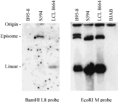
was confirmed to be a viral DNA clone by hybridization to the herpesvirus papio episome present in S594 cells and was sep-arated on an in situ lysing gel (Fig. 1, left panel). As expected, this herpesvirus papio DNA probe did not hybridize to the EBV episome in B95-8 cells under stringent conditions and hybridized only weakly to episomal DNA from the simian herpesvirus naturally infecting rhesus macaques (rhesus EBV in LCL8664 [57]) (Fig. 1, left panel). In contrast, an EcoRI M probe from a putatively noncoding region of the herpesvirus papio genome cross-hybridizes well to the episomes of EBV, herpesvirus papio, and rhesus EBV under similar conditions as previously suggested by Heller et al. (Fig. 1, right panel) (22). Partial nucleotide sequencing from both ends of the DNA fragment revealed that it was approximately 60% homologous to the EBV BamHI Nhet DNA beginning at nucleotide 166,001 and approximately 40% homologous to the EBV
on November 9, 2019 by guest
http://jvi.asm.org/
BamHI Nhet DNA ending at nucleotide 168,020. Thus, this
viral DNA fragment includes the putative herpesvirus papio homologs for the promoter and first exon of EBV LMP2A and the 39untranslated portion of the LMP1 transcript.
The herpesvirus papio DNA was used as a probe on North-ern blots with total RNA from cells infected with EBV (B95-8), herpesvirus papio (S594), and rhesus EBV (LCL8664) which were uninduced and induced for viral replication by transfec-tion with the EBV BZLF1 transactivator expressed from the SV40 early promoter. The herpesvirus papio genomic DNA probe detects multiple transcripts in herpesvirus papio-in-fected cells. A 2.5-kb transcript is readily detected, and a 2.3-kb transcript can also be detected on longer exposures (Fig. 2A and data not shown). Neither of these transcripts increases in abundance upon induction of viral replication, suggesting that these transcripts represent herpesvirus papio homologs for the latent membrane proteins LMP1 and LMP2A. EBV BZLF1 activity is confirmed with all three primate herpesvirus-infected cell lines by the induction and detection of early antigen expression on Western blot by cross-reactive human serum antibodies (Fig. 2C). EBV BZLF1 transfection also induces viral DNA replication of all three primate herpesvi-ruses, as determined by Southern blot analysis (data not shown). Thus, not only are antigenic epitopes of the lytic cycle antigens conserved among these primate herpesviruses, but the activation of the EBV lytic cycle by the BZLF1 transactivator is also functionally conserved between human and simian ho-mologs.
The less readily detected 2.3-kb mRNA is likely to be the LMP2A transcript and is not as easily detected since this viral DNA probe is predicted to contain only the LMP2A first exon. The 2.5-kb mRNA likely represents the herpesvirus papio ho-molog for the EBV latent infection membrane protein 1 (LMP1) since the herpesvirus papio DNA clone is colinear with the region of the EBV BamHI Nhet encoding the 39 untranslated portion of the LMP1 mRNA. The viral DNA probe also detects smaller 0.8-kb transcripts in S594 cells. These are readily detected in the partially permissive S594 cells (Fig. 2A). Although they do not increase with early antigen induction, they likely represent the small early transcripts
co-linear with the 39 end of the LMP1 transcript described by Hudson et al. (26).
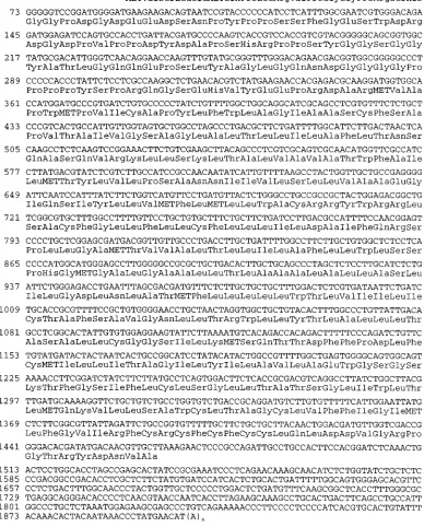
Cloning of a cDNA for the herpesvirus papio LMP2A ho-molog. The herpesvirus papio DNA fragment was used to screen a cDNA library derived from S594 cells. Four identical cDNA clones were obtained, and full-length sequencing re-vealed an open reading frame encoding 482 amino acids with 12 hydrophobic transmembrane domains (Fig. 3 and 4). Over-all, the cDNA sequence shared 54% amino acid identity with the EBV LMP2A protein (Fig. 4). The unique first exon of LMP2A shares only 31% identity with the herpesvirus papio homolog, but the remaining sequence consisting of the multi-ple transmembrane spanning domains common to both LMP2A and LMP2B and the carboxyl-terminal cytoplasmic domain is well conserved (59% identity). The carboxyl-termi-nal cytoplasmic tails demonstrate 44% amino acid identity. The sequence of the herpesvirus papio LMP2 cDNA also dem-FIG. 1. Hybridization of herpesvirus papio BamHI DNA clones to EBV,
[image:3.612.81.274.71.238.2]herpesvirus papio, and rhesus EBV episomes. EBV, herpesvirus papio, and rhesus EBV episomes present in B95-8, S594, and LCL8664 cells, respectively, were separated on an in situ lysing gel and transferred to nylon membranes. Duplicate blots were hybridized with a radiolabelled herpesvirus papio BamHI L8 or EcoRI M probe and washed under similar stringent conditions as described in Materials and Methods. The location of the wells and migration of linear genomes are indicated by the origin and linear designations.
FIG. 2. Northern and Western blots of EBV-, herpesvirus papio; and rhesus EBV-infected cells induced and noninduced for viral replication. Northern blots of total RNA from cells infected with EBV (B95-8), herpesvirus papio (S594), and rhesus EBV (LCL8664) were hybridized with herpesvirus papio BamHI L8 (A) and herpesvirus papio LMP2A cDNA (B) probes under stringent conditions, as described in Materials and Methods. Migrations of 28S, 18S, and 5S rRNAs are indicated by the dashes, and EBV-specific transcripts of interest are identified by their respective sizes (C). The cells used for mRNA were noninduced or induced for viral replication by transfection with the vector or SVN plasmid. (C) Western blots of the transfected cells probed with EBV-immune sera demonstrate induction of viral early lytic gene expression. EBNA-1, migration of the human EBV EBNA-1 protein. Early antigens of human and simian EBV are identified by their increased abundance upon induction of viral replication and comparable molecular sizes.
on November 9, 2019 by guest
http://jvi.asm.org/
[image:3.612.319.549.74.418.2]onstrates that the splicing pattern across the terminal repeats is conserved so that expression can only occur from circularized genomes, similar to that of EBV LMP2 (36). When this her-pesvirus papio cDNA was used to probe Northern blots, a 2.3-kb RNA which did not increase with viral replication was readily detected (Fig. 2B). A larger 4.5-kb RNA which in-creases in abundance with replication was also detected (Fig. 2B). This is similar to the EBV LMP2 probe which detects a 4.8-kb lytic transcript corresponding to the BNRF1 open read-ing frame (25, 27, 62). This lytic transcript is detected by the herpesvirus papio LMP2A cDNA probe which contains re-gions not encoded within the original genomic probe.
Interest-ingly, a 2.0-kb mRNA corresponding to the LMP2B gene is not detected with the herpesvirus papio LMP2A cDNA, which should be colinear with the last eight exons of LMP2B.
Kinase interaction motifs in the LMP2A first exon are con-served in the herpesvirus papio homolog.LMP2A associates with cellular tyrosine kinases, is tyrosine phosphorylated, and can act as a dominant negative regulator of B-cell receptor-mediated induction of the EBV lytic cycle (48). src family tyrosine kinases phosphorylate two tyrosine residues in a YXXL(N)7YXXL motif identified in the cytoplasmic domains
[image:4.612.113.501.88.570.2]of the Igaand Igbcomponents of the sIg signal-transducing receptor complex and the amino-terminal cytoplasmic domain FIG. 3. Nucleotide and amino acid sequence of the herpesvirus papio LMP2A cDNA. The nucleotide sequence of the longest herpesvirus papio LMP2A cDNA is shown, with translation of the longest open reading frame.
on November 9, 2019 by guest
http://jvi.asm.org/
of LMP2A (3, 49) (for a review see references 10 and 67). This motif is well conserved despite the significant amino acid se-quence divergence of the first exon between EBV and herpes-virus papio (Fig. 4), suggesting that the src phosphorylation sites have been conserved in herpesvirus papio. A YEEA motif in the EBV LMP2A first exon has been recognized as similar to the YEEI recognition site for SH2 binding domains (3). This motif is relatively well conserved as a YEEP in the papio LMP2A (Fig. 4). A third well-conserved region (PPPPYSPR) is also identified within the LMP2A first exon (Fig. 4). Al-though not previously recognized, this sequence contains a repeated PXXP motif derived for SH3 binding domains (61). Absolute conservation of this tetraproline repeat between the human and baboon LMP2A proteins suggests a potentially important functional role for this domain.
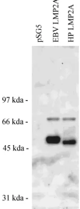
Herpesvirus papio LMP2A is tyrosine phosphorylated in human EBV-negative B cells and induces tyrosine phosphor-ylation similar to that of EBV LMP2A.Conservation of the kinase interaction domains suggests that the herpesvirus papio LMP2A, like the human EBV LMP2A, would be tyrosine phosphorylated in human B cells. The herpesvirus papio LMP2A cDNA was cloned under control of the SV40 early promoter and transfected into the human EBV-negative tumor B-cell line BJAB. Expression of tyrosine-phosphorylated pro-teins after transient transfection was determined by Western blot analysis with polyclonal antisera specific for tyrosine-phos-phorylated proteins. As previously demonstrated, transient transfection of the EBV LMP2A results in tyrosine phosphor-ylation of the overexpressed LMP2A 54 kDa protein (43) (Fig. 5). EBV LMP2A transfection has been previously described to also result in the tyrosine phosphorylation of a 72-kDa cell protein representing tyrosine-phosphorylated syk (49).
Expres-sion of this 72-kDa tyrosine-phosphorylated cell protein is sim-ilarly induced by both EBV and herpesvirus papio LMP2A transfection (Fig. 5). Other less abundant tyrosine-phosphory-lated proteins described in association with LMP2A transfec-tion or EBV infectransfec-tion cannot be visualized under these con-ditions without immunoprecipitation. Transfection of the herpesvirus papio LMP2A cDNA clones results in the abun-dant expression of a tyrosine-phosphorylated 51-kDa protein (Fig. 5), slightly smaller than the EBV LMP2A protein as predicted by the 15 fewer amino acids in the herpesvirus papio homolog. Thus, despite the nucleotide and associated amino acid divergence of the LMP2A first exon encoded within this region of the EBV and herpesvirus papio genome, the splicing, tyrosine kinase binding sites, and tyrosine phosphorylation of LMP2A have been conserved between the simian and human herpesviruses.
[image:5.612.60.293.72.301.2]EBNA-2 responsiveness of the EBV LMP2A promoter is conserved in the herpesvirus papio LMP2A promoter. The 617-nucleotide region of the herpesvirus papio BamHI L8 se-quence 59 to our longest cDNA is 59% homologous to the corresponding region of the EBV genome containing the EBV LMP2A promoter (Fig. 6). The EBV LMP2A promoter is notable for its upregulation by the EBV EBNA-2 gene product (70). The transcriptional transactivator EBNA-2 does not bind DNA directly but is recruited to EBNA-2-responsive promot-ers by its interaction with the DNA binding protein, CBP or Jk, which recognizes a consensus GTGGGAA sequence present in various EBNA-2-responsive viral and cellular promoters (20, 23, 42, 69). Two GTGGGAA consensus binding sites in the EBV LMP2A promoter are repeated within close proximity, and the 59GTGGGAA motif is conserved in the herpesvirus papio LMP2 promoter (Fig. 6). A single nucleotide substitu-tion has been made in the herpesvirus papio sequence corre-sponding to the EBV GTGGGAA motif more proximal to the transcriptional start site. However, the herpesvirus papio se-quence does reveal a second consensus GTGGGAA motif 125 FIG. 4. Comparison of herpesvirus papio and EBV LMP2A protein
se-quences. The herpesvirus papio amino acid sequence is numbered relative to the longest open reading frame. Three kinase interaction motifs within the first exon and conserved in EBV and herpesvirus papio are boxed. The hydrophobic trans-membrane domains are underlined. The methionine residue at the beginning of exon 2 common to both EBV LMP2A and LMP2B is indicated by an asterisk at amino acid 120. A previously described HLA-A2-restricted cytotoxic T-cell epitope in EBV LMP2 is identified by the double underline. :, amino acid identity; ., amino acid similarity.
FIG. 5. Antiphosphotyrosine Western blot of cell extracts after transient transfection of BJAB cells with EBV and herpesvirus papio LMP2 expression vectors. EBV-negative, human B-lymphoma cells (BJAB) were transfected with vector control plasmid (pSG5) or the same plasmid containing an EBV LMP2A cDNA (EBV LMP2A) or the herpesvirus papio LMP2 cDNA (HP LMP2) and analyzed 3 days after transfection. Western blots were probed with a rabbit antiserum specific for phosphotyrosine-containing proteins.
on November 9, 2019 by guest
http://jvi.asm.org/
[image:5.612.394.475.455.666.2]nucleotides 39to the first herpesvirus papio GTGGGAA motif. Thus, dual GTGGGAA motifs have been conserved in the herpesvirus papio LMP2 promoter region.
We tested whether EBNA-2 responsiveness has been func-tionally conserved in the herpesvirus papio LMP2A promoter by cloning the promoter in front of a CAT reporter gene and transfecting it into human EBV-negative B cells with or with-out an EBNA-2 expression vector. As described previously, 2 expression can induce CAT activity from an EBNA-2-responsive promoter such as the LMP1 promoter but has little effect on an EBNA-2-nonresponsive promoter such as the herpes simplex virus thymidine kinase promoter (Table 1) (65). As shown in Table 1, activity of the herpesvirus papio LMP2A
promoter CAT construct is similarly induced severalfold in the presence of EBV EBNA-2, suggesting that LMP2A transcrip-tional regulation has been conserved among these herpesvi-ruses.
DISCUSSION
These studies demonstrate that despite the evolutionary di-vergence of the EBV BamHI Nhet nucleotide sequence, sev-eral properties of the EBV LMP2A, such as the multiple hy-drophobic transmembrane domains, protein kinase interaction motifs, tyrosine phosphorylation, splicing, and EBNA-2-in-duced transcriptional regulation, have been conserved in the herpesvirus papio homolog. We first cloned a herpesvirus pa-pio viral DNA fragment which was colinear with the EBV
BamHI Nhet fragment. We found that this simian viral DNA
clone did not cross-hybridize well to the EBV episome, in contrast to other regions of the genome, and these findings were complementary to the observations of Heller et al., who studied simian herpesvirus genomes by using EBV DNA clones (22). Surprisingly, the herpesvirus papio DNA clone also did not cross-hybridize well to the rhesus EBV episome. This suggests nearly as much sequence divergence in this re-gion between herpesviruses from two Old World monkeys in the same family as between herpesvirus papio and human EBV. Recent cloning and sequencing of a rhesus EBV DNA clone colinear with EBV BamHI Nhet support these findings (16a). The geographic separation of certain Old World non-human primates to Africa (e.g., baboons) and Southeast Asia (e.g., rhesus macaques) may contribute to the evolutionary differences between these herpesviruses. However, Heller et al. also noted significant differences in the BamHI Nhet regions between herpesvirus pan (chimpanzees) and herpesvirus pa-pio, herpesviruses which infect different families of Old World primates from a common location, i.e., Africa (22).
Surprisingly, a probe using the full herpesvirus papio cDNA clone does not detect a 2.0-kb mRNA in herpesvirus papio-infected cells, which would correspond to an LMP2B homolog. This probe should be nearly as sensitive for LMP2B as for LMP2A, since eight of nine exons are shared by the two mRNAs (36, 62); however, a 2.0-kb mRNA was not detected on repeated Northern blots, despite readily detectable LMP2A expression. This suggests that LMP2B is expressed either much less or not at all in herpesvirus papio-infected cells. A func-tional role for LMP2B remains unclear. This membrane pro-tein might be predicted to aggregate or patch, since it contains multiple transmembrane domains but would not associate with tyrosine kinases because of the absence of the first 119 amino acids unique to LMP2A (5, 43). The LMP2B transcriptional start site is positioned so that the EBNA-2-responsive LMP1 regulatory region acts as a bidirectional promoter for LMP1 and LMP2B (35, 62). In contrast to the LMP2A first exon, the LMP2B first exon is noncoding. It will be interesting to confirm whether LMP2B expression is unique to human EBV. Unfor-tunately, our herpesvirus papio DNA clone does not extend to the region corresponding to the putative LMP2B first exon to provide sequence information for a detailed analysis.
It is interesting that the most dramatic nucleotide and amino acid divergence between the herpesvirus papio and EBV LMP2A is in the first exon, since the first 167 amino acids are sufficient for tyrosine phosphorylation and association with cellular tyrosine kinases (5, 43). Busson et al. observed a sim-ilar predisposition for mutations in the LMP2A first exon in their analysis of LMP2A sequences from nasopharyngeal car-cinomas and other EBV isolates (6, 7). One might have pre-dicted that regions containing important functional domains FIG. 6. Homology of EBV and herpesvirus papio LMP2A upstream
[image:6.612.57.297.660.726.2]regula-tory regions. Nucleotide sequence of the herpesvirus papio BamHI L8 viral DNA clone from the BamHI site to the most 59nucleotide of the longest herpesvirus papio LMP2 cDNA is shown. The herpesvirus papio sequence is numbered with the BamHI site of the viral DNA clone as nucleotide 1. The alignment with the homologous region of the EBV genome is shown with conserved residues indi-cated by a bar. GTGGGAA motifs are underlined.
TABLE 1. EBNA-2 responsiveness of the herpesvirus papio LMP2A promoter
Promoter reporter construct Average fold increase in activity with EBNA-2
Standard deviation
EBV LMP1 CAT 8.6 60.8
Herpesvirus papio LMP2 CAT 7.1 60.2
Herpes simplex virus thymidine kinase CAT
1.4 60.1
on November 9, 2019 by guest
http://jvi.asm.org/
would be more well conserved than other nonessential do-mains. However, in general, sequence variation among EBV strains is more often associated with coding rather than non-coding regions. Indeed, the most dramatic EBV sequence vari-ation is associated with the transformvari-ation-essential EBNA-2 gene, i.e., EBNA-2A and EBNA-2B alleles (30). EBV strain variation has also been demonstrated in other latent EBV genes, e.g., EBERs (2), EBNA-3s (59), LMP1 (21, 51), and LMP2 (6). Similarly, significant nucleotide divergence between EBV and herpesvirus papio is more closely associated with latent genes rather than with noncoding regions or lytic genes, e.g., EBERs (24), EBNA-2 (42), EBNA-3 genes-BamHI E (22), LMP2A first exon, and LMP1-BamHI Nhet (this report, unpublished observations, and reference 22). Immunologic pressure has been implicated as a cause of EBV strain varia-tion since some laboratories have reported strain-specific EBNA-3B nucleotide changes associated with a defined cyto-toxic T-cell epitope (9). LMP2A is expressed by circulating EBV-infected peripheral blood B cells (55), and cytotoxic T cells directed against LMP2A can be frequently observed in EBV-immune individuals (52). However, in human studies so far, an EBV LMP2A epitope presented to cytotoxic T cells by a relatively common HLA-A2.1 genetic background has been mapped to the fairly well-conserved 11th transmembrane do-main at amino acids 426 to 434 rather than to the amino-terminal cytoplasmic domain (37) (Fig. 4). Another possibility is that the cell proteins which interact with the EBV latent proteins are not well conserved among primates and force evolution of the viral sequence. However, we find that the herpesvirus papio LMP2A is tyrosine phosphorylated and in-duces tyrosine phosphorylation of at least one cell protein in common with EBV LMP2A in human B cells despite the marked amino acid divergence in the EBV and herpesvirus papio LMP2A first exons. Similarly, Ling and Hayward and Ling et al. demonstrated that despite the significant sequence divergence of the herpesvirus papio EBNA-2 sequence, the carboxyl-terminal transactivation domains and certain critical sequences for EBNA-2 interaction with CBP(Jk) were con-served and functional in human B cells (41, 42). Thus, the pressures driving the sequence divergence preferentially in the first LMP2A exon are unclear, and what we understand so far of the EBV immune response and the cell proteins interacting with viral latent proteins does not appear sufficient to explain these observations.
The three conserved kinase motifs we have recognized in the herpesvirus papio LMP2 first exon are also conserved in a recent PCR analysis of the LMP2A first exon sequence from 28 different EBV isolates (6). Previous reports have recognized the potential importance of the YXXL(N)7YXXL and YEEI
motifs in the LMP2A amino-terminal cytoplasmic domain (3, 5, 43, 49). Although not previously recognized, the conserva-tion of a PXXP tetraproline repeat within this region suggests that it may also have an important functional role. SH3 mains have been observed to bind to short proline-rich do-mains with a PXXP motif, and specificity of the SH3 domain bound may be determined by the context of the proline-rich domain (58, 68). A tetraproline repeat is well conserved in human immunodeficiency virus (HIV) nef sequences, and this
nef PXXP motif has recently been shown to bind the SH3
domains of hyc and lyn. In addition, mutation of this PXXP tetraproline repeat in recombinant HIV results in lower viral titers similar to the phenotype observed with nef-deleted vi-ruses (61). Thus, the PXXP motif appears to play a positive role in HIV replication associated with cellular activation and signal transduction. In cells latently infected with EBV, cell activation and induction of EBV replication would be
poten-tially detrimental to long-term virus survival and/or latency, so that the PXXP and other kinase interaction motifs appear to be playing a negative role in EBV replication (48).
Functional conservation of the herpesvirus papio LMP2A gene and promoter suggests that the basic molecular mecha-nisms of these simian and human herpesviruses are similar. This lends further support to the concept that these simian herpesviruses will be a valid model for studying EBV patho-genesis. Although the factors contributing to the species re-strictions on human and simian EBV infection remain to be determined, it should be possible to infect Old World primates with the herpesvirus which naturally infects their own species. Indeed, we have evidence from recent experiments that it is possible to infect selected rhesus monkeys with a rhesus EBV isolate (65a). This would provide a novel system for studying the biology and pathogenesis of latent EBV infection in vivo. The similarities of the herpesvirus papio LMP2A and the EBV LMP2A described here suggest that this Old World primate model may be useful for identifying the in vivo importance of a well-conserved latent EBV gene which is dispensable for in vitro infection and B-cell immortalization.
ACKNOWLEDGMENTS
This work was supported by grants from the U.S. Public Health Service (CA52244 and CA58051). Michael Franken was supported by a grant from the Dr. Mildred-Scheel Stiftung fu¨r Krebsforschung.
We thank Cheryl Miller for valuable advice and assistance with the phosphotyrosine assays. We are especially grateful to Elliott Kieff for invaluable advice and support.
REFERENCES
1. Ablashi, D. V., P. Gerber, and J. Easton. 1979. Oncogenic herpesviruses of nonhuman primates. Comp. Immunol. Microbiol. Infect. Dis. 2:229–241. 2. Arrand, J. R., L. S. Young, and J. D. Tugwood. 1989. Two families of
sequences in the small RNA-encoding region of Epstein-Barr virus (EBV) correlate with EBV types A and B. J. Virol. 63:983–986.
3. Beaufils, P., D. Choquet, R. Z. Mamoun, and B. Malissen. 1993. The (YXXL/I)2 signalling motif found in the cytoplasmic segments of the bovine leukaemia virus envelope protein and Epstein-Barr virus latent membrane protein 2A can elicit early and late lymphocyte activation events. EMBO J. 12:5105–5112.
4. Brooks, L., Q. Y. Yao, A. B. Rickinson, and L. S. Young. 1992. Epstein-Barr virus latent gene transcription in nasopharyngeal carcinoma cells: coexpres-sion of EBNA1, LMP1, and LMP2 transcripts. J. Virol. 66:2689–2697. 5. Burkhardt, A. L., J. B. Bolen, E. Kieff, and R. Longnecker. 1992. An
Epstein-Barr virus transformation-associated membrane protein interacts with src family tyrosine kinases. J. Virol. 66:5161–5167.
6. Busson, P., R. H. Edwards, T. Tursz, and N. Raab-Traub. 1995. Sequence polymorphism in the Epstein-Barr virus latent membrane protein (LMP)-2 gene. J. Gen. Virol. 76:139–145.
7. Busson, P., R. McCoy, R. Sadler, K. Gilligan, T. Tursz, and N. Raab-Traub. 1992. Consistent transcription of the Epstein-Barr virus LMP2 gene in na-sopharyngeal carcinoma. J. Virol. 66:3257–3262.
8. Countryman, J., H. Jenson, R. Seibl, H. Wolf, and G. Miller. 1987. Poly-morphic proteins encoded within BZLF1 of defective and standard Epstein-Barr viruses disrupt latency. J. Virol. 61:3672–3679.
9. de Campos-Lima, P. O., R. Gavioli, Q. J. Zhang, L. E. Wallace, R. Dolcetti, M. Rowe, A. B. Rickinson, and M. G. Masucci.1993. HLA-A11 epitope loss isolates of Epstein-Barr virus from a highly A111population. Science 260: 98–100.
10. DeFranco, A. L. 1993. Structure and function of the B cell antigen receptor. Annu. Rev. Cell. Biol. 9:377–410.
11. Dillner, J., H. Rabin, N. Letvin, W. Henle, G. Henle, and G. Klein. 1987. Nuclear DNA-binding proteins determined by the Epstein-Barr virus-related simian lymphotropic herpesviruses H. gorilla, H. pan, H. pongo and H. papio. J. Gen. Virol. 68:1587–1596.
12. Dunkel, V. C., T. W. Pry, G. Henle, and W. Henle. 1972. Immunofluorescence tests for antibodies to Epstein-Barr virus with sera of lower primates. J. Natl. Cancer Inst. 49:435–440.
13. Falk, L., F. Deinhardt, M. Nonoyama, L. G. Wolfe, and C. Bergholz. 1976. Properties of a baboon lymphotropic herpesvirus related to Epstein-Barr virus. Int. J. Cancer 18:798–807.
14. Falk, L., L. Wolfe, F. Deinhardt, J. Paciga, L. Dombos, G. Klein, W. Henle, and G. Henle.1974. Epstein-Barr virus: transformation of non-human pri-mate lymphocytes in vitro. Int. J. Cancer 13:363–376.
on November 9, 2019 by guest
http://jvi.asm.org/
15. Feichtinger, H., P. Putkonen, C. Parravicini, S. T. Li, E. E. Kaya, D. Bottiger, and P. Biberfeld.1990. Malignant lymphomas in cynomolgus monkeys in-fected with simian immunodeficiency virus. Am. J. Pathol. 137:1311–1315. 16. Frank, A., W. A. Andiman, and G. Miller. 1976. Epstein-Barr virus and
nonhuman primates: natural and experimental infection. Adv. Cancer Res. 23:171–201.
16a.Franken, M., and F. Wang. Unpublished data.
17. Gardella, T., P. Medveczky, T. Sairenji, and C. Mulder. 1984. Detection of circular and linear herpesvirus DNA molecules in mammalian cells by gel electrophoresis. J. Virol. 50:248–254.
18. Gerber, P., J. W. Branch, and E. N. Rosenblum. 1969. Attempts to transmit infectious mononucleosis to rhesus monkeys and marmosets and to isolate herpeslike virus. Proc. Soc. Exp. Biol. Med. 130:14–19.
19. Goldberg, R. J., E. M. Scolnick, W. P. Parks, L. A. Yakovleva, and B. A. Lapin. 1974. Isolation of a primate type-C virus from a lymphomatous baboon. Int. J. Cancer 14:722–730.
20. Grossman, S. R., E. Johannsen, X. Tong, R. Yalamanchili, and E. Kieff. 1994. The Epstein-Barr virus nuclear antigen 2 transactivator is directed to response elements by the J kappa recombination signal binding protein. Proc. Natl. Acad. Sci. USA 91:7568–7572.
21. Hatfull, G., A. T. Bankier, B. G. Barrell, and P. J. Farrell. 1988. Sequence analysis of Raji Epstein-Barr virus DNA. Virology 164:334–340.
22. Heller, M., P. Gerber, and E. Kieff. 1981. Herpesvirus papio DNA is similar in organization to Epstein-Barr virus DNA. J. Virol. 37:698–709. 23. Henkel, T., P. D. Ling, S. D. Hayward, and M. G. Peterson. 1994. Mediation
of Epstein-Barr virus EBNA2 transactivation by recombination signal-bind-ing protein J kappa. Science 265:92–95.
24. Howe, J. G., and M. D. Shu. 1988. Isolation and characterization of the genes for two small RNAs of herpesvirus papio and their comparison with Epstein-Barr virus-encoded EBER RNAs. J. Virol. 62:2790–2798.
25. Hudson, G. S., A. T. Bankier, S. C. Satchwell, and B. G. Barrell. 1985. The short unique region of the B95-8 Epstein-Barr virus genome. Virology 147: 81–98.
26. Hudson, G. S., P. J. Farrell, and B. G. Barrell. 1985. Two related but differentially expressed potential membrane proteins encoded by the EcoRI Dhet region of Epstein-Barr virus B95-8. J. Virol. 53:528–535.
27. Hummel, M., and E. Kieff. 1982. Epstein-Barr virus RNA. VIII. Viral RNA in permissively infected B95-8 cells. J. Virol. 43:262–272.
28. Kalter, S. S., R. L. Heberling, and J. J. Ratner. 1972. EBV antibody in sera of non-human primates. Nature (London) 238:353–534.
29. Kalter, S. S., R. L. Herberling, and J. J. Ratner. 1973. EBV antibody in monkeys and apes. Bibl. Haematol. 39:871–875.
30. Kieff, E., and D. Liebowitz. 1990. Epstein-Barr virus and its replication. D. Knipe, B. N. Fields and R. Chanock (ed.), Virology. Raven Press, New York. 31. Kim, O. J., and J. L. Yates. 1993. Mutants of Epstein-Barr virus with a selective marker disrupting the TP gene transform B cells and replicate normally in culture. J. Virol. 67:7634–7640.
32. Landon, J. C., L. B. Ellis, V. H. Zeve, and D. P. Fabrizio. 1968. Herpes-type virus in cultured leukocytes from chimpanzees. J. Natl. Cancer Inst. 40:181– 192.
33. Landon, J. C., and L. B. Malan. 1971. Seroepidemiologic studies of Epstein-Barr virus antibody in monkeys. J. Natl. Cancer Inst. 46:881–884. 34. Lapin, B. A. 1974. The epidemiologic and genetic aspect of an outbreak of
leukemia among baboons of the Sukhumi monkey colony in Dutcher and Chieco-Bianchi. Unifying concepts of leukemia. Bibliophyl. Haematol. 39: 263–268.
35. Laux, G., A. Economou, and P. J. Farrell. 1989. The terminal protein gene 2 of Epstein-Barr virus is transcribed from a bidirectional latent promoter region. J. Gen. Virol. 70:3079–3084.
36. Laux, G., M. Perricaudet, and P. J. Farrell. 1988. A spliced Epstein-Barr virus gene expressed in immortalized lymphocytes is created by circulariza-tion of the linear viral genome. EMBO J. 7:769–774.
37. Lee, S. P., W. A. Thomas, R. J. Murray, F. Khanim, S. Kaur, L. S. Young, M. Rowe, M. Kurilla, and A. B. Rickinson.1993. HLA A2.1-restricted cytotoxic T cells recognizing a range of Epstein-Barr virus isolates through a defined epitope in latent membrane protein LMP2. J. Virol. 67:7428–7435. 38. Levine, P. H., S. A. Leiseca, J. F. Hewetson, K. A. Traul, A. P. Andrese, D. J.
Granlund, P. Fabrizio, and D. A. Stevens.1980. Infection of rhesus monkeys and chimpanzees with Epstein-Barr virus. Arch. Virol. 66:341–351. 39. Li, S. L., H. Feichtinger, E. Kaaya, P. Migliorini, P. Putkonen, G. Biberfeld,
J. M. Middeldorp, P. Biberfeld, and I. Ernberg.1993. Expression of Epstein-Barr-virus-related nuclear antigens and B-cell markers in lymphomas of SIV-immunosuppressed monkeys. Int. J. Cancer 55:609–615.
40. Li, Z. H., S. Mahajan, M. M. Prendergast, J. Fargnoli, X. Zhu, S. Klages, D. Adam, G. L. Schieven, J. Blake, J. B. Bolen, et al.1992. Cross-linking of surface immunoglobulin activates src-related tyrosine kinases in WEHI 231 cells. Biochem. Biophys. Res. Commun. 187:1536–1544.
41. Ling, P. D., and S. D. Hayward. 1995. Contribution of conserved amino acids in mediating the interaction between EBNA2 and CBF1/RBPJk. J. Virol. 69:1944–1950.
42. Ling, P. D., J. J. Ryon, and S. D. Hayward. 1993. EBNA-2 of herpesvirus papio diverges significantly from the type A and type B EBNA-2 proteins of
Epstein-Barr virus but retains an efficient transactivation domain with a conserved hydrophobic motif. J. Virol. 67:2990–3003.
43. Longnecker, R., B. Druker, T. M. Roberts, and E. Kieff. 1991. An Epstein-Barr virus protein associated with cell growth transformation interacts with a tyrosine kinase. J. Virol. 65:3681–3692.
44. Longnecker, R., C. L. Miller, X. Q. Miao, A. Marchini, and E. Kieff. 1992. The only domain which distinguishes Epstein-Barr virus latent membrane protein 2A (LMP2A) from LMP2B is dispensable for lymphocyte infection and growth transformation in vitro; LMP2A is therefore nonessential. J. Virol. 66:6461–6469.
45. Longnecker, R., C. L. Miller, X. Q. Miao, B. Tomkinson, and E. Kieff. 1993. The last seven transmembrane and carboxy-terminal cytoplasmic domains of Epstein-Barr virus latent membrane protein 2 (LMP2) are dispensable for lymphocyte infection and growth transformation in vitro. J. Virol. 67:2006– 2013.
46. Longnecker, R., C. L. Miller, B. Tomkinson, X. Q. Miao, and E. Kieff. 1993. Deletion of DNA encoding the first five transmembrane domains of Epstein-Barr virus latent membrane proteins 2A and 2B. J. Virol. 67:5068–5074. 47. Menezes, J., W. Leibold, and G. Klein. 1975. Biological differences between
Epstein-Barr virus (EBV) strains with regard to lymphocyte transform-ing ability, superinfection and antigen induction. Exp. Cell Res. 92:478– 484.
48. Miller, C. L., A. Burkhardt, J. H. Lee, B. Stealey, R. Longnecker, J. B. Bolen, and E. Kieff.1995. Integral membrane protein 2 of Epstein-Barr virus reg-ulates reactivation from latency through dominant negative effects on pro-tein-tyrosine kinases. Immunity 2:155–166.
49. Miller, C. L., J. H. Lee, E. Kieff, and R. Longnecker. 1994. An integral membrane protein (LMP2) blocks reactivation of Epstein-Barr virus from latency following surface immunoglobulin crosslinking. Proc. Natl. Acad. Sci. USA 91:772–776.
50. Miller, G., J. C. Niederman, and D. A. Stitt. 1972. Infectious mononucleosis: appearance of neutralizing antibody to EpsteBarr virus measured by in-hibition of formation of lymphoblastoid cell lines. J. Infect. Dis. 125:403–406. 51. Miller, W. E., R. H. Edwards, D. M. Walling, and N. Raab-Traub. 1994. Sequence variation in the Epstein-Barr virus latent membrane protein 1. J. Gen. Virol. 75:2729–2740.
52. Murray, R. J., M. G. Kurilla, J. M. Brooks, W. A. Thomas, M. Rowe, E. Kieff, and A. B. Rickinson.1992. Identification of target antigens for the human cytotoxic T cell response to Epstein-Barr virus (EBV): implications for the immune control of EBV-positive malignancies. J. Exp. Med. 176: 157–168.
53. O’Gara, R. W., R. H. Adamson, M. G. Kelly, and D. W. Dalgard. 1971. Neoplasms of the hematopoietic system in nonhuman primates: report of one spontaneous tumor and two leukemias induced by procarbazine. J. Natl. Cancer Inst. 46:1121–1130.
54. Persson, B., and P. Argos. 1994. Prediction of transmembrane segments in proteins utilising multiple sequence alignments. J. Mol. Biol. 237:182–192. 55. Qu, L., and D. T. Rowe. 1992. Epstein-Barr virus latent gene expression in
uncultured peripheral blood lymphocytes. J. Virol. 66:3715–3724. 56. Rabin, H., R. H. Neubauer, R. Hopkins, E. K. Dzhikidze, Z. V. Shevtsova,
and B. A. Lapin.1977. Transforming activity and antigenicity of an Epstein-Barr-like virus from lymphoblastoid cell lines of baboons with lymphoid disease. Intervirology 8:240–249.
57. Rangan, S. R., L. N. Martin, B. E. Bozelka, N. Wang, and B. J. Gormus. 1986. Epstein-Barr virus-related herpesvirus from a rhesus monkey (Macaca mulatta) with malignant lymphoma. Int. J. Cancer 38:425–432.
58. Ren, R., Z. S. Ye, and D. Baltimore. 1994. Abl protein-tyrosine kinase selects the Crk adapter as a substrate using SH3-binding sites. Genes Dev. 8:783– 795.
59. Rowe, M., L. S. Young, K. Cadwallader, L. Petti, E. Kieff, and A. B. Rick-inson.1989. Distinction between Epstein-Barr virus type A (EBNA 2A) and type B (EBNA 2B) isolates extends to the EBNA 3 family of nuclear pro-teins. J. Virol. 63:1031–1039.
60. Ryon, J. J., E. D. Fixman, C. Houchens, J. Zong, P. M. Lieberman, Y. N. Chang, G. S. Hayward, and S. D. Hayward.1993. The lytic origin of herpes-virus papio is highly homologous to Epstein-Barr herpes-virus ori-Lyt: evolutionary conservation of transcriptional activation and replication signals. J. Virol. 67:4006–4016.
61. Saksela, K., G. Cheng, and D. Baltimore. 1995. Proline-rich (PxxP) motifs in HIV-1 Nef bind to SH3 domains of a subset of Src kinases and are required for the enhanced growth of Nef1viruses but not for down-regulation of CD4. EMBO J. 14:484–491.
62. Sample, J., D. Liebowitz, and E. Kieff. 1989. Two related Epstein-Barr vi-rus membrane proteins are encoded by separate genes. J. Virol. 63:933– 937.
63. Schable, C. A., B. L. Murphy, K. R. Berquist, C. R. Gravelle, and J. E. Maynard.1974. Inability to detect hepatitis B virus or specific antigens in transformed chimpanzee lymphocytes. Infect. Immun. 10:1443–1444. 64. Stevens, D. A., T. W. Pry, E. A. Blackham, and R. A. Manaker. 1970.
Comparison of antigens from human and chimpanzee herpes-type virus-infected hemic cell lines. Proc. Soc. Exp. Biol. Med. 133:678–683. 65. Tsang, S. F., F. Wang, K. M. Izumi, and E. Kieff. 1991. Delineation of the
on November 9, 2019 by guest
http://jvi.asm.org/
cis-acting element mediating EBNA-2 transactivation of latent infection membrane protein expression. J. Virol. 65:6765–6771.
65a.Wang, F. Unpublished observations.
66. Wang, F., C. Gregory, C. Sample, M. Rowe, D. Liebowitz, R. Murray, A. Rickinson, and E. Kieff.1990. Epstein-Barr virus latent membrane protein (LMP1) and nuclear proteins 2 and 3C are effectors of phenotypic changes in B lymphocytes: EBNA-2 and LMP1 cooperatively induce CD23. J. Virol. 64:2309–2318.
67. Weiss, A., and D. R. Littman. 1994. Signal transduction by lymphocyte antigen receptors. Cell 76:263–274.
68. Yu, H., J. K. Chen, S. Feng, D. C. Dalgarno, A. W. Brauer, and S. L.
Schreiber.1994. Structural basis for the binding of proline-rich peptides to SH3 domains. Cell 76:933–945.
69. Zimber-Strobl, U., E. Kremmer, F. Grasser, G. Marschall, G. Laux, and G. W. Bornkamm.1993. The Epstein-Barr virus nuclear antigen 2 interacts with an EBNA2 responsive cis-element of the terminal protein 1 gene pro-moter. EMBO J. 12:167–175.
70. Zimber-Strobl, U., K. O. Suentzenich, G. Laux, D. Eick, M. Cordier, A. Calender, M. Billaud, G. M. Lenoir, and G. W. Bornkamm.1991. Epstein-Barr virus nuclear antigen 2 activates transcription of the terminal protein gene. J. Virol. 65:415–423.