JOURNAL OFVIROLOGY, Mar. 2009, p. 2518–2530 Vol. 83, No. 6 0022-538X/09/$08.00⫹0 doi:10.1128/JVI.02227-08
A Basic Patch on
␣
-Adaptin Is Required for Binding of Human
Immunodeficiency Virus Type 1 Nef and Cooperative
Assembly of a CD4-Nef-AP-2 Complex
䌤
†
Rittik Chaudhuri,
1,2Rafael Mattera,
1O. Wolf Lindwasser,
1Margaret S. Robinson,
2and Juan S. Bonifacino
1*
Cell Biology and Metabolism Program, Eunice Kennedy Shriver National Institute of Child Health and Human Development, National Institutes of Health, Bethesda, Maryland 20892,1and Cambridge Institute for Medical Research, University of
Cambridge, Wellcome Trust/MRC Building, Addenbrooke’s Hospital, Hills Road, Cambridge CB2 0XY, United Kingdom2
Received 22 October 2008/Accepted 26 December 2008
A critical function of the human immunodeficiency virus type 1 Nef protein is the downregulation of CD4 from the surfaces of infected cells. Nef is believed to act by linking the cytosolic tail of CD4 to the endocytic machinery, thereby increasing the rate of CD4 internalization. In support of this model, weak binary inter-actions between CD4, Nef, and the endocytic adaptor complex, AP-2, have been reported. In particular, dileucine and diacidic motifs in the C-terminal flexible loop of Nef have been shown to mediate binding to a combination of the␣and2 subunits of AP-2. Here, we report the identification of a potential binding site for the Nef diacidic motif on␣-adaptin. This site comprises two basic residues, lysine-297 and arginine-340, on the ␣-adaptin trunk domain. The mutation of these residues specifically inhibits the ability of Nef to bind AP-2 and downregulate CD4. We also present evidence that the diacidic motif on Nef and the basic patch on␣-adaptin are both required for the cooperative assembly of a CD4-Nef-AP-2 complex. This cooperativity explains how Nef is able to efficiently downregulate CD4 despite weak binary interactions between components of the tripartite complex.
CD4, a type I transmembrane glycoprotein that serves as a coreceptor for major histocompatibility complex class II (MHC-II) molecules, is expressed on the surfaces of helper T lymphocytes and cells of the monocyte/macrophage lineage (8). Primate immunodeficiency viruses gain access to these cells by virtue of the interaction of the viral envelope glyco-protein (Env) with a combination of CD4 and a chemokine receptor (63). This interaction causes a conformational change within the Env protein that promotes the fusion of the viral envelope with the plasma membrane. Upon the delivery of the viral genetic material into the cytoplasm of the host cells, one of the first virally encoded proteins to be expressed is Nef, an accessory factor that modulates specific signal transduction and protein-trafficking pathways in a manner that optimizes the intracellular environment for viral replication (reviewed in references 21, 39, and 65). Perhaps the best characterized function of Nef is the downregulation of CD4 from the sur-faces of the host cells (6, 22, 29, 45). CD4 downregulation prevents superinfection (6, 41) and enhances virion release (19, 38, 48, 66, 76), thereby contributing to the establishment of a robust infective state (24, 72).
The mechanism used by the Nef protein of human immu-nodeficiency virus type 1 (HIV-1) to downregulate CD4 has
been the subject of extensive study, but only recently have the molecular details of this process begun to be unraveled. It is generally acknowledged that HIV-1 Nef accelerates the inter-nalization of CD4 from the plasma membrane by linking the cytosolic tail of the receptor to the clathrin-associated endo-cytic machinery (1, 12, 20, 34, 40, 64). In support of this model, a hydrophobic pocket comprising W57 and L58 on the folded core domain of Nef binds with millimolar affinity to the cyto-solic tail of CD4 (28) (all residues and numbers correspond to the NL4-3 variant of HIV-1 Nef used in this study). In addi-tion, a dileucine motif (ENTSLL, residues 160 to 165) (10, 16, 26) and a diacidic motif (D174 and D175) (2) on the C-termi-nal flexible loop of Nef mediate an interaction of micromolar affinity with the clathrin-associated, heterotetrameric (␣-
2-2-2) adaptor protein 2 (AP-2) complex (12, 20, 40, 49). These interactions draw CD4 into clathrin-coated pits that eventually bud inwards as clathrin-coated vesicles (11, 27). Internalized CD4 is subsequently delivered to endosomes and then to lysosomes for degradation (3, 23, 59, 64).
Despite progress in the understanding of the mechanism of Nef-induced CD4 downregulation, several important aspects remain to be elucidated. Previous studies have shown that the Nef dileucine and diacidic motifs interact with a combination of the ␣ and 2 subunits of AP-2 (referred to as the ␣-2 hemicomplex) (12, 20, 40, 49), but the precise location of the Nef binding sites is unknown. It also remains to be determined whether Nef can actually bind CD4 and AP-2 at the same time. Indeed, the formation of a tripartite CD4-Nef-AP-2 complex in which Nef links the cytosolic tail of CD4 to AP-2 has long been hypothesized but has never been demonstrated experi-mentally. Given the relatively weak affinity of Nef for the CD4
* Corresponding author. Mailing address: Cell Biology and Metab-olism Program, Eunice Kennedy Shriver National Institute of Child Health and Human Development, Building 18T, Room 101, National Institutes of Health, Bethesda, MD 20892. Phone: (301) 496-6368. Fax: (301) 402-0078. E-mail: [email protected].
† Supplemental material for this article may be found at http://jvi .asm.org/.
䌤Published ahead of print on 7 January 2009.
2518
on November 8, 2019 by guest
http://jvi.asm.org/
tail (28) and AP-2 (12, 40), it is unclear how such a complex could assemble and function in CD4 downregulation.
In this study, we have addressed these issues by using a combination of yeast hybrid, in vitro binding, and in vivo CD4 downregulation assays. We report the identification of a can-didate binding site for the Nef diacidic motif on the AP-2 complex. This site, a basic patch comprising K297 and R340 on
␣-adaptin, is specifically required for Nef binding and Nef-induced CD4 downregulation. We also show that the Nef di-acidic motif and the␣-adaptin basic patch are required for the cooperative assembly of a tripartite complex composed of the CD4 cytosolic tail, Nef, and the␣-2 hemicomplex. The coop-erative manner in which this complex is formed explains how Nef is able to efficiently downregulate CD4 from the plasma membrane despite weak binary interactions between the com-ponents of this complex.
MATERIALS AND METHODS
Y3H assays.The pBridge.Nef.2, pBridge.tyrosinase.2, and pGADT7.␣ plas-mids have been described previously (12, 33, 40, 49). All mutations of these plasmids were carried out by site-directed mutagenesis, using the QuikChange II kit (Stratagene, La Jolla, CA) and appropriate primers. The open reading frames (ORF) of these and all other constructs used in this study were verified by nucleotide sequence analysis. Yeast three-hybrid (Y3H) assays were performed
as described before (12, 33, 40, 49). Briefly, theSaccharomyces cerevisiaeHF7c
strain was cotransformed with pBridge- and pGADT7-based vectors by the lithium acetate procedure, and double transformants were selected on dropout
agar plates lacking Leu, Trp, and Met (⫹His plates). After 4 days, pooled
colonies from each transformation were resuspended in water and the suspen-sions were normalized to equivalent optical densities (ODs; 0.1 OD units at 600
nm) and transferred onto three sets of plates:⫹His plates; those lacking Leu,
Trp, Met, and His (⫺His plates); and those lacking Leu, Trp, Met, and His and
supplemented with 1 mM 3-amino-1,2,4-triazole (3AT;⫺His ⫹3AT plates).
Wild-type HIV-1 NL4-3 Nef (or the cytosolic tail of mouse tyrosinase) was expressed as a GAL4BD fusion protein from the pBridge vector (Clontech,
Mountain View, CA), along with wild-type or mutant rat2-adaptin. Wild-type
or mutant rat␣C-adaptin was expressed as a GAL4AD fusion protein from
pGADT7 (Clontech). The amino acid sequences of rat and human2 are 100%
identical, while the trunk domains of rat and human␣C are 99.1% identical and
99.8% similar. Colony growth on the three sets of plates was analyzed 3 days
later. The growth of the yeast on the⫺His plates was indicative of an interaction
between the GAL4BD and GAL4AD fusion proteins. Growth on the⫺His⫹
3AT plates indicated stronger interactions. Each Y3H experiment was per-formed a minimum of three times.
Y4H assays.To generate pBridge.Nef, an EcoRI-SalI fragment containing the Nef ORF was subcloned from pNL4-3 Nef.IRES.GFP (12, 40) into the corre-sponding sites of multiple cloning site 1 (MCS1) of pBridge (Clontech). The
pBridge.Nef.2 plasmid was the same as that used for the Y3H experiments, and
alternative versions of this vector (expressing the Nef G2A, WL57,58AA, EEEE62-65AAAA, PP72,75AA, LL164,165AA, and DD174,175AA mutant forms) have been described before (12, 40). To generate the pAD series of vectors, the pMET25-MCS2-PGKT cassette was excised from the pBridge vector by ApaI digestion, polished with DNA polymerase I (New England Biolabs, Ipswich, MA), and inserted into the PvuII site of pGAD424 (Clontech) by blunt-end ligation. This process resulted in a new vector, hereinafter referred to as pAD, which has two MCSs: one downstream of the ADH1 promoter and the GAL4AD sequence (MCS1) and one downstream of the independently con-trolled MET25 promoter (MCS2). The proteins encoded by genes inserted into these MCSs are both targeted to the yeast nucleus by the presence of nuclear localization signals. The cytosolic tail of human CD4 was amplified by PCR, digested with EcoRI and SalI, and ligated into the corresponding sites of pAD
MCS1 to generate pAD.CD4. To create pAD.CD4.␣, the␣C ORF was amplified
by PCR from pGADT7.␣by using primers containing NotI and BamHI
restric-tion sites, digested with the appropriate enzymes, and ligated into the NotI-BglII
site of pAD.CD4 MCS2. The pAD.CD4.␣KR297,340EE plasmid was prepared
by a similar procedure, using pGADT7.␣KR297,340EE as the PCR template.
Assays using these plasmids (including yeast two-hybrid [Y2H], Y3H, and yeast four-hybrid [Y4H] analyses) were carried out according to the Y3H protocol
described above, with only one modification: following the selection of positive transformants, the densities of the corresponding yeast suspensions were nor-malized to 1.6 OD units (at 600 nm), and the suspensions were then serially
diluted to 0.1 OD units before transfer to the⫹His and⫺His dropout agar
plates. Each Y4H experiment was performed a minimum of three times.
Recombinant protein expression and GST pull-down experiments.The
plas-mid pST.AP-2 core (expressing residues 1 to 621 of rat␣C-adaptin [␣trunk] with
a C-terminal fusion to glutathioneS-transferase [GST], residues 1 to 591 of rat
2-adaptin [2 trunk] with an N-terminal hexahistidine [His6] tag, residues 1 to
141 of rat2-adaptin [2-N], and residues 1 to 142 of rat2-adaptin [full-length
2]) has been described previously (12, 40). pST.AP-2 core␣KR297,340EE was
generated by site-directed mutagenesis. AP-2 core protein complexes were
ex-pressed inEscherichia coliBL21(DE3) cells (Novagen, San Diego, CA) and
purified as described before (12, 40). To compare the abilities of wild-type␣and
KR297,340EE mutant␣(␣KR297,340EE) to associate with the other AP-2 core
subunits, the two GST fusion proteins were isolated from bacterial lysates by binding to glutathione-Sepharose beads and analyzed by sodium dodecyl
sulfate-polyacrylamide gel electrophoresis (SDS-PAGE). His6-tagged Nef was expressed
from the pHis-Parallel2-Nef vector and purified as described previously (12, 40).
The interaction of His6-Nef with the wild-type AP-2 core and AP-2 core␣
KR927,340EE was analyzed by GST pull down (12, 40). Briefly, saturating
amounts (⬃5g) of the wild-type and mutant AP-2 core complexes were
im-mobilized onto 30l of glutathione-Sepharose beads, and the beads were
incu-bated with 3g of His6-Nef for 2 h at 4°C in a final volume of 1 ml of binding
buffer (15 mM HEPES [pH 7.0], 75 mM NaCl, 0.25% [vol/vol] Triton X-100, 0.15% [wt/vol] bovine serum albumin) supplemented with a protease inhibitor cocktail (EDTA-free Complete tablets; Roche Applied Science, Indianapolis, IN). After incubation, the beads were washed three times by resuspension in 1.3 ml of binding buffer lacking bovine serum albumin, followed by centrifugation for
2 min at 2,000⫻g and 4°C. Proteins bound to the beads were eluted by
resuspension in 50l of 2⫻Laemmli buffer and incubation for 10 min at 90°C.
The eluted samples were separated from the beads by centrifugation for 2 min at
16,000⫻gand subjected to SDS-PAGE and immunoblotting.
Cells and tissue culture. HeLa cells (American Type Culture Collection,
Manassas, VA) were cultured under a humidified atmosphere (5% CO2) in
Dulbecco’s modified Eagle medium and passaged every 3 to 4 days. The medium
was supplemented with 100 U/ml penicillin, 0.1g/ml streptomycin, 2 mM
L-glutamine, and 10% (vol/vol) fetal bovine serum.
RNA interference (RNAi) and RNAi-resistant reagents. To simultaneously
silence endogenous␣A and␣C expression in HeLa cells, we designed a single
small interfering RNA (siRNA) duplex (sense sequence, 5⬘GAGCAUGUGCA
CGCUGGCCATT; Qiagen, Valencia, CA) targeting nucleotides 1053 to 1072 in
the human␣A and␣C ORFs. An siRNA-resistant version of␣C-adaptin
(here-inafter referred to as␣R) was generated by introducing three silent substitutions
into the rat␣C cDNA (C3T, C3T, and G3C at nucleotides 1053, 1059, and
1065, respectively, of the rat␣C sequence). These substitutions resulted in a total
of four mismatches between␣R and the siRNA-sensitive human␣A/␣C
se-quences (the rat␣C cDNA contains an additional A3G substitution at position
1062 compared to the human␣A/␣C sequences). The sequence for a V5 epitope
tag was fused to the 3⬘ end of the␣R ORF, and the␣R-V5 cassette was
subcloned into the SalI-BamHI sites of pIRES2.EGFP (hereinafter referred to
as pIRES.GFP; Clontech) to generate p␣R-V5.IRES.GFP. Site-directed
mu-tagenesis of p␣R-V5.IRES.GFP yielded p␣R-KR297,340EE-V5.IRES.GFP. The
presence of an internal ribosome entry site in these plasmids allowed for the
independent translation of␣R-V5 (or the KR297,340EE mutant form of␣R
[␣R-KR297,340EE] fused to V5) and enhanced green fluorescent protein (GFP)
from the same bicistronic transcript.
␣-Adaptin RNAi and rescue assays.In HeLa cells, the expression of
endog-enous␣-adaptin was silenced by transfection with siRNA and the endogenous
␣-adaptin was replaced with␣R according to a 7-day protocol. On day 1, HeLa
cells grown on six-well plates (⬃15% confluence) were either left untreated or
transfected with siRNA (100 nM) by using Lipofectamine 2000 (Invitrogen, Carlsbad, CA) and Opti-MEM I (Invitrogen). On day 3, the cells were split at a
1:3 ratio onto fresh six-well plates. On day 4, the cells (at⬃50% confluence) were
transfected with a second round of siRNA and/or a unique combination of DNA
plasmids (pCMV.CD4; pCI or pCI.Nef; and pIRES.GFP, p␣R-V5.IRES.GFP,
or p␣R-KR297,340EE-V5.IRES.GFP) (Table 1) by using Lipofectamine 2000
and Opti-MEM I. The plasmids pCI (empty vector) and pCI.Nef and the pCMV.CD4 plasmid (kindly provided by Klaus Strebel, NIAID, NIH) have been described previously (12, 40). On day 6, the cells were split again onto fresh six-well plates at a 1:2 ratio. On day 7, the cells were detached with 0.5 ml of 2 mM EDTA in phosphate-buffered saline, harvested, and either stained with
fluorescent antibodies (⬃80% of the cell suspension) or lysed for
on November 8, 2019 by guest
http://jvi.asm.org/
ting (⬃20%). All silencing and rescue experiments were repeated a minimum of three times.
Antibodies.Allophycocyanin (APC)-conjugated mouse monoclonal antibody to human CD4 (Caltag, Burlingame, CA) and phycoerythrin (PE)-conjugated mouse monoclonal antibody to human CD71 (transferrin receptor [TfR]) (Sigma-Aldrich, St. Louis, MO) were used for fluorescence-activated cell sorter (FACS) analysis. Unconjugated rabbit anti-HIV-1 Nef (NIH AIDS Research and Ref-erence Reagent Program; originally deposited by Ronald Swanstrom) (70),
mouse anti-␣-adaptin clone numbers 100/2 (Sigma-Aldrich) and 8/Adaptin␣
(BD Biosciences, Franklin Lakes, NJ), mouse V5 (Invitrogen), mouse
anti-␣-tubulin (Sigma-Aldrich), and horseradish peroxidase-conjugated secondary
antibodies (GE Healthcare, Piscataway, NJ) were used for immunoblotting.
FACS analysis.HeLa cells stained with the appropriate antibodies were pre-pared for FACS analysis as described previously (12, 33, 40). Untransfected and unstained HeLa cells were used as a negative control for antibody labeling. GFP fluorescence expressed from the pIRES.GFP series of plasmids was used to identify and gate around transfected cells. The amount of cell-associated APC and PE fluorescence in GFP-positive cells was quantified by a FACSCalibur flow cytometer and analyzed using CellQuest software (BD Biosciences).
Immunoblotting.HeLa cells were lysed for 30 min at 4°C in lysis buffer (1% [vol/vol] NP-40 in phosphate-buffered saline) supplemented with a protease inhibitor cocktail (Roche Applied Science). The lysates were then centrifuged for
15 min at 16,000⫻gand 4°C, and the supernatants were stored at⫺70°C.
SDS-PAGE, transfer onto nitrocellulose membranes, labeling with primary an-tibodies, and detection with secondary antibodies were performed as described previously (12, 40).
RESULTS
Rationale. The Nef flexible loop contains two conserved sequence motifs, a dileucine motif (i.e., ENTSLL in NL4-3, EXXXLL consensus for all HIV-1 variants, where X is any amino acid) and a diacidic motif (i.e., DD in NL4-3, (D/E)D consensus for all variants), both of which are required for binding to AP-2 (12, 20, 40, 49) and for CD4 downregulation (2, 10, 12, 16, 26, 33, 40). The dileucine motif, but not the diacidic motif, also mediates the binding of Nef to the homol-ogous AP-1 (␥-1-1-1) and AP-3 (␦-3-3-3) complexes (33, 40). The Nef dileucine motif belongs to the class of dileucine-based (D/E)XXXL(L/I) sorting signals that are present in many host cell proteins (e.g., the melanosomal en-zyme tyrosinase) and that interact with AP-1, AP-2, and/or AP-3 (9). Unlike Nef, other proteins that have (D/E)XXXL (L/I) class signals do not require an additional diacidic motif for binding to AP-2 or the other AP complexes (40). The importance of this diacidic motif in the recognition of Nef by
AP-2 explains the binding sensitivity to high concentrations of salt and indicates that electrostatic forces are critical determi-nants of this interaction (40). The unique requirement of the Nef diacidic motif for interaction with AP-2 makes it a poten-tial target for specific inhibition of Nef action without inter-ference with the sorting of host cell proteins mediated by dileucine-based signals. These considerations prompted us to map the binding site for the Nef diacidic motif on AP-2 and examine its role in promoting the assembly of a putative CD4-Nef-AP-2 complex.
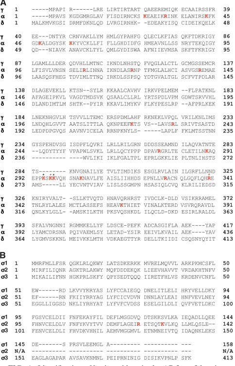
Identification of basic residues on the AP-2␣subunit that are required for interaction with HIV-1 Nef. Based on the above-described considerations, we hypothesized that the Nef diacidic motif must interact with basic residues on AP-2 that are not conserved in the other two AP complexes. We searched for such residues by comparing the amino acid sequences of the subunits that make up the AP-1␥-1, AP-2␣-2, and AP-3
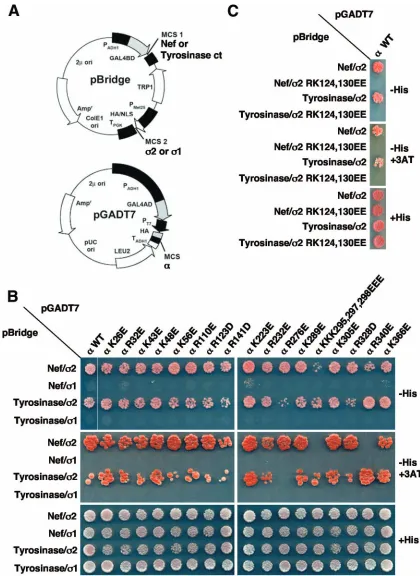
␦-3 hemicomplexes, which contain the binding sites for Nef (12, 20, 33, 40, 49). In this way, we identified 21 arginine and lysine residues on␣-2 that are not present on the other two hemicomplexes (Fig. 1). We then mutated these residues to either glutamate or aspartate and assayed for the loss of bind-ing of␣-2 to wild-type Nef using the Y3H system (12, 40, 49) (Fig. 2A). Several mutations of the␣-2 hemicomplex, includ-ing a triple mutation of ␣ residues K295, K297, and K298 (initially mutated en bloc because of the close proximity of these residues) to three glutamate residues (yielding the ␣ KKK295,297,298EEE construct) (Fig. 2B), a single mutation of␣ residue R340 to glutamate (yielding the␣ R340E con-struct) (Fig. 2B, see both⫺His and⫺His⫹3AT rows), and a double mutation of2 residues R124 and K130 to two gluta-mate residues (yielding the2 RK124,130EE construct), im-paired the binding of Nef (Fig. 2C). To determine whether this loss of binding was due to the disruption of the Nef binding site or to more global effects on the hemicomplex, we also tested the␣-2 mutant constructs for their abilities to bind the cyto-solic tail of mouse tyrosinase. The tyrosinase tail interacts with
␣-2 in a dileucine-dependent manner that, unlike Nef, does not require a diacidic motif (40). We found that the ␣ KKK295,297,298EEE and␣ R340E constructs bound to the tyrosinase tail with relatively strong avidities (Fig. 2B), indicat-ing that the mutation of the correspondindicat-ing residues specifically interfered with the interaction between Nef and AP-2. In con-trast, the2 RK124,130EE mutant failed to bind to the tyrosi-nase tail (Fig. 2C), consistent with an adverse effect of these substitutions on either the folding of the 2 subunit or the stability of the␣-2 hemicomplex. The remaining mutants, all of which were generated by mutation of the␣subunit, inter-acted equally with Nef and the tyrosinase tail (Fig. 2B and C). To determine whether the interactions detected in the Y3H system were genuine, we tested each of the ␣ mutants for self-activation by substituting 1 for 2 in the Y3H assay. Given that␣and1 do not interact with each other (58), this pair should not be able to bind either Nef or the tyrosinase tail and should not be able to stimulate yeast growth on selective media. Indeed, when combined with1, none of the␣mutants showed evidence of interaction with Nef or the tyrosinase tail (Fig. 2B), indicating that (i) the mutations do not cause self-activation, (ii) the observed interactions between the mutant
[image:3.585.43.285.89.201.2]␣-2 hemicomplexes and the ligands are genuine, and (iii) the
TABLE 1. Combinations of siRNA and DNA used for transfection of HeLa cells in AP-2␣rescue assaysa
Row siRNA DNA 1 DNA 2 DNA 3
1 None pIRES.GFP pCI pCMV.CD4
2 None pIRES.GFP pCI.Nef pCMV.CD4
3 ␣-Adaptin pIRES.GFP pCI pCMV.CD4
4 ␣-Adaptin pIRES.GFP pCI.Nef pCMV.CD4 5 ␣-Adaptin paR-V5.IRES.GFP pCI pCMV.CD4 6 ␣-Adaptin paR-V5.IRES.GFP pCI.Nef pCMV.CD4 7 ␣-Adaptin
paR-KR297,340EE-V5.IRES.GFP
pCI pCMV.CD4
8 ␣-Adaptin paR-KR297,340EE-V5.IRES.GFP
pCI.Nef pCMV.CD4
a
The various combinations of siRNA duplexes and DNA plasmids used to
transfect HeLa cells on day 4 of the␣-adaptin RNAi and rescue assays are
shown. For each condition, 0.4g of DNA 1, 0.3g of DNA 2, and 0.3g of
DNA 3 were used. Cells corresponding to rows 3 to 8 were previously treated
with siRNA targeting␣-adaptin on day 1 of the assay.
2520 CHAUDHURI ET AL. J. VIROL.
on November 8, 2019 by guest
http://jvi.asm.org/
only lysine and arginine residues specific to the␣-2 hemicom-plex that potentially contribute to Nef binding are ␣ K295, K297, K298, and R340.
The␣-adaptin K297 and R340 residues are part of a basic patch that is required for the binding of HIV-1 Nef.To analyze the individual contributions of␣K295, K297, K298, and R340 to Nef binding, we generated several additional constructs by mutating these residues to alanine or glutamate and then used the new constructs in the Y3H assay described above. In line with the results described above, the mutation of␣R340 alone
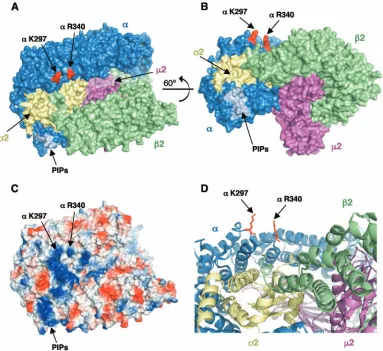
or the combined mutation of all three lysine residues impaired the ability of Nef to bind the ␣-2 hemicomplex (Fig. 3). Individual mutation of␣K295 and K298 revealed that these residues do not significantly contribute to the interaction with Nef, while the mutation of␣K297 produced as significant a defect in Nef binding as the combined mutation of all three lysine residues (Fig. 3). Consistent with this finding, the double mutation of␣K297 and R340 caused approximately the same decrease in Nef binding as the quadruple mutation of␣K295, K297, K298, and R340 (Fig. 3). Thus,␣K297 and R340 are key residues for the interaction of␣-2 with Nef. Although the mutation of␣K297 and R340 to alanine decreased Nef bind-ing (⫺His⫹3AT plates in Fig. 3), changing these residues to glutamate had a more dramatic effect (Fig. 3), probably due to electrostatic repulsion or to a requirement for features other than the charge of the basic side chains (40). In line with the latter explanation, the mutation of the second aspartate (D175) in the Nef diacidic motif to glutamate impaired both AP-2 binding and CD4 downregulation (40), suggesting that the precise length of the side chain, in addition to its charge, determines the strength of the interaction. All of the mutants involving ␣ K297 and R340 bound the tyrosinase tail with wild-type affinity in the presence of2, while none bound to either Nef or the tyrosinase tail in the presence of1 (Fig. 3). These controls demonstrated that the replacement of␣K297 and R340 specifically affected the ability of ␣-2 to interact with Nef in the Y3H system. Mapping of␣K297 and R340 on the three-dimensional structure of AP-2 (15) showed that these residues are in close proximity to each other and are part of a large basic patch on the surface of␣-adaptin (Fig. 4).
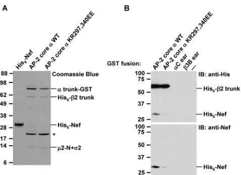
The ␣-adaptin K297 and R340 residues are required for direct binding of HIV-1 Nef. Because interactions detected with yeast hybrid systems are not necessarily direct, we per-formed in vitro binding experiments using recombinant pro-teins expressed inE. coli. We previously showed that an AP-2 core construct lacking the hinge and ear domains of␣- and
2-adaptin and the C-terminal domain of 2 is able to bind Nef with micromolar affinity (12, 40). This AP-2 core construct was mutated to substitute glutamate residues for K297 and R340 (yielding the AP-2 core ␣ KR297,340EE construct). SDS-PAGE followed by Coomassie blue staining of the puri-fied AP-2 core constructs, which included a C-terminal GST tag on␣and an N-terminal His6tag on2, indicated that the
␣KR297,340EE mutation did not affect the assembly of the AP-2 core complex (Fig. 5A). However, GST pull-down assays showed that the␣KR297,340EE mutation markedly impaired the binding of His6-Nef, as detected by immunoblotting with
either anti-His6 (Fig. 5B, top panel) or anti-Nef (Fig. 5B,
bottom panel) antibodies. These observations confirmed the results of the Y3H assays and demonstrated that the require-ment of␣residues K297 and R340 for interaction with Nef is direct.
The ␣-adaptin K297 and R340 residues are required for Nef-induced CD4 downregulation.We next examined whether
[image:4.585.43.285.71.448.2]␣ residues K297 and R340 are necessary for Nef-mediated downregulation of CD4 in HeLa cells by using an RNAi and rescue approach. We used siRNA to deplete cells of␣-adaptin, a manipulation that destabilizes the AP-2 complex and causes various degrees of depletion of the other subunits. The loss of AP-2 inhibits clathrin-dependent internalization of a subset of
FIG. 1. Identification of basic residues in the AP-2␣-2 hemicom-plex that are not conserved in the homologous subunits of AP-1 and AP-3. (A) Sequence alignment of the trunk domains of human AP-1␥ (␥1 isoform; accession no. AAH36283), AP-2␣(␣C isoform; accession no. AH06155), and AP-3␦(accession no. AAC51761). (B) Sequence alignment of human AP-11 (1A isoform; accession no. AAA37243), AP-22 (accession no. AAP36470), and AP-3 63 (63A isoform; ac-cession no. EAW48952). Alignments were performed using the Clust-alW2 program (available at http://www.ebi.ac.uk/Tools/clustalw2/index .html). Amino acid numbers are indicated. Lysine and arginine residues present on AP-2␣and2 but not on the corresponding AP-1 and AP-3 subunits are highlighted in red. These residues were mutated to either aspartate or glutamate (Fig. 2). Black and red asterisks indicate residues that were also mutated to alanine (Fig. 3). Red asterisks denote AP-2␣residues K297 and R340, which were found to be required for the interaction with HIV-1 Nef. N/A, not applicable.
on November 8, 2019 by guest
http://jvi.asm.org/
FIG. 2. Y3H analysis of the interaction of HIV-1 Nef with AP-2␣-2 hemicomplexes having substitutions for nonconserved basic residues. AP-2␣and2 basic residues that are not conserved in the homologous subunits of AP-1 (␥and1) and AP-3 (␦and3) (Fig. 1) were mutated, individually or in combination, to glutamate or aspartate. The resulting␣-2 constructs were tested for interaction with HIV-1 Nef or the mouse tyrosinase cytosolic tail by using a Y3H assay, as described in Materials and Methods. (A) Schematic representation of the plasmids used in the Y3H assay. Nef or the tyrosinase cytosolic tail was expressed as a GAL4BD fusion from MCS1 of pBridge,2 was expressed from MCS2 of pBridge, and␣was expressed as a GAL4AD fusion from the MCS of pGADT7. Cotransformations of yeast cells with Nef or the tyrosinase tail and discordant combinations of AP-2␣and AP-11 constructs were used as self-activation/ specificity controls. (B and C) Y3H assay results. Cotransformants were plated onto medium lacking Leu, Trp, Met, and His (⫺His) in the absence or presence of 1 mM 3AT (⫺His⫹3AT) to detect interaction throughHIS3reporter gene activation. 3AT is a competitive inhibitor of the His3 protein (imidazoleglycerol-phosphate dehydratase) that increases the stringency of the assay. Cotransformants were also plated onto medium lacking Leu, Trp, and Met (⫹His) to control for growth and loading. Growth on⫺His or⫺His⫹3AT plates is indicative of interactions. WT, wild type.
2522
on November 8, 2019 by guest
http://jvi.asm.org/
transmembrane proteins from the plasma membrane (31, 32, 46, 50, 51). FACS analysis showed that the depletion of en-dogenous␣-adaptin increased the surface levels of one such transmembrane protein, TfR (Fig. 6A, left panel), in agree-ment with previous findings (31, 32, 51). This result was con-sistent with the presence of an endocytic, tyrosine-based sort-ing signal (i.e., YTRF) in the TfR cytosolic tail (14, 47) that interacts with the2 subunit of AP-2 (53, 56). Interestingly,
␣-adaptin depletion also increased the surface levels of CD4 (Fig. 6A, right panel), probably by reducing the basal rate of CD4 endocytosis mediated by a dileucine-based sorting signal (i.e., SQIKRLL) in the CD4 cytosolic tail (1, 61). We then tested for the rescue of AP-2 function in␣-adaptin-depleted cells by transfection with RNAi-resistant versions of wild-type or KR297,340EE mutant␣-adaptin. We found that both con-structs reduced surface TfR (Fig. 6B, left panel) and CD4 (Fig. 6B, right panel) to approximately normal levels, indicating that
␣K297 and R340 are not required for endocytosis mediated by tyrosine- and dileucine-based sorting signals. We next used the RNAi-rescue assay with HeLa cells expressing CD4 with or without Nef. The depletion of␣-adaptin completely eliminated the ability of Nef to downregulate CD4 from the cell surface (Fig. 6C). Importantly, the expression of the wild-type ␣R construct in ␣-depleted cells restored the ability of Nef to downregulate CD4 (Fig. 6D, left panel), whereas the expres-sion of the KR297,340EE mutant␣construct did not (Fig. 6D, right panel). Immunoblot analysis of the transfected cells in-dicated that this disparity in Nef function was not due to
differences in the silencing of endogenous␣, the expression of
␣R constructs, or the expression of Nef itself (Fig. 6E). These analyses thus demonstrated that␣-adaptin residues K297 and R340 are specifically required for Nef-induced downregulation of CD4.
Requirement of AP-2␣-adaptin residues K297 and R340 for cooperative assembly of a tripartite Nef-CD4-AP-2 complex.
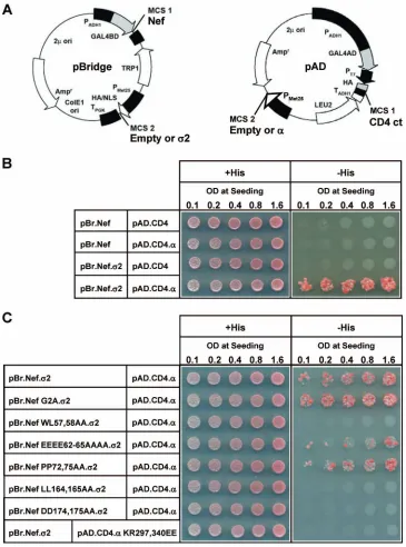
Nef is believed to downregulate CD4 by binding to the cyto-solic tail of this receptor and linking it to AP-2, thereby accel-erating the rate of CD4 endocytosis. Although previous work has provided evidence for weak binary interactions between Nef and CD4 (7, 28, 62, 67), Nef and AP-2 (12, 40), and CD4 and AP-2 (30), the occurrence of a tripartite complex involving all three components has not yet been demonstrated. We an-alyzed the possible formation of such a complex by using a combination of Y2H, Y3H, and Y4H assays. Yeast were trans-formed with the pBridge and pAD vectors, each of which contained two MCSs under the control of independent pro-moters (Fig. 7A). In this way, we were able to express up to four proteins, each of which was targeted to the nucleus by nuclear localization signals. We tested for the interaction be-tween Nef (expressed as a GAL4BD fusion protein) and the cytosolic tail of CD4 (expressed as a GAL4AD fusion protein) either alone or in the presence of the AP-2␣and2 subunits. We observed that, in the absence of␣and2, Nef and CD4 did not detectably interact in our system (Fig. 7B). This result differed from previous Y2H findings (67), probably because of the greater stringency of our assay. Moreover, individual
ex-FIG. 3. AP-2␣residues K297 and R340 contribute to the interaction between␣-2 and HIV-1 Nef. The effects of single or combined mutations of AP-2␣K295, K297, K298, and/or R340 to glutamate or alanine on the interaction of AP-2␣-2 with HIV-1 Nef or the mouse tyrosinase cytosolic tail were investigated. The discordant␣and1 pair was used as a negative control. Analyses were performed using the Y3H system as described in the legend to Fig. 2 and in Materials and Methods.
on November 8, 2019 by guest
http://jvi.asm.org/
[image:6.585.136.449.72.348.2]pression of either␣ or2 failed to promote interaction be-tween Nef and CD4 (Fig. 7B). However, when both␣and2 were expressed, Nef bound to the CD4 cytosolic tail (Fig. 7B). The increased avidity of CD4 for Nef in the presence of the
␣-2 hemicomplex indicates that a CD4-Nef-␣-2 complex is formed by cooperative assembly. To test whether the assembly of this complex is dependent on the known binary interactions between its components, we analyzed several mutants by using a Y4H assay (Fig. 7C). Mutations that inhibit the binding of Nef to either CD4 (Nef WL57,58AA) (28) or AP-2 (Nef LL164,165AA and DD174,175AA) (12, 20, 40) prevented the formation of the tripartite complex (Fig. 7C). Significantly for this study, the mutation of␣-adaptin residues K297 and R340, which are required for the binding of AP-2 to Nef (Fig. 3 and 5), yielded the same result (Fig. 7C). In contrast, mutations of three Nef motifs that are not required for binding to either CD4 or AP-2 (Nef G2A, EEEE62-65AAAA, and PP72,75AA) (28, 62) did not significantly affect the formation of the com-plex. Thus, the assembly of a CD4-Nef-AP-2 complex depends
on the known binary interactions between its individual com-ponents, verifying the specificity of the assays.
DISCUSSION
[image:7.585.101.484.68.419.2]Our search for a binding site for the Nef diacidic motif on AP-2 has identified two basic residues, K297 and R340, on the surface of the␣-adaptin subunit that are specifically required for both Nef binding (Fig. 2, 3, and 5) and CD4 downregula-tion (Fig. 6). In contrast, these basic residues are dispensable for binding to the cytosolic tail of tyrosinase (Fig. 2 and 3), which is entirely dependent on a dileucine-based sorting signal (40). Moreover, the mutation of these basic residues does not affect the ability of AP-2 to maintain steady-state levels of TfR and CD4 at the cell surface (Fig. 6), which is dependent on endocytosis mediated by tyrosine-based (14, 47, 53, 56) and dileucine-based (1, 30, 61) signals, respectively. Finally, these basic residues are not conserved in the homologous AP-1␥and AP-3␦subunits, which, in combination with the corresponding
FIG. 4. Location of AP-2␣K297 and R340 on the three-dimensional structure of the AP-2 core complex (accession no. 1GW5 and 2VGL) (15). (A and B) Surface representations with the␣,2,2, and2 subunits colored in blue, green, magenta, and gold, respectively; the binding site for polyphosphoinositides (PIPs) in the␣subunit is depicted in light blue, and the␣K297 and R340 residues (including their side chains) are shown in red. The image in panel B corresponds to a 60° rotation around thexaxis of that shown in panel A. Residues␣K297 and R340 correspond to K298 and R341 in polymer 1 of the crystal structure (15) (C) Surface representation of the image in panel A colored according to electrostatic potential (contoured as red to blue from⫺74 to 74 kT). (D) Magnified ribbon diagram of the region surrounding residues K297 and R340. All images were drawn with PyMOL (17).
2524 CHAUDHURI ET AL. J. VIROL.
on November 8, 2019 by guest
http://jvi.asm.org/
1 and3 subunits, also bind dileucine-based sorting signals (12, 20, 33, 40). Thus, Nef appears to exploit a unique feature of AP-2 in order to engage the endocytic machinery. Like the Nef diacidic motif, the ␣-adaptin site comprising K297 and R340 is therefore a potential target for pharmacologic inter-ference with Nef-induced CD4 downregulation (21).
Characteristics of the ␣-adaptin basic patch containing K297 and R340.Residues K297 and R340 on␣-adaptin are part of a large basic patch that rivals in size the binding site for polyphosphoinositides near the N terminus of the protein (15) (Fig. 4C). K297 is contained within a loop that also comprises
␣-adaptin residues K295 and K298 (Fig. 1A) (15). Although the individual mutation of K295 or K298 did not affect Nef binding in the Y3H assay (Fig. 3), we cannot rule out the possibility that the double mutation of those residues may have some effect. It is notable that AP-1␥and AP-3␦lack not only residues homologous to K297 and R340 but also a basic loop like that containing K295, K297, and K298 in␣-adaptin (Fig. 1A). Remarkably, all of these basic residues are conserved on both the ␣C and ␣A mammalian isoforms, as well as in
␣-adaptins from a wide range of metazoan species, including the genetic model organismsDrosophila melanogasterand
Cae-norhabditis elegans(see Fig. S1 in the supplemental material). Since Nef-encoding immunodeficiency viruses infect only pri-mates, this phylogenetic conservation hints at a more general function of these basic residues. Perhaps this site is involved in the recognition of clusters of acidic residues, either alone or in combination with other sorting signals. The cytosolic tail of the mammalian endopeptidase furin has a stretch of acidic resi-dues interspersed with serine resiresi-dues (i.e., SDSEEDE) that are substrates for casein kinase II-dependent phosphorylation. Together with sequences resembling tyrosine-based and dileucine-based sorting signals, this acidic cluster contributes to the localization of furin to the trans-Golgi network (35, 68, 78) and to its sorting to the basolateral plasma membrane domain of polarized epithelial cells (71). Remarkably, the acidic cluster is intrinsically capable of mediating clathrin-de-pendent endocytosis in the absence of any other signal (43, 78). Similar acidic clusters are found in the cytosolic tails of furin homologs from all metazoans. Although the phosphorylated mammalian furin cluster has been shown previously to bind a protein named PACS-1 (79), in light of our results, it would be of interest to test whether this cluster also mediates binding to AP-2 via the basic patch described here.
Possible location of the dileucine binding site on AP-2.The mutational approach used here for identifying the binding site for the Nef diacidic motif could also be applied to the mapping of the binding site for the Nef dileucine motif or other dileucine-based sorting signals on AP-2. In fact, the demon-stration that␣-adaptin residues K297 and R340 are required for Nef binding constrains the area of␣-2 that is likely to contain the dileucine binding site. The Nef dileucine motif (i.e., ENTSLL in NL4-3 and consensus sequence EXXXLL in all HIV-1 variants) comprises not only L164 and L165 but also E160, which is partially required for AP-2 binding (12). The two residues that make up the Nef diacidic motif, D174 and D175, lie 14 and 15 residues, respectively, in the C-terminal direction from E160 along the Nef flexible loop. Assuming a distance of 3.8 Å between contiguous␣-carbon atoms in a linear peptide chain, E160 would be at most 53 Å from D174 and 57 Å from D175. Based on these consider-ations, we scanned the three-dimensional structure of AP-2 (15) for basic residues that are located up to 57 Å from
␣-adaptin residues K297 and R340 and that are conserved in AP-1 and AP-3 (since these complexes also bind the Nef dileucine motif). Within this range are two residues in close proximity to each other,␣R21 and2 R15, the mutation of which impairs the binding of both the Nef and tyrosinase dileucine motifs (R. Chaudhuri, unpublished observations). Nestled between the two regions of AP-2 that contain␣R21 and 2 R15 and ␣ K297 and R340 (Fig. 4A and B) are several2 residues where the binding site for the dileucine pair may be located.
[image:8.585.45.284.65.239.2]While this paper was under revision, Kelly et al. reported the crystal structure of AP-2 in a complex with a variant dileucine-based signal from CD4 (36). The CD4 signal differs from typical (D/E)XXXL(L/I) signals in that a phosphoserine-glu-tamine pair substitutes for the acidic residue in the canonical motif (9). The crystal structure indicates that the phospho-serine-glutamine moiety likely binds to the␣R21 and2 R15 residues and that the two leucines bind to adjacent pockets lined by several hydrophobic2 residues, including V88 and
FIG. 5. The replacement of␣K297 and R340 impairs the binding of the recombinant AP-2 core to Nef in vitro. (A) Recombinant Nef and AP-2 core constructs used in the in vitro binding experiments were produced as described in Materials and Methods and subjected to SDS-PAGE on 4 to 12% acrylamide gradient gels, followed by Coo-massie blue staining. Lanes corresponding to the AP-2 core complexes bearing wild-type (WT) and KR297,340EE mutant␣chains show the ␣trunk-GST, His6-2 trunk,2-N, and full-length2 subunits in order
of increasing mobilities (2-N and2 comigrate in this gel system). The band at⬃21 kDa (asterisk) in the lanes corresponding to wild-type and mutant AP-2 cores represents a GST degradation product. (B) Equal amounts of GST fusion proteins with wild-type (WT) or␣ KR297,340EE-containing AP-2 cores, or the ear domains of AP-2␣C (57) or AP-33B (18) (the latter two as negative controls), were immobilized on glutathione-Sepharose beads, and the beads were in-cubated with recombinant His6-Nef for 2 h at 4°C. Bound proteins
were eluted and subjected to SDS-PAGE on 10% acrylamide gels, followed by immunoblotting (IB) with antibodies to the His6tag
(up-per panel) or to Nef (lower panel). The last lane on the right (labeled ⫺) shows binding to unfused GST. The⬃60-kDa band labeled by anti-His6in the upper blot corresponds to the His6-tagged2 trunk in
the recombinant AP-2 core and served as an internal loading control. Numbers to the left indicate the positions of molecular mass markers (in kilodaltons).
on November 8, 2019 by guest
http://jvi.asm.org/
2526
on November 8, 2019 by guest
http://jvi.asm.org/
L103 (36). These findings are consistent with the results of our mutational analysis and explain why both ␣ and 2 are re-quired for interactions with dileucine-based signals in Y3H assays (12, 40) and in vitro binding assays (20). We hypothesize that the Nef dileucine motif binds to the same site on␣-2 as the CD4 dileucine signal and that the unique Nef diacidic motif extends the interaction to the distal part of␣, conferring greater affinity and specificity.
Cooperative assembly of a tripartite complex comprising the CD4 tail, Nef, and AP-2.To our knowledge, the Y4H assay results reported here represent the first demonstration of the occurrence of a tripartite complex comprising the CD4 tail, Nef, and AP-2. The detection of this complex is dependent on the known determinants of bimolecular interactions between CD4 and Nef (i.e., Nef residues W57 and L58 that bind to the CD4 tail) and between Nef and AP-2 (i.e., the Nef dileucine and diacidic motifs and ␣-adaptin residues K297 and R340) (Fig. 7). Furthermore, its detection is independent of Nef res-idues that do not participate in binding to CD4 or AP-2, such as the myristoylation site (G2), the acidic cluster (EEEE62-65), and the polyproline motif (comprising P72 and P75) (Fig. 7). Importantly, these results correlate with previously pub-lished functional data: mutations that disrupt the formation of the CD4-Nef-AP-2 complex also prevent the downregulation of CD4 from the plasma membrane (1, 2, 12, 40, 73, 75; this study), while mutations that do not affect the assembly of the complex are not essential for CD4 downregulation (1, 12, 44, 73). The only exception to this correlation is the Nef G2A mutant, which fails to downregulate CD4 from the cell surface because it is not myristoylated and therefore cannot efficiently associate with the plasma membrane (7, 73, 81). However, myristoylation of Nef is not necessary for its incorporation into the CD4-Nef-AP-2 complex in the Y4H assay, as all the pro-teins in this complex are targeted to the yeast nucleus by the presence of heterologous nuclear localization signals. Given the requirement of the dileucine motif in the CD4 cytosolic tail for both constitutive and Nef-induced CD4 downregulation (1, 64), it will now be important to examine the role of this motif
in the formation of the CD4-Nef-AP-2 complex. Another acidic residue on Nef, D123, has also been shown previously to be required for CD4 downregulation (42, 55), and it would be of interest to test if this requirement reflects a role in the assembly of the CD4-Nef-AP-2 complex or, as previously pro-posed, in Nef oligomerization (4) or interaction with another host protein (13).
The assembly of a CD4-Nef-AP-2 complex is consistent with the previous proposal that Nef links the tail of CD4 to AP-2 (54, 60). In addition, our data show that the␣-2 hemicomplex promotes the interaction of the CD4 tail with Nef (Fig. 7). Thus, the formation of the CD4-Nef-AP-2 complex involves cooperative interactions among all of its components. A pos-sible explanation for this cooperativity is that the binding of AP-2 to one site on Nef increases the affinity of a second site on Nef for the CD4 tail, a phenomenon that is formally known as positive heterotropic cooperativity (69). In this model, AP-2 does not make direct contact with the CD4 tail but causes a conformational change in Nef that enhances binding to the CD4 tail. An alternative explanation is that each component of the complex makes simultaneous contacts with the other two, thereby enhancing the overall stability of the tripartite com-plex. This model requires that both Nef and AP-2 bind to the CD4 tail directly and at the same time. Further biochemical and structural analyses will be needed to distinguish between these models and to identify all the determinants of assembly of the CD4-Nef-AP-2 complex.
In T cells, CD4 exists in a complex with the protein tyrosine kinase Lck (77; reviewed in reference 54). In this complex, the CD4 cytosolic tail and the N terminus of Lck form a folded zinc clasp structure (37). The engagement of the CD4 tail by Nef likely requires (or induces) the dissociation of Lck (25), with subsequent unfolding of the CD4 tail (37). The assembly of a stable CD4-Nef-AP-2 complex may preclude the reassociation of Lck with CD4, thereby promoting the internalization and eventual lysosomal degradation of CD4 (64).
Nef has also been shown previously to engage in cooperative interactions with other receptors and AP complexes. For
ex-FIG. 6. AP-2␣residues K297 and R340 are required for Nef-induced CD4 downregulation. HeLa cells were subjected to a 7-day siRNA-cDNA transfection protocol as described in Materials and Methods and in Table 1. Control and␣siRNA-treated cells were cotransfected with three plasmids: one expressing CD4, one lacking (⫺) or expressing (⫹) Nef, and one lacking or expressing either wild-type ␣R (␣R-WT) or ␣R-KR297,340EE. The cells were then prepared for FACS analysis and immunoblotting. Cells prepared for FACS analysis were either left unlabeled as a control for background fluorescence (shaded curves in all plots) or stained with PE-conjugated anti-human TfR or APC-conjugated anti-human CD4 antibodies. (A) Treatment with␣siRNAs increases cell surface TfR and CD4 levels in the absence of Nef. (B) Both wild-type ␣R and␣R-KR297,340EE prevent the increase in the levels of TfR and CD4 caused by treatment with␣siRNA in the absence of Nef. (C) Nef expression downregulates CD4 in control but not␣siRNA-treated HeLa cells. (D) Rescue of Nef-induced downregulation of CD4 by wild-type ␣R but not␣R-KR297,340EE in␣siRNA-treated HeLa cells. The results shown are representative of data from three independent experiments with similar results. Statistical analyses of the data from these three experiments showed that Nef-dependent CD4 downregulation (expressed as the ratio of geometric means in the absence or presence of Nef) was 4.81⫾0.70 for control cells, 1.51⫾0.24* for␣siRNA-treated cells, 4.60⫾ 0.94 for␣ siRNA-treated cells expressing wild-type␣R, and 1.50⫾0.04*† for␣siRNA-treated cells expressing␣R-KR297,340EE (mean⫾ standard error of the mean;n⫽3), with the symbols * and † indicating values that are significantly different (P⬍0.05) from those for control cells and␣siRNA-treated cells expressing wild-type␣R, respectively, as calculated by analysis of variance followed by two-tail Dunnett’s test. (E) Aliquots of the transfected cells from all experimental groups were lysed and subjected to SDS-PAGE and immunoblotting (IB) with the antibodies indicated to the right. All cells were transfected with a plasmid encoding CD4, together with the plasmids and oligonucleotides indicated in the grid above the lanes (⫹, present;⫺, absent; KREE, KR297,340EE mutant). Notice that the anti-AP-2␣ clone 100/2 recognized both endogenous isoforms of␣-adaptin,␣A and␣C (apparent as an⬃100-kDa doublet in which the upper and lower bands represent␣A and␣C, respectively) (5), as well as a nonspecific band (⬃85 kDa). The anti-AP-2␣clone 8/Adaptin␣, however, recognized only endogenous␣A-adaptin since it was raised against a fragment of this isoform. TheV5-tagged, RNAi-resistant␣C rescue constructs were detected by both the 100/2 anti-␣ antibody and the anti-V5 antibody. Immunoblotting with anti-␣-tubulin was used as a loading control. Numbers to the left indicate the positions of molecular mass markers (in kilodaltons).
on November 8, 2019 by guest
http://jvi.asm.org/
ample, the binding of HIV-1 Nef to the cytosolic tail of MHC-I increases the affinity of a tyrosine-based motif in this tail for the 1 subunit of AP-1, thus stabilizing the assembly of an MHC-I-Nef-AP-1 complex (52, 80). In addition, the binding of
simian immunodeficiency virus Nef to the cytosolic domain of the CD3-chain of the T-cell antigen receptor increases the affinity of this domain for AP-2, leading to the stabilization of a CD3--Nef-AP-2 complex (74). These cooperative
interac-FIG. 7. Assembly of a tripartite complex of Nef, the cytosolic tail of CD4, and the AP-2␣-2 hemicomplex as demonstrated by yeast hybrid assays. (A) Plasmids used in the Y2H, Y3H, and Y4H assays. In all assays, HIV-1 Nef was expressed from pBridge as a GAL4BD fusion protein, while the cytosolic tail of human CD4 (CD4 ct) was expressed from pAD as a GAL4AD fusion protein. In the Y2H assay, no other proteins were expressed from these vectors; in the Y3H assay, either2-adaptin or␣C-adaptin was expressed from pBridge or pAD, respectively; in the Y4H assay, both2 adaptin and␣C adaptin were coexpressed. (B) Y2H, Y3H, and Y4H analyses of the interaction between GAL4BD-Nef and GAL4AD-CD4 in the absence or presence of one or both components of the␣-2 hemicomplex. The plasmids used in the yeast hybrid experiments are noted to the left of the panel, with the pBridge (pBr)-based vectors in the first column and the pAD-based vectors in the second column. Row 1 corresponds to the Y2H assay, rows 2 and 3 correspond to the Y3H assays, and row 4 corresponds to the Y4H assay. Yeast from all assays were seeded onto⫹His and⫺His plates at increasing levels of OD. Yeast growth on the⫺His plates indicates an interaction between GAL4BD-Nef and GAL4AD-CD4. (C) Y4H analysis of the effect of several Nef and␣mutants on the interaction of GAL4BD-Nef and GAL4AD-CD4 in the presence of the␣-2 hemicomplex.
2528 CHAUDHURI ET AL. J. VIROL.
on November 8, 2019 by guest
http://jvi.asm.org/
[image:11.585.110.475.67.559.2]tions enable Nef to downregulate MHC-I from an intracellular location and the CD3-chain from the cell surface. Together with our findings, these observations indicate that the estab-lishment of cooperative interactions with various host cell pro-teins is a general property of Nef that underlies its effects on protein trafficking.
ACKNOWLEDGMENTS
We thank X. Zhu and N. Tsai for excellent technical assistance; A. Rojas, B. Beach, Y. Kato, K. Strebel, and the AIDS Research and Reference Reagent Program (NIAID, NIH) for kind gifts of reagents; and J. Hurley for helpful discussions.
This work was supported by the intramural program of NICHD, the NIH Intramural AIDS Targeted Antiviral Program (IATAP), and the Wellcome Trust. R.C. was supported by the NIH-Cambridge and Gates-Cambridge graduate scholarships.
REFERENCES
1.Aiken, C., J. Konner, N. R. Landau, M. E. Lenburg, and D. Trono.1994. Nef induces CD4 endocytosis: requirement for a critical dileucine motif in the
membrane-proximal CD4 cytoplasmic domain. Cell76:853–864.
2.Aiken, C., L. Krause, Y. L. Chen, and D. Trono.1996. Mutational analysis of HIV-1 Nef: identification of two mutants that are temperature-sensitive for
CD4 downregulation. Virology217:293–300.
3.Anderson, S. J., M. Lenburg, N. R. Landau, and J. V. Garcia.1994. The cytoplasmic domain of CD4 is sufficient for its down-regulation from the cell
surface by human immunodeficiency virus type 1 Nef. J. Virol.68:3092–3101.
4.Arold, S., F. Hoh, S. Domergue, C. Birck, M. A. Delsuc, M. Jullien, and C. Dumas.2000. Characterization and molecular basis of the oligomeric
struc-ture of HIV-1 nef protein. Protein Sci.9:1137–1148.
5.Ball, C. L., S. P. Hunt, and M. S. Robinson.1995. Expression and
localiza-tion of alpha-adaptin isoforms. J. Cell Sci.108:2865–2875.
6.Benson, R. E., A. Sanfridson, J. S. Ottinger, C. Doyle, and B. R. Cullen.1993. Downregulation of cell-surface CD4 expression by simian immunodeficiency
virus Nef prevents viral super infection. J. Exp. Med.177:1561–1566.
7.Bentham, M., S. Mazaleyrat, and M. Harris.2003. The di-leucine motif in the cytoplasmic tail of CD4 is not required for binding to human immuno-deficiency virus type 1 Nef, but is critical for CD4 down-modulation. J. Gen.
Virol.84:2705–2713.
8.Bierer, B. E., B. P. Sleckman, S. E. Ratnofsky, and S. J. Burakoff.1989. The biologic roles of CD2, CD4, and CD8 in T-cell activation. Annu. Rev.
Immunol.7:579–599.
9.Bonifacino, J. S., and L. M. Traub.2003. Signals for sorting of
transmem-brane proteins to endosomes and lysosomes. Annu. Rev. Biochem.72:395–
447.
10.Bresnahan, P. A., W. Yonemoto, S. Ferrell, D. Williams-Herman, R. Geleziu-nas, and W. C. Greene.1998. A dileucine motif in HIV-1 Nef acts as an internalization signal for CD4 downregulation and binds the AP-1 clathrin
adaptor. Curr. Biol.8:1235–1238.
11.Burtey, A., J. Z. Rappoport, J. Bouchet, S. Basmaciogullari, J. Guatelli, S. M. Simon, S. Benichou, and A. Benmerah.2007. Dynamic interaction of HIV-1 Nef with the clathrin-mediated endocytic pathway at the plasma
membrane. Traffic8:61–76.
12.Chaudhuri, R., O. W. Lindwasser, W. J. Smith, J. H. Hurley, and J. S. Bonifacino.2007. Downregulation of CD4 by human immunodeficiency virus type 1 Nef is dependent on clathrin and involves direct interaction of Nef
with the AP2 clathrin adaptor. J. Virol.81:3877–3890.
13.Cohen, G. B., V. S. Rangan, B. K. Chen, S. Smith, and D. Baltimore.2000. The human thioesterase II protein binds to a site on HIV-1 Nef critical for
CD4 down-regulation. J. Biol. Chem.275:23097–23105.
14.Collawn, J. F., M. Stangel, L. A. Kuhn, V. Esekogwu, S. Q. Jing, I. S. Trowbridge, and J. A. Tainer.1990. Transferrin receptor internalization sequence YXRF implicates a tight turn as the structural recognition motif
for endocytosis. Cell63:1061–1072.
15.Collins, B. M., A. J. McCoy, H. M. Kent, P. R. Evans, and D. J. Owen.2002. Molecular architecture and functional model of the endocytic AP2 complex.
Cell109:523–535.
16.Craig, H. M., M. W. Pandori, and J. C. Guatelli.1998. Interaction of HIV-1 Nef with the cellular dileucine-based sorting pathway is required for CD4 down-regulation and optimal viral infectivity. Proc. Natl. Acad. Sci. USA
95:11229–11234.
17.DeLano, W. L.2002. The PyMOL molecular graphics system. DeLano Sci-entific, Palo Alto, CA. http://www.pymol.org.
18.Dell’Angelica, E. C., J. Klumperman, W. Stoorvogel, and J. S. Bonifacino.
1998. Association of the AP-3 adaptor complex with clathrin. Science280:
431–434.
19.de Ronde, A., B. Klaver, W. Keulen, L. Smit, and J. Goudsmit.1992. Natural
HIV-1 NEF accelerates virus replication in primary human lymphocytes.
Virology188:391–395.
20.Doray, B., I. Lee, J. Knisely, G. Bu, and S. Kornfeld.2007. The gamma/ sigma1 and alpha/sigma2 hemicomplexes of clathrin adaptors AP-1 and AP-2
harbor the dileucine recognition site. Mol. Biol. Cell18:1887–1896.
21.Foster, J. L., and J. V. Garcia.2008. HIV-1 Nef: at the crossroads.
Retro-virology5:84.
22.Garcia, J. V., and A. D. Miller.1991. Serine phosphorylation-independent
downregulation of cell-surface CD4 by nef. Nature350:508–511.
23.Giolo, G., F. Neri, N. Casartelli, M. Potesta, F. Belleudi, M. R. Torrisi, and M. Doria.2007. Internalization and intracellular retention of CD4 are two separate functions of the human immunodeficiency virus type 1 Nef protein.
J. Gen. Virol.88:3133–3138.
24.Glushakova, S., J. Munch, S. Carl, T. C. Greenough, J. L. Sullivan, L. Margolis, and F. Kirchhoff.2001. CD4 down-modulation by human immu-nodeficiency virus type 1 Nef correlates with the efficiency of viral replication
and with CD4⫹T-cell depletion in human lymphoid tissue ex vivo. J. Virol.
75:10113–10117.
25.Goldsmith, M. A., M. T. Warmerdam, R. E. Atchison, M. D. Miller, and W. C. Greene. 1995. Dissociation of the CD4 downregulation and viral infectivity enhancement functions of human immunodeficiency virus type 1
Nef. J. Virol.69:4112–4121.
26.Greenberg, M., L. DeTulleo, I. Rapoport, J. Skowronski, and T. Kirch-hausen.1998. A dileucine motif in HIV-1 Nef is essential for sorting into
clathrin-coated pits and for downregulation of CD4. Curr. Biol.8:1239–1242.
27.Greenberg, M. E., S. Bronson, M. Lock, M. Neumann, G. N. Pavlakis, and J. Skowronski.1997. Co-localization of HIV-1 Nef with the AP-2 adaptor protein complex correlates with Nef-induced CD4 down-regulation. EMBO
J.16:6964–6976.
28.Grzesiek, S., S. J. Stahl, P. T. Wingfield, and A. Bax.1996. The CD4 determinant for downregulation by HIV-1 Nef directly binds to Nef.
Map-ping of the Nef binding surface by NMR. Biochemistry35:10256–10261.
29.Guy, B., M. P. Kieny, Y. Riviere, C. Le Peuch, K. Dott, M. Girard, L. Montagnier, and J. P. Lecocq.1987. HIV F/3⬘orf encodes a phosphorylated
GTP-binding protein resembling an oncogene product. Nature330:266–269.
30.Honing, S., D. Ricotta, M. Krauss, K. Spate, B. Spolaore, A. Motley, M. Robinson, C. Robinson, V. Haucke, and D. J. Owen.2005. Phosphatidylino-sitol-(4,5)-bisphosphate regulates sorting signal recognition by the
clathrin-associated adaptor complex AP2. Mol. Cell18:519–531.
31.Huang, F., A. Khvorova, W. Marshall, and A. Sorkin.2004. Analysis of clathrin-mediated endocytosis of epidermal growth factor receptor by RNA
interference. J. Biol. Chem.279:16657–16661.
32.Janvier, K., and J. S. Bonifacino.2005. Role of the endocytic machinery in
the sorting of lysosome-associated membrane proteins. Mol. Biol. Cell16:
4231–4242.
33.Janvier, K., Y. Kato, M. Boehm, J. R. Rose, J. A. Martina, B. Y. Kim, S. Venkatesan, and J. S. Bonifacino. 2003. Recognition of dileucine-based
sorting signals from HIV-1 Nef and LIMP-II by the AP-1␥-1 and AP-3
␦-3 hemicomplexes. J. Cell Biol.163:1281–1290.
34.Jin, Y. J., C. Y. Cai, X. Zhang, H. T. Zhang, J. A. Hirst, and S. J. Burakoff.
2005. HIV Nef-mediated CD4 down-regulation is adaptor protein complex 2
dependent. J. Immunol.175:3157–3164.
35.Jones, B. J., L. Thomas, S. S. Molloy, C. D. Thulin, M. D. Fry, K. A. Walsh, and G. Thomas.1995. Intracellular trafficking of furin is modulated by the phosphorylation state of a casein kinase II site in its cytoplasmic tail. EMBO
J.14:5869–5883.
36.Kelly, B. T., A. J. McCoy, K. Spa¨te, S. E. Miller, P. R. Evans, S. Ho¨nin, and D. J. Owen. 2008. A structural explanation for the binding of endocytic
dileucine motifs by the AP2 complex. Nature456:976–979.
37.Kim, P. W., Z. Y. Sun, S. C. Blacklow, G. Wagner, and M. J. Eck.2003. A zinc clasp structure tethers Lck to T cell coreceptors CD4 and CD8. Science
301:1725–1728.
38.Lama, J., A. Mangasarian, and D. Trono.1999. Cell-surface expression of CD4 reduces HIV-1 infectivity by blocking Env incorporation in a Nef- and
Vpu-inhibitable manner. Curr. Biol.9:622–631.
39.Lindwasser, O. W., R. Chaudhuri, and J. S. Bonifacino.2007. Mechanisms of CD4 downregulation by the Nef and Vpu proteins of primate
immuno-deficiency viruses. Curr. Mol. Med.7:171–184.
40.Lindwasser, O. W., W. J. Smith, R. Chaudhuri, P. Yang, J. H. Hurley, and J. S. Bonifacino.2008. A diacidic motif in human immunodeficiency virus
type 1 Nef is a novel determinant of binding to AP-2. J. Virol.82:1166–1174.
41.Little, S. J., N. L. Riggs, M. Y. Chowers, N. J. Fitch, D. D. Richman, C. A. Spina, and J. C. Guatelli.1994. Cell surface CD4 downregulation and re-sistance to superinfection induced by a defective provirus of HIV-1. Virology
205:578–582.
42.Liu, L. X., N. Heveker, O. T. Fackler, S. Arold, S. Le Gall, K. Janvier, B. M. Peterlin, C. Dumas, O. Schwartz, S. Benichou, and R. Benarous.2000. Mutation of a conserved residue (D123) required for oligomerization of human immunodeficiency virus type 1 Nef protein abolishes interaction with human thioesterase and results in impairment of Nef biological functions.
J. Virol.74:5310–5319.
43.Lubben, N. B., D. A. Sahlender, A. M. Motley, P. J. Lehner, P. Benaroch, and
on November 8, 2019 by guest
http://jvi.asm.org/
M. S. Robinson.2007. HIV-1 Nef-induced down-regulation of MHC class I requires AP-1 and clathrin but not PACS-1 and is impeded by AP-2. Mol.
Biol. Cell18:3351–3365.
44.Mangasarian, A., V. Piguet, J. K. Wang, Y. L. Chen, and D. Trono.1999. Nef-induced CD4 and major histocompatibility complex class I (MHC-I) down-regulation are governed by distinct determinants: N-terminal alpha helix and proline repeat of Nef selectively regulate MHC-I trafficking. J.
Vi-rol.73:1964–1973.
45.Mariani, R., and J. Skowronski.1993. CD4 down-regulation by nef alleles isolated from human immunodeficiency virus type 1-infected individuals.
Proc. Natl. Acad. Sci. USA90:5549–5553.
46.McCormick, P. J., J. A. Martina, and J. S. Bonifacino.2005. Involvement of clathrin and AP-2 in the trafficking of MHC class II molecules to
antigen-processing compartments. Proc. Natl. Acad. Sci. USA102:7910–7915.
47.McGraw, T. E., and F. R. Maxfield.1990. Human transferrin receptor in-ternalization is partially dependent upon an aromatic amino acid on the
cytoplasmic domain. Cell Regul.1:369–377.
48.Miller, M. D., M. T. Warmerdam, I. Gaston, W. C. Greene, and M. B. Feinberg.1994. The human immunodeficiency virus-1 nef gene product: a positive factor for viral infection and replication in primary lymphocytes and
macrophages. J. Exp. Med.179:101–113.
49.Mitchell, R. S., R. Chaudhuri, O. W. Lindwasser, K. A. Tanaka, D. Lau, R. Murillo, J. S. Bonifacino, and J. C. Guatelli.2008. A competition model for the upregulation of the major histocompatibility complex class II-associated
invariant chain by human immunodeficiency virus type 1 Nef. J. Virol.82:
7758–7767.
50.Motley, A., N. A. Bright, M. N. Seaman, and M. S. Robinson.2003.
Clathrin-mediated endocytosis in AP-2-depleted cells. J. Cell Biol.162:909–918.
51.Motley, A. M., N. Berg, M. J. Taylor, D. A. Sahlender, J. Hirst, D. J. Owen, and M. S. Robinson.2006. Functional analysis of AP-2␣and2 subunits.
Mol. Biol. Cell17:5298–5308.
52.Noviello, C. M., S. Benichou, and J. C. Guatelli.2008. Cooperative binding of the class I major histocompatibility complex cytoplasmic domain and human immunodeficiency virus type 1 Nef to the endosomal AP-1 complex
via its mu subunit. J. Virol.82:1249–1258.
53.Ohno, H., J. Stewart, M. C. Fournier, H. Bosshart, I. Rhee, S. Miyatake, T. Saito, A. Gallusser, T. Kirchhausen, and J. S. Bonifacino.1995. Interaction of tyrosine-based sorting signals with clathrin-associated proteins. Science
269:1872–1875.
54.Oldridge, J., and M. Marsh.1998. Nef—an adaptor adaptor? Trends Cell
Biol.8:302–305.
55.O’Neill, E., L. S. Kuo, J. F. Krisko, D. R. Tomchick, J. V. Garcia, and J. L. Foster.2006. Dynamic evolution of the human immunodeficiency virus type
1 pathogenic factor, Nef. J. Virol.80:1311–1320.
56.Owen, D. J., and P. R. Evans.1998. A structural explanation for the
recog-nition of tyrosine-based endocytotic signals. Science282:1327–1332.
57.Owen, D. J., Y. Vallis, M. E. Noble, J. B. Hunter, T. R. Dafforn, P. R. Evans, and H. T. McMahon. 1999. A structural explanation for the binding of
multiple ligands by the alpha-adaptin appendage domain. Cell97:805–815.
58.Page, L. J., and M. S. Robinson.1995. Targeting signals and subunit
inter-actions in coated vesicle adaptor complexes. J. Cell Biol.131:619–630.
59.Piguet, V., F. Gu, M. Foti, N. Demaurex, J. Gruenberg, J. L. Carpentier, and D. Trono.1999. Nef-induced CD4 degradation: a diacidic-based motif in Nef functions as a lysosomal targeting signal through the binding of beta-COP in
endosomes. Cell97:63–73.
60.Piguet, V., and D. Trono.1999. The Nef protein of primate lentiviruses. Rev.
Med. Virol.9:111–120.
61.Pitcher, C., S. Honing, A. Fingerhut, K. Bowers, and M. Marsh.1999. Cluster of differentiation antigen 4 (CD4) endocytosis and adaptor complex binding require activation of the CD4 endocytosis signal by serine
phosphor-ylation. Mol. Biol. Cell10:677–691.
62.Preusser, A., L. Briese, A. S. Baur, and D. Willbold.2001. Direct in vitro
binding of full-length human immunodeficiency virus type 1 Nef protein to
CD4 cytoplasmic domain. J. Virol.75:3960–3964.
63.Ray, N., and R. W. Doms.2006. HIV-1 coreceptors and their inhibitors. Curr.
Top. Microbiol. Immunol.303:97–120.
64.Rhee, S. S., and J. W. Marsh.1994. Human immunodeficiency virus type 1 Nef-induced down-modulation of CD4 is due to rapid internalization and
degradation of surface CD4. J. Virol.68:5156–5163.
65.Roeth, J. F., and K. L. Collins.2006. Human immunodeficiency virus type 1 Nef: adapting to intracellular trafficking pathways. Microbiol. Mol. Biol. Rev.
70:548–563.
66.Ross, T. M., A. E. Oran, and B. R. Cullen.1999. Inhibition of HIV-1 progeny virion release by cell-surface CD4 is relieved by expression of the viral Nef
protein. Curr. Biol.9:613–621.
67.Rossi, F., A. Gallina, and G. Milanesi.1996. Nef-CD4 physical interaction
sensed with the yeast two-hybrid system. Virology217:397–403.
68.Scha¨fer, W., A. Stroh, S. Bergho¨fer, J. Seiler, M. Vey, M. L. Kruse, H. F. Kern, H. D. Klenk, and W. Garten.1995. Two independent targeting signals in the cytoplasmic domain determine trans-Golgi network localization and
endosomal trafficking of the proprotein convertase furin. EMBO J.14:2424–
2435.
69.Segel, I. H.Enzyme kinetics, p. 348–390. John Wiley and Sons, New York, NY. 70.Shugars, D. C., M. S. Smith, D. H. Glueck, P. V. Nantermet, F. Seillier-Moiseiwitsch, and R. Swanstrom. 1993. Analysis of human
immunodefi-ciency virus type 1nefgene sequences present in vivo. J. Virol.67:4639–4650.
71.Simmen, T., M. Nobile, J. S. Bonifacino, and W. Hunziker.1999. Basolateral sorting of furin in MDCK cells requires a phenylalanine-isoleucine motif
together with an acidic amino acid cluster. Mol. Cell. Biol.19:3136–3144.
72.Stoddart, C. A., R. Geleziunas, S. Ferrell, V. Linquist-Stepps, M. E. Moreno, C. Bare, W. Xu, W. Yonemoto, P. A. Bresnahan, J. M. McCune, and W. C. Greene.2003. Human immunodeficiency virus type 1 Nef-mediated down-regulation of CD4 correlates with Nef enhancement of viral pathogenesis.
J. Virol.77:2124–2133.
73.Stove, V., I. Van de Walle, E. Naessens, E. Coene, C. Stove, J. Plum, and B. Verhasselt.2005. Human immunodeficiency virus Nef induces rapid
inter-nalization of the T-cell coreceptor CD8␣. J. Virol.79:11422–11433.
74.Swigut, T., M. Greenberg, and J. Skowronski.2003. Cooperative interactions
of simian immunodeficiency virus Nef, AP-2, and CD3-mediate the
selec-tive induction of T-cell receptor-CD3 endocytosis. J. Virol.77:8116–8126.
75.Swigut, T., A. J. Iafrate, J. Muench, F. Kirchhoff, and J. Skowronski.2000. Simian and human immunodeficiency virus Nef proteins use different sur-faces to downregulate class I major histocompatibility complex antigen
ex-pression. J. Virol.74:5691–5701.
76.Terwilliger, E. F., E. Langhoff, D. Gabuzda, E. Zazopoulos, and W. A. Haseltine.1991. Allelic variation in the effects of the nef gene on replication
of human immunodeficiency virus type 1. Proc. Natl. Acad. Sci. USA88:
10971–10975.
77.Veillette, A., M. A. Bookman, E. M. Horak, and J. B. Bolen.1988. The CD4 and CD8 T cell surface antigens are associated with the internal membrane
tyrosine-protein kinase p56lck. Cell55:301–308.
78.Voorhees, P., E. Deignan, E. van Donselaar, J. Humphrey, M. S. Marks, P. J. Peters, and J. S. Bonifacino.1995. An acidic sequence within the cytoplasmic domain of furin functions as a determinant of trans-Golgi network
localiza-tion and internalizalocaliza-tion from the cell surface. EMBO J.14:4961–4975.
79.Wan, L., S. S. Molloy, L. Thomas, G. Liu, Y. Xiang, S. L. Rybak, and G. Thomas. 1998. PACS-1 defines a novel gene family of cytosolic sorting
proteins required for trans-Golgi network localization. Cell94:205–216.
80.Wonderlich, E. R., M. Williams, and K. L. Collins. 2008. The tyrosine binding pocket in the adaptor protein 1 (AP-1) mu1 subunit is necessary for Nef to recruit AP-1 to the major histocompatibility complex class I
cytoplas-mic tail. J. Biol. Chem.283:3011–3022.
81.Yu, G., and R. L. Felsted.1992. Effect of myristoylation on p27 nef
subcel-lular distribution and suppression of HIV-LTR transcription. Virology187:
46–55.
2530 CHAUDHURI ET AL. J. VIROL.