0022-538X/89/093852-06$02.00/0
Copyright C) 1989,AmericanSocietyforMicrobiology
The 1:1 N-NS
Protein
Complex
of
Vesicular
Stomatitis Virus Is
Essential
for
Efficient Genome
Replication
FRANK M. LA FERLAAND RICHARD W. PELUSOt*
Department ofMicrobiology, University of Minnesota, Minneapolis, Minnesota 55455
Received1November 1988/Accepted 31 May 1989
Westudied theeffectpHhadonthe N-NSprotein complextodetermine its rolein vesicularstomatitis virus
(VSV) genome replication, as we had previously shown that VSVgenome replication in vitro requires the interactionof theviralNandNSproteinsintoa1:1complex.Apreviousreport showed that thegrowth of VSV inLcells wassensitivetothepHoftheenvironment (M. Fiszman, J. B. Leaute,C.Chany, and M. Girard,
J.Virol. 13:801-808, 1974). Wehypothesizedthat lowpH might disruptthe N-NSprotein complex,andsowe
investigatedthe moleculareventsleadingtoinhibitionofviral RNAreplicationinvitro from extracts thatwere
preparedfromVSV-infectedcells incubatedatpH6.6. We found that viralgenomeRNAsynthesisin vitrowas
reduced wheninfected cellswere maintained atpH 6.6. Through immunoprecipitation analysisof theviral solubleprotein pool,wefound thatacomplexthatusuallyexists between the N andNSproteinsatpH7.4was altered inextractsfrominfectedcellsmaintainedatpH 6.6,and thiswasresponsiblefor theobserved effects onviralreplication.Theeffect of lowpHontheN-NSprotein complexcould not beabolished byincreasing the
concentration ofthealteredcomplex, indicatingthat the effect ismorethansimplyadecrease inthe level ofthe
protein complexinthecell. Ourdataprovideadditionalevidence that the 1:1 N-NSprotein complex,and not
theNprotein alone, serves asthe substrateforviralRNA replicationinvivo.
The genome ofvesicularstomatitis virus (VSV), a rhab-dovirus, consists ofsingle-stranded RNA of negative polar-ity. The full-length RNA is tightly complexed with the nucleocapsid (N) protein and less tightly associated with two other viral proteins, forming a helical nucleocapsid that is the functionaltemplatefor transcription and replication (1). Toanalyze thereplicative process, several in vitro systems have been developed that support the replication of VSV genomic RNA(4, 6, 8, 9, 17). The system we useisderived from VSV-infected cells and supports full-length genomic RNA synthesis and nucleocapsid assembly (17). In this system, a complex between the N and NS proteins in a 1:1 molar ratio has been observed to be necessary to support replication and encapsidation of the VSV genome (16, 18, 19). Otherforms of the complex with various ratios of N to NS have been detected (5, 14, 16), but their function in genomicreplication is unclear.
The growthof VSV in tissue culture cells has been shown
to be affected by the pH at which the cells are incubated, with anoptimum yield of virus at pH 7.4 (7, 10). Incubation of VSV-infected L cellsatpH 6.6 resulted inareduction in the production of infectious viral particles compared to incubation at pH 7.4. Further study revealed that the lower pH was selectively preventing the replication of the viral genome into nucleocapsids, with little effect on mRNA synthesis.
We performed a series of experiments to examine the possible effect of low pH on the N-NS protein complex, since this complex has been shown to contain the activity needed for the replication and encapsidation of genomic
RNA molecules but is not required for transcription of the viral genome. We hypothesized that low pH might be affecting the N-NS protein complex, resulting in its
destabi-*Corresponding author.
tPresent address: Department of Microbiology, Mount Sinai School ofMedicine, One Gustave Levy Place, New York, NY 10029.
lization, leading to the observed effect on RNA synthesis. Our results show that in vitro RNA synthesis was reduced whenvirallyinfected cells wereincubated at the more acidic pH, withapreferential effectonreplication. Protein synthe-siswasonly slightlyreduced whenthemediumwaslowered
to pH 6.6. However, when extracts from infected cells incubatedatpH 6.6wereimmunoprecipitatedwithananti-N monoclonal antibody, the same amount of N protein was
precipitated,withareduced amountof NSprotein coprecip-itated. Our resultscorrelate a decreased ability of extractsto
support RNA replication with a decrease in the amount of the 1:1 N-NS protein complex at pH 6.6. Low pH was
affecting more thanjust the level of the 1:1 N-NS protein complex, since increasingtheconcentration of soluble pro-teinsfromcells incubated atpH 6.6 didnotrestore replica-tion to control levels.
MATERIALS ANDMETHODS
Virusgrowth, plaque assays,andinfectionandpreparation
ofcytoplasmicextracts.Theheat-resistant (HR) strain of the Indiana serotype of VSV (21) and the MS-T defective interfering (DI)particle(11)were grownasdescribed previ-ously (15). Plaque assays were performed as described previously (17).
Subconfluent monolayercultures ofbaby hamsterkidney (BHK) cells(_107cells) seeded in 100-mmpetri plateswere
infected with 10 PFU of HR-VSV per cell. Cells were
infected in 1mlof minimal Eagle medium lacking
bicarbon-ate and buffered at pH 7.4 with 25 mM HEPES (N-2-hydroxyethylpiperazine-N'-2-ethanesulfonic acid)-10 mM
PIPES [piperazine-N-N'-bis(2-ethanesulfonic acid)]-10 mM
TES
[N-tris(hydroxymethyl)methyl-2-aminoethanesulfonic
acid] and incubated at 37°C underatmospheric conditions. This procedure wasnecessary in orderto maintaina stable pH in the cultures. At 1 h postinfection (hpi), themedium
wasreplaced with 5mlof the above mediumbufferedatthe selectedpH,containing2% fetal calfserumand dactinomy-cin(2 ,ug/ml). Themediumbuffered atpH 6.6wasprepared
3852
on November 10, 2019 by guest
http://jvi.asm.org/
daily. At 3 or 4hpi,cytoplasmic extracts were preparedand
in vitro RNA synthesis was measured as described previ-ously (17).
Western blotting. Western blotting (immunoblotting) of proteins was performed under conditions that allowed quan-titative electrotransfer of proteins onto GeneScreen Plus nylon filter paper (Dupont, NEN Research Products, Bos-ton,Mass.) asdescribed previously (20). Viral proteins were detected with anti-VSV serumand 125I-protein A.
Reconstitution assays. BHK cells were coinfected with 10 PFUof HR-VSV per cell and an amount of MS-T that gave -95% inhibition of VSV production at pH 7.4. At 1 hpi, fresh medium buffered at pH 7.4 or pH 6.6 containing 2% fetal calf serum and dactinomycin (2 ,ug/ml) was added. Cytoplasmic extracts were prepared at 4 hpi and then fractionated by centrifugation through 30% glycerol onto a 99% glycerol cushion in a Beckman SW55 rotor at 50,000 rpmfor90 min at4°C (17). The supernatant fluid containing the soluble protein synthesized at either pH 7.4 or pH 6.6 wasthen reconstituted with the pellet containing the nucleo-capsid fraction and incubated for 90 min at 30°C in the presenceof
[5,6-3H]UTP
(35Ci/mmol; 100 ,uCi per reaction) to allow replication and encapsidation of MS-T and VSV genomicRNA (17). The products of each reaction were then treated with micrococcal nuclease for 60 min followed by proteinase K digestion, phenol-chloroform extraction, and ethanol precipitation. The newly synthesized and encapsi-dated RNA products were analyzed by electrophoresis through a 1.5%agarose-acid-urea gel (12).Immunoprecipitation of VSV proteins. VSV-infected cells were incubated for 30 min at 3.5 hpi in bicarbonate-free minimalEagle medium lacking methionine. Cells were then labeledwith
L-[355]methionine
(50pCi
perdish)for 20 min and then chased for 2 h in the presence ofcycloheximide (16). Cytoplasmic extracts were prepared asdescribed pre-viously (17). Soluble proteins free of nucleocapsids were obtainedby centrifugationat50,000rpmfor 75 minat4°Cina Beckman SW55 rotor. The supernatant fluid was then layeredonto a5 to20%glycerol gradient and centrifuged at 32,000 rpmin a Beckman SW41 rotorfor 22 h. Fractions (1 ml)werethencollected andimmunoprecipitated with rabbit anti-VSV serum or anti-N monoclonal antibody as described previously(16, 18, 19).
RESULTS
Growth of VSV atdifferentpHs in BHK cells. Ithas been previouslyreported thatchanging the pHofthemedium has significant consequences for the growth cycle ofVSV in L cells.Fiszmanetal.(7) showed that viraltiterswerereduced 10,000-fold when infected Lcells were incubatedin pH 6.6
bufferedmedium. Weexamined the effect of lowpHonboth BHKand L cells todetermine whether VSV growth inour
cells was similarly affected. Our results showed that VSV titers were reduced 35-fold in BHK cells and 70-fold in L
cells when cultures were maintained at pH 6.6. The
differ-encein the magnitude of the effect of pH may be duetothe fact thatweinitiatedinfectionatpH7.4and then shifted the pH whereas Fiszmanetal. infected cells at pH6.6.
Effect ofdifferent pHs onVSV genomic RNA synthesisin vitro.We wantedtostudytheeffect pH hadonVSV genome replicationbyusingasystem that has beenpreviouslyshown
to support genomic RNA replication and transcription in vitro (17). Experiments were performed to measure the effect ofaltering the medium pH of VSV-infected cells on
viral RNA synthesis in vitro.
a._
u
0
Q.,
4-D
._
.0Ui
(l $H.
100000
80000
60000
40000
20000
5 10 15 20 25
Fraction
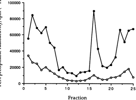
FIG. 1. EffectofdifferentpH values on in vitro genomic RNA synthesis. VSV wasallowedtoabsorbto BHKcells for 1 hat pH 7.4.Thecellswerethenreplenished withnewmediaatthe appro-priatepHand incubated foranadditional3h. At 4hpi,cytoplasmic extractswereprepared and incubated with [3H]UTP in the presence ofdactinomycin for90min at30°Cto measuregenomic replication. RNA specieswere separated byCsCl densitycentrifugation, frac-tionated, and precipitatedwithtrichloroacetic acid(TCA),andthe radioactivity wasplotted. Thenucleocapsid peak is in fractions 15 through 18. Sedimentation is from left to right. *, Extracts from cellsincubatedatpH 7.4for4hbeforepermeabilizing; 0,extracts produced from cells incubatedfor 1 h at pH 7.4 and then 3 h atpH 6.6before permeabilizing.
Since virus adsorption or uncoating was reduced when BHK cells were infected at pH 6.6 (datanot shown), cells wereinfected withVSV for 1 h with medium buffered at pH 7.4.Afterthis hourofadsorption,mediumbufferedatpH 7.4
or pH 6.6 was added to the cells and the infection was allowed to continue. Extracts were made at 4 hpi and incubatedin vitrowith[3H]UTPinthepresenceof
dactino-mycin (17). The replication products were separated from viral mRNAsbycentrifugation on a 20 to 40% CsCl gradient, where VSV nucleocapsids band at their buoyantdensity of
1.31g/cm3and mRNAs sediment to the pellet (2, 13). Figure 1 shows the differentamounts of[3H]UTP incor-porated in vitro into acid-precipitable material by extracts
thatwereprepared from infected cells incubatedatdifferent pHs. When VSV-infected cells were maintained at pH 6.6 for 3 h(Fig. 1, opencircles),therewas a33-foldreduction in incorporation of [3H]UTP into acid-precipitable material with adensity ofVSV nucleocapsids.
Quantitative analysisof viralproteinsatdifferentpHs. The
disparity between the RNA synthetic ability of the two extracts (pH7.4 versus pH 6.6) mighthave been attributed
to a decline in protein synthesis at the lower pH, since replication of genomic RNA is dependent upon protein synthesis (22).Previousreports, however,showed thatviral
protein synthesis levels remain unaltered if the infection
proceedsatpH 6.6(3, 7).Those methods usedradiolabeling techniques, and it ispossiblethatlowpHmodifieduptakeof
theisotope,preventingaquantitative
analysis.
Toavoid this problem, we determined the protein levels at the differentpH valuesthrough quantitative Westernblotting(20). Extracts were preparedat 4hpifrom infected cellsat pH
7.4 and pH 6.6 and fractionated by centrifugation into a
soluble protein fraction that was free ofnucleocapsid
tem-plates. The proteins were electrophoresed,
on November 10, 2019 by guest
http://jvi.asm.org/
[image:2.612.317.553.77.251.2]100 80 -60 40 20
I" ._
-4
a)
0
M N NS
FIG. 2. Western blot analysis of viral proteins. Cytoplasmic extractsfrom VSV-infected cells incubatedateitherpH 7.4 (N)or
pH 6.6 (El)werepreparedat4hpi. An equal portion of each reaction mixture was electrophoresed through an 11% polyacrylamide gel andelectrotransferredontonylon filterpaper.Theblotwasblocked
with BLOTTO, probed with anti-VSV serum,and then allowedto reactwith 1251-proteinA. The M, N, and NS proteins in the blot
wereexcised, and the radioactivitywasdetermined and plotted.
ferred to nylon membranes, probed with anti-VSV serum,
and reacted with 1251I-protein A. Figure 2 showsa compari-sonof the levels of the M, N, and NS proteins produced in infectedcells maintainedatpH 7.4or6.6.These dataarethe
averageof threeseparate determinations. There is less than a 40%inhibition ofsynthesis ofany of these viral proteins duetothe lowerpH. These resultssuggestedthat the block in RNA replication and nucleocapsid assembly observed at
pH 6.6 was probably not due to a decrease in total viral
protein synthesis at the lower pH.
Reconstitution assays with nucleocapsids and soluble
pro-teinsproducedatdifferent pHs.Infected cellextractscanbe
separated into one fraction containing the viral soluble
proteins and another fraction containingthe nucleocapsids;
thesefractionscanthen be recombinedtoreconstituteRNA
replication (17). We performed reconstitution experimentsto determine whether thereductioningenomeRNAreplication atpH6.6 correlated withadeficiencyinthe viralproteins,in the nucleocapsid templates, or in both. Accordingly, we
prepared cytoplasmicextracts at4 hpi fromBHK cellsthat were coinfected with VSV and the MS-T DI particle and maintainedatpH7.4or6.6. Theextractswereseparated by
centrifugation, and theresulting soluble proteinand
nucleo-capsidfractions were then mixedheterologously. The pres-ence ofdoublet micrococcalnuclease-resistant 19SRNA is indicativeofgenomeRNAsynthesis and encapsidation (17). Asanticipated, the recombination of pH 7.4 nucleocapsids withpH7.4soluble proteins resulted in the highest levels of replication (Fig. 3, lane 8). The mixture ofpH 6.6 soluble proteinswith pH 6.6 nucleocapsids (Fig. 3,lane 1) resulted
in very little RNA replication, suggesting that either the
protein or template fractions, or both, were defective. To determine whethertheeffect of lower pH on genome RNA
replicationandnucleocapsid assembly wasduetodefective soluble protein, we performed a series of reconstitution reactions with pH 7.4 nucleocapsids containing decreasing
amountsofpH7.4 solubleproteinandcompared the level of
genome RNAencapsidation toareaction containingpH 7.4
nucleocapsidsandpH6.6soluble proteins (Fig. 3,lanes 4to 8). pH7.4 solubleprotein levels of68, 50, and25% (Fig. 3, lanes 7, 6, and 5, respectively) ofthe control reaction all
6.6 7.4 7.4 6.6 7.4 7.4 74 7.4 Soluble
Protein 1I0 1'00 10) 10(X) 25 50 68 1}00 %SP
6.6 6.6 4x 6.6 7.4 7.4 7.4 7.4 7.4
I I I 1l _Ntlc_ocapsids
FIG. 3. Reconstitution ofMS-T genome RNA replication with nucleocapsids and soluble proteins. Cytoplasmic extracts were prepared fromMS-TplusVSVcoinfectedcells that wereincubated for4hwithmediabufferedateither pH7.4 orpH6.6andseparated into a soluble protein fraction and a nucleocapsid fraction by ultracentrifugation. Thenucleocapsid and soluble protein fractions were thenheterologously recombined andincubatedat30°C for 90 mininthe presenceof[3H]UTP.Theproducts of this reactionwere incubated with micrococcal nuclease for 1 h and then protease treated. Newly replicated genomic RNA was then extracted and analyzed byelectrophoresis on a1.5%agarose-acid-urea gel. The region ofthegelcontaining the plusand minus strands of the DI genomic RNA is shown here. Lanes (from left to right): pH 6.6 nucleocapsids reconstituted with pH 6.6 soluble proteins, pH6.6 nucleocapsids reconstituted with pH 7.4 soluble proteins, pH 7.4 soluble proteins reconstituted with pH 6.6 nucleocapsids concen-tratedfour times,pH 7.4nucleocapsids reconstituted with100% of the pH 6.6solubleproteins, pH7.4nucleocapsids reconstituted with 25% of thepH 7.4 solubleproteins, pH 7.4nucleocapsids reconsti-tuted with50%of thepH 7.4 solubleproteins, pH7.4nucleocapsids reconstituted with 68% of pH 7.4 soluble proteins, and pH 7.4 nucleocapsids reconstituted with100% ofpH 7.4soluble proteins (100% denotesthat amountofmaterial from 107cells).
supported levels of RNA replication higher than that seen
with pH7.4nucleocapsids and100%pH 6.6soluble protein (Fig. 3, lane 4). Since the level of viralproteinsincellsatpH 6.6isnotreducedbyeven50%(Fig. 2), these reconstitution experiments demonstrate that the defect in pH 6.6 soluble
protein is not simply due toa quantitative decrease in viral proteins but, rather, must be due to aqualitative changein
this fraction. Also shown in Fig. 3 (lane 3) is a reaction in
which the concentration of nucleocapsids from cells incu-bated at pH 6.6 was increased fourfold, demonstrating increased synthesis of 19S RNA relative to the reaction in
lane 2. This demonstrates that pH 6.6 nucleocapsids are
replication competent but are limited in amount in the
infected cell under these conditions of altered pH.
We performed another series of reconstitution reactions designedtoinvestigatethe effectofanincreased
concentra-tion of soluble proteinfrom infected cells at pH 6.6on the replication of nucleocapsids from cells atpH 7.4. Figure4
demonstrates that increasing the concentration of pH 6.6 soluble protein fourfold indeed stimulated replication of
nucleocapsid RNA but only to 56% of the control level (compare lanes5and2). This result shows that the effect of pH on soluble protein is not simply quantitative, since increasing the concentration of soluble protein from cells at
pH 6.6 fourfold (a minimum increase of viral proteins of 240%; Fig. 2) did not result in genome RNA replication comparable to 100% of pH 7.4 soluble protein with an
identical amountof pH7.4 nucleocapsids.
Immunoprecipitation of VSV proteins made at different
pHs.Since the lower level ofreplicationatpH 6.6 correlated withadeficiency inthesolubleprotein fraction,weanalyzed this fraction in greater detail. We previously reported the kinetic profile of VSV proteins by gradient fractionation
AEL ANIM&
I
on November 10, 2019 by guest
http://jvi.asm.org/
[image:3.612.70.299.69.223.2] [image:3.612.323.565.72.175.2]6.6 7.4
0
6.6 4x 6.6
Protein
6.6
7.4
7.4
7.4
7.4
I ] 1
__I__1_Nuclocapsids
U
e eISEI, "H74e-S-9
NS _
N __
B Chose, pH74,a-VSV
NS-
_-N
[image:4.612.316.552.67.336.2]M __
FIG. 4. Reconstitution of DI genomic RNA replication. This experiment was performed as described in the legend to Fig. 3. Lanes (from left to right): pH 6.6nucleocapsids reconstituted with pH 6.6 solubleproteins, pH 7.4 nucleocapsids reconstituted with pH
7.4solubleproteins, pH 7.4 nucleocapsids reconstituted with buffer, pH 7.4nucleocapsids reconstituted with pH 6.6 soluble proteins, and pH 7.4nucleocapsids reconstituted with pH 6.6 soluble proteins
concentrated fourtimes.
C Pulse,pH66,a-VSV D Chase, pH6.6,a-VSV
coupledwithimmunoprecipitation assays (16). Using similar techniques, we wanted to determine whether low pH af-fected the associations of the viral proteins. Infected cells incubated at pH 7.4 or pH 6.6 were pulse-labeled with [35S]methionine for20 min andchased for2h. Extracts were then prepared, and the soluble protein fraction was centri-fuged on 5 to 20% glycerol gradients. The gradients were fractionated, and the viral proteins were immunoprecipitated with either anti-VSV serum oranti-N monoclonal antibody. The immunoprecipitations with the anti-VSV serum showed that the same viral proteins were synthesized and located in the samegradient fractions regardless of the pH at which the infected cells were incubated (Fig. 5A and C). Afterthe 2-h chase, the majority of the Nprotein immuno-precipitated by the anti-VSV serum was found as a faster-sedimenting form in the pH 7.4 extracts (Fig. 5B). These results are consistent with those previously reported (16). However, the N protein was more evenly distributed throughout the gradient after the 2-h chase of the cells maintainedatpH 6.6using anti-VSV serum(Fig. SD) or an anti-N monoclonalantibody(Fig. 6D).This suggests that the maturation oftheviralN proteinisaltered at pH 6.6.
Peluso and Moyer (18, 19) reported that incubation of infected cell extracts with an anti-N monoclonal antibody precipitated boththe N andNS proteins,demonstrating that these two proteins exist in a complex. When we reacted solubleproteins frominfected cells incubatedatpH 7.4with
ananti-N monoclonalantibody, both the N and NS proteins
were precipitated as expected (Fig. 6A). However, when fractionated extracts from VSV-infected cells that were maintained atpH 6.6 for 3 h were assayed with the same
monoclonal antibody, the Nproteinwasprecipitated witha
reducedamountoftheNSprotein (Fig.6C).Quantitation of the N and NSproteins in gradient fractions1 to5ofthegels shownin panels 6Aand 6C revealeda twofolddecrease in the amount of NS protein relative to N protein atpH 6.6.
Thus, the lower pH appears to affect the associations of theseproteins duringtheirsynthesisorafter their formation. Also seen was an effectoflower pH on theform of the N
proteinpresent after 2 h of chase (compare panels B and D). Lastly,wewantedtodetermine whether the N-NSprotein complex formed in cells at pH 7.4 could be disrupted by incubation of cell extracts at pH 6.6. Hence, extracts that
werederivedfrom cells maintainedatpH7.4 were
immuno-NS
--N __ N
---FIG. 5. Immunoprecipitation of fractionated viral proteins with
anti-VSV serum. Infected cells were labeled with [355]methionine
for20 min atpH7.4(A) or 6.6 (C) or chased for anadditional 2 h at pH7.4(B)or6.6(D). Cytoplasmic extractswere prepared, and the
soluble proteinfractionwasobtainedand separated on a 5 to20%
glycerol gradient. An equal portion of each gradient fraction was immunoprecipitated withanti-VSV serum and analyzed by sodium dodecyl sulfate-polyacrylamide gel electrophoresis and fluorog-raphy. Fraction 1, the top of each gradient, is on the left. The
positionsofthe viralpolypeptides areindicated.
precipitated in buffer at pH 6.6. The results, shown in Fig. 7, indicatethatsimply incubatingthe N-NSproteincomplexin pH 6.6 buffer does not disrupt it once ithasformed.
DISCUSSION
Ourrationaleforincubating infected cellsatpH6.6was to
provide a means to disrupt or alter the formation of the N-NSprotein complexandexamineitssubsequent effecton
replication. We were interested in finding conditions that would perturbtheassociation ofthese twoproteinsinvivo,
so that we could determine whether the data from in vitro analysesof infected cellextracts demonstratingthe
require-mentofthe N-NSproteincomplex in genome RNA replica-tion (16, 18, 19) reflected accurately the requirements for RNA replication in the intact cell. In view of evidence demonstrating the essential role of the 1:1 N-NS protein complex in supporting VSV and DI genomic RNA replica-tion (16, 18, 19),we
hypothesized
thatoneeffect of lowpH
wouldbe the disruption or prevention offormation of this complex.Wefirst showed that viralRNA
replication
invitrowas reduced in extracts derived from infected cells incu-bated at pH 6.6, leading us to suspect that either the viral
proteinsortemplates,orperhapsboth,wereaffectedbythe decrease inpH.Throughreconstitution assays,wenarrowed the defect to the viral soluble protein fraction. Thus the solubleproteinfraction from infected cells maintainedat
pH
6.6 exhibits a decreased ability to support replication in
on November 10, 2019 by guest
http://jvi.asm.org/
[image:4.612.52.296.70.184.2](hase,pHi-A,4itN B
Cis.e, iI (,. 6.e' \
D
I . ...
E .-- 011o_ * _m _. w_ _ v
-FIG. 6. Immunoprecipitation of fractionated viral proteins with an anti-N monoclonal antibody. infected cells were labeled with
[35S]methioninefor 20minatpH 7.4(A)or6.6(C)orchasedforanadditional2hatpH7.4(B)or6.6(D).Cytoplasmicextractswereprepared,
and the solubleprotein fractionwasobtained andseparatedon a5to20%glycerol gradient.Anequal portionofeachgradientfractionwas
immunoprecipitated with anti-N monoclonal antibody and analyzed by sodium dodecyl sulfate-polyacrylamide gel electrophoresis and fluorography. Fraction1,thetopof each gradient,is ontheleft.Thepositions ofthe viral polypeptidesareindicated.
vitro, evenwhen supplied with nucleocapsids from infected
cells maintained at pH 7.4. This could have been due to
eitheraquantitative change in the pH 6.6 soluble proteinsor a qualitative change in this fraction. We addressed this
question by increasing the concentration of the pH 6.6 soluble proteins in the reconstitution assays (Fig. 4, lane 5)
but we were still unable to reconstitute replication to the levelsat pH 7.4. This result showed that the negative effect of low pH on the soluble protein fraction of VSV-infected cells was not simply due to a quantitative decrease in the amountof viral proteinspresent;low pHmustbealtering the
structure of one or more viral proteins or altering their
association with each other. We then examined the pool of viral proteins from infected cells incubated at pH 6.6 and showed that theamountof NS protein complexed with the N proteinwasdecreased. Thus,analteration in the association
of these two proteins in the cell has a negative effect on
genome RNA replication both in vivo and in vitro. It is
possible that the alteration in the N-NS protein complex is
one ofseveral effects ofpH, since Clinton et al. (3) have
shown that the phosphorylation state of the NS protein 7.4
NS
N
'a-.
FIG. 7. Immunoprecipitation of unfractionated viral proteins with an anti-N monoclonal antibody in pH 7.4 or 6.6 buffer. VSV-infected cellswerelabeled with [35S]methionine for 30minat pH 7.4. Unfractionated soluble proteins were immunoprecipitated
andwashedfive times in buffer(pH 7.4or6.6)plus0.5%Nonidet
P-40 andanalyzed by sodium dodecyl sulfate-polyacrylamide gel
electrophoresisandfluorography.
varied between the two pH values with an alteration in its
association with the ribonucleoprotein. We are currently
investigatingthestate ofphosphorylation of theNS protein in the N-NS protein complex at pH 6.6 in an effort to
determine whether there arealterations.
Interestingly, when fractionated pH 7.4 soluble proteins
arechased for 2 h, theN protein is found predominantly in thelowergradient fractions. Whencells werepulse-chased at pH 6.6, theN protein wasdistributed evenly throughout
the gradient. This suggests that low pH, in addition to
affectingtheprotein associations,isaffectingthematuration of theproteins. We havepreliminaryevidence that suggests
that the faster-sedimenting form of the N protein from
VSV-infected cellsat pH 7.4 is associated with small RNA species. However,we areunabletodetectanyRNAspecies
associatedwiththe Nproteinfrominfectedcellschased for 2 hatpH 6.6. We arecurrently intheprocessof character-izing these RNA species.
It should be noted that in systems for studying VSV genome RNA replication based on programming rabbit
re-ticulocytelysateswith VSV mRNAs, multiple forms of the N:NS protein complex have been demonstrated (5, 14). Indeed, we have detected complexes of N and NS with NINS ratios ofbetween 2:1 and8:1 ininfected cells (16),yet wehave been unableto demonstrate the abilityofanything
other than a 1:1 complex of N and NS to support genome RNA replication. These previous results, along with the resultspresented in this paper, leadusto conclude that the
substrate for genome RNA replication and nucleocapsid assembly in infected cells is in fact the 1:1 N:NS protein complexand thateventhoughitispossible fortheseproteins to associate in other ways, the relevance to replication of these alteredcomplexes isat bestuncertain.
ACKNOWLEDGMENTS
This research was supported by Public Health Service grant
A122116 from the National Institute of Allergy and Infectious Diseases.
A
.C
'p- I
on November 10, 2019 by guest
http://jvi.asm.org/
[image:5.612.126.502.72.283.2] [image:5.612.118.256.573.656.2]We thankAnne Deatly and Nancy Schultfor helpful comments and suggestions in preparing the manuscript.
LITERATURE CITED
1. Banerjee, A. K. 1987. Transcription andreplication of rhabdo-viruses. Microbiol. Rev. 51:66-87.
2. Blumberg, B. M., and D. Kolakofsky. 1981. Intracellular vesic-ular stomatitis virus leader RNAs are found in nucleocapsid structures. J.Virol.40:568-576.
3. Clinton, G. W., B. Burge, and A. S. Huang. 1978. Effects of phosphorylationand pH on the association of NS protein with vesicular stomatitis virus cores. J. Virol. 27:340-348.
4. Condra, J. H., and R. A. Lazzarini. 1980. Replicative RNA synthesis and nucleocapsid assembly in vesicular stomatitis virus-infected permeabilized cells. J. Virol. 36:796-804. 5. Davis, N. L., H. Arnheiter, and G. W. Wertz. 1986. Vesicular
stomatitisvirusNandNS proteins form multiplecomplexes. J. Virol. 59:751-754.
6. Davis, N. L., and G. W. Wertz. 1982. Synthesis of vesicular stomatitis virus negative-strandRNA in vitro: dependence on viralprotein synthesis.J. Virol. 41:821-832.
7. Fiszman, M., J. B. Leaute, C. Chany, and M. Girard. 1974. Mode of action of acid pH values on the development of vesicularstomatitisvirus. J.Virol. 13:801-808.
8. Ghosh, K., and H. P. Ghosh. 1982. Synthesis in vitro of full length genomic RNA and assembly of the nucleocapsid of vesicularstomatitis virus in acoupledtranscription-translation system.Nucleic AcidsRes.10:6341-6351.
9. Hill, V. M., L. Marnell, and D. F. Summers. 1981. In vitro replication and assembly of vesicular stomatitis virus nucleo-capsids. Virology113:109-118.
10. Huang, A., and E. L. Palma. 1974. Vesicularstomatitisvirus: defectiveness anddisease, p.87-100. In W.S. Robinsonand C. F. Fox (ed.), Mechanisms of virus disease. ICN-UCLA Sym-posia. W.A. Benjamin,Inc., MenloPark, Calif.
11. Leamson, R. W., and M. E. Reichmann. 1974. The RNA of defective vesicular stomatitisvirusparticlesin relation toviral
cistrons.J. Mol. Biol.85:551-568.
12. Lehrach,H., D.Diamond, J. M. Wozney, and H. Boedtker. 1977. RNA molecular weight determinations by gel electrophoresis underdenaturingconditions, a critical reexamination. Biochem-istry16:4743-4751.
13. Leppert, M., L. Rittenhouse, J. Perrault, D. F. Summers, and D. Kolakofsky. 1979. Plus andminus strandleaderRNAsin nega-tive strandvirus-infectedcells. Cell 18:735-747.
14. Masters, P. S., and A. K. Banerjee. 1988.Resolutionofmultiple complexesofphosphoprotein NS with nucleocapsidprotein N of vesicular stomatitis virus.J. Virol. 62:2651-2657.
15. Moyer, S. A., and S. H.Gatchell. 1979. Intracellular events in the replication of defective interfering particles of vesicular stomatitis virus.Virology92:168-179.
16. Peluso, R. W. 1988. Kinetic, quantitative,andfunctional anal-ysis ofmultiple forms ofthevesicular stomatitis virus nucleo-capsidproteinininfected cells.J. Virol. 62:2799-2807. 17. Peluso, R. W., and S. A.Moyer. 1983. Initiationandreplication
of vesicular stomatitis virusgenome RNA in acell-freesystem. Proc.Natl.Acad. Sci. USA80:3198-3202.
18. Peluso, R. W., and S. A.Moyer.1984. Vesicularstomatitis virus proteins required forthein vitroreplication of defective inter-feringparticlegenomeRNA,p. 153-160. In D. H. L.Bishopand R. W. Compans (ed.), Nonsegmented negative strand viruses. Academic Press, Inc.,NewYork.
19. Peluso, R. W., and S. A.Moyer. 1988. Viralproteins required for the in vitro replication of vesicular stomatitis virus defective interferingparticlegenome RNA. Virology 162:369-376. 20. Peluso, R. W., and G. Rosenberg. 1987. Quantitative
electro-transfer of proteins from sodium dodecyl
sulfate-polyacryl-amide gels onto positively charged nylon membrane. Anal. Biochem. 162:389-398.
21. Prevec, L., and C. Y. Kang. 1970. Homotypicandheterotypic
interference by defective interferingparticles of vesicular
sto-matitisvirus. Nature(London) 228:25-27.
22. Wertz, G. W., and M. Levine.1973. RNAsynthesis by vesicular stomatitis virus andasmallplaquemutant:effectsof
cyclohex-imide.J. Virol. 12:253-264.


![FIG. 6.andfluorography.immunoprecipitated[35S]methionine Immunoprecipitation of fractionated viral proteins with an anti-N monoclonal antibody](https://thumb-us.123doks.com/thumbv2/123dok_us/1324668.86249/5.612.118.256.573.656/andfluorography-immunoprecipitated-methionine-immunoprecipitation-fractionated-proteins-monoclonal-antibody.webp)
