Copyright © 2004, American Society for Microbiology. All Rights Reserved.
LMP2A Does Not Require Palmitoylation To Localize to
Buoyant Complexes or for Function
Rebecca B. Katzman and Richard Longnecker*
Department of Microbiology-Immunology, Northwestern University Feinberg School of Medicine, Chicago, Illinois
Received 22 March 2004/Accepted 26 May 2004
Epstein-Barr virus (EBV) latent membrane protein 2A (LMP2A) is expressed constitutively in lipid rafts in latently infected B lymphocytes. Lipid rafts are membrane microdomains enriched in cholesterol and sphin-golipids selective for specific protein association. Lipid rafts have been shown to be necessary for B-cell receptor (BCR) signal transduction. LMP2A prevents BCR recruitment to lipid rafts, thereby abrogating BCR function. As LMP2A is palmitoylated, whether this fatty acid modification is necessary for LMP2A to localize to lipid rafts and for protein function was investigated. LMP2A palmitoylation was confirmed in latently infected B cells. LMP2A was found to be palmitoylated on multiple cysteines only by S acylation. An LMP2A mutant that was not palmitoylated was identified and functioned similar to wild-type LMP2A; unmodified LMP2A localized to lipid rafts, was tyrosine phosphorylated, was associated with LMP2A-associated proteins, was ubiquitinated, and was able to block calcium mobilization following BCR cross-linking. Therefore, pal-mitoylation of LMP2A is not required for LMP2A targeting to buoyant complexes or for function.
Epstein-Barr virus (EBV) is a human herpesvirus that is able to establish and maintain latency in B lymphocytes (18, 20, 32). EBV infection is associated with the hematopoietic cancers African Burkitt’s lymphoma, Hodgkin’s lymphoma, and certain T-cell lymphomas. EBV is also associated with the epithelial diseases nasopharyngeal carcinoma, oral hairy leukoplakia, and gastric carcinoma. As a ubiquitous virus, EBV persists la-tently in about 0.5 to 50 cells per million B lymphocytes in over 95% of adults (39). Latent membrane protein 2A (LMP2A) is one of 10 EBV latent genes expressed by the virus in lympho-blastoid cell lines (LCLs). LMP2A is the most consistently de-tected transcript of EBV found in circulating B cells latently infected with EBV. Appropriately, LMP2A is thought to ensure EBV latency and may play a role in EBV-associated diseases. LMP2A is a highly hydrophobic protein with 12 putative transmembrane domains (26). The amino terminus of the pro-tein consists of a 119-amino-acid cytoplasmic tail. This tail con-tains functional domains and tyrosine residues responsible for recruiting B-cell signaling proteins such as Lyn and Syk; Lyn binds to tyrosine 112 of LMP2A, and Syk binds to the tyrosine residues 74 and 85 that make up the immunoreceptor tyrosine-based activation motif (ITAM) of LMP2A (11–13). Cells ex-pressing LMP2A are unable to mobilize intracellular calcium or to induce EBV lytic activation following B-cell receptor (BCR) activation (27, 28). Expression of LMP2A mutants deficient for Lyn or Syk binding shows normal calcium moblization and EBV lytic activation following BCR cross-linking (11–13).
LMP2A aggregates in patches in the plasma membrane (21). More recently, LMP2A has been shown to constitutively local-ize to lipid rafts (8). In vitro, it was demonstrated that LMP2A prevents recruitment of an activated BCR to lipid rafts, theby blocking BCR signaling; normally an activated BCR is
re-cruited to lipid rafts to activate BCR signal transduction (6, 8). Lipid rafts are microdomains in the membrane of unique pro-tein and lipid content, as well as having high concentrations of cholesterol (2, 37). One function associated with lipid rafts is that they are sorting sites for proteins and lipids required for specific signal transduction pathways to signal. Normally the BCR is recruited to lipid rafts, following antigen stimulation, where a complex of signaling proteins at the plasma membrane called the BCR signalosome can then be organized. However, LMP2A blocks BCR translocation to lipid rafts, thereby block-ing BCR signal transduction (8).
The mechanism of integral membrane protein localization to lipid rafts is unclear, although several possibilities have been demonstrated and hypothesized (4, 23, 35, 36). Protein affinity for lipid rafts is often determined by the anchoring of two saturated fatty acyl chains to the protein, as is the case for both glycosylphosphatidylinositol-anchored proteins and doubly acylated proteins. Hydrophobic transmembrane domains and/ or fatty acid modification may confer affinity of membrane-spanning proteins for lipid rafts. For example, lipid raft local-ization of some transmembrane proteins requires the post-translational modification of the protein by the fatty acid palmitate on cysteine residues by S acylation. The T-cell linker protein Lat and the influenza virus glycoprotein hemagglutinin (HA) both require palmitoylation of tandem cysteine residues in order to localize to lipid rafts (23, 41). However, the hydro-phobicity conferred by modifications is not the only character-istic responsible for protein localization within the membrane, as prenylated proteins do not associate with lipid rafts (23). Therefore, it seems that specific characteristics are important for lipid raft localization by dictating a protein-lipid packing order for this liquid-ordered (lo) phase of the lipid bilayer (3).
Transient expression of LMP2A in 293 and B cells has shown that LMP2A is a palmitoylated protein (14, 22). We analyzed a series of LMP2A mutants first to determine the site(s) of modification of the protein by palmitic acid and * Corresponding author. Mailing address: Department of
Microbi-ology-Immunology, Northwestern University Medical School, Chicago, IL 60611. Fax: (312) 503-139. E-mail: [email protected].
10878
on November 8, 2019 by guest
http://jvi.asm.org/
second to determine if modification by palmitic acid is required for LMP2A buoyant complex localization and function.
MATERIALS AND METHODS
Cell culture and transient transfections.B-cell lines were maintained in RPMI 1640 medium supplemented with 10% fetal bovine serum, 1,000-U/ml penicillin, and 1,000-g/ml streptomycin (complete RPMI 1640 medium). ES1 is an LMP2A⫺EBV-transformed lymphoblastoid cell line (LCL), and LCL1 is an
LCL transformed with wild-type EBV (27). BJAB is an EBV-negative Burkitt’s lymphoma cell line obtained from the American Type Culture Collection (Ma-nassas, Va.). A total of 107BJAB cells were transiently transfected with the Gene Pulser (Bio-Rad) at 220 V and 960F of capacitance.
Plasmids and LMP2A site-directed mutagenesis.L10-GFP is a lipid raft-targeted green fluorescent protein (GFP) expression construct and was kindly provided by William Rodgers (33). HA-conjugated ubiquitin (HA-Ub) was also described previously (40). pDsRed1-N1 was used for red fluorescent protein (RFP) expression (Clontech). RL168 is a plasmid containing HA-tagged LMP2A in a pSG5 backbone (13). The deletion mutants⌬168-365,⌬168-497, and⌬ 312-497 were described previously (21).⌬105-167 was made by removing the se-quence from the PmlI site of pY122F (13) to the MscI site in the LMP2A cDNA sequence.⌬426-497 is a truncation of LMP2A caused by a frameshift at amino acid 426 and the addition of 19 amino acids before a stop codon. Cysteine-to-alanine site-directed mutagenesis of RL168 was performed, except where noted, using a QuickChange site-directed mutagenesis kit (Stratagene) according to the manufacturer’s recommendations. Primers used for the mutagenesis also in-serted a silent restriction enzyme site that was used to confirm the mutations by restriction digestion. Mutations were also confirmed by sequencing. The 5⬘33⬘ primers used are listed below, and the 3⬘35⬘primers used were the reverse complement of the respective 5⬘33⬘primer. Bold type represents mismatched nucleotides; underlined nucleotides comprise the restriction enzyme recognition sequence; the corresponding restriction enzyme is in parentheses. pC3XXA, pC3XX/4XXA, and pC-ALL were made by a PCR-based cloning strategy with-out the kit.
The following primers were used: C1243A, 5⬘-GGGCAGAGGAAGTAT GAATCCAGTAGCGCTGCCTGTAATTGTTGCG-3⬘(Eco47III); C2453A (also used on pC124A to make pC124/245A), 5⬘-GGAGATGGCGCCGTTTG ACTGTTGCCGGCGGCATCA-3⬘ (NaeI); C339/342/3453A (also used on pC4XXA to make pC3XX/4XXA and on pC124/245A plus pC4XXA to make mutant pC-ALL), 5⬘-GGTTCTCCTGATTGCCTCTTCGGCGTCTTCAGCCC CACTGAGC-3⬘(BsaHI); C4263A (used on pC472/473/476/477/480A to make pC4XXA), 5⬘-CGGTCCAGTTTTTATGGCCCTCGGGGGCCTGCTCACC-3⬘ (AvaI); C472/4733A 5⬘-GGGGTCATTAGAGCCGCGCGCTACTGCTGCTA CTACTGC-3⬘(BssHII); C4763A (also used on pC472/473A to make pC472/ 473/476A), 5⬘-GTCATTAGAGCCGCGCGGTACGCCTGCTACTACTGCCT TAC-3⬘(eliminates BssHII site from pC472/3A); C4773A (also used on pC472/ 473/476A to make pC472/473/476/477A, 5⬘-GTCATTAGAGCCGCGCGCTAC GCCGCCTACTACTGCCTTAC-3⬘(BssHII); C4803A (also used on pC472/ 473/476/477A to make pC472/473/476/477/480A, 5⬘-GCCGCCTACTACGCCC TTACTCTAGAAAGTGAGGAGCGCCCACC-3⬘(XbaI). pC-ALL was made by PCR amplification first of two fragments separately with the following primer sets on the specified LMP2A mutant DNA: a primer for the T7 bacteriophage promoter and the reverse complement of C339/342/3453A with pC124/245A and C339/342/3453A and pSG5-1088 (recognizes sequences downstream of the multiple cloning site in pSG5) with pC4XXA.
Immunoprecipitation and Western blotting.Cells were lysed in 1 ml of 1% Triton X-100 lysis buffer (50 mM Tris-HCl [pH 7.4], 150 mM NaCl, 10% glycerol, 2 mM EDTA) containing the protease and phosphatase inhibitors (1M pep-statin, 1M leupeptin, 0.5 mM phenylmethylsulfonyl fluoride, 1 mM sodium orthovanadate, 10 mM sodium fluoride). Transiently transfected cells were lysed 20 h after transfection. The insoluble material was removed by centrifugation at 4°C. Cleared lysates were incubated with the indicated antibodies, and immune complexes were captured with protein G Sepharose for 1 h at 4°C, washed three times in 1% Triton X-100 lysis buffer, and mixed with 2⫻sodium dodecyl sulfate (SDS) sample buffer. Samples were incubated at 70°C for 5 min and separated by SDS-polyacrylamide gel electrophoresis (PAGE). Proteins were transferred to Immobilon membranes (Millipore). Membranes were blocked in 4% milk in Tris-buffered saline-Tween (TBST) for 1 h at room temperature. Three percent bovine serum albumin was used when antibody to phosphotyrosine was used. Membranes were next incubated with primary antibody in TBST for 1 h in TBST, washed three times, incubated with horseradish peroxidase (HRP)-linked sec-ondary antibody for 30 min at room temperature, and washed four times in TBST, and the proteins were detected by enhanced chemiluminescence (ECL;
Amersham Bioscience) using HyperfilmMP (Amersham Bioscience) and pro-cessed through a Kodak film developer.
Rat monoclonal primary antibody was used to immunoprecipitate LMP2A and for Western blotting for LMP2A (14B7) (11). Biotinylated 14B7 was used for Western blots of immunoprecipitated LMP2A. NeutrAvidin-HRP was pur-chased from Pierce. The HA epitope tag was detected with mouse monoclonal HA.11 (BabCO). Polyclonal Lyn was purchased from Santa Cruz. Rabbit poly-clonal serum directed against AIP-4 was described previously (17). RC20-HRP recognizes tyrosine-phosphorylated proteins and was purchased from Transduc-tion Laboratories. BD Living ColorsA.v. peptide antibody was used to detect GFP (BD Biosciences). The HRP-conjugated secondary antibodies used were goat anti-rat immunoglobulin G (IgG), F(ab)2-specific species (Jackson Immu-noResearch Laboratories), anti-mouse Ig (Amersham), and anti-rabbit IgG (Cell Signaling Technology).
[3H]palmitic acid and [35S]methionine metabolic labeling.Cells were first
washed two times in serum-free medium. For the [3H]palmitate label, the cells were resuspended in 4 ml of RPMI 1640 medium containing 2% dialyzed serum and 750Ci of [3H]palmitic acid (Amersham Bioscience). For the [35 S]methi-onine labeling, the cells were resuspended in 4 ml of methiS]methi-onine, cysteine, and
L-glutamine-free RPMI 1640 medium (Sigma) containing 2% dialyzed serum, 4
mML-glutamine, and 150-Ci/ml [35S]methionine (Amersham Bioscience). The cells were incubated at 37°C for 4 h with occasional rocking. The labeled cells were then lysed in 1% Triton X-100 lysis buffer for immunoprecipitation as described above.
Fluorography and hydroxylamine treatment of SDS-PAGE gels.Following SDS-PAGE of metabolically labeled immunoprecipitates, gels were fixed with 10% acetic acid–25% isopropanol–65% water for 30 min and then treated with Amplify (Amersham Bioscience) for 30 min. For hydroxylamine treatment of palmitate gels, parallel gels were treated overnight with 2 M hydroxylamine, 2 M Tris-HCl, pH 7.0, or 2 M Tris-HCl, pH 7.0, and rinsed three times for 5 min each with water prior to treatment with Amplify.
Isolation of lipid raft proteins and protein electrophoresis.Lipid rafts were isolated from transiently transfected BJAB cells 15 h after 107cells were elec-troporated with RL168, mutant LMP2A, or empty vector plus L10-GFP as de-scribed previously (6). The cells were lysed in 1 ml of 1% Triton X-100 lysis buffer in TNE (10 mM Tris-HCl [pH 7.5], 150 mM NaCl, 5 mM EDTA) with protease and phosphatase inhibitors (1M pepstatin, 1M leupeptin, 0.5 mM phenyl-methylsulfonyl fluoride, 1 mM sodium orthovanadate, and 10 mM sodium fluo-ride) on ice for 30 min. Lysates were diluted 1:1 with 1 ml of cold 85% sucrose in TNE in the bottom of a Beckman 14- by 89-mm centrifuge tube. The lysate mixture was then overlayed with 6 ml of 35% sucrose in TNE followed by 3.5 ml of 5% sucrose in TNE. Gradients were centrifuged at 200,000⫻gin an SW41 rotor for 18 h at 4°C. One-milliliter fractions were collected from the top of the gradient and diluted 1:1 with 2⫻SDS sample buffer. Equal volumes from each 1-ml fraction collected were loaded onto SDS-polyacrylamide gels, and protein localization was then analyzed by Western blotting.
Calcium flux assay.To assay for calcium flux, BJAB cells were first cotrans-fected with 20g of RFP plus 20g of LMP2A, mutant LMP2A, or empty vector. Ten hours posttransfection, changes in intracellular calcium concentra-tion were measured as described before (27). Briefly, 2⫻106cells per sample were loaded with the ratiometric calcium-binding fluorescent dye indo-1 (1M; Molecular Probes) in 1% serum containing RPM1 for 30 min at 37°C. The samples were washed and then resuspended in fresh 1% RPMI without dye for 30 min at 37°C. Cells were examined with a Beckman Coulter Elite ESP flow cytometer, using Elite version 4.02 software. Fluorescence was measured at 395 and 525 nm, and the ratio of the two readings was calculated by the software. Changes in the ratio over time reflect a calcium flux. Each sample was analyzed for 5 min and stimulated with 10-g/ml goat anti-human Ig (IgM⫹IgG⫹IgA, H⫹L; Southern Biotechnologies) after approximately 40 s. Samples were main-tained at 37°C throughout the entire procedure.
RESULTS
LMP2A is fatty acid modified by palmitic acid in LCLs.
LMP2A constitutively associates with lipid rafts in B lympho-cytes and blocks BCR recruitment to lipid rafts following BCR activation (8). It has been reported that LMP2A is modified by the fatty acid palmitic acid in transient transfection assays of 293 and B cells (14, 22). Since these previous experiments used transfected cells, we first wanted to confirm these results, using B cells latently infected with EBV. Metabolic labeling
on November 8, 2019 by guest
http://jvi.asm.org/
periments using two LCL lines latently infected with EBV were performed with [35S]methionine to label all proteins or
[3H]palmitic acid to specifically detect palmitoylated
pro-tein. The cell lines used were LCL1, which expresses LMP2A, and ES1, which is null for LMP2A. Labeled cell lysates were immunoprecipitated for LMP2A. The top panel in Fig. 1 shows a fluorogram of a gel with [3H]palmitic acid-labeled LMP2A
immunoprecipitates, and the middle panel shows that of a gel with [35S]methionine-labeled immunoprecipitates (Fig. 1A and
B). A unique band appears at the molecular mass equivalent to LMP2A (55 kDa) only in the lane representing immunopre-cipitated proteins from LCL1. A parallel Western blot con-firms that the band is indeed LMP2A, thereby confirming LMP2A palmitoylation in LCLs (Fig. 1C).
LMP2A is multiply palmitoylated in different domains.In
order to determine where LMP2A is palmitoylated, a series of LMP2A deletion mutants accounting for all transmembrane regions were analyzed (Fig. 2A). The EBV-negative B-cell line, BJAB, was used for these experiments. BJAB cells were elec-troporated with vector control, wild-type, or mutant LMP2A and then metabolically labeled with [3H]palmitic acid.
Immu-noprecipitates from cells expressing the different LMP2A con-structs were analyzed by fluorography and also by Western FIG. 1. LMP2A is fatty acid modified by palmitic acid in LCLs. LCLs ES1 (LMP2A negative [⫺]) and LCL1 (LMP2A positive [⫹]) were incubated with [3H]palmitic acid (3H-Palm) or with [35
S]methi-onine (35S-Met) for 4 h at 37°C while rocking and then lysed. Lysates
were immunoprecipitated for LMP2A with a monoclonal rat antibody and then prepared for SDS-PAGE. Gels were treated for fluorography before exposing them to film. Fluorography of half of the [3H]palmitic
acid-labeled immunoprecipitated proteins (A). [35
S]methionine-immu-noprecipitated proteins (B). Half of the [3H]palmitic acid-labeled
[image:3.603.70.253.70.328.2]im-munoprecipitated proteins were analyzed on a parallel gel, transferred to Immobilon membrane, and immunoblotted for LMP2A with a bi-otin-conjugated monoclonal rat antibody (␣LMP2A) (C). Represen-tative data of three independent experiments are shown.
FIG. 2. LMP2A deletion mutants are palmitoylated. EBV-negative BJAB B cells were transiently transfected with full-length and LMP2A deletion constructs (shown in panel A). After 4 h, the cells were incu-bated with [3H]palmitic acid (3H-Palm) for an additional 4 h and then
lysed. Lysates were immunoprecipitated for metabolically labeled LMP2A followed by SDS-PAGE and autoradiography (B, top). West-ern blot analysis was performed in parallel with antibodies to LMP2A (␣LMP2A) (B, bottom). Each deletion mutant maintained palmitoyl-ation like full-length LMP2A, whereas no bands were detected in the pSG5 vector control lane. The data shown represent at least three independent experiments.
on November 8, 2019 by guest
http://jvi.asm.org/
blotting. Figure 2B shows that all of the deletion constructs re-tained palmitoylation similar to wild-type LMP2A. Surprising-ly, even mutants lacking the C-terminal tail domain of LMP2A (⌬168-497,⌬312-497, and⌬426-497) were palmitoylated in BJAB cells. This differed from what was observed previously in 293 cells, where it was shown that the cysteines in the cytoplasmic carboxy terminus were the sites for palmitoylation (22). Based on these data, we concluded that LMP2A is likely modified by palmitic acid and in several different domains and that there may be cell-type-specific palmitoylation of LMP2A.
LMP2A is palmitoylated on multiple cysteine residues.
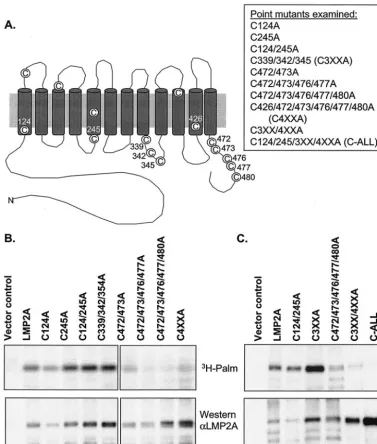
[image:4.603.106.483.71.515.2]Gen-erally cysteine residues close to the inner leaflet of the plasma membrane are major targets for posttranslational modification by palmitic acid (1, 19). LMP2A has 15 cysteine residues in its sequence, 11 of which lie close to the inner leaflet according to the putative structure of the protein (Fig. 3A). Therefore, a series of point mutants were constructed in which the 11 cys-teines close to the inner leaflet were mutated to alanine either singly or in combination with other cysteine-to-alanine muta-tions (Fig. 3A). As palmitic acid is unable to form a thioester FIG. 3. LMP2A cysteine-to-alanine point mutants retain palmitoylation. The putative structure of LMP2A showing where the cysteine residues of LMP2A are located is shown in panel A. The position in the sequence of LMP2A of the cysteines mutated to alanine is indicated in the diagram, and all of the point mutants constructed are listed. EBV-negative BJAB B cells were transiently transfected with full-length and mutant LMP2A constructs (B and C). After 4 h, the cells were incubated with [3H]palmitic acid (3H-Palm) for an additional 4 h and then lysed. Lysates were
im-munoprecipitated for metabolically labeled LMP2A followed by SDS-PAGE and autoradiography. Western blot analysis was performed in parallel using antibodies to LMP2A (␣LMP2A). Relative palmitoylation (Rel Palm) was calculated following densitometry by dividing the ratio of tritiated protein to protein detected by Western blotting by the ratio determined for wild-type LMP2A. The ratio for wild-type LMP2A was thus set to 1.0. Representative data from four independent experiments are shown.
on November 8, 2019 by guest
http://jvi.asm.org/
bond with alanine, mutating a potential site of palmitoylation to alanine would eliminate modification of that residue.
Following site-directed mutagenesis, the LMP2A point mu-tants were transiently transfected into BJAB cells and com-pared with the vector control and wild-type LMP2A. The trans-fected cells were metabolically labeled with [3H]palmitic acid,
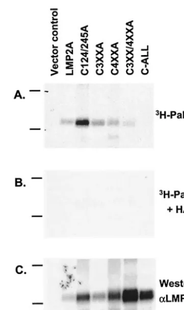
and immunoprecipitates were analyzed again by fluorography and Western blotting. Relative palmitoylation was determined by densitometry with wild-type LMP2A set to 1.0 (Fig. 3B). The majority of the mutants were palmitoylated similar to wild-type LMP2A. When several of the sites were mutated in the same construct, significantly less palmitoylation was ob-served (Fig. 3B, mutants C472/473A, C472/473/476/477A, C472/ 473/476/477/480A, and C4XXA). Therefore, LMP2A mutants containing a greater number of cysteine-to-alanine mutations were constructed, including one mutant in which all of the highlighted cysteines were mutated to alanine (Fig. 3C, mu-tants C3XX/4XXA and C-ALL). The mutant in which all of the cysteines were mutated to alanine (C-ALL) had only back-ground levels of labeling. These data indicate that in pC-ALL all of the sites of cysteine palmitoylation have been eliminated and this mutant of LMP2A is no longer palmitoylated. This was further confirmed by treating a parallel gel with the nu-cleophile hydroxylamine (pH 7.0) prior to exposure to film. Hydroxylamine treatment cleaves thioester bonds, thus con-firming S acylation of a protein, most likely by the addition of palmitic acid to cysteine residues. Hydroxylamine treatment eliminated the signal from wild-type LMP2A and the palmi-toylated mutants, as would be expected if LMP2A is modified by palmitic acid (Fig. 4). These data confirm that the LMP2A signal detected by fluorography is due to modification of LMP2A only by S acylation and not from metabolic conversion of the [3H]palmitic acid label.
Reduced palmitoylation is not required for LMP2A buoyant
complex localization.Several proteins require multiple
modi-fications by palmitic acid in order to localize to lipid rafts (1). However, for some lipid raft-localized integral membrane pro-teins, the means of lipid raft localization is unclear. To deter-mine if palmitoylation is necessary for constitutive buoyant complex localization of LMP2A, different LMP2A constructs were cotransfected into BJAB cells along with a lipid raft-localized GFP marker. L10-GFP contains the first 10 amino
acids of Lck and is thus constitutively targeted to lipid rafts (33). L10-GFP therefore serves as both a transfection control
and a lipid raft marker.
Transfected BJAB cells were lysed, and lysates were sub-jected to sucrose gradient density ultracentrifugation. Lipid rafts are insoluble in 1% Triton X-100 at 4°C. These detergent-resistant membranes (DRMs) migrate to lower-density frac-tions in a sucrose gradient, whereas soluble lipids and proteins will remain at the bottom of the ultracentrifuge tube. One-milliliter fractions were collected from the top of the tube and mixed 1:1 with SDS-sample buffer. The fraction samples were then analyzed by SDS-PAGE and transferred to Immobilon membranes. The Immobilon membranes were cut in strips based on the migration of the molecular mass markers before probing for the indicated protein. Lyn, a src family protein tyrosine kinase, constitutively localizes to DRMs due to N-terminal dual acylation by both myristic and palmitic acid (Fig. 5A, fractions 3, 4, and 5). When L10-GFP was transfected into
cells, GFP was found in the same DRM fractions as Lyn (Fig. 5A, fractions 3, 4, and 5). CD45, a soluble integral membrane protein, was consistently seen only in soluble fractions (Fig. 5A, fractions 11 and 12). When wild-type LMP2A was cotrans-fected into cells with L10-GFP, LMP2A cofractionated with
GFP (Fig. 5B, fractions 4, 5, and 6). The localization of all of the LMP2A cysteine-to-alanine point mutants was tested with this transient assay. They were all found to retain lipid raft localization similar to wild-type LMP2A (Fig. 5C and D) (data not shown). Even when the mutant lacking palmitoylation, C-ALL, was expressed in B cells, it too cofractionated with the lipid raft-targeted GFP (Fig. 5D). Additionally, conventional immunofluorescence microscopy did not show any dramatic difference in the localization of the LMP2A point mutants compared to wild-type LMP2A (data not shown). These data indicate that palmitoylation is not required for the constitutive lipid raft localization of LMP2A.
Palmitoylation is not required for LMP2A phosphorylation,
ubiquitination, or protein association.To further test the
[image:5.603.329.510.65.370.2]func-tional significance of LMP2A palmitoylation, a series of the FIG. 4. LMP2A is modified by S acylation. [3H]palmitic acid (3
H-Palm)-labeled immunoprecipitates of transiently transfected BJAB cells with the indicated constructs were divided into three parts for SDS-PAGE. After fixing the gels, the first gel was treated with 2 M Tris-HCl, pH 7.0, overnight (A), whereas the second gel was treated with 2 M hydroxylamine in 2 M Tris-HCl, pH 7.0, overnight prior to fluorography (B). The third gel was transferred to Immobilon mem-brane for Western blotting with antibodies to LMP2A (␣LMP2A) (C). Shown are representative data from two independent experiments. The positions of the 66 and 45 molecular size markers in kilodaltons are shown to the left of each gel.
on November 8, 2019 by guest
http://jvi.asm.org/
LMP2A point mutants were tested for their ability to associate with LMP2A-associated signaling proteins and were assayed for tyrosine phosphorylation and Ub conjugation. LMP2A re-quires association with Lyn to become tyrosine phosphorylated and requires association with Lyn and Syk to block BCR
acti-vation (12, 13). LMP2A also associates with the Nedd4 family of E3 Ub ligases, AIP-4. This association results in the ubiq-uitination of LMP2A and LMP2A-associated proteins (15, 17). To examine whether or not palmitoylation is required for LMP2A posttranslational modification and protein associa-FIG. 5. Palmitoylation is not required for LMP2A buoyant complex localization. EBV-negative B cells were transiently cotransfected with empty vector, LMP2A, or mutant LMP2A constructs, and with L10-GFP, lysed with 1% Triton X-100 lysis buffer after 15 h, and subjected to density gradient
ultracentrifugation. Samples from each fraction were analyzed by immunoblotting for wild-type or mutant LMP2A. L10-GFP cofractionates with Lyn
in lower-density fractions representing DRMs (A, fractions 3 to 6), but not with CD45, which remains in the high-density soluble protein fractions (A, fractions 10 to 12). Wild-type LMP2A cofractionates with L10-GFP in DRM fractions (B). Representative data of the LMP2A cysteine-to-alanine
point mutants (C and D). Each mutant assayed was found in DRM fractions. The blots shown are representative of two independent experiments.
on November 8, 2019 by guest
http://jvi.asm.org/
tion, BJAB cells were transiently transfected with wild-type LMP2A and a series of the LMP2A point mutants. Lysates from these cells were immunoprecipitated either for Lyn (Fig. 6A) or for LMP2A (Fig. 6B). Immunoprecipitates were ana-lyzed by SDS-PAGE and transferred to Immobilon mem-branes for Western blotting for the indicated proten or protein modification. All of the LMP2A mutants analyzed coimmuno-precipitated with Lyn, similar to wild-type LMP2A (Fig. 6A). Also, all of the mutants were tyrosine phosphorylated and able to bind AIP-4 (Fig. 6B).
Finally, BJAB cells were cotransfected with HA-Ub and either vector control, LMP2A, or the C-ALL mutant. The cells were lysed, and LMP2A was immunoprecipitated with a mono-clonal antibody for LMP2A. The immunoprecipitated LMP2A was analyzed for Ub modification. Membranes were probed with an antibody to HA. Both monoubiquinated LMP2A and polyubiquinated LMP2A were detected as indicated by the arrows in Fig. 6C. Therefore, the C-ALL mutant demonstrates that palmitoylation is not required for the ubiquitination of LMP2A.
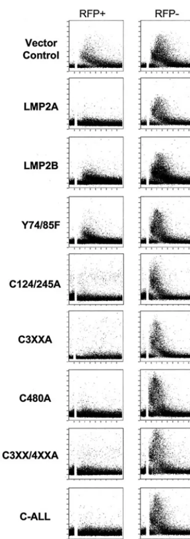
Palmitoylation is not required for the LMP2A-induced block of calcium mobilization following BCR cross-linking.
LMP2A blocks calcium mobilization following BCR cross-link-ing in B lymphocytes (28). To investigate whether or not pal-mitoylation of LMP2A is required for LMP2A to block BCR signaling, calcium mobilization was assessed with a flow cytom-etry-based assay utilizing the ratiometric fluorescent dye indo-1. The different LMP2A mutants were transiently expressed in BJAB cells. RFP was cotransfected into the cells as a marker for transfected cells, and RFP-positive versus RFP-negative cells were compared. Untransfected, RFP-negative cells serve as an internal control for calcium flux.
[image:7.603.326.510.70.584.2]As shown in Fig. 7, intracellular calcium mobilization was observed in both the RFP-positive and -negative channels when the cells were electroporated with the vector control (Fig. 7). However, when wild-type LMP2A was expressed in the cells, there was no mobilization of intracellular calcium in RFP-positive cells relative to the cells not expressing RFP where a calcium flux was observed (Fig. 7). Therefore, RFP-expressing cells are indicative of LMP2A-RFP-expressing cells. All of the LMP2A cysteine-to-alanine point mutants tested were also able to block calcium mobilization similar to wild-type LMP2A, including C-ALL, which was no longer palmitoylated. LMP2B and Y74/85F were both used as negative controls. LMP2B lacks the cytoplasmic amino terminus of LMP2A, which contains the motifs for Lyn and Syk binding to LMP2A. Y74/85F contains a tyrosine-to-phenylalanine mutation at both tyrosines 74 and 85 of the LMP2A amino acid sequence. As a result of this mutation, Syk is unable to bind to LMP2A and LMP2A no longer blocks calcium mobilization following BCR activation. A calcium flux was observed in both the RFP-pos-itive and -negative channels when LMP2B or Y74/85F was expressed in BJAB B cells. The calcium response was some-what muted in the negative controls (LMP2B and Y74/85F) when compared to the vector control-transfected cells and the RFP-negative cells. This may be due to overexpression of exogenous protein negatively affecting the cells. Regardless, there is a substantial difference when the calcium responses are compared to those of wild-type LMP2A or the LMP2A cys-teine mutants. Modification of LMP2A by palmitic acid is
FIG. 6. Palmitoylation is not required for LMP2A protein associ-ation, tyrosine phosphorylassoci-ation, and ubiquitination. Wild-type LMP2A or mutant LMP2A was transiently transfected into BJAB cells. Fifteen hours after transfection, the cells were lysed. Lysates were immuno-precipitated (IP) for LMP2A, and immunoprecipitates were analyzed by SDS-PAGE, transferred to Immobilon membrane, and probed for either LMP2A (A and B; LMP2A), phosphorylated tyrosine residues (B; APT), or AIP-4 coassociation (B; AIP-4). Lyn immunoprecipitates were analyzed by Western blotting for Lyn and for LMP2A coassocia-tion (A; Lyn and LMP2A). BJAB cells were also cotransfected with HA-Ub (C). Lysates were immunoprecipitated for LMP2A, and mem-branes were probed with an antibody for the HA tag to detect higher-molecular-weight LMP2A species representing ubiquitinated LMP2A (C, arrows). Empty vector was included as a control. Representative immunoblots of two independent experiments are shown.
on November 8, 2019 by guest
http://jvi.asm.org/
therefore not necessary for LMP2A to block signaling down-stream of the BCR.
DISCUSSION
Modification of a protein by palmitic acid has been the ma-jor means identified for conferring lipid raft affinity for trans-membrane proteins. Therefore, the palmitoylation of LMP2A in B cells was investigated both in stable LCLs and in tran-siently transfected cell lines. Also, whether or not palmitoyl-ation is functionally significant for LMP2A was analyzed.
LMP2A is palmitoylated in multiple domains, as indicated by the analysis of the LMP2A deletion mutants. Several cysteine-to-alanine mutations were made in the LMP2A se-quence. While the majority of LMP2A palmitoylation is in the C-terminal cytoplasmic tail of LMP2A, other cysteine residues in LMP2A close to the inner leaflet of the plasma membrane are palmitoylated. One mutant of LMP2A, C-ALL, had only background levels of palmitoylation. While a short labeling time was used to prevent metabolism of the tritium label, it is possible that the background level of labeling reflected modi-fication of LMP2A by a means other than S acylation or that another cysteine residue in LMP2A is palmitoylated (Fig. 3A). Previously it was reported that in 293 cells LMP2A is palmi-toylated only in the C terminus (22). It is possible that LMP2A is posttranslationally modified differently in epithelial cells than it is in B cells. As little is known about the mechanism by which proteins become palmitoylated and no definitive palmi-toylase has yet been identified, it is possible that LMP2A is palmitoylated at different positions in different cell types (19). Palmitate is a 16-carbon chain added posttranslationally to cysteine residues via a stable thioester bond (29, 34). The fatty acid is a hydrophobic moiety that is thought to regulate many functions of integral membrane proteins, such as protein sort-ing into lipid rafts (1). Therefore, whether or not the loss of the palmitate modification resulted in the loss of LMP2A lipid raft localization was analyzed. The localization of LMP2A in lipid rafts appears to block BCR signaling following receptor acti-vation by preventing recruitment of the activated BCR into lipid rafts. The data presented here show that palmitoylation is not required for LMP2A buoyant complex localization; C-ALL and L10-GFP both cofractionated in DRM-containing
frac-tions following sucrose gradient ultracentrifugation. Each of the point mutants tested floated in sucrose gradients, indicat-ing that they are associated with DRMs. Similar to LMP2A, palmitoylation of EBV LMP1 is not required for the con-stitutive lipid raft association of this latent EBV protein (14). LMP2A must therefore associate with lipid rafts by some means other than palmitoylation.
[image:8.603.65.256.64.603.2]Lipid raft-associated proteins have also been shown to be myristylated. While LMP2A has three ATG start codons, the ATG used by LMP2A in LCLs does not contain a glycine res-idue in position 2 (16). Therefore, LMP2A is probably not modified by myristic acid.
FIG. 7. Palmitoylation is not required for LMP2A to block Ca mobilization. BJAB cells were cotransfected with RFP and the indi-cated LMP2A expression construct or vector control. Ten hours after transfection, the cells were loaded with the calcium-sensitive dye indo-1 indo-1.5 h prior to analysis by flow cytometry. The cells were stimulated with goat anti-human Ig about 40 s after observing baseline calcium levels (indicated by a break in the histogram). Events were collected over 5 min. Thexaxis represents time, and theyaxis represents the ratio of fluorescence at 395 nm to that at 525 nm (ratio of calcium-bound indo-1 to uncalcium-bound indo-1). Transfected cells were determined by gating on RFP-positive cells. RFP-negative cells represent untrans-fected cells and serve as an internal control. LMP2B lacks the cyto-plasmic amino terminus of LMP2A, and Y74/85F is defective for Syk
binding; therefore, both proteins are unable to block BCR signaling. Calcium fluxes of 5-min time courses show representative data of at least three independent experiments.
on November 8, 2019 by guest
http://jvi.asm.org/
It is also thought that some proteins associate with lipid rafts via an association with another protein, as has been proposed for HA (7, 10, 30). LMP2A associates with Lyn, and this as-sociation is essential for LMP2A to block BCR signaling. Lyn constitutively localizes with lipid rafts as it is a dual acylated protein; Lyn is modified by both myristic acid and palmitic acid at the N terminus (31). It has been demonstrated previously that LMP2A does not require Lyn binding in order to localize to lipid rafts, as an LMP2A mutant deficient for Lyn binding constitutively localizes with DRMs (8). It is possible, however, that LMP2A could be tethered to lipid rafts via an interaction with another binding partner that is localized to lipid rafts. Future experiments will address this possibility with proteins that are currently known to interact with LMP2A, or perhaps these data are indicative of protein associations that are not yet known for LMP2A.
Palmitoylation has also been shown to have other functions such as in protein trafficking and protein turnover (1). Palmi-toylation can be responsible for protein trafficking from the ER to the plasma membrane. Therefore, whether or not the loss of palmitoylation of LMP2A interfered with LMP2A function was investigated. However, all of the point mutants analyzed herein, including the nonpalmitoylated mutant C-ALL, were able to associate with Lyn and were consequently tyrosine phosphorylated. Also, the mutants were able to bind with AIP-4 and were found to be ubiquitinated similar to wild-type LMP2A. And finally, the cysteine-to-alanine mutants were able to function normally, as demonstrated by their ability to block the mobilization of intracellular calcium. It was surprising that even the C-ALL mutant, with 11 cysteine residues mutated to alanine, was able to function normally in the function assays performed. Palmitoylation of LMP2A therefore does not seem to be involved in any of its known functions able to be assessed in vitro.
It seems unlikely that a viral protein would be multiply modified by the fatty acid for no functional purpose. There-fore, future experiments aim to discern if palmitoylation of LMP2A is required for other functions of LMP2A. One such possibility is for the LMP2A-mediated survival signal, of which little is known. It has been demonstrated in LMP2A-expressing transgenic mice, however, that B cells expressing LMP2A by-pass normal developmental checkpoints and survive in the periphery without a fully developed BCR (5). It has further been demonstrated in the LMP2A transgenic mice that this survival signal requires an intact ITAM in LMP2A, the ty-rosine kinase Btk, and the scaffold protein BLNK (9, 24, 25). Finally, in vitro the proto-oncogene Akt, which is necessary for cell survival, is constitutively phosphorylated in LMP2A-ex-pressing cell lines (38). Therefore it has been hypothesized that LMP2A provides a constitutive signal allowing for the devel-opmental bypass and survival of B lymphocytes that would otherwise apoptose. Palmitoylation of LMP2A might play a role in regulating a protein interaction or localization required for LMP2A-mediated cell survival.
ACKNOWLEDGMENTS
We would like to thank members of the Longnecker lab for their help in these studies and Mary Paniagua for help with calcium analysis. R.L. is supported by Public Health Service grants CA62234, CA73507, and CA93444 from the National Cancer Institute and by
grant DE13127 from the National Institute of Dental and Craniofacial Research. R.L. is a Stohlman Scholar of the Leukemia and Lymphoma Society of America. R.B.K. was supported in part by a training grant from the National Institutes of Health (T32CA009560).
REFERENCES
1. Bijlmakers, M. J., and M. Marsh.2003. The on-off story of protein palmi-toylation. Trends Cell Biol.13:32–42.
2. Brown, D. A., and K. Jacobson.2001. Microdomains, lipid rafts and caveolae (San Feliu de Guixols, Spain, 19–24 May 2001). Traffic2:668–672. 3. Brown, D. A., and E. London.1998. Structure and origin of ordered lipid
domains in biological membranes. J. Membr. Biol.164:103–114. 4. Brown, D. A., and J. K. Rose.1992. Sorting of GPI-anchored proteins to
glycolipid-enriched membrane subdomains during transport to the apical cell surface. Cell68:533–544.
5. Caldwell, R. G., J. B. Wilson, S. J. Anderson, and R. Longnecker.1998. Epstein-Barr virus LMP2A drives B cell development and survival in the absence of normal B cell receptor signals. Immunity9:405–411.
6. Cheng, P. C., M. L. Dykstra, R. N. Mitchell, and S. K. Pierce.1999. A role for lipid rafts in B cell antigen receptor signaling and antigen targeting. J. Exp. Med.190:1549–1560.
7. Cheong, K. H., D. Zacchetti, E. E. Schneeberger, and K. Simons.1999. VIP17/MAL, a lipid raft-associated protein, is involved in apical transport in MDCK cells. Proc. Natl. Acad. Sci. USA96:6241–6248.
8. Dykstra, M., S. K. Pierce, and R. Longnecker.2001. Epstein-Barr virus coopts lipid rafts to block the signaling and antigen transport functions of the BCR. Immunity14:57–67.
9. Engels, N., M. Merchant, R. Pappu, A. C. Chan, R. Longnecker, and J. Wienands.2001. Epstein-Barr virus latent membrane protein 2A (LMP2A) employs the SLP-65 signaling module. J. Exp. Med.194:255–264. 10. Fiedler, K., F. Lafont, R. G. Parton, and K. Simons.1995. Annexin XIIIb: a
novel epithelial specific annexin is implicated in vesicular traffic to the apical plasma membrane. J. Cell Biol.128:1043–1053.
11. Fruehling, S., S. K. Lee, R. Herrold, B. Frech, G. Laux, E. Kremmer, F. A. Grasser, and R. Longnecker.1996. Identification of latent membrane pro-tein 2A (LMP2A) domains essential for the LMP2A dominant-negative effect on B-lymphocyte surface immunoglobulin signal transduction. J. Virol. 70:6216–6226.
12. Fruehling, S., and R. Longnecker.1997. The immunoreceptor tyrosine-based activation motif of Epstein-Barr virus LMP2A is essential for blocking BCR-mediated signal transduction. Virology235:241–251.
13. Fruehling, S., R. Swart, K. M. Dolwick, E. Kremmer, and R. Longnecker. 1998. Tyrosine 112 of latent membrane protein 2A is essential for protein tyrosine kinase loading and regulation of Epstein-Barr virus latency. J. Virol. 72:7796–7806.
14. Higuchi, M., K. M. Izumi, and E. Kieff.2001. Epstein-Barr virus latent-infection membrane proteins are palmitoylated and raft-associated: protein 1 binds to the cytoskeleton through TNF receptor cytoplasmic factors. Proc. Natl. Acad. Sci. USA98:4675–4680.
15. Ikeda, M., A. Ikeda, L. Longan, and R. Longnecker.2000. The Epstein-Barr virus latent membrane protein 2A PY motif recruits WW domain-containing ubiquitin-protein ligases. Virology268:178–191.
16. Ikeda, M., A. Ikeda, and R. Longnecker.2002. Lysine-independent ubiquiti-nation of Epstein-Barr virus LMP2A. Virology300:153–159.
17. Ikeda, M., A. Ikeda, and R. Longnecker.2001. PY motifs of Epstein-Barr virus LMP2A regulate protein stability and phosphorylation of LMP2A-associated proteins. J. Virol.75:5711–5718.
18. Kieff, E., and A. Rickinson.2001. Epstein Barr virus and its replication, p. 2511–2573.InD. Knipe, P. Howley, R. Chanock, J. Melnick, T. Monath, B. Roizman, and S. Straus (ed.), Fields virology, 4th ed., vol. 2. Lippincott Williams and Wilkins, Philadelphia, Pa.
19. Linder, M. E., and R. J. Deschenes.2003. New insights into the mechanisms of protein palmitoylation. Biochemistry42:4311–4320.
20. Longnecker, R.1998. Molecular biology of Epstein-Barr virus, p. 135–174.In D. McCance (ed.), Human tumor viruses. ASM Press, Washington, D.C. 21. Longnecker, R., B. Druker, T. M. Roberts, and E. Kieff.1991. An
Epstein-Barr virus protein associated with cell growth transformation interacts with a tyrosine kinase. J. Virol.65:3681–3692.
22. Matskova, L., I. Ernberg, T. Pawson, and G. Winberg.2001. C-terminal domain of the Epstein-Barr virus LMP2A membrane protein contains a clustering signal. J. Virol.75:10941–10949.
23. Melkonian, K. A., A. G. Ostermeyer, J. Z. Chen, M. G. Roth, and D. A. Brown.1999. Role of lipid modifications in targeting proteins to detergent-resistant membrane rafts. Many raft proteins are acylated, while few are prenylated. J. Biol. Chem.274:3910–3917.
24. Merchant, M., R. G. Caldwell, and R. Longnecker.2000. The LMP2A ITAM is essential for providing B cells with development and survival signals in vivo. J. Virol.74:9115–9124.
25. Merchant, M., and R. Longnecker.2001. LMP2A survival and developmen-tal signals are transmitted through Btk-dependent and Btk-independent pathways. Virology291:46–54.
on November 8, 2019 by guest
http://jvi.asm.org/
26. Merchant, M., R. Swart, R. B. Katzman, M. Ikeda, A. Ikeda, R. Longnecker, M. L. Dykstra, and S. K. Pierce.2001. The effects of the Epstein-Barr virus latent membrane protein 2A on B cell function. Int. Rev. Immunol.20:805– 835.
27. Miller, C. L., J. H. Lee, E. Kieff, and R. Longnecker.1994. An integral membrane protein (LMP2) blocks reactivation of Epstein-Barr virus from latency following surface immunoglobulin crosslinking. Proc. Natl. Acad. Sci. USA91:772–776.
28. Miller, C. L., R. Longnecker, and E. Kieff.1993. Epstein-Barr virus latent membrane protein 2A blocks calcium mobilization in B lymphocytes. J. Vi-rol.67:3087–3094.
29. Mumby, S. M.1997. Reversible palmitoylation of signaling proteins. Curr. Opin. Cell Biol.9:148–154.
30. Puertollano, R., F. Martin-Belmonte, J. Millan, M. C. de Marco, J. P. Albar, L. Kremer, and M. A. Alonso.1999. The MAL proteolipid is necessary for normal apical transport and accurate sorting of the influenza virus hemag-glutinin in Madin-Darby canine kidney cells. J. Cell Biol.145:141–151. 31. Resh, M. D.1994. Myristylation and palmitylation of Src family members:
the fats of the matter. Cell76:411–413.
32. Rickinson, A., and E. Kieff.2001. Epstein-Barr virus, p. 2575–2627.InD. Knipe, P. Howley, R. Chanock, J. Melnick, T. Monath, B. Roizman, and S. Straus (ed.), Fields virology, 4th ed., vol. 2. Lippincott Williams and Wilkins, Philadelphia, Pa.
33. Rodgers, W.2002. Making membranes green: construction and
character-ization of GFP-fusion proteins targeted to discrete plasma membrane do-mains. BioTechniques32:1044–1046, 1048, 1050–1051.
34. Schmidt, M. F., and G. R. Burns.1989. Hydrophobic modifications of mem-brane proteins by palmitoylation in vitro. Biochem. Soc. Trans.17:625–626. 35. Schroeder, R., E. London, and D. Brown.1994. Interactions between satu-rated acyl chains confer detergent resistance on lipids and glycosylphosphati-dylinositol (GPI)-anchored proteins: GPI-anchored proteins in liposomes and cells show similar behavior. Proc. Natl. Acad. Sci. USA91:12130–12134. 36. Schroeder, R. J., S. N. Ahmed, Y. Zhu, E. London, and D. A. Brown.1998. Cholesterol and sphingolipid enhance the Triton X-100 insolubility of gly-cosylphosphatidylinositol-anchored proteins by promoting the formation of detergent-insoluble ordered membrane domains. J. Biol. Chem.273:1150–1157. 37. Simons, K., and D. Toomre.2000. Lipid rafts and signal transduction. Nat.
Rev. Mol. Cell Biol.1:31–39.
38. Swart, R., I. K. Ruf, J. Sample, and R. Longnecker.2000. Latent membrane protein 2A-mediated effects on the phosphatidylinositol 3-kinase/Akt path-way. J. Virol.74:10838–10845.
39. Thorley-Lawson, D. A.2001. Epstein-Barr virus: exploiting the immune sys-tem. Nat. Rev. Immunol.1:75–82.
40. Treier, M., L. M. Staszewski, and D. Bohmann.1994. Ubiquitin-dependent c-Jun degradation in vivo is mediated by the delta domain. Cell78:787–798. 41. Zhang, W., J. Sloan-Lancaster, J. Kitchen, R. P. Trible, and L. E. Samelson. 1998. LAT: the ZAP-70 tyrosine kinase substrate that links T cell receptor to cellular activation. Cell92:83–92.