Int. J. Electrochem. Sci., 13 (2018) 1241 – 1249, doi: 10.20964/2018.02.18
International Journal of
ELECTROCHEMICAL
SCIENCE
www.electrochemsci.org
Voltammetric Determination of Caffeic Acid Using Co
3O
4Microballs Modified Screen Printed Carbon Electrode
Settu Ramki1, Paramasivam Balasubramanian1, Shen-Ming Chen* 1, Tse-Wei Chen1, Tien-Wen Tseng1, Bih-Show Lou2,3*
1
Department of Chemical Engineering and Biotechnology, National Taipei University of Technology, Taipei 106, Taiwan, ROC
2
Chemistry Division, Center for General Education, Chang Gung University, Taoyuan 333, Taiwan, ROC.
3
Department of Nuclear Medicine and Molecular Imaging Center, Chang Gung Memorial Hospital, Taoyuan 333, Taiwan, ROC.
*
E-mail: [email protected] (S.M.Chen) ; [email protected] (B.S.Lou)
Received: 3 October 2017 / Accepted: 30 November 2017 / Published: 28 December 2017
Herein, we report a novel electrochemical sensor for the determination of caffeic acid based on the cobalt oxide microballs modified screen printed electrode. The cobalt oxide microballs is synthesized through a facile sonochemical strategy. Furthermore, as-synthesized Co3O4 microballs were systematically characterized by X-ray diffraction method (XRD), Fourier transformer infrared spectroscopy (FT-IR) and scanning electron microscope (SEM). The electrocatalytic activity of Co3O4 microballs modified electrode evaluated by the determination of caffeic acid using cyclic voltammetry (CV). Differential pulse voltammetry (DPV) was used to derive the analytical curve. The proposed sensor exhibited superior electrocatalytic activity towards caffeic acid determination in terms of decent sensitivity, a broad dynamic range with a lower limit of detection 48 nM. In addition, the proposed sensor offers good selectivity, good reproducibility, and decent stability. Moreover, the real sample analysis were carried out in wine samples and obtained an acceptable recovery rate.
Keywords: cobalt oxide microballs, sonochemical, caffeic acid, decent sensitivity.
1. INTRODUCTION
quantitative detection of CA attains great significance to comprehend our daily diet. Moreover, the determination of CA in wines received a considerable importance in quality control analysis. Hitherto, various analytical methods have been developed to determine the CA, including capillary gas chromatographic method [5], liquid chromatography–electro spray ionization mass spectrometry [6], high pressure liquid chromatography (HPLC) [7,8], capillary electrophoresis [9] and voltammetric method [10]. These reported methods need sophisticated facilities and highly skilled technicians to operate those instruments; furthermore, they are time-consuming process. Among the aforementioned techniques, the electrochemical methods are more preferred due to the excellent sensitivity, quick response, excellent selectivity, low cost, reliability and its simplicity. Considering the fact that the selectivity and sensitivity of electrochemical detections are strongly dependent on the nanostructures and properties of electrode materials, researchers are now focusing on the use of novel nanostructured materials or chemically modified electrodes.
The nanostructures of metal oxides like TiO2, Fe3O4, NiO, MnO2 and Co3O4 have been used as catalysts in various applications, including lithium ion batteries, super-capacitors, solar cells, fuel cells and catalysis to overcome the high cost of metals [11,12]. Recently, electrodes modified with metal oxide nanostructures have been thoroughly investigated for the electrochemical determination of several biologically important analytes because of their interesting electrocatalytic properties. Among the various metal oxides, Cobalt oxide nanoparticles are promising electrode materials for the development of electrochemical sensors owing to their high specific area, low-cost, earth abundance, good electrochemical properties, large surface area and possess good ability of promoting electron transfer reactions. Controlled synthesis processes of nanomaterials to implement desired structure, shape, and size have guided to many applications and the morphology has profound impact on the performance of polyhedrons structured cobalt oxide [13]. Because of their good electrochemical features, cobalt nanoparticles have been used to modify on electrode surfaces for various electrochemical applications. For instance, electrophoretic deposition of CoOx nanoparticles on electrode surface, the electrode was used for fast, accurate sensitive sensing of As(III) species and H2O2 over a large dynamic range [14]. Salimi et al. Showed that hemoglobin can be immobilized on a Co3O4-modified glassy electrode which reveals the Fe(III)/Fe(II)redox signal related hemoglobin. The hemoglobin/Co3O4/glassy carbon electrode can be used for the sensitive detection of hydrogen peroxide [15]. Recently, cobalt oxide nanoparticles have been used to decorate on reduced graphite oxide-modified glassy carbon electrode. The two nanomaterials work in synergy to yield a very sensitive sensor for hydrogen peroxide [16]. The combination of the same two nanomaterials into a new 3D material showed very promising results in supercapacitors and non-enzymatic glucose sensors [17].
2. EXPERIMENTAL SECTION 2.1 Chemicals and Instruments
Cobalt acetate, Ascorbic acid, sodium hydroxide, disodium hydrogen phosphate, and sodium dihydrogen phosphate were purchased from sigma Aldrich. All chemicals and reagents used in this study analytical rank and used as received without additional refinement. All the electrochemical experiments were carried out by using CHI 900 work station. The structural behavior of the Co3O4 was analyzed by X-ray diffraction pattern analysis (XRD), XPERT-PRO with Cu Kα radiation (λ = 1.5406 Å). Fourier transform infrared (FT-IR) spectra were taken from KBr window in JASCO FT/IR-6600 spectrophotometer. The morphology of the Co3O4 was observed by scanning electron microscopy (SEM) on HITACHI under the accelerating voltage of 20 kV. The pH measurements were carried out using a clean pH meter (pH500) with a combined pH glass electrode. The conventional three electrode system was used for the electrochemical studies, the modified SPCE was used as a working electrode, saturated silver/ silver chloride as a reference electrode and platinum wire used as a counter electrode.
2.2 Synthesis of cobalt oxide microballs
In a typical synthesis, Cobalt acetate (0.1 M) solutions were prepared in 10 ml of water and stirred for 5 minutes at room temperature to obtain a completely dissolved clear solution. Next, 5 ml of 1 M NaOH solution was added to the above mixture and then allowed to stirring for 6 h at 80 °C. The resultant precipitate was rinsed with copious amount of water and ethanol to remove unreacted reagents and transferred to hot air oven and dried at 60 °C. Subsequently, the obtained dried product was calcined about 2 h at 300 °C with a ramping rate of 5 °C min-1 and attained the final product cobalt oxide microballs.
2.3 Fabrication of modified electrode
2 mg of cobalt oxide microballs was dispersed in 1 mL of DI water and ultrasonicated for 10 min. Then allowed to form a homogeneous solution, after that about 6 μL of the solution was drop casted on SPCE and dried at room temperature. This Co3O4 modified electrode was used for the further electrochemical measurements.
3. RESULTS AND DISCUSSIONS 3.1 Surface characterization of Co3O4
[image:4.596.89.508.265.618.2]
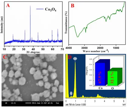
(JCPDS no. 42-1467) [18]. Such sharp diffraction peaks indicate the well crystallization of Co3O4 microballs. FT-IR spectrum of Co3O4 showed two vibration peaks at 534 and 688 cm–1 that were originated from Co3O4 (Figure 2B). A broad band appeared at around 3395 and 1634 cm-1, which are due to the –OH stretching vibration associated with deformation vibration of H2O because of the sample always contact with an environment and absorb the moistures [19]. The morphology of Co3O4 microballs was examined by SEM (Figures 1C), and the images of Co3O4 microballs displayed obvious spherical shaped ball-like micro-particles with good uniformity. The microballs structure, roughed surface and high crystallinity jointly furnished large surface area, which is highly beneficial in electrochemical sensing. The EDX profile of Co3O4 microballs featured with O and Co element with atomic percentages of 67.9 and 32.1 %, respectively (Figures 1D).
Figure 1. XRD pattern (A), FT-IR spectrum (B), SEM image (C), EDAX analysis (D) of Co3O4
3.2 Electrochemical behavior of CA
appeared on the Co3O4 modified SPCE at various peak potential in addition of 500 μM CA. The observed redox peaks are attributed to the formation of o-quinone, these redox reactions of CA were followed via two-electron transfer process [20]. This high electrocatalytic oxidation of CA at Co3O4/SPCE indicated that the Co3O4 has a good electrocatalytic activity toward the CA detection.
Figure 2. (A) CV of bare SPCE (a) and Co3O4/SPCE (b) in the presence of 500 μM CA in 0.05 M PB solution (pH 7.0) with scan rate 50 mVs-1 (B) CVs of Co3O4/SPCE in 500 μM CA solutions in various pHs. Scan rate: 0.05 mVs−1. (C) CV responses obtained at Co3O4/SPCEin the presence of 500 μM CA as a function of scan rate ranging from 20 to 200 mVs-1
in 0.05 M PB solution (pH 7.0) (D) corresponding calibration plot of current vs. scan rate.
3.3 Effect of scan rate and pH
[image:5.596.85.512.155.514.2]
altering flux is drastically lower at the electrode surface when sweeping the potential at lower scan rates, hence, the peak potential was shifted. In addition, the peak current of CA oxidation was plotted against the scan rate and shown in Figure 3D. The observed plot indicated that the CA oxidation had a good linearity (R2 = 0.9978 and 0.9986). This study indicated that the electrocatalytic oxidation of CA at Co3O4/SPCE was controlled by the adsorption controlled process [21].
The pH of the electrolyte was considerably influenced the electrochemical behavior of CA. Hence, the oxidation of CA was studied by CV in the various pH solutions containing 500 μM CA at scan rate of 50 mV/s. Figure 2B, shows the CV responses of the CA oxidation at Co3O4/SPCE in different pH ranging from 3.0 to 11.0. It can be seen that the peak potentials of the CA oxidation curves were shifted to the more negative potential when increase the pHs [22]. The anodic peak potentials (Epa) did not follow the linear relationship against the pHs. The Co3O4/SPCE exhibited an utmost redox peak current for the pH 7.0. This investigation resulted that the CA oxidation at various pHs didn’t show any appreciable linearity over the pHs. This is because of the surface confined adsorption of the CA at the Co3O4/SPCE surface.
3.4 Determination of CA on Co3O4/SPCE
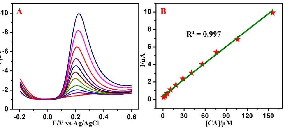
Figure 3. (A) DPVs at Co3O4/SPCE in PBS (pH 7.0) containing various concentrations of caffeic acid (0.2 – 272 µM). (B) Calibration plot of caffeic acid sensor.
[image:6.596.64.543.365.582.2][image:7.596.47.541.189.405.2]
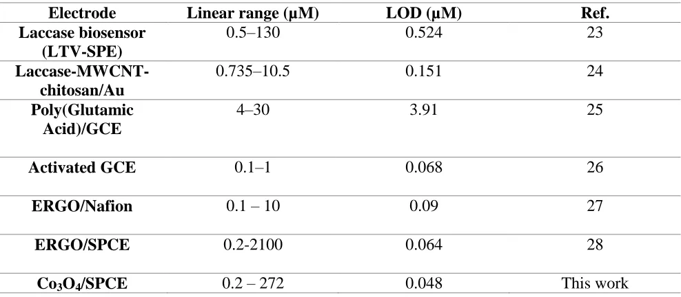
of sensitivity, broad linear range and lower detection limits were compared with previously reported CA sensors and shown in Table. 1. The superior electrocatalytic activity may be attributed to the uniform size of Co3O4 microballs structure.
Table 1. Comparison of the electrocatalytic performance of the Co3O4/SPCE for the CA detection with earlier reported sensor.
Electrode Linear range (µM) LOD (µM) Ref.
Laccase biosensor (LTV-SPE)
0.5–130 0.524 23
Laccase-MWCNT-chitosan/Au
0.735–10.5 0.151 24
Poly(Glutamic Acid)/GCE
4–30 3.91 25
Activated GCE 0.1–1 0.068 26
ERGO/Nafion 0.1 – 10 0.09 27
ERGO/SPCE 0.2-2100 0.064 28
Co3O4/SPCE 0.2 – 272 0.048 This work
3.5 Selectivity, reproducibility and stability
In order to evaluate the selectivity of the proposed sensor was studied by DPV method in the presence of structurally similar compounds and some common anion and cations. The anti-interference ability of the proposed sensor was studied by DPV in the presence of CA with 10-fold excess of interfering ions such as catechol, dopamine, ferulic acid, resorcinol, ascorbic acid, glucose and uric acid. The obtained interference analysis with those compounds results specified that almost there is no interference current was observed. Additionally, the anti–interference ability was assessed in 100 times excess of Na+, K+, Mg2+, Ni2+, Cu2+, Cl−, NO3
−
, and SO4 2−
3.6 Real sample analysis
Practical applicability of the fabricated Co3O4/SPCE sensor was estimated to detect the CA in red wine. Two different red wines samples were used for the real sample analysis and each sample was detected three times in parallel. Under optimized experimental conditions, certain amount of raw wine were directly added into the 0.05 M PBS and calculated the amount of CA. The concentration of CA determined by the voltammetric method to be 97.1 μM for sample 1 with the RSD of 2.8 %. Similar analytical procedure were followed for another samples and obtained recovery results were shown in Table 2.
Table 2. Real time determination of CA in Red wine samples by using proposed method
Red wine Found (µM) RSD (%)
Sample 1 97.1 2.8
Sample 2 96.2 3.1
4. CONCLUSION
In summary, we have reported a novel electrochemical sensor for the determination of caffeic acid based on the cobalt oxide microballs modified screen printed electrode. The cobalt oxide microballs were synthesized via a facile sonochemical method. The electrocatalytic activity of the prepared Co3O4 microballs evaluated by the determination caffeic acid using cyclic voltammetry (CV). Differential pulse voltammetry (DPV) was used to derive the analytical curve. The proposed sensor exhibited superior electrocatalytic activity towards caffeic acid determination in terms of decent sensitivity, a broad dynamic range with a lower LOD of 48 nM. In addition, the proposed sensor offers good selectivity, nice reproducibility, and decent stability. Moreover, the real sample analysis were carried out in wine samples and obtained an acceptable recovery rate.
ACKNOWLEDGEMENT
This project was supported by the Ministry of Science and Technology and the Ministry of Education of Taiwan (Republic of China). The financial support from the Chung Gung Memorial Hospital through contract no. BMRP 280 to B.S. Lou is also acknowledged.
References
1. L. Zhen, J. Xu, R. Yue, T. Yang, and L. Gao, Electrochim. Acta. 196 (2016) 1. 2. F. Wang, and J. Yang, Food Sci. Technol. 46 (2012) 239.
3. Z. Bo, X. Shuai, S. Mao, H. Yang, J. Qian, J. Chen, and K. Cen, Sci. Rep. 4 (2014) 1.
4. M. Hirose, Y. Takesada, H. Tanaka, S. Tamano, T. Kato, and T. Shirai, Carcinogenesis. 19 (1998) 207.
7. C. Michailof, P. Manesiotis, and C. Panayiotou, J. Chromatogr. A. 1182 (2008) 25.
8. Y. Xing, H.Y. Peng, M.X. Zhang, X. Li, W.W. Zeng, and X.E. Yang, J. Zhejiang Univ. Sci. B: Biomed. Biotechnol. 13 (2012) 487.
9. B. Mancek, and S. Kreft, Talanta, 66 (2005) 1094.
10.F.R.F. Leite, W.D.J.R. Santos, and L.T. Kubota, Sens. Actuator B-Chem. 193 (2014) 238. 11.L. Jiang, S.Gu, Y.Ding, F.Jiang, and Z.Zhang, Nanoscale, 6 (2014) 207.
12.M.H. Asif, F.Elinder, and M.Willander, J. Anal.Bioanal.Tech. (2011) 9.
13.A. Numan, M.M. Shahid, F.S. Omar, K. Ramesh, and S. Ramesh, Sens.Actuators, B, 238 (2017) 1043.
14.A. Salimi, H. Mamkhezri, R. Hallaj, S. Soltani, Sens.Actuators, B, 129 (2008) 246. 15.A. Salimi, R. Hallaj, S. Soltanian, Biophys.Chem. 130 (2007) 122.
16.S.J. Li, J.M. Du, J.P. Zhang, M.J. Zhang, J. Chen, Microchim.Acta 181 (2014) 631.
17.Xi. C. Dong, H. Xu, X.W. Wang, Y.X. Huang, Mary B.C. Park, H. Zhang, L.H. Wang, W. Huang, and P. Chen, ACS Nano 6 (2012) 3206.
18.J. Wang, N. Yang, H. Tang, Z. Dong, Q. Jin, M. Yang, D. Kisailus, H. Zhao, Z. Tang, and D. Wang, Angewandte Chemie, 125 (2013) 6545.
19.V.K. Patel, J.R. Saurav, K. Gangopadhyay, S. Gangopadhyay, and S. Bhattacharya, RSC Advances, 5 (2015) 21471.
20.G. Di Carlo, A. Curulli, R.G. Toro, C. Bianchini, T. De Caro, G. Padeletti, D. Zane, and G.M. Ingo. Langmuir 28 (2012) 5471.
21.T. Kokulnathan, N. Raja, S.M. Chen, and W.C. Liao, J. colloid interface sci. 501 (2017) 77. 22.A.T.E. Vilian, S.M. Chen, Y.H. Chen, M.A. Ali, and F.M.A. Al-Hemaid, J. colloid interface sci.
423 (2014) 33.
23.P. Ibarra-Escutia, J.J. Gómez, C. Calas-Blanchard, J.L. Marty and M.T. Ramirez-Silva, Talanta, 81 (2010) 1636
24.M. Diaconu, S.C. Litescu and G.L. Radu, Sens. Actuators B. 145 (2010) 800.
25.D.P. Santos, M.F. Bergamini, A.G. Fogg and M.V.B. Zanoni, Microchim. Acta. 151 (2005) 127. 26.G. Magarelli, J.G. Da Silva, I.A. De Sousa Filho, I.S.D. Lopes, J.R. Souza De, L.V. Hoffmann and
C.S.P. Castro, Microchem. J. 109 (2013) 23.