This is a repository copy of A method for quantitative analysis of regional lung ventilation using deformable image registration of CT and hybrid hyperpolarized gas/H-1 MRI. White Rose Research Online URL for this paper:
http://eprints.whiterose.ac.uk/105957/ Version: Accepted Version
Article:
Tahir, B.A., Swift, A.J., Marshall, H. et al. (8 more authors) (2014) A method for quantitative analysis of regional lung ventilation using deformable image registration of CT and hybrid hyperpolarized gas/H-1 MRI. Physics in Medicine and Biology, 59 (23). pp. 7267-7277. ISSN 0031-9155
https://doi.org/10.1088/0031-9155/59/23/7267
[email protected] https://eprints.whiterose.ac.uk/ Reuse
Unless indicated otherwise, fulltext items are protected by copyright with all rights reserved. The copyright exception in section 29 of the Copyright, Designs and Patents Act 1988 allows the making of a single copy solely for the purpose of non-commercial research or private study within the limits of fair dealing. The publisher or other rights-holder may allow further reproduction and re-use of this version - refer to the White Rose Research Online record for this item. Where records identify the publisher as the copyright holder, users can verify any specific terms of use on the publisher’s website.
Takedown
If you consider content in White Rose Research Online to be in breach of UK law, please notify us by
A method for quantitative analysis of regional lung ventilation
using deformable image registration of CT and hybrid
hyperpolarised gas/
1H MRI
Bilal A. Tahir1,2, Andrew Swift2, Helen Marshall2, Juan Parra-Robles2, Matthew Q. Hatton1, Ruth Hartley3, Richard Kay4, Christopher E. Brightling3, Wim Vos5, Jim M. Wild2 and Rob H. Ireland1,2
Academic Units of 1Clinical Oncology and 2Academic Radiology, University of Sheffield, UK
3Institute for Lung Health, University of Leicester, UK 4Novartis, Basel, Switzerland
5FluidDA nv2, Kontich, Belgium
Correspondence
Rob Ireland, Academic Unit of Clinical Oncology, Weston Park Hospital University of Sheffield, S10 2SJ, UK
Email[email protected]
Short Title
Abstract
Hyperpolarized gas MRI generates highly detailed maps of lung ventilation and
physiological function while CT provides corresponding anatomical and structural
information. Fusion of such complementary images enables quantitative analysis of
pulmonary structure-function. However, direct image registration of hyperpolarized
gas MRI to CT is problematic, particularly in lungs whose boundaries are difficult to
delineate due to ventilation heterogeneity. This study presents a novel indirect
method of registering hyperpolarized gas MRI to CT utilizing 1H-structural MR images that are acquired in the same breath-hold as the gas MRI. The feasibility of
using this technique for regional quantification of ventilation of specific pulmonary
structures is demonstrated for the lobes.
The direct and indirect methods of hyperpolarized gas MRI to CT image registration
were compared using lung images from 15 asthma patients. Both affine and
diffeomorphic image transformations were implemented. Registration accuracy was
evaluated using the target registration error (TRE) of anatomical landmarks identified
on 1H MRI and CT. Wilcoxon signed-rank test was used to test statistical significance.
For the affine transformation, the indirect method of image registration was
significantly more accurate than the direct method (TRE=14.7±3.2 vs. 19.6±12.7mm,
p=0.036). Using a deformable transformation, the indirect method was also more
accurate than the direct method (TRE=13.5±3.3 vs. 20.4±12.8mm, p=0.006).
Accurate image registration is critical for quantification of regional lung ventilation
with hyperpolarized gas MRI within the anatomy delineated by CT. Automatic
deformable image registration of hyperpolarized gasMRI to CT via same breath-hold
1H MRI is more accurate than direct registration. Potential applications include
improved multi-modality image fusion, functionally-weighted radiotherapy planning,
and quantification of lobar ventilation in obstructive airways disease.
Keywords
1. Introduction
Computed tomography is the gold standard for high-resolution structural imaging of
the lung but has limited functional sensitivity and soft tissue contrast. The quality of
proton structural MRI of the lungs has improved in recent years through the use of
MR sequences (Wild et al., 2012) such as ultra-short echo time (Lederlin and
Crémillieux, 2013) and balanced steady state free precession (Rajaram et al., 2012)
to the point where the scans have diagnostic utility in certain conditions.
Hyperpolarized gas (3He and 129Xe) MRI provides highly detailed images of lung ventilation and regional pulmonary function with sensitivity to different aspects of
pulmonary physiology and lung disease (Van Beek et al., 2004; Fain et al., 2010).
Visualization and quantification of this functional data superimposed upon
anatomical images may be advantageous in order to maximize the complementary
information relating to form and function.
In practice, fused hyperpolarized gas and anatomical lung imaging could be
achieved either by the application of image registration or same breath-hold data
acquisition. While hyperpolarized gas and 1H MRI is routinely performed in the same imaging session, coil and pulse sequence designs have only recently realized the
capability for same breath-hold acquisition of hyperpolarized gas and 1H lung MRI (Wild et al., 2011; Wild et al., 2013). Additionally, superior analytical interpretation
and quantification is provided by registration of the hyperpolarized gas MRI not only
to 1H MRI but also to the more detailed images of lung structure provided by high-resolution x-ray CT. However, there is little prospect of a hardware solution for
combined MR/CT imaging and as such an image registration solution is required.
Registration of hyperpolarized gas MRI and CT has been shown to facilitate
functionally weighted radiotherapy treatment planning for lung cancer patients
(Ireland et al., 2007a) and enable comparison of CT and 3He MRI measures of ventilation (Mathew et al., 2012). Clinical investigations of other obstructive lung
diseases, such as cystic fibrosis, asthma and emphysema (van Beek et al., 2004),
could also potentially benefit from the fusion of hyperpolarized gas ventilation MRI
with matched anatomical images. In addition, multimodality image fusion of
ventilation for specific pulmonary structures such as the lobes which cannot be
identified on hyperpolarized gas MR images (Tahir et al., 2014).
Preliminary work on hyperpolarized gas MRI to CT image registration demonstrated
the feasibility of fusing the images semi-automatically using control point rigid
registration (Ireland et al., 2007a). Subsequently, an enhanced acquisition and
registration protocol was reported (Ireland et al., 2008). Although hyperpolarized gas
MRI to CT registration accuracy was improved, the method still requires the input of
operator landmarks that have an associated inter-observer variability. Ideally the
registration would be performed fully automatically.
In this study, our first hypothesis is that as the 1H MRI acquired in the same breath-hold as hyperpolarized gas MRI is independent of ventilation defects and structurally
similar to the CT then it can facilitate automatic hyperpolarized gas MRI/CT image
registration. This is analogous to the way CT images from PET/CT can be used as
the intermediary for PET image registration (Ireland et al., 2007b).
A secondary issue with registering hyperpolarized gas MRI to corresponding
anatomical images is that quantitative evaluation of registration error is difficult
(Ireland et al., 2008), particularly if significant ventilation defects exist. A method that
enables calculation of the target registration error (TRE) of corresponding expert
defined anatomical landmarks (Murphy et al., 2011) would be beneficial for
assessing hyperpolarized gas MRI registration, especially for distinguishing between
registration algorithms. Therefore, in this study, our second hypothesis is that
inclusion of same breath-hold 1H MRI can provide a method for quantifying the TRE for hyperpolarized gasMR image registration.
In this work, we evaluate the role of synchronously acquired 1H MRI in facilitating automatic hyperpolarized gas MRI/CT image registration and quantitative TRE
analysis of hyperpolarized gas MR image registration. In so doing, we demonstrate
the feasibility of regional quantification of hyperpolarized gas MR derived ventilation
2. Materials and Methods
2.1. Image acquisition
The study was performed with national research ethics committee approval.
Between February and November 2012, 15 asthma patients gave written informed
consent to undergo both hyperpolarized 3He MRI and CT during breath hold at functional residual capacity (FRC) + 1L and total lung capacity (TLC), respectively.
1H MRI were acquired in the same breath hold as the 3He MRI acquisition (Figure 1)
(Wild et al., 2011), on a GE HDx 1.5T MR scanner (GE Healthcare, Princeton, NJ,
USA). Hyperpolarized 3He and 1H MR images were acquired with in-plane resolutions of 3 × 3 mm2 and 3 × 6 mm2 and voxel matrices of 128 × 128 × 24 and 128 × 64 × 24, respectively, and slice thicknesses of 10 mm for both. CT in-plane
[image:6.595.77.525.432.600.2]resolution was approximately 0.86 × 0.86 mm2 with a pixel matrix of 512 × 512. CT slice thickness was 1 mm with approximately 600 slices for each patient.
Figure 1. 3He and 1H MRI acquired during the same breath-hold for patient 2.
2.2. Image registration
Image registration was performed using the antsRegistration tool incorporated
as part of the Advanced Normalization Tools (ANTs) (Avants et al., 2011; Avants et
registered to CT (fixed image) using affine and diffeomorphic image transformations.
CT was resampled to a matrix size of 512 × 512 × 160 voxels to reduce the
computational time in performing registrations.
A coarse pre-alignment rigid transform was applied to align the centers of mass of
the CT and MR image intensities. The resulting transform was then applied to the
affine stage. Due to the multimodal nature of the problem, the mutual information
similarity metric was used with 32 histogram bins optimized via the gradient descent
algorithm with a step size of 0.1. A multi-resolution Gaussian pyramid with 5 levels
was used with down-sampling factors 8x6x4x2x1 and corresponding smoothing
Gaussian sigmas of 4x3x2x1x0 mm. A maximum of 10,000 iterations were set for
each resolution level to ensure convergence (Glocker et al., 2011).
An additional diffeomorphic transformation that copes with large deformations while
preserving topology of anatomical structures (Avants et al., 2008) was also applied
to the resulting transform of the affine pipeline with the same parameters for the
multi-resolution Gaussian pyramid and similarity metric. The Greedy Symmetric
Normalization algorithm (SyN) provided by ANTs registration suite was the chosen
diffeomorphic algorithm as it was the highest performing algorithm at a recent
pulmonary image registration competition (Murphy et al., 2011). A step size of 0.2
was selected for the gradient descent optimization algorithm.
To reduce the computational time in performing the registration, 16 cores via two
Intel Xeon E5-2670 processors @ 2.60 GHz were run in parallel on a 64-bit high
performance Linux server (Iceberg, University of Sheffield) using the multi-threading
options available in Insight Toolkit version 4 (ITK, www.itk.org). Computational times
for the full diffeomorphic pipeline ranged from 28 to 37 minutes, including the rigid
pre-alignment and affine stages.
2.3. Asthma patient study
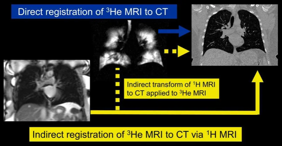
For each of the fifteen patients, the following four registration pipelines were
performed to register 3He MRI to CT either directly or indirectly (Figure 2) via the same breath-hold 1H MR image:
1. Affine direct Affine registration of 3He MRI to CT 2. Affine indirect Affine registrationof 1H MRI to CT
3. Diffeomorphic direct Diffeomorphic registration of 3He MRI to CT 4. Diffeomorphic indirect Diffeomorphic registration of 1H MRI to CT.
For all the registration pipelines, only the transform (affine or diffeomorphic) and
[image:8.595.73.523.369.603.2]moving image (3He MRI for direct and 1H MRI for indirect) were varied.
2.4. Registration evaluation
The registration accuracy of each registration pipeline was assessed quantitatively
where pairs of anatomical landmarks were identified by one of the authors (BAT)
using Slicer 4 (Fedorov et al., 2012), on the 1H MR and CT images and then reviewed independently by a chest radiologist. Landmark locations included the apex
and base of the lungs, arch of the aorta and bifurcations of the trachea and blood
vessels. The transformations derived from the direct 3He MRI to CT and indirect 1H MRI to CT registrations were applied to the landmark coordinates to enable the
mean target registration error (TRE) of the corresponding landmarks to be calculated
for all points. TRE is defined as the Euclidean distance between two corresponding
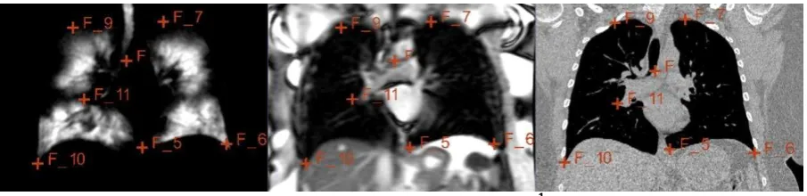
points (Murphy et al., 2011). Figure 3 shows an example of corresponding landmarks
[image:9.595.72.525.390.500.2]identified on 1H MRI and CT, alongside the 3He MRI that was acquired in the same breath hold as the 1H MRI.
Figure 3. An example of corresponding landmarks identified on 1H MRI (middle) and CT (right)
images, alongside the 3He MRI (left) that was acquired in the same breath hold as the 1H MRI.
2.5. Statistics
The Wilcoxon signed-rank test implemented in IBM SPSS (version 20.0; Chicago, Il,
USA) was used to test the statistical significance of the differences between the
direct and indirect registration of 3He MRI to CT. In addition, the TRE for the indirect diffeomorphic pipeline was compared to all other pipelines. A p value less than0.05
2.6. Lobar segmentation and regional ventilation quantification
As an example to demonstrate the feasibility of quantification of 3He MRI ventilation of pulmonary structures identifiable only on CT, the percentage ventilation per lobe
was calculated by taking the ratio of 3He MRI volume in a given lobe to the total 3He MRI volume in the lungs. Medical image segmentation software (Mimics; Materialise,
Leuven, Belgium) was used to segment the lobes of CT by identification of fissures
in both lungs. These lobar segmentations were used to mask the registered 3He MRI which were segmented via the Otsu histogram based method which separates the
foreground ventilated volume from signal void hypo-ventilated regions and
background noise (Otsu, 1979).
3. Results
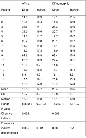
A median of 31 anatomical landmarks per patient (range 22 to 42) were identified on
both the 1H MRI and CT images. For this group of patients, the mean±SD target registration error for the direct affine, indirect affine methods, direct diffeomorphic
and indirect diffeomorphic algorithms were 19.6±12.7mm, 14.7±3.2mm,
20.4±12.8mm and 13.5±3.3mm, respectively. Table 1 displays the TRE results for all
15 patients. The Wilcoxon signed-rank test demonstrates a statistically significant
difference between the direct and indirect affine (p=0.036) and diffeomorphic
(p=0.006) methods of image registration. The TRE of 13.5±3.3mm for the
Affine Diffeomorphic
Patient Direct Indirect Direct Indirect
1 11.6 12.9 12.1 11.5
2 13.4 15.4 11.3 12.0
3 23.6 14.1 26.3 10.6
4 22.0 19.6 20.7 18.7
5 14.0 11.7 12.7 10.2
6 23.7 19.8 25.1 19.7
7 14.9 14.8 15.1 14.5
8 15.3 17.3 15.0 15.9
9 62.6 16.8 63.4 15.8
10 20.3 12.3 22.4 12.1
11 13.0 9.7 14.6 8.8
12 13.9 16.6 13.7 15.4
13 8.8 9.2 13.1 8.9
14 18.5 16.1 20.8 13.9
15 18.0 14.3 19.7 14.0
Mean 19.6 14.7 20.4 13.5
SD 12.7 3.2 12.8 3.3
Median 15.3 14.8 15.1 13.9
Range 8.8,62.6 9.2,19.8 11.3,63.4 8.8,19.7
P value
Direct vs
indirect
0.036 0.006
P value vs
indirect
diffeomorphic
[image:11.595.127.470.100.647.2]0.005 0.001 0.006 N/A
Table 1. Mean target registration errors (in mm) for the direct and indirect affine and
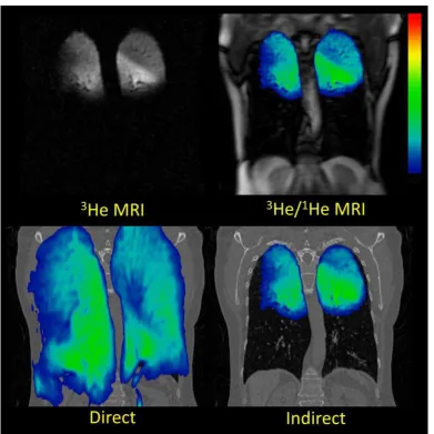
An example in which the direct method failed was for patient 9, which was
characterized by extreme hypoventilation including no ventilation in both the left and
right lower lungs (Figure 4). The direct registration broke down for the affine and
diffeomorphic methods as the amount of ventilation was insufficient to permit
[image:12.595.104.496.181.573.2]multimodal alignment.
Figure 4. Corresponding coronal slices of patient 9 showing registered 3He MRI fused with CT via
the direct (bottom left) and indirect (bottom right) diffeomorphic methods with preregistered 3He
MRI (top left) and 1H MRI fused with 3He MRI (top right). The direct method breaks down,
attempting to stretch the 3He images which show ventilation in the upper lobes only, across the
whole lung.
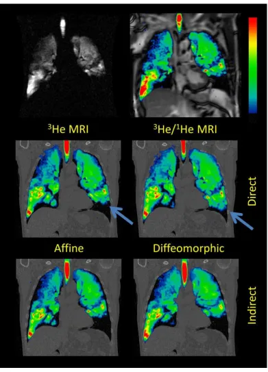
Although not as extreme an example, Figure 5 (blue arrow) demonstrates where the
direct method inaccurately registers 3He MRI to CT by increasing the ventilation signal near the base of the left lung. Conversely, the indirect method yields a more
Figure 5. Corresponding coronal slices of patient 14 showing registered 3He MRI fused with CT via
the direct (middle row) and indirect (bottom row) affine and diffeomorphic methods with
preregistered 3He MRI (top left) and 1H MRI fused with 3He MRI (top right). The blue arrows near
the base of the left lung indicates a registration error in the direct affine and diffeomorphic
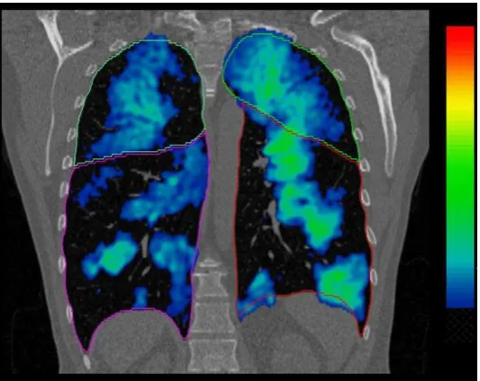
The potential for providing regional quantitative ventilation of specific anatomical
structures is demonstrated in Figure 6 for patient 11 where the pulmonary CT lobar
masks are superimposed on the registered 3He MRI. For this patient, the percentage ventilation for each lobe was RUL=18.39% RML=14.71%, RLL=25.06%,
[image:14.595.166.433.184.396.2]LUL=27.20%, and LLL=14.64%.
Figure 6. Example slice of patient 11 showing the CT lobar masks fused with 3He MRI after
registration.
4. Discussion
Image registration of hyperpolarized gas MRI has previously been published in the
context of perfusion MRI using controlled gas administration in pigs (Rizi et al., 2003;
Hong et al., 2005) while human in vivo 3He MRI to CT image registration has been reported as part of a study on the feasibility of lung cancer treatment planning with
3He MRI (Ireland et al., 2007a). Furthermore, the importance of the image acquisition
procedures on the resultant image registration of 3He MRI to CT has been demonstrated (Ireland et al., 2008). When there are major differences in patient
positioning and posture between the MRI and CT image acquisitions, there can be
large errors in image registration when a rigid algorithm is applied.
registration accuracy was improved in that work by having the patient positioning
match as closely as possible between the MRI and CT acquisitions. In the current
work matching MRI and CT patient positioning was not part of the study protocol.
The CT was performed in a standard manner, involving a breath hold that was
different to that used for the MRI acquisition and with a curved diagnostic bed. The
objective of the current study was not to improve upon the radiotherapy level of
registration accuracy, but rather to investigate the use of the same breath-hold 1H MRI within the image registration and whether it enables target registration error
(TRE) analysis to be performed along with quantification of regional ventilation.
In the present work, the additional 1H MRI is used as an intermediate step in the registration of hyperpolarized gas MRI to CT. The method of automatic registration
assumes intrinsic spatial registration of gas MRI and 1H MRI, preferably due to same breath-hold acquisition (Wild et al., 2011), although the method is still potentially
viable if hyperpolarizedgas and 1H MR image registration is required from separate breath-holds (Ireland et al., 2009).
In addition, application of the image registration transformation that is calculated
from hyperpolarized gas MRI to CT to the 1H MRI landmarks provides a method of assessing the accuracy of gas MRI to CT registration with TRE analysis. Although
high quality hyperpolarized gas MR images often contain structural detail that could
potentially be used for TRE analysis (Ireland et al., 2008), the advantage of the same
breath-hold 1H MRI method is that it allows for identification of spatially correlated anatomical landmarks that may not be seen on the hyperpolarized gas MR images
due to ventilation defects and partial volume effects.
The indirect affine and diffeomorphic methods demonstrated statistically significant
improvements in TRE compared to their corresponding direct methods. The
diffeomorphic indirect method of image registration, using 1H MRI, was more accurate than the other direct and indirect methods. Due to the differences in
breathing maneuver and physiological variations between inter-session CT and MRI
scans, the diffeomorphic deformable algorithm was able to account better for large
deformations than the affine transformation.
With the direct hyperpolarized gas MRI to CT image registration method, there was
direct affine and diffeomorphic registration broke down for one patient in the study
(Figure 3) and yielded clinically unrealistic deformations of 3He MRI for several patients (Figure 4).
One limitation of our study data is that there is a difference in resolution between the
1H MRI and CT images especially in the z direction (10mm vs. 1mm). This may
cause observer errors in landmark identification due to partial volume effects at low
resolutions. The slice thickness of 10mm was chosen to provide high signal to noise
and to ensure full volumetric lung coverage within a breath-hold time achievable by
patients, important considerations when both 3He and 1H images are acquired within the same breath. However, this limitation may be mitigated by recent improvements
in MRI technology that provide superior resolution for both same-breath acquired
nuclei (Horn et al., 2014).
For this study, only the transform (affine and diffeomorphic) and the moving image
(3He MRI for direct and 1H MRI for indirect) were varied for each patient while all other registration parameters such as similarity metrics, multiresolution stages and
optimization remained constant. Due to inter-patient variations in ventilation
heterogeneity and respiratory states, further improvements in registration may be
gained by patient specific parameterization.
There are many potential clinical applications of improved hyperpolarized gas MR
image registration since accurate registration of anatomical and functional images
can enhance both image interpretation and quantification. For a variety of lung
diseases, registration of hyperpolarized gas MRI to CT would enable the pulmonary
ventilation to be assessed against the underlying anatomical CT structure, which
serves as the gold standard. The feasibility of using the indirect registration
technique for regional quantification of ventilation of specific pulmonary structures is
demonstrated in Figure 6. In addition, registration to CT is critical in radiotherapy for
the implementation of functionally weighted treatment planning (Ireland et al., 2007a;
Bates et al., 2009; Partridge et al., 2010).
5. Conclusion
This study demonstrates the benefit of a method of automatic image registration of
breath-hold as gas MRI data. This study also shows that inclusion of same
breath-hold 1H MRI enables TRE quantification of hyperpolarized gas MRI to CT image registration. Evaluation in 15 clinical cases demonstrates that TRE analysis is
practical and that image registration accuracy can be improved when the additional
anatomical 1H MRI information is incorporated. Accurate image registration is critical for quantification of regional ventilation using hyperpolarized gas MRI and CT.
Acknowledgements
The High Performance Computing Service was provided by The University of
Sheffield Corporate Information and Computing Services. Salman Siddiqui and Marie
Laurencin provided assistance for the AirPROM study design and patient
recruitment. Funding was provided by EU FP7 AirPROM, Novartis, University of
Sheffield James Morrison Fund, Sheffield Hospitals Charity and Weston Park
Hospital Cancer Charity. Conflicts of interest: None.
References
Avants B, Tustison N, Song G, Wu B, Stauffer M, McCormick M, Johnson H and Gee J 2012 A unified image registration framework for ITK Lect. Notes Comput. Sc. 7359 266-75
Avants B B, Epstein C L, Grossman M and Gee J C 2008 Symmetric diffeomorphic image registration with cross-correlation: evaluating automated labeling of elderly and neurodegenerative brain Med. Image Anal. 12 26-41
Avants B B, Tustison N J, Song G, Cook P A, Klein A and Gee J C 2011 A reproducible evaluation of ANTs similarity metric performance in brain image registration NeuroImage 54 2033-44 Bates E L, Bragg C M, Wild J M, Hatton M Q and Ireland R H 2009 Functional image-based
radiotherapy planning for non-small cell lung cancer: A simulation study Radiother. Oncol. 93
32-6
Fain S, Schiebler M L, McCormack D G and Parraga G 2010 Imaging of lung function using hyperpolarized helium-3 magnetic resonance imaging: Review of current and emerging translational methods and applications Magn. Reson. Imag. 32 1398-1408
Fedorov A, Beichel R, Kalpathy-Cramer J, Finet J, Fillion-Robin J, Pujol S, Bauer C, Jennings D,
Fennessy F, Sonka M, Buatti J, Aylward S, Miller J, Pieper S and Kikinis R 2012 3D Slicer as an image computing platform for the Quantitative Imaging Network Magn. Reson. Imaging 30
1323-41
Glocker B, Sotiras A, Komodakis N and Paragios N 2011 Deformable medical image registration: setting the state of the art with discrete methods Annu. Rev. Biomed. Eng. 13 219-44 Hong C, Leawoods J C, Yablonskiy D A, Leyendecker J R, Bae K T, Pilgram T K, Woodard P K, Conradi
M S and Zheng J 2005 Feasibility of combining MR perfusion, angiography, and 3He
ventilation imaging for evaluation of lung function in a porcine model Acad. Radiol. 12 202-9 Horn F C, Tahir B A, Stewart N J, Collier G J, Norquay G, Leung G, Ireland R H, Parra-Robles J, Marshall
Ireland R H, Woodhouse N, Swinscoe J A, Hatton M Q and Wild J M 2009 Towards automatic image registration of hyperpolarized 3He MRI and x-ray CT images of the lung Proc. Intl. Soc. Mag. Reson. Med. 17 2197
Ireland R H, Bragg C M, McJury M, Woodhouse N, Fichele S, van Beek E J, Wild J M and Hatton M Q 2007a Feasibility of image registration and intensity-modulated radiotherapy planning with hyperpolarized helium-3 magnetic resonance imaging for non-small-cell lung cancer Int. J. Radiat. Oncol. Biol. Phys. 68 273-81
Ireland R H, Dyker K E, Barber D C, Wood S M, Hanney M B, Tindale W B, Woodhouse N, Hoggard N, Conway J and Robinson M H 2007b Nonrigid image registration for head and neck cancer radiotherapy treatment planning with PET/CT Int. J. Radiat. Oncol. Biol. Phys. 68 952-7 Ireland R H, Woodhouse N, Hoggard N, Swinscoe J A, Foran B H, Hatton M Q and Wild J M 2008 An
image acquisition and registration strategy for the fusion of hyperpolarized helium-3 MRI and x-ray CT images of the lung Phys. Med. Biol. 53 6055-63
Lederlin M and Crémillieux Y 2013 Three-dimensional assessment of lung tissue density using a clinical ultrashort echo time at 3 tesla: A feasibility study in healthy subjects J. Magn. Reson. Imaging doi: 10.1002/jmri.24429
Mathew L, Wheatley A, Castillo R, Castillo E, Rodrigues G, Guerrero T and Parraga G 2012
Hyperpolarized (3)He magnetic resonance imaging: comparison with four-dimensional x-ray computed tomography imaging in lung cancer Acad. Radiol. 19 1546-53
Murphy K, van Ginneken B, Reinhardt J M, Kabus S, Ding K, Deng X, Cao K, Du K, Christensen G E, Garcia V, Vercauteren T, Ayache N, Commowick O, Malandain G, Glocker B, Paragios N, Navab N, Gorbunova V, Sporring J, de Bruijne M, Han X, Heinrich M P, Schnabel J A, Jenkinson M, Lorenz C, Modat M, McClelland J R, Ourselin S, Muenzing S E, Viergever M A, De Nigris D, Collins D L, Arbel T, Peroni M, Li R, Sharp G C, Schmidt-Richberg A, Ehrhardt J, Werner R, Smeets D, Loeckx D, Song G, Tustison N, Avants B, Gee J C, Staring M, Klein S, Stoel B C, Urschler M, Werlberger M, Vandemeulebroucke J, Rit S, Sarrut D and Pluim J P 2011 Evaluation of registration methods on thoracic CT: the EMPIRE10 challenge IEEE Trans. Med. Imaging 30 1901-20
Otsu N 1979 A threshold selection method from gray-level histograms IEEE Trans. Syst. Man Cybern.
9 62-6
Partridge M, Yamamoto T, Grau C, Hoyer M and Muren L P 2010 Imaging of normal lung, liver and parotid gland function for radiotherapy Acta Oncol. 49 997-1011
Rajaram S, Swift A J, Capener D, Telfer A, Davies C, Hill C, Condliffe R, Elliot C, Hurdman J, Kiely D G and Wild J M 2012 Lung morphology assessment with balanced steady-state free precession MR imaging compared with CT Radiology 263 569-77
Rizi R R, Saha P K, Wang B, Ferrante M A, Lipson D, Baumgardner J and Roberts D A 2003
Co-registration of acquired MR ventilation and perfusion images--validation in a porcine model Magn. Reson. Med. 49 13-8
Tahir B A, Holsbeke C V, Ireland R H, Swift A J, Marshall H, Parra-Robles J, Hartley R, Laurencin M, Kay R, Siddiqui S, Brightling C E, Backer J D, Vos W and Wild J M 2014 Comparison of CT-based lobar ventilation models with helium-3 MRI ventilation measurements in asthmatics Am. J. Respir. Crit. Care Med. 189 A2391
van Beek E J, Wild J M, Kauczor H U, Schreiber W, Mugler J P, 3rd and de Lange E E 2004 Functional MRI of the lung using hyperpolarized 3-helium gas J. Magn. Reson. Imaging 20 540-54 Wild J M, Ajraoui S, Deppe M H, Parnell S R, Marshall H, Parra-Robles J and Ireland R H 2011
Synchronous acquisition of hyperpolarised 3He and 1H MR images of the lungs - maximising mutual anatomical and functional information NMR Biomed. 24 130-4