A 44-year-old man with eye,
kidney, and brain dysfunction
The Harvard community has made this
article openly available.
Please share
how
this access benefits you. Your story matters
Citation
Vodopivec, Ivana, Derek H. Oakley, Cory A. Perugino, Nagagopal
Venna, E. Tessa Hedley-Whyte, and John H. Stone. 2016. “A
44-Year-Old Man with Eye, Kidney, and Brain Dysfunction.” Annals of
Neurology 79 (4) (March 7): 507–519. doi:10.1002/ana.24583.
Published Version
10.1002/ana.24583
Citable link
http://nrs.harvard.edu/urn-3:HUL.InstRepos:37045423
Terms of Use
This article was downloaded from Harvard University’s DASH
A 44-Year-Old Man with Eye, Kidney, and Brain Dysfunction
Ivana Vodopivec, MD, PhD1,2, Derek H. Oakley, MD, PhD1,3, Cory A. Perugino, DO1,4,
Nagagopal Venna, MD, MRCP1,2, E. Tessa Hedley-Whyte, MD1,3, and John H. Stone, MD,
MPH1,4
1Harvard Medical School, Boston, USA
2Massachusetts General Hospital, Department of Neurology, Boston, USA
3Massachusetts General Hospital, Department of Pathology (Neuropathology), Boston, MA, USA
4Massachusetts General Hospital, Rheumatology Unit, Boston, MA, USA
Abstract
Retinal vasculopathy with cerebral leukodystrophy (RVCL) is a rare, autosomal dominant condition caused by mutations of the three-prime repair exonuclease-1 (TREX1). The phenotypic expressions range from isolated retinal involvement to varying degrees of retinopathy, cerebral infarction with calcium depositions, nephropathy, and hepatopathy. We report a case of RVCL caused by a novel TREX1 mutation. This patient’s multisystem presentation, retinal involvement interpreted as “retinal vasculitis”, and improvement of neuroimaging abnormalities with
dexamethasone led to the accepted diagnosis of a rheumatologic disorder resembling Behçet’s disease. Clinicians should consider RVCL in any patient with retinal capillary obliterations associated with tumefactive brain lesions or nephropathy.
Keywords
retinal vasculopathy with cerebral leukodystrophy; cerebral calcifications; nephropathy
Case Presentation (Dr. Cory Perugino)
A 44-year-old man was admitted to another hospital with visual field defects, intermittent confusion, apraxia, extinction to double simultaneous stimulation on the left, and mild left-sided weakness. For two days prior to admission, he had been confused, unable to recall recent events and execute certain actions, such as tying his shoes.
Corresponding author: Ivana Vodopivec, MD, PhD, Department of Neurology, Massachusetts General Hospital, 55 Fruit Street,
Boston, MA 02114, Phone: 617-726-7565, 617-573-3412, [email protected].
This work was presented in part at the Combined Neurology-Rheumatology Grand Rounds at Massachusetts General Hospital, Boston, MA, on May 5, 2015.
Potential Conflicts of Interest
The authors report no disclosures relevant to the manuscript.
Author Contributions
HHS Public Access
Author manuscript
Ann Neurol
. Author manuscript; available in PMC 2016 May 05.Published in final edited form as:
Ann Neurol. 2016 April ; 79(4): 507–519. doi:10.1002/ana.24583.
A
uthor Man
uscr
ipt
A
uthor Man
uscr
ipt
A
uthor Man
uscr
ipt
A
uthor Man
uscr
His medical history was complex. Retinal vasculopathy and chronic kidney disease of unclear etiology had been diagnosed in Germany six years prior to admission. Hypertension was diagnosed a year later, followed by hypertensive cardiomyopathy approximately two years after the onset of visual symptoms. The retinal vasculopathy had been termed posterior uveitis with retinal vasculitis. Over the succeeding six years, the patient had been treated with several immunosuppressive medications, including prednisone, cyclosporine, mycophenolate mofetil, adalimumab, methotrexate, and interferon-alpha. Additional treatments included laser photocoagulation, intravitreal glucocorticoids, and bevacizumab. Five months before admission, he developed acute heart failure, headache, and visual field defects, and all treatments had been discontinued.
The patient had undergone two kidney biopsies in the years before this hospitalization, at four years and then again at three years before admission. Both of these biopsies had been interpreted as thrombotic microangiopathy affecting the interlobular arteries and afferent arterioles with focal segmental glomerulosclerosis and mild nephrosclerosis. No tubulointerstitial disease was present.
He underwent extensive diagnostic studies, including evaluations of the blood and cerebrospinal fluid (CSF), computed tomography (CT) of the head, brain magnetic resonance imaging (MRI), and a whole-body 18F-fluorodeoxyglucose positron emission tomography/computed tomography (FDG-PET/CT) study. The initial non-contrast MRI of the brain revealed a tumefactive lesion in the right temporo-parieto-occipital area with confluent T2/fluid-attenuated inversion recovery (FLAIR) signal abnormalities extending through the splenium of the corpus callosum into the left periventricular white matter (Fig 1A). An additional T2 hyperintense white matter lesion was noted in the left cerebellar hemisphere. Scattered punctate areas of restricted diffusion [hyperintense areas on diffusion-weighted imaging (DWI) with corresponding apparent diffusion coefficient (ADC)
hypointensities] were present in the right parietal lobe (Fig 1B).
The CT scan of the brain demonstrated focal calcifications in the right frontal, left frontal, and right parietal white matter (Fig 1C). Review of a head CT scan obtained for evaluation of headaches 2 years earlier revealed no abnormalities. A head CT that had been obtained five months before admission for evaluation of the headache and the visual field defects reportedly showed multiple punctate areas of calcification with surrounding vasogenic edema in the right superior frontal gyrus. That imaging study was not available for review.
Whole-body FDG-PET/CT imaging showed no evidence of lymphadenopathy or malignancy. Laboratory investigations were remarkable only for abnormalities in renal function. The serum creatinine was 3.1 mg/dL (normal <1.3 mg/dL), and a urinalysis revealed 2+ proteinuria, several dysmorphic red blood cells (RBCs), and a few granular casts in the urine sediment. The CSF analysis documented total nucleated cell counts of 1 and 5 cells/µL (normal <6 cells/µL), a total CSF protein of 58 mg/dL (normal 5–55 mg/dL), and CSF glucose of 64 mg/dL. The CSF IgG index was 0.39 (normal <0.77), and angiotensin converting enzyme (ACE) level was 1.2 U/L (normal <2.5 U/L).
A
uthor Man
uscr
ipt
A
uthor Man
uscr
ipt
A
uthor Man
uscr
ipt
A
uthor Man
uscr
The patient then underwent a biopsy of the right hemispheric lesion. This revealed markedly abnormal white matter but no definite diagnosis. A second biopsy, performed two weeks later, was similarly interpreted as being non-diagnostic.
High-dose dexamethasone was initiated for extensive cerebral edema noted on imaging. After five weeks at the outside hospital, the patient was transferred to our institution for further management.
The patient was a molecular biologist who had lived for many years in Germany and France. He drank alcohol moderately and did not smoke. He was married but had no children. The patient’s father died aged 36 years from Hodgkin’s lymphoma and his paternal uncle died in his early forties from an unclear cause accompanied by renal dysfunction. The family history on his mother’s side was unremarkable. The patient denied any history of oral, genital, or skin lesions, sicca symptoms, musculoskeletal, respiratory or gastrointestinal symptoms.
On examination, the patient was afebrile with an irregularly irregular pulse at a rate of 98 beats per minute and blood pressure of 146/98 mmHg. The respiratory rate was 14 per minute. There were no cardiac murmurs. The mouth, lung, abdominal, skin, and joint examinations were unremarkable. He had 3+ pitting edema of the lower extremities. The corrected visual acuity was reduced to 20/80 in the right eye and 20/50-1 in the left eye, with bilateral dyschromatopsia. A relative afferent pupillary defect was not present. A dilated ophthalmoscopic examination showed dehemoglobinized blood in the left inferior vitreous, moderate bilateral symmetric optic atrophy, epiretinal membranes, cotton wool spots in the peripapillary area, retinal arteriole obstructions with sclerosis and resulting ghost vessels that were most obvious around the optic discs. Extensive pan-retinal photocoagulation scars were present bilaterally. Neurological examination on admission was remarkable for non-specific visual field defects on confrontation testing, mild left lower facial weakness, mild left pronator drift, and unsteady gait. Confusion, apraxia, and extinction to double
simultaneous stimulation on the left, documented five weeks earlier were no longer present.
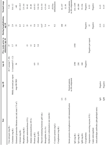
Table 1 shows the patient’s routine laboratory results upon transfer to our hospital and the results of similar examinations obtained 4 years, 3 years, and 5 weeks before the transfer. A skin pathergy test, performed at our institution, was negative.
Fluorescein angiography revealed extensive retinal ischemia with enlargement of the foveal avascular zones, perivascular fluorescein leakage, hyperfluorescence of vessel walls, and numerous scars from pan-retinal photocoagulation (Fig 2). The perivascular leakage of the contrast and the hyperfluorescence of vessel walls were repeatedly interpreted as retinal vasculitis.
High-dose dexamethasone was continued and weekly adalimumab was added 8 weeks after the onset of neurological symptoms, with the working diagnosis of Behçet’s disease. After a 10-week course of dexamethasone, complete resolution of the mass effect and decrease in the extent of the T2 hyperintensity within the cerebral hemispheres was noted on brain MRI (Fig 4). However, the patient’s visual acuity continued to deteriorate and there was persistent perivascular leakage with retinal ischemia documented by fluorescein angiography.
A
uthor Man
uscr
ipt
A
uthor Man
uscr
ipt
A
uthor Man
uscr
ipt
A
uthor Man
uscr
The brain biopsies were reviewed. A laboratory test was requested.
Neuropathological Discussion (Dr. Tessa Hedley-Whyte)
The original slides of the renal biopsies were unavailable for review. The brain biopsies were markedly abnormal with areas of circumscribed pallor. There was little or no tissue reaction surrounding areas of necrosis and extensive calcium deposition (Fig 3A). PAS and elastic stains highlighted thickened vessel walls (Fig 3B, C) but otherwise demonstrated no specific abnormalities. Adjacent to the areas of necrosis, several angiomatoid vascular proliferations associated with a mild, chronic, T cell-predominant inflammatory infiltrate were present (Fig 3D). Neurofilament staining revealed loss of axons and axonal spheroid formations within the lesions. The lesions seemed to be ischemic but the lack of tissue response was reminiscent of the necrosis seen in radiation damage. Although the small vessels were abnormal with thickened vessel walls, there was no evidence of vasculitis or arteriosclerosis.
Discussion (Dr. Ivana Vodopivec)
This is a very important and instructive case from many perspectives. I am aware of the diagnosis as I was involved in this patient’s care and obtained the confirmatory laboratory test. I will discuss the case by applying the logic followed while caring for this patient. First, I will address the working diagnosis and other initial differential considerations in a
systematic manner, from the perspective of the history, the physical examination, the available diagnostic evaluations and their interpretations. I will then proceed to discussing the underlying pathophysiology and the treatment that we offered to the patient.
To summarize, this is a 44-year-old man who presented with two days of right frontal and parietal lobe dysfunction stemming from the tumefactive lesion involving almost the entire right hemisphere. Since age 38, he had suffered from progressive retinal vasculitis refractory to numerous immunosuppressive medications and chronic kidney disease. The hypertension developed a year after the renal insufficiency had been noted, and eventually resulted in hypertensive cardiomyopathy complicated by paroxysmal atrial fibrillation. Five months prior to the presentation, headache had developed and triggered the neuroimaging that demonstrated unexplained right frontal lobe vasogenic edema associated with punctate calcifications.
We initially considered two broad categories of neurological disease: immune-mediated, related to the suspected “uveitis”; and infectious, particularly those caused by opportunistic pathogens.
The retinal vasculopathy in combination with tumefactive cerebral lesions raised the possibility of Behçet’s disease. This chronic systemic inflammatory disease, characterized by variable-vessel vasculitis,2 is a common cause of posterior uveitiswith retinal vasculitis. Behçet’s disease typically causes mucocutaneous lesions, including oral and genital ulcerations. In addition, papulopustular and nodular skin lesions, the latter of which mimics erythema nodosum, frequently occur in Behçet’s disease. Less common manifestations include arthralgias, venous and arterial thrombosis, epididymitis, cardiac (e.g., pericarditis or myocarditis), gastrointestinal lesions that mimic Crohn’s disease, and a variety of central
A
uthor Man
uscr
ipt
A
uthor Man
uscr
ipt
A
uthor Man
uscr
ipt
A
uthor Man
uscr
nervous system pathologies that often affect the white matter. Parenchymal lesions, some with mass effect as seen in this case, are among the most common manifestations of neuro-Behçet’s disease.3 In the absence of biomarkers, the definitive diagnosis is established using clinical criteria that include the presence of recurrent oral ulcerations plus two of the following: recurrent genital ulcerations, skin lesions, eye lesions, and a positive pathergy test.4 Oral aphthous ulcers are often regarded as a sine qua non of Behçet’s disease.5 In a small minority of patients, however, obvious oral ulcerations are observed only after the disease has become manifest in other organs. Thus, absolute insistence on oral aphthous ulcers before rendering a diagnosis of Behçet’s disease can lead to diagnostic errors. Genital aphthous ulcers, which have the same clinical appearance as the oral lesions, occur in about 80% of patients. The absence of both oral and genital lesions despite lengthy follow-up and frequent questioning over this time period made the diagnosis of Behçet’s disease
improbable. Does the additional diagnostic testing support the diagnosis of Behçet’s disease? No. Both the pathergy test and a test for HLA-B51 were negative. However, the pathergy test has low sensitivity that is reduced further by immunosuppressive treatments.6, 7 HLA-B51, the strongest genetic susceptibility factor for Behçet’s disease,8 is present in only 59% of patients. Its absence, therefore, does not exclude the diagnosis. The patient’s renal disease was also difficult to link with a diagnosis of Behçet’s disease. Renal involvement in Behçet’s disease is unusual and, when present, occurs years after initial symptoms, rather than as the initial presentation. The renal disease observed most commonly in Behçet’s disease is secondary amyloidosis,9 but the renal biopsies had not suggested that diagnosis. In summary, the patient’s clinical presentation, the negative pathergy and HLA-B51 antigen testing, the nature of his renal disease, and the apparent absence of treatment response all argued against Behçet’s disease.
The patient’s kidney biopsies suggested a thrombotic microangiopathy and focal segmental glomerulosclerosis. One cause of thrombotic microangiopathy is thrombotic
thrombocytopenic purpura (TTP). TTP can be associated with both central nervous system disease (strokes) as well as renal dysfunction.10 Yet the patient’s time course for TTP is improbable and he had none of the other classic features of TTP: fever, thrombocytopenia, and purpura.
Sarcoidosis is on the differential diagnosis for any patient with simultaneous retinal and cerebral manifestations, especially in the setting of retinal vasculitis. Parenchymal neurosarcoidosis infrequently presents with mass lesions accompanied by calcifications.11 The definitive diagnosis is made by tissue biopsy. The histopathological changes observed in this case of a microvasculopathy in the absence of granulomatous inflammation excluded sarcoidosis.
The diagnosis of tubulointerstitial nephropathy and uveitis (TINU) syndrome was suspected early in the course. TINU presents with bilateral acute anterior (rather than posterior) uveitis, often in children (median age 15 years).12 Although our patient had both ocular and renal dysfunction, the details of his case were incompatible with the diagnosis of TINU.
Granulomatosis with polyangiitis (GPA) is an anti-neutrophil cytoplasmic antibody (ANCA)-associated small vessel vasculitis with predilection for the respiratory tract and
A
uthor Man
uscr
ipt
A
uthor Man
uscr
ipt
A
uthor Man
uscr
ipt
A
uthor Man
uscr
kidneys. Common ocular manifestations include necrotizing scleritis and orbital
inflammatory disease.13 The cerebral vessels are involved in 3–5% patients. At least two features argued against this diagnosis in our patient: the radiographic findings of leukoencephalopathy with tumefactive lesions14, 15 and the repeatedly negative ANCA.
Eales’ disease is an inflammatory venous occlusive disorder of peripheral retina resulting in retinal angiogenesis and vitreous hemorrhage.16 It is typically associated with tuberculous infection and/or hypersensitivity. Only rare case reports of simultaneous or sequential cerebral abnormalities in Eales’ disease exist, and Eales’ disease would not account for the patients’ end-stage renal disease.17
Diagnostic possibilities were also broadened by considering opportunistic infections in light of the chronic immunosuppression. Toxoplasmosis and progressive multifocal
leukoencephalopathy were refuted based on the analysis of the serum and the CSF.
It became apparent that none of the aforementioned potential diagnoses discussed truly explained the patient’s retinal, renal, and brain parenchymal disease. We therefore re-visited the assumptions surrounding the case and re-examined the available pieces of clinical information devoid of their interpretations.
Returning to the diagnostic evaluations, it was noteworthy that the fluorescein angiogram was interpreted consistently as “retinal vasculitis” over the patient’s entire course across multiple hospitals. The brain histopathology, however, unequivocally refuted the blood vessel wall as a focus of inflammation. Rather, the biopsy confirmed the presence of vasculopathy as opposed to a vasculitis – a clue suggesting that the retinal disease might be secondary to a vasculopathy, as well. Recognition that the nature of the retinal disease was more likely to be a vasculopathy than a true vasculitis was the final argument leading to the rejection of Behcet’s disease.
Additional histopathological features that appeared pertinent, although interpreted as non-specific, were ischemic necrosis with calcium deposition. The calcifications were present even on the CT of the head that had been obtained five months before the hospital
admission. Dystrophic calcifications, i.e., calcification occurring in degenerated or necrotic tissue, are rarely seen after ischemic stroke.18–21 The existing case reports and small case series suggest that dystrophic calcifications occur at least weeks, usually months to years after the ischemic insult. Accelerated and exaggerated deposition of calcium is typically reported in patients with uremia with hyperphosphatemia and/or hypercalcemia, none of which had been present in this case.
Was there any other evidence to uphold the hypothesis of the underlying inflammation or autoimmunity? First, except for the ophthalmologic condition, his history and physical examination were unremarkable, as discussed earlier. Second, there were no signs of inflammation in the posterior ocular segment, such as cells in the vitreous, vitreous haze, retinal or choroidal inflammatory lesions. Third, laboratory blood testing had not demonstrated any clear evidence of inflammatory disease throughout the 6-year disease course. Even the notion that the patient’s condition had responded to immunosuppression could be disputed. In fact, his had been refractory to numerous immunosuppressive
A
uthor Man
uscr
ipt
A
uthor Man
uscr
ipt
A
uthor Man
uscr
ipt
A
uthor Man
uscr
medications. Although the “improvement” in his brain imaging abnormalities with dexamethasone had been considered a surrogate marker of cerebral inflammation, similar effects can be observed in response to the administration of glucocorticoids in settings associated with vasogenic edema from any cause, including primary and metastatic tumors as well as high-altitude cerebral edema.22–24
Given the lack of clinical data supporting the hypothesis that the illness was, in fact, due to a systemic autoimmune or any other inflammatory condition, we sought an alternative differential diagnosis. We performed a PubMed search for the four most prominent features, “vasculopathy”, “retinopathy”, “nephropathy”, and “cerebral calcifications.” The first matching entity to appear was Retinal Vasculopathy with Cerebral Leukodystrophy (RVCL), which is a rare, autosomal dominant condition caused by mutations of TREX1 gene.
RVCL encompasses the following phenotypes or clinical syndromes, named originally according to the organ of dominant involvement: hereditary vascular retinopathy (HVR), cerebroretinal vasculopathy (CRV), hereditary endotheliopathy with retinopathy, nephropathy, and stroke (HERNS), and hereditary systemic angiopathy (HSA). RVCL patients typically present in their thirties or forties with vision complaints or neurological symptoms, often accompanied by asymptomatic renal insufficiency.1 RVCL is caused by heterozygous frameshift mutations in the carboxyl-terminus (C-terminus) of three-prime repair exonuclease-1 (TREX1), the major mammalian 3′ to 5′ DNA exonuclease.25 TREX1 degrades aberrant single-stranded DNA (ssDNA) and double-stranded (dsDNA) molecules that are products of reverse transcriptase activity. The aberrant endogenous nucleic acids can upregulate type I interferon (IFN) signaling through binding to a DNA sensor cyclic GMP-AMP synthase (cGAS) in a manner similar to that of viral DNA.26 TREX1 thus functions as a negative regulator of interferon-stimulatory DNA (ISD) responses.27
To date, only 12 families and 3 individual patients with RVCL harboring 8 C-terminal frameshift mutations have been identified (Table 2).25, 28–38 One out of 3 individual cases carried a de novo mutation.32 In RVCL, the mutations alter the intracellular localization of the enzyme without impairing its enzymatic activity or causing a toxic gain of function.25 Mutations that disrupt the enzymatic sites in TREX1 are associated with a variety of different syndromes including Aicardi-Goutières syndrome (AGS), systemic lupus
erythematosus (SLE), and familial chilblain lupus, the pathophysiology of which is distinct from RVCL.39 The loss of exonuclease function causes accumulation of the endogenous nucleic acids that trigger constitutive upregulation of type I interferon signaling. The three conditions are characterized by increased IFN-α as well as other markers of inflammation and autoimmunity.
The link between the impairment of TREX1 localization and the vasculopathy in patients with RVCL remains elusive. Recently, upregulation of type I IFN–inducible genes in the peripheral blood mononuclear cells and normal serum IFN-α levels in a patient with mutation-proven RVCL has been described.35 A broader immunological anomaly related to dysfunctional microglia that triggers the vasculopathic process has been proposed.1
A
uthor Man
uscr
ipt
A
uthor Man
uscr
ipt
A
uthor Man
uscr
ipt
A
uthor Man
uscr
The microvasculopathy leads to end-organ damage that initially manifests with retinopathy and/or neurological symptoms. Gradually progressive loss of vision is accompanied by several features that are indicative of retinal microvascular injury. These include capillary obliterations, enlargement of the foveolar avascular zone, cotton-wool spots, telangiectatic vessels, and microaneurysms.1, 40 The resulting neovascularization necessitates panretinal laser photocoagulation for prevention of vitreous hemorrhage.
Neuroimaging features encompass tumor-like lesions with surrounding vasogenic edema, white matter involvement reminiscent of demyelinating disease, and focal
calcifications.37, 41 Thickened vessels with white matter ischemia, necrosis, and dystrophic calcifications are characteristic neuropathological features.1 Renal insufficiency is present in about 75% patients. The characteristic histopathological renal abnormality is diffuse arteriolopathy with resulting arteriolonephrosclerosis and glomerulosclerosis. Less frequently, hepatocellular injury with or without cholestasis and nodular regenerative hyperplasia may develop. It thus became apparent that all the elements in this patient’s history, ophthalmologic and neurologic examination, neuroimaging, laboratory findings, renal and brain histopathology were congruent with the features of RVCL.
In considering the possibility of an autosomal dominant condition, it surfaced again that his father had died of Hodgkin’s disease at age when RVCL would have been latent. We also note that although the cause of his paternal uncle’s early death in his mid-forties remains unexplained, the uncle’s demise was associated with renal failure.
Laboratory Diagnosis
A sample of the patient’s blood was sent for TREX1 gene analysis to Denver Genetic Laboratories at Children’s Hospital Colorado. It demonstrated a sequence variant in the TREX1 gene, c.830–833dupAGGA that translates into p.Asp278Glufs*48 (D278Efs*48). This is a novel heterozygous frameshift mutation in the C-terminus of the protein.
Final Diagnosis
The diagnosis of RVCL was established in this case based on the clinical, imaging, and histopathological features, combined with the confirmatory genetic analysis.25
Discussion of Management
The prognosis for patients with RVCL is poor. The disease currently leads uniformly to death 5 to 10 years after symptom onset. Therapeutic options are limited. Glucocorticoids stall the vasogenic edema and the resulting mass effect, as seen in this patient, but do not alter the underlying pathophysiology. The patient, a molecular biologist by training, understood the incomplete state of knowledge with regard to RVCL and the poor prognosis associated with this diagnosis. Nevertheless, he was committed to pursuing an aggressive approach to his disease based on current knowledge.
We initiated therapy with tofacitinib and hydroxychloroquine. This particular drug combination was selected for its potential steroid-sparing effects and the possibility that it
A
uthor Man
uscr
ipt
A
uthor Man
uscr
ipt
A
uthor Man
uscr
ipt
A
uthor Man
uscr
might target disease-specific mechanisms. Tofacitinib is an inhibitor of Janus kinase 1 (JAK1), the IFN-α receptor-associated protein tyrosine kinase that is activated by the binding of IFN-α to its receptor.42 Tofacitinib is designed to suppress the downstream intracellular effects of IFN-α. Hydroxychloroquine, an anti-malarial agent that is known to inhibit activation of endosomal Toll-like receptors 7 and 9 by exogenous nucleic acid ligands43, has been recently shown to inhibit cGAS and thus block downstream IFN-β
expression.44 We also continued the patient’s maintenance dexamethasone because the full clinical effects of tofacitinib and hydroxychloroquine were expected to require several months. Unfortunately, 6 months after the initial hospital admission, the patient’s clinical course was complicated by multiple medical issues related to iatrogenic immunosuppression (herpes zoster, S. aureus pneumonia), renal failure that eventually progressed to end-stage renal disease, hypertensive cardiomyopathy, and atrial fibrillation with several episodes of flash pulmonary edema. Our intention to treat him regularly with tofacitinib and
hydroxychloroquine, unfortunately, was interrupted by intercurrent complications, particularly infections, that were likely the result of his longstanding glucocorticoid treatment.
Serial brain MRIs demonstrated the absence of tumefactive lesions, but also progression of the hemispheric white matter with conspicuous diffuse cerebral atrophy. Nine months after the initial hospital admission, he became dependent on others for all activities of daily living. Three months later, the patient was bed-bound, unable to communicate, and increasingly somnolent. A month later, 13 months after the initial hospitalization, all treatments were discontinued in light of his family’s decision to move his care to hospice.
This case precludes any conclusions about the effect of the tofacitinib and
hydroxychloroquine in management of RVCL. Numerous secondary complications of iatrogenic immunosuppression and end-organ damage were already irreversible at the time of their initiation. An alternative, potentially therapeutic compound that was recently shown to correct defects in TREX1 mutant cells and Trex1−/− knock-out mouse model is
aclacinomycin.45 The clinical data supporting its use are still lacking. We hope that better molecular characterization of the link between C-terminal TREX1 mutations and microvasculopathy will help in identification of potential therapeutic targets and defining targeted treatments for patients with this rare condition.
Conclusion
In summary, this case is instructive from many perspectives. First, it highlights the principles of diagnostic reasoning and reveals several clinical heuristics as sources of cognitive error.46 The multisystem organ involvement and the retinal abnormalities led to acceptance of a rheumatologic, Behçet-like disease. The interpretation of fluorescein angiography as indicative of “retinal vasculitis” and “posterior uveitis” led to the faulty assumption that the patient’s multi-organ disorder was caused by an inflammatory “-itis”, thereby creating a cognitive anchor that precluded the appropriate assimilation of subsequent data. Further, the absence of indicators of underlying inflammation in serum and CSF was attributed
incorrectly to chronic immunosuppressive therapy. Improvement of brain imaging
abnormalities following the initiation of dexamethasone was considered a surrogate marker
A
uthor Man
uscr
ipt
A
uthor Man
uscr
ipt
A
uthor Man
uscr
ipt
A
uthor Man
uscr
of cerebral inflammation. The case thus underscores the importance of re-visiting previous assumptions when the various facts do not “add up”. Second, it demonstrates the utility of PubMed in diagnosing rare diseases. Finally, this case expands the genetic landscape of RVCL and provides the detailed clinical, radiological and histopathological description of this rare autosomal dominant condition.1 The callosal involvement has not been described previously. The infratentorial lesions observed in this case have heretofore been deemed rare.1
Acknowledgments
We thank Dr. Kathryn Kronquist (Denver Genetic Laboratories at Children’s Hospital Colorado) for interpretation of the genetic testing. We acknowledge Dr. Bruce Tronic (Lahey Hospital and Medical Center, Burlington, MA) for providing the slides of the brain biopsies for histopathological analysis. We thank Drs. George Papaliodis and Evan Gragoudas (Massachusetts Eye and Ear Infirmary, Boston, MA) for interpretation of the fluorescein angiography.
References
1. Kolar GR, Kothari PH, Khanlou N, Jen JC, Schmidt RE, Vinters HV. Neuropathology and genetics of cerebroretinal vasculopathies. Brain Pathol. 2014 Sep; 24(5):510–518. [PubMed: 25323666] 2. Jennette JC, Falk RJ, Bacon PA, et al. 2012 revised International Chapel Hill Consensus Conference
Nomenclature of Vasculitides. Arthritis Rheum. 2013 Jan; 65(1):1–11. [PubMed: 23045170] 3. Saip S, Akman-Demir G, Siva A. Neuro-Behçet syndrome. Handb Clin Neurol. 2014; 121:1703–
1723. [PubMed: 24365442]
4. Criteria for diagnosis of Behçet's disease. International Study Group for Behçet's Disease. Lancet. 1990 May; 335(8697):1078–1080. [PubMed: 1970380]
5. Alpsoy E, Donmez L, Onder M, et al. Clinical features and natural course of Behçet's disease in 661 cases: a multicentre study. Br J Dermatol. 2007 Nov; 157(5):901–906. [PubMed: 17711526] 6. Davatchi F, Chams-Davatchi C, Ghodsi Z, et al. Diagnostic value of pathergy test in Behcet's disease
according to the change of incidence over the time. Clin Rheumatol. 2011 Sep; 30(9):1151–1155. [PubMed: 21365194]
7. Tüzün Y, Altaç M, Yazici H, et al. Nonspecific skin hyperreactivity in Behçet's disease. Haematologica. 1980 Jun; 65(3):395–398. [PubMed: 6778794]
8. Gul A, Ohno S. HLA-B*51 and Behçet Disease. Ocul Immunol Inflamm. 2012 Feb; 20(1):37–43. [PubMed: 22188278]
9. Akpolat T, Dilek M, Aksu K, et al. Renal Behçet's disease: an update. Semin Arthritis Rheum. 2008 Dec; 38(3):241–248. [PubMed: 18221990]
10. George JN. Clinical practice. Thrombotic thrombocytopenic purpura. N Engl J Med. 2006 May; 354(18):1927–1935. [PubMed: 16672704]
11. Shah R, Roberson GH, Curé JK. Correlation of MR imaging findings and clinical manifestations in neurosarcoidosis. AJNR Am J Neuroradiol. 2009 May; 30(5):953–961. [PubMed: 19193748] 12. Mandeville JT, Levinson RD, Holland GN. The tubulointerstitial nephritis and uveitis syndrome.
Surv Ophthalmol. 2001 Nov-Dec;46(3):195–208. 2001. [PubMed: 11738428]
13. Pakrou N, Selva D, Leibovitch I. Wegener's granulomatosis: ophthalmic manifestations and management. Semin Arthritis Rheum. 2006 Apr; 35(5):284–292. [PubMed: 16616151] 14. Nishino H, Rubino FA, DeRemee RA, Swanson JW, Parisi JE. Neurological involvement in
Wegener's granulomatosis: an analysis of 324 consecutive patients at the Mayo Clinic. Ann Neurol. 1993 Jan; 33(1):4–9. [PubMed: 8388187]
15. Holle JU, Gross WL. Neurological involvement in Wegener's granulomatosis. Curr Opin Rheumatol. 2011 Jan; 23(1):7–11. [PubMed: 21124081]
16. Das T, Pathengay A, Hussain N, Biswas J. Eales' disease: diagnosis and management. Eye (Lond). 2010 Mar; 24(3):472–482. [PubMed: 20075970]
A
uthor Man
uscr
ipt
A
uthor Man
uscr
ipt
A
uthor Man
uscr
ipt
A
uthor Man
uscr
17. Biswas J, Sharma T, Gopal L, Madhavan HN, Sulochana KN, Ramakrishnan S. Eales disease--an update. Surv Ophthalmol. 2002 May-Jun;47(3):197–214. 2002. [PubMed: 12052408]
18. Wityk RJ, Lapeyrolerie D, Stein BD. Rapid brain calcification after ischemic stroke. Ann Intern Med. 1993 Sep; 119(6):490–491. [PubMed: 8357115]
19. Parisi J, Place C, Nag S. Calcification in a recent cerebral infarct--radiologic and pathologic correlation. Can J Neurol Sci. 1988 May; 15(2):152–155. [PubMed: 3383027]
20. Kuzuhara S, Naito Y, Namura Y, Takahashi R, Chiba K. CT demonstration of calcification within old cerebral infarcts. J Comput Assist Tomogr. 1985 Mar-Apr;9(2):268–271. 1985. [PubMed: 3973149]
21. Kapila A. Calcification in cerebral infarction. Radiology. 1984 Dec; 153(3):685–687. [PubMed: 6494464]
22. Galicich JH, French LA, Melby JC. Use of dexamethasone in treatment of cerebral edema associated with brain tumors. J Lancet. 1961 Feb.81:46–53. [PubMed: 13703072]
23. Dietrich J, Rao K, Pastorino S, Kesari S. Corticosteroids in brain cancer patients: benefits and pitfalls. Expert Rev Clin Pharmacol. 2011 Mar; 4(2):233–242. [PubMed: 21666852] 24. Ferrazzini G, Maggiorini M, Kriemler S, Bärtsch P, Oelz O. Successful treatment of acute
mountain sickness with dexamethasone. Br Med J (Clin Res Ed). 1987 May; 294(6584):1380– 1382.
25. Richards A, van den Maagdenberg AM, Jen JC, et al. C-terminal truncations in human 3'–5' DNA exonuclease TREX1 cause autosomal dominant retinal vasculopathy with cerebral leukodystrophy. Nat Genet. 2007 Sep; 39(9):1068–1070. [PubMed: 17660820]
26. Crow YJ, Manel N. Aicardi-Goutières syndrome and the type I interferonopathies. Nat Rev Immunol. 2015 Jul; 15(7):429–440. [PubMed: 26052098]
27. Stetson DB, Ko JS, Heidmann T, Medzhitov R. Trex1 prevents cell-intrinsic initiation of autoimmunity. Cell. 2008 Aug; 134(4):587–598. [PubMed: 18724932]
28. Grand MG, Kaine J, Fulling K, et al. Cerebroretinal vasculopathy. A new hereditary syndrome. Ophthalmology. 1988 May; 95(5):649–659. [PubMed: 3174024]
29. Storimans CW, Van Schooneveld MJ, Oosterhuis JA, Bos PJ. A new autosomal dominant vascular retinopathy syndrome. Eur J Ophthalmol. 1991 Apr-Jun;1(2):73–78. 1991. [PubMed: 1821204] 30. Terwindt GM, Haan J, Ophoff RA, et al. Clinical and genetic analysis of a large Dutch family with
autosomal dominant vascular retinopathy, migraine and Raynaud's phenomenon. Brain. 1998 Feb; 121(Pt 2):303–316. [PubMed: 9549508]
31. Winkler DT, Lyrer P, Probst A, et al. Hereditary systemic angiopathy (HSA) with cerebral calcifications, retinopathy, progressive nephropathy, and hepatopathy. J Neurol. 2008 Jan; 255(1): 77–88. [PubMed: 18204807]
32. DiFrancesco JC, Novara F, Zuffardi O, et al. TREX1 C-terminal frameshift mutations in the systemic variant of retinal vasculopathy with cerebral leukodystrophy. Neurol Sci. 2015 Feb; 36(2):323–330. [PubMed: 25213617]
33. Weil S, Reifenberger G, Dudel C, Yousry TA, Schriever S, Noachtar S. Cerebroretinal vasculopathy mimicking a brain tumor: a case of a rare hereditary syndrome. Neurology. 1999 Aug; 53(3):629–631. [PubMed: 10449133]
34. Jen J, Cohen AH, Yue Q, et al. Hereditary endotheliopathy with retinopathy, nephropathy, and stroke (HERNS). Neurology. 1997 Nov; 49(5):1322–1330. [PubMed: 9371916]
35. Schuh E, Ertl-Wagner B, Lohse P, et al. Multiple sclerosis-like lesions and type I interferon signature in a patient with RVCL. Neurol Neuroimmunol Neuroinflamm. 2015 Feb.2(1):e55. [PubMed: 25566545]
36. Cohn AC, Kotschet K, Veitch A, Delatycki MB, McCombe MF. Novel ophthalmological features in hereditary endotheliopathy with retinopathy, nephropathy and stroke syndrome. Clin
Experiment Ophthalmol. 2005 Apr; 33(2):181–183. [PubMed: 15807828]
37. Dhamija R, Schiff D, Lopes MB, Jen JC, Lin DD, Worrall BB. Evolution of brain lesions in a patient with TREX1 cerebroretinal vasculopathy. Neurology. 2015 Nov; 85(18):1633–1634. [PubMed: 26527794]
38. Gutmann DH, Fischbeck KH, Sergott RC. Hereditary retinal vasculopathy with cerebral white matter lesions. Am J Med Genet. 1989 Oct; 34(2):217–220. [PubMed: 2817001]
A
uthor Man
uscr
ipt
A
uthor Man
uscr
ipt
A
uthor Man
uscr
ipt
A
uthor Man
uscr
39. Rice GI, Rodero MP, Crow YJ. Human disease phenotypes associated with mutations in TREX1. J Clin Immunol. 2015 Apr; 35(3):235–243. [PubMed: 25731743]
40. Qian Y, Kosmorsky G, Kaiser PK. Retinal manifestations of cerebroretinal vasculopathy. Semin Ophthalmol. 2007 Jul-Sep;22(3):163–165. 2007. [PubMed: 17763237]
41. Mateen FJ, Krecke K, Younge BR, et al. Evolution of a tumor-like lesion in cerebroretinal vasculopathy and TREX1 mutation. Neurology. 2010 Sep; 75(13):1211–1213. [PubMed: 20876473]
42. Ivashkiv LB, Donlin LT. Regulation of type I interferon responses. Nat Rev Immunol. 2014 Jan; 14(1):36–49. [PubMed: 24362405]
43. Kuznik A, Bencina M, Svajger U, Jeras M, Rozman B, Jerala R. Mechanism of endosomal TLR inhibition by antimalarial drugs and imidazoquinolines. J Immunol. 2011 Apr; 186(8):4794–4804. [PubMed: 21398612]
44. An J, Woodward JJ, Sasaki T, Minie M, Elkon KB. Cutting edge: Antimalarial drugs inhibit IFN-β
production through blockade of cyclic GMP-AMP synthase-DNA interaction. J Immunol. 2015 May; 194(9):4089–4093. [PubMed: 25821216]
45. Hasan M, Fermaintt CS, Gao N, et al. Cytosolic Nuclease TREX1 Regulates
Oligosaccharyltransferase Activity Independent of Nuclease Activity to Suppress Immune Activation. Immunity. 2015 Sep; 43(3):463–474. [PubMed: 26320659]
46. Vickrey BG, Samuels MA, Ropper AH. How neurologists think: A cognitive psychology perspective on missed diagnoses. Ann Neurol. 2010 Apr; 67(4):425–433. [PubMed: 20437577]
A
uthor Man
uscr
ipt
A
uthor Man
uscr
ipt
A
uthor Man
uscr
ipt
A
uthor Man
uscr
Figure 1.
Neuroimaging on hospital admission. Axial fluid-attenuated inversion recovery (FLAIR; A) magnetic resonance imaging (MRI) of the brain showed a left cerebellar lesion and a right temporo-parieto-occipital tumefactive lesion with confluent white matter hyperintensities that extended through the splenium of the corpus callosum into the left periventricular white matter. Axial diffusion-weighted imaging (DWI; B left) paired with apparent diffusion coefficient (ADC; B right) map revealed foci of restricted diffusion. Gadolinium was not administered due to renal insufficiency. Computerized tomography (CT) of the head
A
uthor Man
uscr
ipt
A
uthor Man
uscr
ipt
A
uthor Man
uscr
ipt
A
uthor Man
uscr
demonstrated punctate calcifications scattered throughout the frontal white matter (arrowheads).
A
uthor Man
uscr
ipt
A
uthor Man
uscr
ipt
A
uthor Man
uscr
ipt
A
uthor Man
uscr
Figure 2.
Fluorescein angiography of the left eye on admission to our hospital. Extensive retinal ischemia with enlargement of the foveal avascular zone (arrowhead), perivascular fluorescein leakage (curved arrow), hyperfluorescence of vessel walls (arrows), and numerous scars in the retinal periphery from pan-retinal photocoagulation are illustrated.
A
uthor Man
uscr
ipt
A
uthor Man
uscr
ipt
A
uthor Man
uscr
ipt
A
uthor Man
uscr
Figure 3.
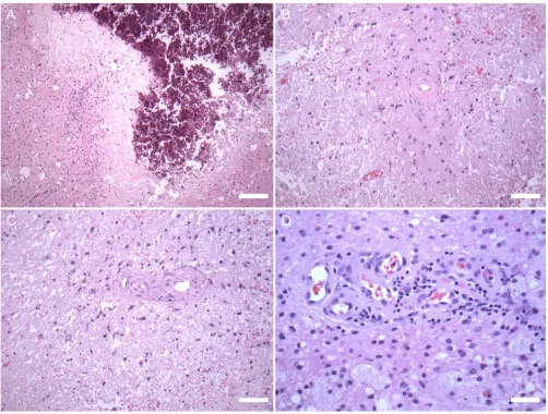
Brain histopathology, hematoxylin and eosin. The white matter has ill-defined areas of necrosis with dystrophic calcification, with margins that appear to have ischemic changes (A; scale bar: 150 µm). The small blood vessels in the tissue surrounding the areas of necrosis have abnormally thickened walls (B, C; scale bar: 75 µm). Other angiomatoid vascular proliferations are associated with mild chronic inflammatory infiltrate (D; scale bar: 40 µm).
A
uthor Man
uscr
ipt
A
uthor Man
uscr
ipt
A
uthor Man
uscr
ipt
A
uthor Man
uscr
Figure 4.
Axial FLAIR MRI of the brain following a 10-week course of high-dose dexamethasone. Note the prominent decrease in the white matter edema with resolution of the mass effect, and postsurgical changes in the right parietal lobe (arrowhead).
A
uthor Man
uscr
ipt
A
uthor Man
uscr
ipt
A
uthor Man
uscr
ipt
A
uthor Man
uscr
A
uthor Man
uscr
ipt
A
uthor Man
uscr
ipt
A
uthor Man
uscr
ipt
A
uthor Man
uscr
ipt
T ab le 1Serum and urine laboratory e
v
aluations since age 40
T est Age 40 Age 41 Fi v
e weeks prior to
admission, age 44
During hospitalization,
age 44
Normal range
Urea nitrogen (mg/dL)
20.7 (normal 7–20)
37 75 7–18 Creatinine (mg/dL) Mildly ele v
ated per report
1.4 (normal 0.7–1.2)
3.7
2.9
0.6–1.2
Estimated glomerular f
iltration rate (mL/min/1.73 m
2)
stage III CKD
56 18 23 >60 Calcium (mg/dL) 8.5 8.5–10.5 Phosphorus (mg/dL) 4.1 2.5–4.6
Aspartate aminotransferase (U/L)
15
25
10–42
Alanine aminotransferase (IU/L)
12 31 10–40 Protein (g/dL) 6.9 6.6 5.8 6.0–8.3 Alb umin (g/dL) 3.9 3.8 3.2 3.8–5.0 Th
yroid stimulating hormone (μIU/mL)
6.47
0.4–5.0
Hemoglobin A1c (%)
5.4
4.3–6.4
Erythroc
yte sedimentation rate (mm/hr)
9
0–11
C-reacti
v
e protein (mg/L)
0.2 <8.0 for inflammation Complement (mg/dL) C3 100 81–157 C4 34 12–39
Serum-protein electrophoresis with immunof
ixation
Normal pattern, no M-component Normal pattern, no M-component
IgG (mg/dL) 1390 1550 614–1295 IgA (mg/dL) 250 69–309 IgM (mg/dL) 160 53–334 Cryoglob ulins Ne g ati v e Ne g ati v e Ne g ati v e Angiotensin-con v
erting enzyme (U/L)
Ne
g
ati
v
e per report
15 8–53 Lupus anticoagulant Ne g ati v e Ne g ati v e
Anticardiolipin antibodies (phospholipid units)
IgG Ne g ati v e 11.9 0–15 IgM Ne g ati v e 12.1 0–15 Anti– β
[image:19.612.92.502.117.679.2]A
uthor Man
uscr
ipt
A
uthor Man
uscr
ipt
A
uthor Man
uscr
ipt
A
uthor Man
uscr
ipt
T est Age 40 Age 41 Fi ve weeks prior to
admission, age 44
During hospitalization, age 44 Normal range IgG Ne g ati v e <9.4 IgM Ne g ati v e <9.4 Antinuclear antibodies Ne g ati v e Positi v
e at 1:160,
homogenous pattern
Ne
g
ati
v
e per report
Positi
v
e at 1:40,
speckled pattern Ne g ati v e
Anti–Ro (SSA) antibody
Ne g ati v e Ne g ati v e Ne g ati v e
Anti–La (SSB) antibody
Ne g ati v e Ne g ati v e Ne g ati v e Anti–double-stranded DN A antibodies Ne g ati v e Ne g ati v
e per report
Ne g ati v e Ne g ati v e Anti–neutrophil c ytoplasmic antibodies Ne g ati v e Ne g ati v e Ne g ati v
e per report
Ne
g
ati
v
e
Anti–glomerular basement membrane antibody
Ne g ati v e Ne g ati v e Rheumatoid f actor (IU/mL) Ne g ati v e Ne g ati v
e per report
Ne g ati v e Anti–c
yclic citrullinated peptide antibody
Ne g ati v e Ne g ati v e HLA–B51 Ne g ati v e HLA–Bw5 Ne g ati v e HLA–B27 Ne g ati v e Human immunodef icienc
y virus 1 and 2 antibodies
Ne g ati v e Ne g ati v
e per report
Ne g ati v e Interferon-g
amma release assay
Ne g ati v e Ne g ati v e T
oxoplasma antibodies, IgM and IgG
Ne g ati v e Ne g ati v e T reponema pallidum hemagglutination assay Ne g ati v e Ne g ati v e V
enereal Disease Research Laboratory (VDRL) test
Ne
g
ati
v
e per report
Ne g ati v e Borrelia b ur gdorferi
antibodies, IgG and IgM
Ne g ati v e Ne g ati v
e per report
Ne
g
ati
v
e
Urine total protein, 24-hour (mg)
370
<100
Urine creatinine, 24-hour (g)
1.37
0.90–1.58
Urine total protein (mg/L)
1180
0–135
Urine creatinine (mg/mL)
70
N/A
Urine protein to creatinine ratio
1.69
<0.15
CKD, chronic kidne
A
uthor Man
uscr
ipt
A
uthor Man
uscr
ipt
A
uthor Man
uscr
ipt
A
uthor Man
uscr
[image:21.612.78.494.119.330.2]ipt
Table 2
TREX1 mutations in retinal vasculopathy with cerebral leukodystrophy
Frameshift Mutation and References
Reported Syndromes Subjects
V235fs (V235Gfs*6)25,28
25,29,30
31,32
25
CRV
HVR + migraine + Raynaud's syndrome HSA
not reported
1 family w/ 18 affected members + 2 individual patients 1 family w/ >20 affected members
1 family w/ 5 affected members
2 families, unknown number of affected individuals
T236fs (T236Nfs*5)25,33 CRV 1 family w/ 4 affected members
T249fs (T249Sfs*14)25,34 HERNS 1 family w/ 11 affected members
T270fs (T270Dfs*55)32 HSA 1 individual patient
P275fs (P275Qfs*2)35 RVCL (retina, CNS, kidneys) 1 individual patient, probably brother
D278fs (D278Efs*48) RVCL (retina, CNS, kidneys) 1 individual patient, possibly father and paternal uncle
R284fs (R284Kfs*41)25,36 HERNS 1 family w/ 3 affected members
E285fs (not reported)37 CRV 1 family w/ multiple affected members
L287fs (L287Afs*38)25 not reported 1 family, unknown number of affected individuals
not analyzed38 CRV 1 family w/ 4 affected members