Copyrightq1997, American Society for Microbiology
Assembly of Complete, Functionally Active Herpes Simplex
Virus DNA Replication Compartments and Recruitment
of Associated Viral and Cellular Proteins in
Transient Cotransfection Assays
LING ZHONG1ANDGARY S. HAYWARD1,2*
Molecular Virology Laboratories, Department of Pharmacology and Molecular Sciences1
and Department of Oncology,2Johns Hopkins University School
of Medicine, Baltimore, Maryland 21205
Received 6 August 1996/Accepted 26 December 1996
Early during the herpes simplex virus (HSV) lytic cycle or in the presence of DNA synthesis inhibitors, core viral replication machinery proteins accumulate in intranuclear speckled punctate prereplicative foci, some of which colocalize with numerous sites of host cellular DNA synthesis initiation known as replisomes. At later times, in the absence of inhibitors, several globular or large irregularly shaped replication compartments are formed; these compartments also contain progeny viral DNA and incorporate the IE175(ICP4) transcription factor together with several cellular proteins involved in DNA replication and repair. In this study, we demonstrate that several forms of both prereplication foci and active viral replication compartments that display an appearance similar to that of the compartments in HSV-infected cells can be successfully assembled in transient assays in DNA-transfected cells receiving genes encoding all seven essential HSV replication fork proteins together with oriS target plasmid DNA. Furthermore, bromodeoxyuridine (BrdU)-pulse-labeled DNA synthesis initiation sites colocalized with the HSV single-stranded DNA-binding protein (SSB) in these replication compartments, implying that active viral DNA replication may be occurring. The assembly of complete HSV replication compartments and incorporation of BrdU were both abolished by treatment with phosphonoacetic acid (PAA) and by omission of any one of the seven viral replication proteins, UL5, UL8, UL9, UL42, UL52, SSB, and Pol, that are essential for viral DNA replication. Consistent with the fact that both HSV IE175 and IE63(ICP27) localize within replication compartments in HSV-infected cells, the assembled HSV replication compartments were also able to recruit both of these essential regulatory proteins. Blocking viral DNA synthesis with PAA, but not omission of oriS, prevented the association of IE175 with prereplication structures. The assembled HSV replication compartments also redistributed cotransfected cellular p53 into the viral replication compartments. However, the other two HSV immediate-early nuclear proteins IE110(ICP0) and IE68(ICP22) did not enter the replication compartments in either infected or transfected cells.
During the productive or lytic cycle of herpes simplex virus type 1 (HSV-1) infection, viral DNA replication begins within 4 h in distinctive nuclear viral DNA replication compartments (RC) or factories containing both viral proteins and host cel-lular proteins. Genetic analyses have determined that there are seven essential viral proteins required specifically for DNA replication during infection (28, 58). The isolated genes for these same seven proteins when introduced into cultured mammalian cells by transient DNA transfection procedures are also sufficient for the specific amplification of cotransfected bacterial plasmid DNA containing the HSV origin (oriS or oriL) as assayed byDpnI resistance and Southern blot hybrid-ization (8, 60). These seven essential viral replication proteins are the helicase-primase components UL5, UL8, and UL52, the origin DNA-binding protein UL9, the viral DNA polymer-ase (Pol or UL30), the polymerpolymer-ase accessory protein UL42, and the single-stranded DNA-binding protein (SSB, ICP8, or UL29). UL5, UL8, and UL52 form a stable triplet complex with both helicase and primase activities (12, 63, 64).
Antibody against SSB was initially used to identify and
de-fine various prereplicative structures and RC in indirect im-munofluorescence assays (IFA) (46). Early during viral infec-tion, a number of small punctate nuclear structures referred to as prereplicative sites or foci are observed. These are defined most clearly by specifically blocking viral DNA synthesis with phosphonoacetic acid (PAA). At least some of the prereplica-tive foci (pre-RF) colocalize with initiation sites for host cel-lular DNA synthesis as defined by bromodeoxyuridine (BrdU) pulse-labeling and have been suggested to represent a reorga-nization of the cellular replisomes (14, 47). Eventually, these numerous punctate foci aggregate into several very large, mostly irregularly shaped globular RC containing progeny viral DNA. All seven essential HSV replication proteins are also found to accumulate in the RC (6, 21, 31, 32, 42) as well as some host cellular proteins, including the tumor suppressors Rb and p53 and several proteins related directly to cellular DNA replication such as DNA polymerase delta and DNA ligase (59). The viral core replication proteins UL5, UL8, UL52, and SSB are all also known to be present in the pre-replicative sites in infected cells (6, 7, 21, 31, 32). UL9 may also be required for SSB to localize in prereplicative sites in in-fected cells (6, 21, 31, 32), but only UL5, UL8, and UL52 are sufficient for the localization of SSB in prereplicative sites in DNA-transfected cells (31).
Both the IE175(ICP4) and IE63(ICP27) nuclear regulatory * Corresponding author. Department of Pharmacology and
Molec-ular Sciences, Johns Hopkins University School of Medicine, 725 N. Wolfe St., WBSB 317, Baltimore, MD 21205. Phone: (410) 955-8684. Fax: (410) 955-8685. E-mail: [email protected].
3146
on November 9, 2019 by guest
http://jvi.asm.org/
proteins are essential for efficient synthesis of both replication-related proteins and viral DNA in HSV-1 infection, although neither is essential for viral DNA replication per se in transient replication assays. Nevertheless, IE175 is known to redistribute from an early nuclear diffuse pattern into viral replication factories later during infection (30, 49), and we have recently obtained a similar finding of partial redistribution for IE63 also (62). However, IE175 does not localize within the prereplica-tive sites formed when viral DNA synthesis is blocked (29, 48). IE175 is a DNA-binding protein that behaves as an autoregu-latory repressor of immediate-early (IE) and latency promot-ers and as a transcriptional transactivator of viral delayed-early and late promoters in transient transfection assays (18–20, 40, 41, 47). Furthermore, IE175 deletion or temperature-sensitive mutant viruses are unable to synthesize delayed-early mRNA (16, 43, 56). IE63 is required for normal high levels of viral replication during infection, and some IE63 deletion mutant viruses produce considerably less progeny HSV DNA com-pared to wild-type virus (35). The efficiency of SSB distribution within viral RC was also found to be greatly altered in cells infected with a IE63 null mutant virus (13). Recently, reduced levels of accumulated mRNA of UL5, UL8, UL9, UL42, UL52, and Pol have also been demonstrated with IE63 mu-tants (55). However, it is still not clear whether just the assem-bled HSV RC are sufficient to redistribute IE175 or IE63 during viral infection or whether other factors are involved, nor is it known whether the subnuclear location of either IE175 or IE63 is important for fulfillment of their biological function. In uninfected mammalian cells, multiprotein replication complexes that contain DNA polymerases alpha and delta, DNA primase, topoisomerases I and II, RNase H, proliferating cell nuclear antigen, a DNA-dependent ATPase, replication factor C, DNA ligase I, DNA helicase, and replication protein A have been characterized (1). Cellular replication initiation sites, sometimes called replisomes, become pulse-labeled by biotin-11-dUTP or BrdU in S-phase cells and associate with the nuclear matrix, where replication occurs as the template moves through them (24). Even though HSV has a relatively large (155-kb) genome and encodes numerous viral proteins, HSV apparently also takes advantage of several host cell pro-teins, including RNA polymerase II and associated transcrip-tion factors, and probably also components of the cellular DNA synthesis and replication machinery as well. The expres-sion and distribution of some cellular replication initiation proteins, such as replication protein A, cdc2, cyclin A, and DNA polymerase alpha, are known to change during the G1-S
phase transition in mammalian cells (5), but little is known about how or whether the cellular DNA replication apparatus changes in response to the formation of functional viral DNA RC. The HSV pre-RF formed in the absence of viral DNA synthesis have been shown to colocalize with BrdU-labeled cellular DNA synthesis initiation sites (14, 15) and are more prone to do so in the presence of PAA in infections with mutant viruses that lack one of the replication proteins (31, 32). Because of these results, it has been presumed that several viral DNA replication proteins (particulately the primase-he-licase complex and SSB) may initially target to the preexisting cellular DNA initiation sites before accumulation and assem-bly of the larger functionally active viral RC. However, it is not clear yet whether such structures are actual direct intermedi-ates in the formation of RC or simply represent storage sites. Indeed, Maul et al. (34) have recently argued that input HSV genomes instead localize at cellular protein PML-containing nuclear bodies (ND10 or PODs) and that initial progeny DNA and transcripts are also associated with PODs.
HSV infection is a complicated process in which many genes
are turned on and off in a tightly controlled cascade pattern, and the efficient expression of replication genes is dependent on the presence of several IE regulatory proteins and perhaps virion factors. Furthermore, many other processes, including DNA maturation and capsid assembly, occur simultaneously. To study how HSV-1 replication compartments are formed, together with the impact of assembled HSV RC on the sub-cellular location of other viral and sub-cellular proteins, we chose to introduce by cotransfection all seven HSV-1 replication fork proteins (Rep mixture) expressed under the control of strong heterologous promoters to avoid complications from other HSV-encoded gene products. In this report, we demonstrate, for the first time, that typical large HSV RC, whose character-istics are remarkably similar to the functionally active struc-tures formed in virus-infected cells, can be successfully assem-bled in DNA-transfected Vero cells. Our assay involves the introduction of a complete set of expression plasmids contain-ing the UL5, UL8, UL9, UL42, UL52, Pol, and SSB genes driven by constitutive human cytomegalovirus (HCMV) major IE enhancers-promoters, together with the target plasmid con-taining HSV oriS. In this simplified and easily manipulated system we have (i) identified both the essential and minimal viral replication protein requirements for forming large PAA-sensitive HSV RC; (ii) shown that PAA-PAA-sensitive DNA syn-thesis initiation as defined by pulse-labeled BrdU incorpora-tion occurs within structures formed by the viral HSV replication proteins; (iii) demonstrated that both IE175 and IE63 are efficiently redistributed into assembled RC; and (iv) found that the cellular p53 protein expressed by cotransfection is also recruited into the assembled HSV RC.
MATERIALS AND METHODS
Cells and viruses.Vero cells were grown in Dulbecco’s modified Eagle’s medium (DMEM) containing 10% fetal bovine serum in humidified 5% CO2 incubator. Cells were seeded at 83104cells per well in two-well slide chambers for transfection and in four-well slide chambers for virus infection studies, and 106cells per well were plated in 100-mm-diameter dishes for the transientDpnI DNA replication assays. Stocks of HSV-1(KOS) were prepared by infecting monolayer Vero cells at 0.01 PFU per cell. Infected cells were incubated in DMEM supplemented with 1% calf serum in humidified 5% CO2incubator and were harvested after 2 days. Supernatant virus was collected following three freeze-thaw cycles and centrifuged to pellet cell debris. The clarified viral stocks were titered on monolayer Vero cells by plaque formation in DMEM supple-mented with 1% human serum to neutralize cell-free virus and prevent formation of secondary plaques. Infected cells were fixed with methanol for 10 min at room temperature (RT) and stained with crystal violet for 10 min at RT followed by washing with distilled H2O several times.
For IFA, the titer of the HSV-1(KOS) used was 1.53108PFU per ml. HSV-1(KOS) was used to infect cells in four-well slides at a multiplicity of infection (MOI) of 0.5 PFU per cell. After absorption in phosphate-buffered saline (PBS)–glucose–inactivated calf serum) at RT for 1 h, the cells were incubated in DMEM supplemented with 1% calf serum and incubated for up to 6 h in a humidified 5% CO2incubator. PAA was included at 100mg/ml in the postabsorption medium when viral DNA synthesis was to be blocked. BrdU (Sigma) was added at final concentration of 10mM for incorporation into newly synthesized DNA for 30 min just before the cells were fixed.
Expression plasmids.Plasmid pLZ11 DNA (1mg) carries the IE63 gene driven by its own promoter with four tandemly repeated SNE sites (9) inserted at theBamHI site at2276 upstream of the IE63 promoter (in a fragment from genome map positions 113422 to 115742) to boost basal expression (62). Plasmid pGH114 DNA (0.5mg) contains the IE175 gene driven by cytomegalovirus enhancer-promoter region (38), plasmid pGH92 DNA (1mg) contains the IE110 gene driven by its own promoter (38), and plasmid pGR169 DNA (1mg) contains the IE68 gene driven by its own promoter (40). Plasmid pSVp53 DNA (0.5mg) carrying the human wild-type p53 gene driven by the simian virus 40 enhancer-promoter region was a gift from Ken Kinsler (Johns Hopkins Oncology Center). Transient DNA transfection.Transient DNA transfection assays for IFA were carried out with 83104Vero cells in two-well slide chambers. A mixture of seven DNA plasmids (0.3mg of each) carrying genes encoding UL5, 8, 9, 42, 52, SSB, and Pol driven by the cytomegalovirus enhancer-promoter region, together with plasmid pMC110 (0.3mg) carrying an HSV oriS origin fragment, was used to represent the complete set of replication plasmids in each well (23). The CsCl-purified plasmid DNAs were cotransfected by the calcium phosphate pre-cipitation procedure in BBS buffer (38). pUC18 DNA was used as a carrier to
on November 9, 2019 by guest
http://jvi.asm.org/
normalize the total amount of transfected DNA. Transfected cells were incu-bated in DMEM supplemented with 10% fetal bovine serum in a humidified 3% CO2incubator at 358C overnight. The medium was changed 18 h after transfec-tion, and the slides were placed into a 5% CO2incubator at 378C. To block viral DNA synthesis, PAA at 400mg/ml was included in the medium from 18 h after transfection. Cells were fixed 48 h after transfection for IFA. BrdU was added to the culture medium at final concentration of 10mM for 30 min before fixation when appropriate.
TransientDpnI replication assay.For transient replication assays (8), DNA transfection was carried out as described above for IFA except that 1.8mg of each plasmid DNA carrying UL5, UL8, UL9, UL42, UL52, SSB, Pol, and oriS was used per 100-mm-diameter dish. Transfected Vero cells were harvested 48 h after transfection. PBS was used to wash the transfected cells twice before the cells were scraped into 2 ml of 150 mM NaCl–40 mM Tris (pH 7.5). Pelleted cells were incubated with 100mg of RNase A per ml for 1 h at 378C followed by addition of 2 ml of lysis buffer containing 10 mM Tris (pH 8.0), 10 mM EDTA, 2% sodium dodecyl sulfate (SDS) and 100mg of proteinase K per ml for 2 h at 378C. Lysed cells were subsequently extracted twice with phenol-chloroform-isoamyl alcohol (25:24:1) and once with chloroform-phenol-chloroform-isoamylalcohol (24:1). The upper layer of cellular DNA was precipitated in 70% ethanol containing 0.3 M sodium acetate (pH 5.2) at2208C overnight. DNA was pelleted by centrifuga-tion and washed with 70% ethanol before being resuspended in 300ml of distilled H2O. Each DNA sample (200ml) was digested with 40 U ofHindIII at 378C overnight to generate linear monomers of the target oriS DNA from pMC110. Samples (10mg) of cellular DNA were digested with 30 U ofDpnI at 378C overnight. The digested cellular DNA was resolved by electrophoresis on a 0.8% agarose gel at 1.0 V/cm overnight. The DNA was denatured by incubating the gel in 0.2 M HCl for 10 min at RT followed 0.4 M NaOH and 0.6 M NaCl for 20 min at RT. The DNA was transferred to a NytrAN membrane (Schleicher & Schuell), which had been treated in 103SSC (1.5 M NaCl, 0.15 M sodium citrate) for 20 min at RT, by vacuum transfer for 1 h and cross-linked by UV radiation onto the membrane after air drying for 1 h at RT. The membrane was preincubated in prehybridization buffer (0.75 M NaCl, 0.05 M Na2HPO4, 5 mM Na2EDTA, 5 mg of Carnation nonfat dried milk per ml, 0.5 mg of heparin per ml, 60 mg of polyethylene glycol 8000, 0.2 mg of denatured salmon sperm DNA per ml, 10% formamide, 1% SDS) at 608C for 2 h. A gel-purified 230-bpSmaI oriS fragment (100 ng) from pMC110 DNA was labeled with [a-32P]ATP and Klenow DNA polymerase by random priming to obtain a specific activity of 108cpm permg. The membrane was incubated in hybridization buffer containing 105cpm of the probe DNA per ml at 608C overnight then washed twice with 0.13SSC–0.1% SDS at 658C for 45 min before being exposed to Kodak XAR5 film with an intensifying screen at2808C for 5 days.
IFA.Infected or transfected cells were washed in 13Tris-saline (100 mM NaCl, 10 mM Tris-HCl [pH 7.5]), fixed with 1% paraformaldehyde in PBS for 10 min at RT, and then permeabilized in 0.2% Triton X-100 in PBS for 20 min on ice. To expose incorporated BrdU residues, pulse-labeled cells were incubated with 4 N HCl for 10 min at RT and then washed for 10 min in PBS. The primary mouse monoclonal antibody (MAb) and rabbit polyclonal antibody (PAb) were diluted together in PBS with 2% goat serum for double labeling or diluted separately for single labeling. Primary antibodies were incubated for 1 h at 378C and then incubated with the appropriate combination of fluorescein isothiocya-nate (FITC)-conjugated and rhodamine-conjugated anti-mouse, anti-rabbit, or anti-human secondary antibodies at 1:100 dilution for 30 min at 378C for double-labeling. Rhodamine-conjugated anti-mouse secondary antibody was diluted at 1:100 for single labeling. Antibodies used included mouse anti-p53 MAb-1 (On-cogene Science, Inc.), mouse anti-BrdU MAb (Becton Dickinson), and human nucleoli agent (ANA-N) antibody (Sigma). Mouse SSB 39S MAb, anti-IE175 58S MAb, rabbit anti-IE110(N) PAb, and rabbit anti-anti-IE175(N) peptide PAb were described elsewhere (38). Rabbit anti-IE68(N) peptide PAb was gen-erated by immunization with the keyhole limpet hemocyanin-conjugated peptide (14-KARRPALRSPPLGTRK-29) by procedures described previously (45). Rab-bit anti-SSB PAb 3-83 was generously provided by David Knipe (Harvard Med-ical School). Slides were screened and photographed with a 403oil immersion objective on a Leitz Dialux 20EB epifluorescence microscope, using Kodak T-MAX P3200 and appropriate narrow-band FITC or rhodamine filters.
RESULTS
Characterization of BrdU-labeled replication structures in HSV-infected Vero cells. For an initial examination of the patterns of BrdU incorporation and formation of DNA repli-cation-related structures, we infected Vero cells with HSV-1(KOS) at a relatively low MOI (,1 PFU per cell). In both virus-infected and mock-infected cultures, cellular DNA initi-ation sites that were pulse-labeled by BrdU for 30 min were randomly distributed as nuclear speckles or networks (repli-somes) in 20 to 30% of the cells, presumably representing those in S-phase (Fig. 1b and d). However, in virus-infected cells, some of the BrdU-labeled DNA synthesis initiation sites
instead formed distinctive globular or large irregularly shaped nuclear structures at 6 h postinfection; and these usually colo-calized with HSV RC, as detected by antibody to the viral SSB in double-label IFA experiments (Fig. 1a and b and c and d). The formation of complete HSV replication compartments was inhibited when PAA was added at a final concentration of either 100 or 400 mg/ml to specifically block viral DNA syn-thesis. Under these conditions, all of the larger structures dis-appeared and viral SSB was instead found in relatively small punctate or speckled nuclear structures, which have been shown previously to colocalize with sites of pulse-labeled BrdU incorporation and referred to as prereplication foci (14). The percentages of cells that incorporated BrdU during an infec-tion experiment in Vero cells are shown in Table 1 (experiment A). In uninfected cultures, 29% of the cells incorporated BrdU into replisome speckles at 6 h, whereas 77% of cells infected in the absence of PAA were labeled with BrdU at 6 h, and 89% of the SSB-positive cells and 94% of those containing either globular or large irregular viral replication compartments in-corporated BrdU. However, only 53% of cells with large rep-FIG. 1. SSB and BrdU incorporation into viral RC in HSV-infected Vero cells in the presence or absence of PAA. Infected cells were labeled by 10mM BrdU for 30 min before fixation at 6 h after infection in the absence of PAA (a to d) or in the presence of 400mM PAA (e to h). SSB (UL30 or ICP8) was detected by IFA with FITC-labeled anti-SSB PAb 3-83 in panels a, c, e, and g; incorporated BrdU was detected by rhodamine-labeled anti-BrdU MAb in pan-els b, d, f, and h. Panpan-els a and b, c and d, e and f, and g and h show the paired double-label immunofluorescence images of the same fields. In some virus-infected cells (arrowed), BrdU was incorporated into either complete viral DNA RC (a to d) or pre-RF (g and h).
on November 9, 2019 by guest
http://jvi.asm.org/
lication structures were labeled with BrdU when PAA was used to inhibit viral DNA replication. The higher percentage of cells with replication structures that labeled with BrdU in the absence of PAA indicates that ongoing viral DNA synthesis was probably occurring in these structures. Curiously, the per-centage of cells with SSB in small punctate or speckled pre-RF that incorporated BrdU in a similar pattern remained at just under 90% both before and after PAA treatment. The latter result appears to indicate that these cells may have been un-dergoing cellular but not viral polymerase-driven DNA synthe-sis.
Recognition of two distinct types of viral pre-RF.To further evaluate the effects of PAA on SSB-associated structures ob-served in infected cells, we compared and tabulated the several different categories of IFA patterns observed in parallel sam-ple cultures at 6 h after infection (Table 2). Five distinct SSB patterns were recognized, four in the presence of PAA and two in the absence of PAA. First, nearly 40% of the SSB-positive cells contained between 50 and 200 speckled or micropunctate SSB structures that closely resembled the replisomes seen in uninfected S-phase cells (Fig. 1g). Indeed, these structures were predominately colocalized with strongly BrdU-incorpo-rating speckled patterns in the absence of PAA, and both the SSB and BrdU patterns were totally unaffected by the presence of PAA (Fig. 1h). Another 21% of the SSB-positive cells con-tained a mixture of several larger globules together with the speckled pattern, although only the speckled structures labeled
strongly with BrdU and the globules disappeared in the pres-ence of PAA. Approximately 27% of the infected cells con-tained between 2 and 10 spherical globular SSB-positive struc-tures of various sizes without the speckled background. These structures, referred to as prereplication compartments (pre-RC), usually labeled weakly with BrdU in the absence of PAA but disappeared almost completely in the presence of PAA. Finally, 13% of the SSB-positive cells at this stage of infection displayed large irregular bodies or multilobed structures that sometimes nearly filled all of the nucleoplasm surrounding the nucleolus (similar to the structures shown in Fig. 2d to f or 4a and c). These forms, which we refer to as true fully active vi-rus RC, all labeled strongly with BrdU, and their formation was abolished in the presence of PAA. At later stages of infection, cells with these complete RC became much more abundant.
In contrast, in the presence of PAA, essentially only two types of patterns were observed, either or both of which rep-resent pre-RF. Approximately 57% of the SSB-positive cells contained exactly the same patterns of small numerous BrdU-labeled SSB speckles described above that were unaffected by PAA and which we have interpreted to be associated with cellular S-phase replisomes (Table 2). However, 40% displayed a novel SSB pattern not seen in the absence of PAA, in which between two and five small punctate spots were accompanied by a uniform diffuse background nuclear staining (Fig. 1e). Most of these cells failed to incorporate any BrdU either into the punctate spots or into any speckled patterns and were therefore interpreted to represent non-S-phase cells.
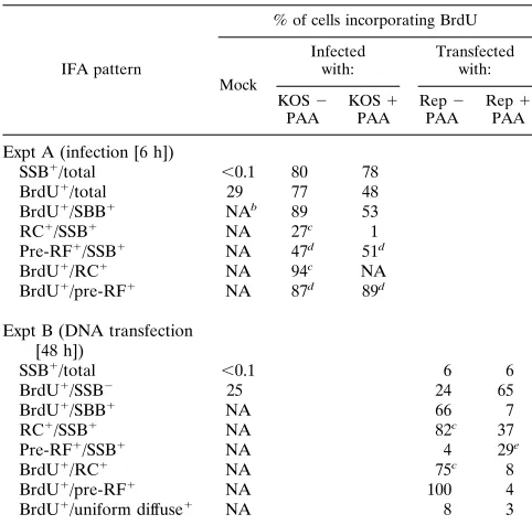
[image:4.612.57.298.98.336.2]Assembly of HSV RC in transient DNA transfection assays. We next asked whether similar viral DNA replication-related structures can be formed in transient expression assays in DNA cotransfected cells receiving just the seven essential HSV rep-lication genes UL5, UL8, UL9, UL42, UL52, SSB, and Pol under the control of the strong constitutive HCMV enhancer-promoter region. Various combinations of these genes were cotransfected into Vero cells together with the HSV oriS target plasmid DNA (pMC110). Initially, a plasmid encoding the viral SSB gene (pSSB) was transfected alone, and the expressed SSB protein (detected with anti-SSB 39S MAb) was found to be distributed in a typical uniform nuclear diffuse pattern at 48 h after transfection (Fig. 2a to c). In contrast, when the
TABLE 2. Alteration in the patterns of SSB- and BrdU-associated replication structures in HSV-infected cells in
the presence and absence of PAA
SSB IFA patterna Level of BrdU incorporation b(%)
Strong Weak Negative
PAA added 2 1 2 1 2 1
Speckled or micropunctatec 30 50 2 1 7 6
Globulesdplus speckledc 17 ,1 4 ,1 ,1 ,1
Spherical globulesconly (5pre-RC) 3 ,1 20 1 4 1
Large irregular bodies (5RC) 13 ,1 ,1 ,1 ,1 ,1 Few punctate plus diffuse (5pre-RF) ,1 ,1 ,1 1 ,1 40
Total 63 50 26 3 11 47
aVero cells at 6 h after infection with HSV-1(KOS) at an MOI of 5; 80% of the cells were positive for SSB; the total numbers of cells scored were 102 in the absence of PAA (2) and 82 in the presence of PAA (1).
bStrong BrdU incorporation represented FITC and rhodamine IFA signals of approximately equal intensity, whereas cells with weak BrdU incorporation had much stronger SSB IFA signals than BrdU signals.
cThe SSB pattern was predominantly colocalized with a speckled replisome-like BrdU pulse-label pattern both before and after PAA.
[image:4.612.316.557.549.654.2]dGlobules show a large range of different sizes. TABLE 1. Comparison of Percentages of SSB-positive cells that
incorporate BrdU during HSV infection or after transient expression in cotransfection assaysa
IFA pattern
% of cells incorporating BrdU
Mock
Infected
with: Transfectedwith: KOS2
PAA KOSPAA1 RepPAA2 RepPAA1
Expt A (infection [6 h])
SSB1/total ,0.1 80 78
BrdU1/total 29 77 48
BrdU1/SBB1 NAb 89 53
RC1/SSB1 NA 27c 1
Pre-RF1/SSB1 NA 47d 51d
BrdU1/RC1 NA 94c NA
BrdU1/pre-RF1 NA 87d 89d
Expt B (DNA transfection [48 h])
SSB1/total ,0.1 6 6
BrdU1/SSB2 25 24 65
BrdU1/SBB1 NA 66 7
RC1/SSB1 NA 82c 37
Pre-RF1/SSB1 NA 4 29e
BrdU1/RC1 NA 75c 8
BrdU1/pre-RF1 NA 100 4
BrdU1/uniform diffuse1 NA 8 3
aAll experiments were carried out in Vero cells in the presence or absence of PAA. DNA transfection involved the complete Rep1oriS plasmid mixture. Mock-infected or mock-transfected cells (Mock) were used as controls. Double-label IFA was performed with rabbit anti-SSB PAb 3-83 and mouse anti-BrdU MAb. A pulse of 10mM BrdU was incorporated for 30 min before fixation for IFA. BrdU1/total5fraction of total cells incorporating BrdU, etc.
bNA, not applicable.
cIncludes both globules (pre-RC) and irregular bodies (full RC).
dPredominantly speckled or micropunctate structures that colocalize with BrdU-labeled replisomes (see Table 2).
eIncludes primarily punctate plus a few speckled or micropunctate forms (see Table 4).
on November 9, 2019 by guest
http://jvi.asm.org/
whole set of seven replication genes and oriS were cotrans-fected into Vero cells, most of the SSB-positive cells formed either globular structures (pre-RC [not shown]) or large irreg-ularly shaped bodies that closely resembled the complete HSV RC obtained in virus-infected cells (Fig. 2d to f). Cotransfected cells receiving plasmids carrying the other six essential HSV core replication genes only, but omitting the UL9 origin DNA-binding protein gene, failed to assemble any large replication-associated structures. Instead, SSB remained in numerous punctate or small globular pre-RF within a diffuse nuclear background (Fig. 2g to i). However, in the absence of oriS, SSB still formed several mid-sized spherical globules in most DNA-transfected cells (Fig. 2j to l), and there were even a few cells that contained large irregular bodies similar to the complete RC assembled in the presence of oriS. Therefore, oriS was not required for assembly of the large globular replication struc-tures formed in transiently transfected cells, but it did appear to increase the efficiency of formation of complete RC. Most of the large globular structures formed in the absence of HSV oriS probably represent intermediates between the pre-RF and active viral RC, and therefore we will also refer to them as pre-RC on the presumption that they do not synthesize viral DNA.
Functional characterization of the HSV replication plas-mids. A transientDpnI resistance replication assay was also carried out by using a32P-labeled oriS-containing DNA
frag-ment as a probe on a Southern blot to confirm that our plasmid DNA containing HSV oriS was replicated in the same cotrans-fected cultures and under the same transfection conditions
used for the assembly of the HSV RC (Fig. 3). The full set of plasmids carrying genes encoding all seven replication proteins and the oriS target plasmid was cotransfected into Vero cells. Cells receiving the same complete Rep plasmid mixture but omitting the plasmid carrying the Pol gene (Rep2Pol) or the oriS-containing plasmid, (Rep 2 oriS) were included in the same replication assay as negative controls. Input oriS-contain-ing DNA plasmid and amplified DNA were cleaved to give a 3.3-kb linear monomer band by digestion withHindIII in cells transfected with Rep (lane 1) or Rep2Pol (lane 2). Since cells transfected with the Rep 2 oriS mixture did not have oriS-containing DNA, no input DNA was detected in that sample (lane 3). Somewhat more monomer linearized oriS-containing DNA was recovered from cells transfected with Rep than that with Rep2Pol, although each received the same amount of input transfected oriS DNA. By double digestion with DpnI andHindIII, amplified DNA that was resistant to methylation-specific digestion byDpnI was also detected as a linear band of 3.3 kb. As expected, a significant amount of oriS-containing DNA was found to be resistant to DpnI digestion from cells transfected with the complete Rep mixture (lane 4), whereas there was no DpnI-resistant replicated DNA detected from cells transfected with Rep2Pol (lane 5) or Rep2oriS (lane 6). This result confirms that the oriS-containing DNA was replicated in those transfected Vero cells receiving our com-plete set of Rep plasmids but not when Pol was absent.
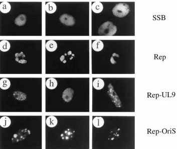
[image:5.612.133.486.67.363.2]Inhibition of viral DNA synthesis with PAA blocks the as-sembly of HSV RC in cotransfected cells.Since the formation of complete RC in HSV-infected cells was inhibited by PAA, FIG. 2. Assembly of HSV RC in cotransfected cells. All panels show three examples of single-label IFA patterns for SSB detected with rhodamine-labeled anti-SSB 39S MAb in transfected Vero cells. (a to c) Typical nuclear diffuse pattern in cells receiving only the plasmid carrying the SSB gene; (d to f) assembled replication compartments in cells cotransfected with the oriS plasmid pMC110 and expression plasmids encoding UL5, UL8, UL9, UL42, UL52, SSB, and Pol; (g to i) diffuse plus pre-RF patterns in cells cotransfected with oriS and plasmids encoding UL5, UL8, UL42, UL52, SSB, and Pol without UL9; (j to l) pre-RC in cells cotransfected with
plasmids encoding UL5, UL8, UL9, UL42, UL52, SSB, and Pol without oriS.
on November 9, 2019 by guest
http://jvi.asm.org/
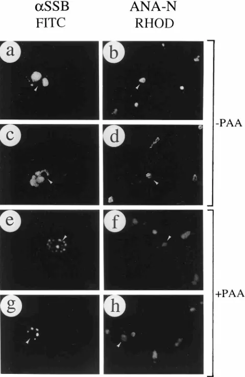
the effect of PAA on the assembly of RC in transfected cells was also studied. Compared with the SSB distribution in rep-lication compartments in the absence of PAA (Fig. 4a and c), the addition of PAA proved to efficiently inhibit the assembly of virtually all large RC and SSB remained predominantly as small nuclear globules or punctate structures in the presence of PAA (Fig. 4e and g). Quantitatively, the proportion of SSB-positive cells with globules or large irregular bodies decreased from 82 to 37%, and those with small punctate structures increased from 4 to 29% in one typical experiment after addi-tion of PAA (Table 1, experiment B).
Because of possible morphological similarities between the assembled pre-RC or RC with large globular nucleoli, anti-SSB 39S MAb and an ANA-N antibody were also used in double-label IFA experiments to compare the two structures in DNA-transfected cells. The results confirmed that the assem-bled globular replication compartments were clearly not asso-ciated with nucleolar domains labeled by the ANA-N antibody (Fig. 4a to d). Similarly, the SSB-positive punctate structures formed in the presence of PAA were quite distinct from nu-cleoli (Fig. 4e to h).
Assembly of HSV RC requires each of the seven HSV essen-tial replication proteins.In transientDpnI replication assays, all seven essential HSV replication gene products, including UL5, UL8, UL9, UL42, UL52, SSB, and Pol, are required for the replication of plasmid DNA containing HSV oriS. Elimi-nation of each of these gene products one at a time was tested to identify the essential protein requirements for assembling HSV replication-associated structures in transient cotrans-fected cells. In the presence of the whole set of replication gene products and oriS, complete HSV replication compart-ments were frequently detected by the anti-SSB 39S MAb (Fig. 5a and b). In the absence of UL5, UL8, or UL52, SSB gave a
somewhat uneven nuclear diffuse distribution only (Fig. 5c to h). However, consistent with previous mutant HSV infection data (31, 33), SSB formed numerous small nuclear micropunc-tate structures in the absence of Pol or UL42 (Fig. 5i to l). Furthermore, SSB gave a similar nuclear punctate pattern in the presence of UL5, UL8, and UL52 only (Fig. 5m and n). Therefore, the helicase-primase complex of UL5, UL8, and UL52 was all that was required for SSB to localize into nuclear micropunctate structures that are similar to some of the pre-RF seen in infected cells in the absence of viral DNA synthesis. However, because each of the seven essential HSV replication gene products was required for SSB to locate in complete assembled RC, the results are fully consistent with the requirements for the amplification of HSV oriS DNA plas-mids in theDpnI replication assay in transfected cells. There-fore, based on the similarities between the assembled compart-ments and those found in HSV-infected cells, it is reasonable to claim that even after transient expression in
[image:6.612.111.245.70.275.2]DNA-cotrans-FIG. 4. Neither complete RC nor pre-RF assembled in the presence of PAA are associated with nucleoli. oriS DNA and the whole set of plasmids encoding UL5, UL8, UL9, UL42, UL52, SSB, and Pol were cotransfected in the absence of PAA (a to d) or in the presence of 400mM PAA (e to h). SSB was detected by rhodamine-labeled anti-SSB MAb 39S in panels a, c, e, and g; nucleoli were detected by FITC-labeled ANA-N antibody in panels b, d, f, and h. Panels a and b, c and d, e and f, and g and h are paired double-label frames for the same fields. Arrowed cells show that nucleoli are localized outside SSB RC in the absence of PAA (a to d) and outside pre-RF in the presence of PAA (e to h).
FIG. 3. HSV oriS-dependent transient DNA replication assay in cotrans-fected Vero cells receiving all seven essential viral replication proteins. Southern blotting to detect oriS DNA was performed with size-fractionated cellular DNA from transfected Vero cells receiving various sets of plasmid DNAs. Lane 1, UL5, UL8, UL9, UL42, UL52, Pol, and SSB genes plus oriS; lane 2, complete sets of plasmids except that the plasmid encoding the Pol gene was omitted; lane 3, complete set of plasmids except that the oriS-containing plasmid was omitted. Each DNA sample (10mg) was digested withHindIII to linearize the input oriS-containing plasmid or digested withHindIII andDpnI to detect amplified un-methylated oriS-containing DNA in transfected cells. A 230-bpSmaI-SmaI frag-ment containing oriS isolated from pMC110 was used as the hybridization probe.
on November 9, 2019 by guest
http://jvi.asm.org/
[image:6.612.317.557.281.649.2]fected cells, SSB can be localized into structures that represent assembled fully active HSV RC and that the presence of UL5, UL8, UL9, UL42, UL52, Pol, and SSB is both necessary and sufficient for the process.
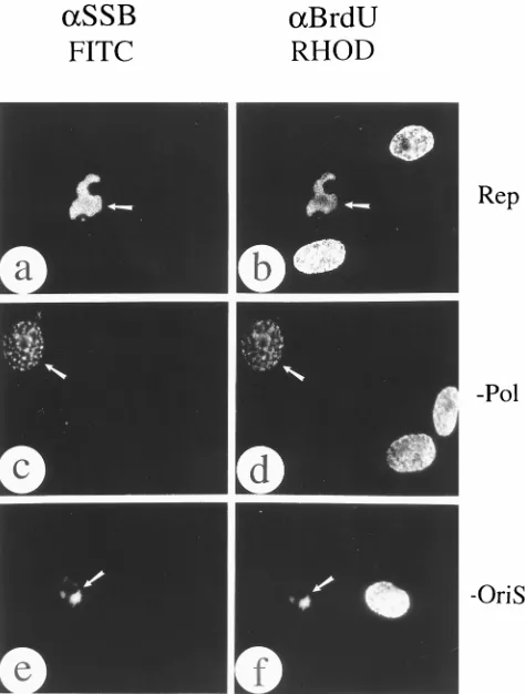
Incorporation of BrdU into assembled HSV replication structures. Because the assembled HSV RC produced by DNA transfection are morphologically similar to those formed in virus-infected cells, we asked whether DNA synthesis initi-ation as defined by BrdU incorporiniti-ation also occurred at these sites. The results revealed that in the presence of all seven replication proteins and the oriS target plasmid DNA (Rep), a 30-min pulse with BrdU was incorporated into newly synthe-sized DNA in 66% of the SSB-positive cells and as many as 75% of the assembled RC (Table 1, experiment B), where it often colocalized with SSB (Fig. 6a and b). However, with the same DNA plasmid mixture in the presence of PAA, BrdU was incorporated into only 7% of the SSB-positive cells (Table 1, experiment B). In the absence of HSV polymerase, a few SSB-positive structures colocalized with DNA synthesis
initia-tion sites, and these resembled micropunctate pre-RF whose distribution was similar to the speckled BrdU patterns ob-tained in untransfected S-phase cells (Fig. 6c and d). Globular pre-RC that still incorporated BrdU were also formed rela-tively frequently in the Rep2oriS mixture (Fig. 6e and f).
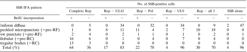
[image:7.612.66.289.67.447.2]To assess the effect of omission of core protein components on the BrdU incorporation patterns of replication structures formed in transient assays, we tabulated the results from a set of experiments similar to these described above (Table 3). Approximately 50 SSB-positive cells were scored in each sam-ple. In the complete Rep control, 64% of the SSB-positive cells contained either globules or irregular bodies with strong colo-calized BrdU patterns, whereas there were very few colocal-ized speckles or punctate structures (6% in Table 1, experi-ment B). In contrast, the Rep2UL42, Rep2Pol, and Rep2 UL9 samples all gave nearly 70% uniform diffuse SSB patterns without any BrdU incorporation (Table 3). However, most of the remaining 30% gave micropunctate SSB, and approxi-mately half of these occurred in cells with S-phase-like speck-led BrdU patterns of which at least some were colocalized (Fig. 6c and d). In the case of the sample receiving UL5, UL8, UL52, and SSB only, 67% of the SSB-positive cells were speck-led or micropunctate and almost 25% colocalized with
[image:7.612.320.557.320.633.2]FIG. 6. Colocalization of assembled RC and BrdU-labeled DNA initiation sites. SSB was detected with FITC-labeled rabbit anti-SSB 3-83 PAb in panels a, c, and e; incorporated BrdU was detected with rhodamine-labeled anti-BrdU MAb in panels b, d, and f. (a and b) oriS DNA and the whole set of plasmids encoding UL5, UL8, UL9, UL42, UL52, SSB and Pol cotransfected together; (c to f) cotransfection of all plasmids except the one encoding Pol (c and d) or the one encoding oriS (e and f). Paired panels show the immunofluorescence images of the same fields labeled with anti-SSB (a, c, and e) and anti-BrdU (b, d, and f) from each transfection. In some cotransfected cells (arrowed), incorporated BrdU colocalized with the SSB protein in either RC (a and b), pre-RF (c and d), or pre-RC (e and f).
FIG. 5. All seven essential viral replication gene products are required for the assembly of complete HSV RC in cotransfected cells. SSB was detected by rhodamine-labeled anti-SSB 39S MAb. (a and b) Two separate single-label frames showing cells cotransfected with oriS DNA and the whole set of plasmids encoding UL5, UL8, UL9, UL42, UL52, SSB, and Pol; (c to l) omission exper-iments showing cotransfection of all plasmids except the one encoding UL5 (c and d), UL8 (e and f), UL52 (g and h), Pol (i and j), or UL42 (k and l); (m and n) cotransfection of plasmids encoding UL5, UL8, UL52, and SSB only.
on November 9, 2019 by guest
http://jvi.asm.org/
S-phase-like BrdU patterns (Table 3). Surprisingly, even when SSB was transfected alone, 98% of the cells gave a uniform diffuse SSB-positive pattern but only 5% of these displayed S-phase BrdU speckles (Table 3), whereas the normal 25% of nonexpressing cells still did so. Evidently expression of SSB in a uniform diffuse pattern occurs only in non-S-phase cells un-der the conditions of our transient assays, whereas assembly into micropunctate or replisome-like patterns together with UL5, UL8, and UL52 shows some preference for S-phase cells. Inhibitory effect of PAA on BrdU incorporation into RC in transient assays.A quantitative comparison of the incorpora-tion of BrdU into the various different SSB-associated repli-cation-related structures in a transient expression assay in the presence and absence of PAA is shown in Table 4. In DNA-transfected Vero cells receiving the full Rep plasmid mixture plus oriS in the absence of PAA, the predominant patterns observed in SSB-positive cells were the typical spherical glob-ules (pre-RC) and large irregular bodies (RC) similar to those found in virus-infected cells. In this experiment, up to 53% of the cells contained globules, with 18% showing strong colocal-ized BrdU staining and another 20% giving a weak BrdU-positive pattern. Among another 29% of the SSB-BrdU-positive cells that we categorized as full replication compartments, 13% had strong colocalized BrdU staining and 9% incorporated BrdU relatively weakly.
Virtually all of the full RC and many of the large prerepli-cation globules disappeared in the presence of PAA (400mg/ ml), leaving a new distribution of SSB-positive nuclei consisting of 34% uniform diffuse, 34% globular, and 24% punctate-plus-diffuse patterns (Table 4). Very few of the SSB-positive cells in the presence of PAA (7% overall [Table 1, experiment B]) incorporated any BrdU at all, and only 5% showed the numer-ous micropunctate foci pattern, of which only a small subset colocalized with a strong BrdU-labeled speckled pattern (1%). In fact, the very rare presence (2% only) of either SSB or BrdU-labeled typical S-phase speckled patterns (even in the absence of PAA) represented the major difference between the IFA results observed in transfected cells compared to virus-infected cells (compare Tables 2 and 4). On the other hand, the 24% of SSB-positive cells in PAA that showed a small number of punctate spots within a uniform diffuse background closely resembled the type of pre-RF seen in infected non-S-phase cells. Furthermore, in the Rep 1oriS experiment described above, where only 1% of the SSB-expressing cells grown in PAA for 48 h gave typical speckled BrdU incorporation pat-terns, the percentage of non-SSB-expressing cells in the same culture that gave strong speckled BrdU-positive patterns in-creased from 25 to 65% (Table 1, experiment B). We conclude that virtually all of the SSB-associated replicating structures
formed in transfected cells are PAA sensitive and that the process of transfection led in some way to synchronization or selection against S-phase characteristics only in those cells that expressed viral proteins.
[image:8.612.62.558.81.187.2]Role of oriS in formation of replication structures obtained in DNA-transfected cells.To examine further the rather sur-prising observation that many prereplication globules and even some full RC-like structures were still generated in the Rep2 oriS mixtures in the transient transfection assay (Fig. 2j to l; Fig. 6e and f), we tabulated the BrdU incorporation results from two separate experiments carried out in the presence and absence of oriS in which the levels of SSB expression were widely different (Table 5). In experiment A, the transfection efficiency was very high, giving globular or irregular bodies in 93% of the SSB-positive cells and uniform diffuse patterns in only 5% or less, whereas in experiment B, with a much lower transfection efficiency, approximately 55% of the SSB-positive cells displayed uniform diffuse patterns only or a mixture of uniform diffuse plus globules. Again, no more than 1 to 2% of the SSB-positive cells in experiment A gave S-phase-like BrdU-pulse-labeled speckles, whereas in both experiments, 20 to 23% of the cells containing viral SSB structures showed strong BrdU incorporation in the presence of oriS, and 11 to 12% did so in the absence of oriS. Only the number of fully active irregular bodies (RCs) incorporating high levels of BrdU appeared to be significantly affected by the absence of oriS (from 12 to 4% or 11 to 5%), whereas the proportion of cells with globular pre-RC incorporating low levels of BrdU were essentially unaffected. The proportion of such structures that
TABLE 4. Alteration in the patterns of SSB- and BrdU-associated replication structures in transient expression assays in
the presence and absence of PAAa
SSB IFA pattern Level of BrdU incorporation (%) High Low Negative PAA added 2 1 2 1 2 1
Uniform diffuse 1 ,1 1 1 12 33
Speckled micropunctate (5pre-RF) 2 1 ,1 1 ,1 3
Globulesbonly (5pre-RC) 18 1 20 3 15 30
Large irregular bodies (5RC) 13 ,1 9 ,1 7 3
Few punctate plus diffuse (5pre-RF) 2 ,1 ,1 ,1 ,1 24
Total 36 2 30 5 34 93
aVero cells received the complete Rep1oriS mixture of plasmid DNAs; 6% of the cells were positive for SSB; the total numbers of cells scored were 259 in the absence of PAA (2) and 134 in the presence of PAA (400mg/ml) (1).
bIncludes mixed globules plus diffuse in some cases. Globules show a large range of different sizes.
TABLE 3. Effect of removing UL42, Pol, or UL9 from the complete transfection plasmid mixturea
SSB IFA pattern No. of SSB-positive cells
Complete Rep Rep2UL42 Rep2Pol Rep2UL9 Rep2all 3 SSB alone
BrdU incorporation 1 2 1 2 1 2 1 2 1 2 1 2
Uniform diffuse 0 5 0 34 0 32 0 34 0 9 2 47
Speckled micropunctate (5pre-RF) 1 0 8 11 11 4 2 7 10 18 0 0
Few punctate (5pre-RF) 2 4 0 2 1 1 0 1 0 2 0 1
Globular (5pre-RC) 16 6 0 1 0 4 0 7 0 4 0 0
Irregular bodies (5RC) 13 3 0 0 0 0 0 0 0 0 0 0
Total (%) 64 36 17 83 22 78 4 96 30 70 4 96
aVero cells received either the complete Rep1oriS mixture of plasmid DNAs or mixtures lacking the components indicated. A total of between 43 and 56 SSB-positive cells were scored in each sample. All cell cultures were pulse-labeled with BrdU for 30 min at 48 h after transfection and then scored for the presence (1) or absence (2) of BrdU incorporation and SSB patterns by double-label IFA.
on November 9, 2019 by guest
http://jvi.asm.org/
[image:8.612.315.559.581.687.2]lacked any BrdU incorporation also increased somewhat (from 43 to 60% or 19 to 25%) in the absence of oriS. Although we have not shown directly that BrdU incorporation in this situ-ation is PAA sensitive, we conclude that even in the absence of the specific oriS plasmid, not only was extensive formation of viral replication associated structures occurring, but some DNA synthesis associated with the viral biochemical machin-ery was probably ongoing also.
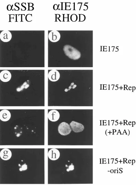
Recruitment of the viral IE175 and IE63 proteins into as-sembled HSV RC.During HSV infection, the viral DNA RC that are formed in many Vero cells by 6 h after infection at low MOI appear to incorporate most of the HSV IE175 protein present at that time into colocalized structures (30, 49). We have also found that some but not all of the IE63 protein present at that time is also incorporated into the RC (62). Since both IE175 and IE63 are essential for viral reproduction, the subnuclear location of both IE175 and IE63 proteins dur-ing viral infection is probably important for their biological function. To test whether the assembled HSV RC in cotrans-fected cells were also able to recruit the IE175 or IE63 pro-teins, double-label IFA of IE175 or IE63 together with SSB and the assembled RC components was performed in Vero cells.
A typical nuclear diffuse distribution was obtained with anti-IE175 antibody when a plasmid expressing the anti-IE175 gene (pGH114) was transfected alone into Vero cells (IE175) (Fig. 7a and b). However, the IE175 protein was found in either globules or irregularly shaped bodies within the nucleus when the IE175 plasmid was cotransfected with the complete set of seven replication gene plasmids encoding the UL5, UL8, UL9, UL42, UL52, SSB, and Pol proteins and the oriS plasmid. These structures were colocalized with the SSB protein in all cells that contained them (Fig. 7c and d). In the presence of PAA, SSB was found only in relatively small punctate struc-tures (Fig. 7e), but IE175 always remained totally nuclear diffuse in the same cells (Fig. 7e and f). Therefore, in cotrans-fected cells, the assembled HSV replication compartments were able to recruit the essential IE175 gene product, whereas IE175 did not enter pre-RF. Surprisingly, IE175 relocalization still occurred within the nucleus in assembled replication
struc-tures (pre-RC and RC-like) in the absence of oriS. However, only 20% of the cells gave complete colocalization (Fig. 7g and h), whereas the majority of the cells gave a mixed pattern of partial colocalization together with a uniform diffuse back-ground.
[image:9.612.58.299.100.268.2]IE63 expressed on its own from plasmid pLZ11 in trans-fected Vero cells is distributed partially in a nuclear diffuse pattern and partly in a punctate colocalized pattern with the SC-35 spliceosome-associated antigen SC35 (44, 51, 52, 62). However, IE63 was also recruited into assembled RC in Vero cells cotransfected with the full set of replication plasmids carrying UL5, UL8, UL9, UL42, UL52, SSB, Pol, and oriS (Fig. 8a and b). When both viral transactivators were cotrans-fected into Vero cells with the full set of replication plasmids, the assembled replication compartments recruited both IE63 (Fig. 8e and f) and IE175 (Fig. 8g and h) into similar struc-tures. Furthermore, the recruitment of either IE63 or IE175 was independent of the presence of oriS (Fig. 8i to l). IE175 always remained in a nuclear diffuse pattern when SSB was not detected in the same cells (Fig. 8l). Interestingly, in the ab-sence of UL42, some IE63 was still redistributed into nuclear punctate pre-RF containing SSB, although some remained in a
[image:9.612.318.557.310.635.2]FIG. 7. Recruitment of the HSV IE175 protein by assembled RC. SSB was detected by FITC-labeled anti-SSB 39S MAb in panels a, c, e, and f; IE175 was detected by rhodamine-labeled anti-IE175(N) PAb in panels b, d, f, and h. (a and b) Two immunofluorescence images of the same field when IE175-encoding plasmid pGH114 was transfected alone; (c and d) cotransfection of IE175 (pGH114), oriS, and the whole set of plasmids encoding UL5, UL8, UL9, UL42, UL52, Pol, and SSB; (e and f) cotransfection of IE175 (pGH114), oriS, and the whole set of plasmids encoding UL5, UL8, UL9, UL42, UL52, SSB, and Pol in the presence of PAA; (g and h) cotransfection of IE175 (pGH114) and the whole set of plasmids encoding UL5, UL8, UL9, UL42, UL52, SSB, and Pol without oriS.
TABLE 5. Relatively small effects of the presence or absence of oriS on BrdU incorporation into various replication structures
generated in transient expression assaysa
SSB IFA pattern Level of BrdU incorporation (%) High Low Negative OriS DNA added 1 2 1 2 1 2
Expt A (high efficiency)
Uniform diffuse ,1 ,1 ,1 ,1 5 1
Speckled micropunctate (5pre-RF) 2 1 ,1 ,1 ,1 ,1
Globules only (5pre-RC) 11 7 16 15 26 30
Large irregular bodies (5RC) 12 4 11 12 17 30
Total 25 12 27 27 48 61
Expt B (low efficiency)
Uniform diffuse ,1 ,1 ,1 ,1 29 31
Diffuse plus globules (5pre-RC) 3 1 13 13 8 13
Globules (5pre-RC) 6 6 13 20 8 11
Large irregular bodies (5RC) 11 5 5 11 3 1
Total 20 12 31 34 48 56
aVero cells received the complete Rep mixture of plasmid DNAs with (1) or without (2) the oriS plasmid; 8 and 1.5% of the cells were positive for SSB in experiments A and B, respectively. The total numbers of SSB-positive cells scored were 122 and 126 in experiment A and 82 and 83 in experiment B.
on November 9, 2019 by guest
http://jvi.asm.org/
nuclear diffuse background as well (Fig. 8m and n). In contrast, in the same cells, IE175 stayed in a nuclear diffuse pattern, even in those cells displaying SSB as a pattern of nuclear punctate pre-RF (Fig. 8o and p).
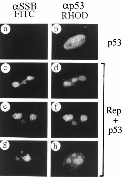
Recruitment of p53 by assembled HSV RC in cotransfected cells.Several cellular proteins, including p53, have been sug-gested to colocalize with HSV RC in HSV-infected cells (59). Since endogenous p53 in Vero cells gives no detectable IFA signal with anti-p53 MAb-1, plasmid pSVp53 expressing the wild-type p53 protein was transfected into Vero cells in the presence or absence of complete set of plasmids carrying UL5, UL8, UL9, UL42, UL52, SSB, Pol, and oriS. Expression of p53 alone produced a typical nuclear diffuse pattern (p53) (Fig. 9a and b), but double-label IFA of cotransfected cells revealed that p53 was efficiently redistributed into the HSV replication compartments together with SSB (Fig. 9c to h). p53 is known to be involved in DNA repair and the G1/S cell cycle control
checkpoint (22) and has been suggested to colocalize with viral DNA replication compartments in HSV-infected cells (59). Therefore, the colocalization between p53 and the assembled HSV RC in transiently cotransfected cells strengthens the idea that these assembled RC are biologically functional and that this model may provide a simplified system to study how host cellular factors contribute to viral DNA replication.
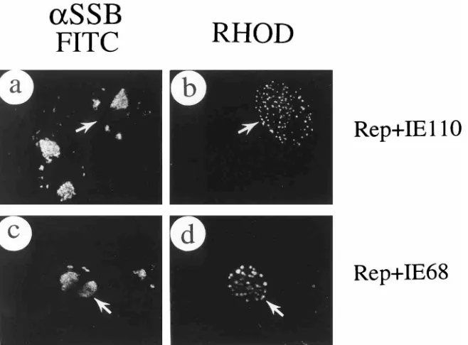
[image:10.612.75.546.68.405.2]Specificity of the recruitment by assembled HSV RC.The assembled HSV RC have many similarities to those found in HSV-infected cells based on the requirement for all seven essential replication gene products, colocalization with cellular DNA synthesis initiation sites, the inhibition effect by PAA, and the ability to recruit both the viral IE175 and IE63 proteins and p53. To test the specificity of the recruitment of HSV nuclear proteins by the assembled HSV RC, the other two HSV IE nuclear proteins, IE110 and IE68, were also tested by cotransfection in the presence of UL5, UL8, UL9, UL42, UL52, SSB, Pol, and oriS. In infected Vero cells, both IE110 and IE68 gave a nuclear punctate distribution, which is unre-lated to the viral RC (61). Similarly in DNA-transfected cells, both IE110 and IE68 remained in typical nuclear punctate patterns despite the presence of the assembled SSB-positive RC in the same cells (Fig. 10). Even though IE110 has been demonstrated to be able to associate with many other HSV proteins in punctate structures in cotransfected cells, including IE175 (38), IE63 (62), IE68 (62), and UL5, UL8, and UL52 (33), as well as with cellular proteins such as p53 and RAG-1 (62), IE110 evidently does not associate with assembled RC in DNA-transfected cells. This result demonstrates both the spec-ificity of recruitment by assembled RC and the selectivity of FIG. 8. Recruitment of the HSV IE63 protein in the presence and absence of IE175 by assembled RC. SSB was detected with FITC-labeled anti-SSB 39S MAb (a, c, e, g, i, k, m, and o); IE63 was detected with rhodamine-labeled anti-IE63(N) PAb (b, f, j, and n); IE175 was detected with rhodamine-labeled anti-IE175(N) PAb (d, h, l, and p). Paired double-label immunofluorescence images of the same fields are shown in panels a and b, c and d, e and f, g and h, i and j, k and l, m and n, and o and p. (a to d) Cotransfection of IE63 (pLZ11), oriS, and plasmids encoding UL5, UL8, UL9, UL42, UL52, SSB and Pol; (e to h) cotransfection of IE63 (pLZ11), IE175 (pGH114), oriS, and plasmids encoding UL5, UL8, UL9, UL42, UL52, SSB, and Pol; (i to l) cotransfection of IE63 (pLZ11), IE175 (pGH114), and plasmids encoding UL5, UL8, UL9, UL42, UL52, SSB and Pol (the IE175 protein remained in a nuclear diffuse pattern when SSB RC were not assembled in the same transfected cells [arrowed cells in panel l]); (m to p) cotransfection of IE63 (pLZ11), IE175 (pGH114), oriS, and plasmids encoding UL5, UL8, UL9, UL52, SSB and Pol.
on November 9, 2019 by guest
http://jvi.asm.org/
colocalization between IE110 and other viral or cellular pro-teins.
DISCUSSION
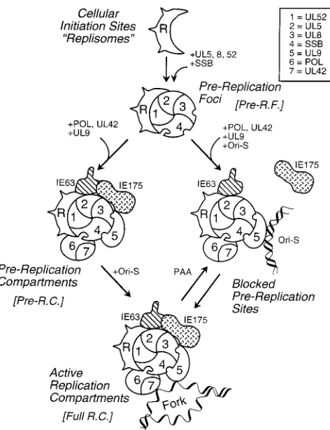
Efficient assembly of functionally active HSV RC in tran-sient expression assays.Since HSV origin-specific DNA rep-lication can be reproduced inDpnI resistance cotransfection assays, and the requirements for viral replication proteins are the same as those in infected cells, we investigated whether the transient transfection system could also be extended visually at the level of single cells to understand how HSV RC are as-sembled. By cotransfection of constitutive expression plasmids encoding only UL5, UL8, UL52, and SSB into Vero cells, we were able to form numerous SSB-containing micropunctate structures, which are similar to the pre-RF described recently by others (31). Importantly, as summarized in the model in Fig. 11, we took this a step further to demonstrate that large glob-ular structures or irregglob-ularly shaped bodies containing SSB were also observed in the nucleus when the complete set of Rep plasmids carrying each of the seven HSV essential repli-cation genes were cotransfected into Vero cells together with the HSV oriS-containing target plasmid. This is the first dem-onstration of the apparently complete assembly of functional HSV RC in DNA-transfected cells.
These SSB RC-like structures have been examined in several different ways to determine whether they are biologically func-tional. The viral protein requirements for assembly were dem-onstrated to be the same as for positive signals in the DpnI DNA replication assay in transfected cells, as well as for viral DNA replication in virus-infected cells (28, 58). Importantly, omission of any one of the seven essential HSV replication proteins abolished assembly of the largest forms of the RC, although much smaller micropunctate structures resembling pre-RF remained, especially in the absence of UL42, Pol, or UL9. Of even greater significance, the largest globular struc-tures and irregular bodies assembled, which were most mor-phologically similar to active viral RC, frequently incorporated high levels of pulse-labeled BrdU, and addition of the HSV DNA polymerase-specific inhibitor PAA abolished both as-sembly of the largest forms and BrdU incorporation into the small globular or punctate structures that remained. There-fore, it appears entirely reasonable to claim that specific viral Pol-dependent DNA synthesis was occurring in these struc-tures and that they are functionally equivalent to the active viral DNA RC generated in HSV-infected cells.
As expected, theDpnI replication assay carried out with the same input plasmids under the same conditions showed that they were competent to carry out replication of the viral oriS target plasmid DNA in an HSV Pol-dependent fashion. Others have previously confirmed the specificity of such assays by showing that similar target plasmids lacking key oriS motifs failed to give detectable replication both in transient cotrans-fection assays and after coincotrans-fection with baculovirus vectors expressing the seven HSV replication proteins (54, 57). All of these pieces of evidence suggest that assembled HSV RC are probably biologically functional and active in synthesizing viral DNA in cotransfected cells.
Do the assembled HSV replication structures initiate at or incorporate cellular replisome sites? Based on the previous observations that PAA-resistant micropunctate structures in infected cells colocalize with cellular BrdU-pulse-labeled speckles or replisomes, and that both complete viral RC and pre-RF apparently contain several cellular replication-related proteins (58), the simple model that cellular S-phase matrix-associated replisomes represent initial sites of formation of
viral pre-RF, which then coalesce into a smaller number of larger bodies, appears both plausible and attractive. However, there is no direct evidence that the micropunctate SSB pre-RF observed both before and after PAA treatment in S-phase cells are actual intermediates in the process. Furthermore, Maul et al. (34) have recently suggested that input HSV genomes are targeted to a small number of punctate matrix-associated in-tranuclear loci referred to as ND10 or PODs that contain the cellular protein PML.
Our observations that infected cells that are not in S phase form a second type of PAA-resistant punctate pattern contain-ing only a small number of pre-RF that do not incorporate BrdU (Table 2), together with our evidence that fully active viral RC form efficiently in DNA-transfected cells that appar-ently lack S-phase characteristics, also suggest that alternative pathways might occur. Indeed, the similarity in number of the punctate SSB foci seen in non-S-phase infected cells in the presence of PAA to the number of complete RC in infected cells (an average of four to five per cell) might make these more likely to be functional intermediates than the far more numerous and smaller replisome-associated structures. How-ever, we do not know whether these few punctate foci are derived from ND10 or PODs, nor do we know whether they contain viral DNA or cellular replication proteins. Further-FIG. 9. Redistribution of cotransfected p53 protein by assembled RC. (a and b) Plasmid pSVp53 transfected alone; (c to h) cotransfection of pSVp53 and the whole set of plasmids encoding UL5, UL8, UL9, UL42, UL52, SSB, and Pol plus oriS. Paired double-label IFA panels show SSB detected with FITC-labeled anti-SSB PAb 3-83 (a, c, e, and g) and p53 detected with rhodamine-labeled anti-p53 MAb in the same field (b, d, f, and h).
on November 9, 2019 by guest
http://jvi.asm.org/
[image:11.612.318.558.67.411.2]more, despite their close resemblance, fewer than 30% of the micropunctate structures formed by transfection of the heli-case-primase (UL5, UL8, and UL52) and SSB components alone or by omission of UL42, UL9, or Pol colocalized with BrdU (Table 3), and therefore they may not all correlate with the predominantly replisome-like speckled pre-RF seen in in-fected S-phase cells in the presence and absence of PAA.
Cellular DNA replication sites in mammalian cells have been visualized by biotin-dUTP pulse-labeling to be able to fuse with each other (25). Therefore, it is possible that viral pre-RF and cellular DNA replication initiation sites also fuse and coalesce into larger globular viral pre-RC containing both viral and cellular proteins. Since viral oriS was not required for targeting of SSB into the globular pre-RC, oriS is probably recruited into the complete RC later, either with or without UL9, to stimulate efficient assembly of viral RC followed by specific replication of viral DNA within these structures.
Diminished role of oriS in the transient replication assay.In experiments using DNA transfection mixtures containing all seven protein components (UL5, UL8, UL9, UL42, UL52, SSB, and Pol) but in the absence of the added oriS plasmid, there were still many large globular and irregularly shaped structures formed, although strong BrdU pulse-labeling of the full RC-like forms was reduced two- to threefold compared to parallel samples in the presence of oriS (Table 5). Clearly, the role of oriS sequences was much less than expected in these assays for reasons that we do not yet understand. Interestingly, in a similar transient cotransfection assembly assay that we have recently described for formation of functionally active HCMV RC (53), the formation of the complete large irregular bodies was much more dependent on the inclusion of ori-lyt plasmid DNA than was the case here with the HSV system.
In both the presence and absence of oriS, low-level pulse-labeled BrdU incorporation still occurred in many of the
as-sembled viral structures, especially the medium-sized globules (pre-RC), implying that they were active at some level in DNA synthesis. Based on the PAA sensitivity of most of these glob-ules in the presence of oriS (Table 4), we presume that this residual DNA synthesis was driven by the viral DNA polymer-ase. Because bacterial plasmids lacking core oriS motifs are not replicated in the transientDpnI assay (54, 57), it is clear that HSV origin-specific DNA replication could not be occurring. Therefore, the question arises as to whether this type of DNA synthesis involves cellular DNA or some unknown cryptic or-igins in our input viral replication gene sequences or perhaps represents some form of repair synthesis. Obviously, we also cannot exclude the possibility that the viral replication machin-ery is also capable of amplifying cellular DNA even in the full RC, as well as in the pre-RC in the presence or absence of oriS, although host DNA synthesis is generally thought to be shut off in wild-type HSV-infected cells at high MOI. We are currently attempting to resolve some of these issues by using combined fluorescence in situ hybridization and antibody IFA proce-dures.
[image:12.612.146.475.70.313.2]Specific recruitment of viral IE175 and IE63 by assembled HSV RC.Two of the four HSV IE gene products IE175 and IE63 are essential for productive HSV-1 replication in virus-infected cells. Although neither of them is involved in viral DNA replication directly, they both relocate into the RC at later times during infection. By using assembled HSV RC, we showed that both IE175 and IE63 could be recruited either independently or simultaneously by the assembled RC (into both globular pre-RC and full-sized RC). Curiously, oriS was found to be dispensable for the redistribution of IE175 and IE63 in transfected cells. This finding implies that they are able to recognize the assembled pre-RC, probably through protein-protein interactions. However, consistent with previous reports of studies using virus-infected cells (29, 48), IE175 was unable FIG. 10. Failure to recruit IE110 or IE68 protein by assembled RC. (a and b) Paired double-label IFA panels showing IE110 (pGH92) cotransfected with oriS and the whole set of plasmids encoding UL5, UL8, UL9, UL42, UL52, SSB, and Pol; (c and d) IE68 (pGR169) cotransfected with oriS and the whole set of plasmids encoding UL5, UL8, UL9, UL42, UL52, SSB, and Pol. SSB was labeled with rhodamine-labeled anti-SSB 39S MAb in panels a and c; IE110 was detected with FITC-labeled anti-IE110(N) PAb in panel b; IE68 was detected with FITC-labeled anti-IE68(N) PAb in panel d. Both the IE110 and IE68 proteins remained in nuclear punctate structures despite the presence of assembled SSB-positive RC in the same cotransfected cells (arrowed).