0022-538X/96/$04.0010
Copyrightq1996, American Society for Microbiology
Functional Order of Assembly of Herpes Simplex Virus DNA
Replication Proteins into Prereplicative Site Structures
LAUREN M. LIPTAK, SUSAN L. UPRICHARD,ANDDAVID M. KNIPE*
Committee on Virology and Department of Microbiology and Molecular Genetics, Harvard Medical School, Boston, Massachusetts 02115
Received 28 September 1995/Accepted 7 December 1995
Herpes simplex virus replicates its DNA within nuclear structures called replication compartments. In contrast, in cells in which viral DNA replication is inhibited, viral replication proteins localize to punctate structures called prereplicative sites. We have utilized viruses individually mutated in each of the seven essential replication genes to assess the function of each replication protein in the assembly of these proteins into prereplicative sites. We observed that four replication proteins, UL5, UL8, UL52, and UL9, are necessary for the localization of ICP8 (UL29) to prereplicative sites under natural infection conditions. Likewise, four of the seven viral DNA replication proteins, UL5, UL52, UL9, and ICP8, are necessary for the localization of UL8 to these sites. In contrast, all six of the other replication proteins are necessary for efficient localization of the viral DNA polymerase to prereplicative sites. On the basis of these results, we present a model for prerepli-cative site formation in infected cells in which the helicase-primase components (UL5, UL8, and UL52), the origin-binding protein (UL9), and the viral single-stranded DNA-binding protein (ICP8) assemble together to initiate the process. This is followed by the recruitment of the viral polymerase into the structures, a step facilitated by the polymerase accessory protein, UL42. Host cell factors can apparently substitute for some of these viral proteins under certain conditions, because the viral protein requirements for prereplicative site formation are reduced in transfected cells and in infected cells treated with drugs that inhibit DNA synthesis.
Seven viral gene products are required for replication of the herpes simplex virus type 1 (HSV-1) DNA genome in infected cells: the viral DNA polymerase (Pol) or UL30 gene product, the Pol accessory protein or UL42 protein, an origin-binding protein or UL9 protein, a single-stranded DNA-binding pro-tein or ICP8 (UL29) propro-tein, and a helicase-primase complex composed of the UL5, UL8, and UL52 proteins (reviewed in references 26 and 46). In transfected cells, these seven viral gene products are sufficient for amplification of a cotransfected plasmid containing a viral origin of replication (6, 48). Numer-ous interactions between these proteins have been docu-mented, consistent with the existence of a replication complex involving these proteins. Biochemical and functional interac-tions between Pol and its accessory protein have been charac-terized extensively (8, 14, 21, 23, 33, 35). The UL5, UL8, and UL52 proteins interact to form a stable complex with helicase-primase activities (9, 49). Evidence for a higher-order structure includes reports of interactions between Pol and ICP8 (7, 36, 42), between UL9 and ICP8 (1, 2), and between UL9 and UL8 (34).
HSV DNA replication takes place in globular nuclear struc-tures (12, 41) that have been called replication compartments (38). The localization of the HSV ICP8 single-stranded DNA-binding protein was originally used to define these structures (38). If DNA replication is blocked by the viral Pol inhibitor phosphonoacetic acid (PAA), hydroxyurea, or a Pol defect, ICP8 localizes to nuclear punctate structures called prerepli-cative sites (12, 38). During the course of infection, ICP8 is initially localized (2 to 3 h postinfection) to a few punctate structures similar to prereplicative sites (12). These structures become larger in size as viral replication begins, and by 3 to 4
h, globular replication compartments, often appearing to be clusters of prereplicative sites, start to form. At later times, the replication compartments nearly fill the nuclei of some cells. In contrast, if viral DNA replication is inhibited, the punctate structures remain the same size and dispersed as the infection progresses, but their number increases. By confocal micros-copy, prereplicative sites and replication compartments appear to extend through a large portion of the nucleus along an axis perpendicular to the culture substrate (13). By both conven-tional and confocal microscopy, replication compartments ap-pear to be clusters of prereplicative sites (13). Thus, several types of data suggest that structures similar or equivalent to prereplicative sites are precursors of replication compart-ments.
In addition to ICP8, several viral proteins localize to viral replication compartments, including UL9 (37), UL30 (3, 20), UL42 (20, 37), and ICP4 (28, 41). Although UL5, UL8, and UL52 were initially reported to show diffuse nuclear staining patterns (37), this study and other recent studies (11, 30) have shown localization of these proteins to replication compart-ments. In addition to ICP8, several viral proteins localize to prereplicative sites, including Pol (3, 20), UL5 (30), UL8 (ref-erence 30 and this work), and UL52 (11, 30). Although they show localization to replication compartments, UL42 (11, 20) and ICP4 (27, 39) do not localize to prereplicative sites but show a diffuse nuclear distribution when viral DNA replication is blocked. Cellular factors also localize to viral DNA replica-tion structures. The cellular proteins proliferating cell nuclear antigen, Rb, p53, and RPA-1 colocalize with ICP8 at prerep-licative sites and replication compartments (47), and the host cell DNA replication apparatus is localized to prereplicative sites (12). The host DNA replication apparatus is active at these sites as evidenced by bromodeoxyuridine (BrdU) label-ling under conditions in which only cellular DNA is being replicated.
In addition to being localized to prereplicative site
struc-* Corresponding author. Mailing address: Department of Microbi-ology and Molecular Genetics, Harvard Medical School, 200 Long-wood Ave., Boston, MA 02115. Phone: (617) 432-1934. Fax: (617) 432-0223. Electronic mail address: [email protected].
1759
on November 9, 2019 by guest
http://jvi.asm.org/
tures, ICP8 is necessary for the redistribution of the host DNA replication apparatus and formation of these structures (12) and for localization of HSV Pol to these sites (3). In contrast, functional Pol is not required for ICP8 localization to prerep-licative sites (3). Calder et al. (4) reported that UL9 and the helicase-primase complex when expressed together under im-mediate-early conditions could localize to punctate structures in the nucleus. Although that study argued that ICP8 is not required for formation of these structures, low-level expression of ICP8 can occur in cells infected under similar immediate-early conditions (40). Therefore, a role for ICP8 in localization of these proteins cannot be ruled out by those experiments.
The assembly of multiple HSV DNA replication proteins at discrete sites in the infected cell nucleus provides a system to study the mechanisms regulating assembly of high-order pro-tein complexes in the cell nucleus. In this study, we have uti-lized viruses defective for each of the DNA replication proteins and examined localization of ICP8, UL8, and Pol in cells in-fected with these mutant viruses in an attempt to define an assembly pathway for the HSV DNA replication proteins in the infected cell nucleus.
MATERIALS AND METHODS
Cells and viruses.African green monkey kidney (Vero) cells were propagated in Dulbecco’s modified Eagle’s medium (Irvine Scientific) containing 10% fetal calf serum (Gibco)–2 mML-glutamine, streptomycin, and penicillin. The wild-type (wt) HSV strain KOS was originally obtained from P. Schaffer (Dana-Farber Cancer Institute). Viruses mutated in the replication protein genes, hr99 (UL52) (49), hr80 (UL82) (5), hr114 (UL522) (19), and hr94 (UL92) (31) were provided by S. Weller (University of Connecticut Health Center). The strain 17 syn1UL42 mutant virus, CgalD42, is a lacZ insertion mutant (25) provided by P. Johnson (University of California, San Diego). The Pol mutant virus, HP66 (32), was provided by D. Coen (Harvard Medical School). The ICP8 gene deletion mutant virus, KOS1.1 d301, has been described previously (16). In all infections, virus was allowed to bind to cells for 1 h at 378C. The inoculum was then removed, and cells were incubated in 199 medium (Gibco) containing 1% calf serum for another 4.5 h prior to fixation (5.5 h postinfection).
Reagents and antibodies.The 39S mouse monoclonal antibody (MAb) (43), which is specific for ICP8, was prepared from supernatant of ascites tumors formed by hybridoma cells obtained from the American Type Culture Collection. This MAb recognizes a conformational epitope on ICP8 that is preferentially found when the protein is localized to prereplicative sites or replication com-partments (10, 18, 44). The 3-83 rabbit serum was prepared by immunization of rabbits with purified ICP8 (27) and at appropriate dilutions recognizes all forms of wt ICP8 (10). The Be5 mouse MAb recognizing the viral polymerase (45) was provided by D. Coen. The 5H11D6 mouse MAb specific for UL42 (15) was provided by M. Gao and R. Colonno (Bristol-Myers Squibb). The 80K rabbit serum recognizing UL8 was generously provided by S. Weller. The secondary antibodies, rhodamine isothiocyanate (RITC)-conjugated goat anti-mouse im-munoglobulin antibody and fluorescein isothiocyanate (FITC)-conjugated goat anti-rabbit immunoglobulin antibody, were obtained from Cappel Laboratories. Expression vector plasmids encoding the HSV DNA replication proteins under the control of the cytomegalovirus immediate-early promoter enhancer were originally constructed by Heilbronn and zur Hausen (22) and provided by D. Hayward (Johns Hopkins School of Medicine).
Indirect immunofluorescence.Cells were grown on coverslips and infected with virus in the presence or absence of PAA (400mg/ml), acyclovir (25mM), hydroxyurea (10 mM), or aphidicolin (10mg/ml) as indicated. At 5.5 h postin-fection, cells were fixed in 2% formaldehyde in phosphate-buffered saline (PBS [pH 7.6]) and permeabilized in 100% acetone for 2 min at2208C or in 0.2% Triton X-100 for 10 min at room temperature. Cells were then washed in PBS and reacted with primary antibody (3-83 diluted 1:50; 39S diluted 1:30; 80K diluted 1:20; Be5 diluted 1:10; 5H11D6 diluted 1:30) for 30 min at 378C. Cells were then rinsed three times for 5 min in PBS and reacted with the appropriate secondary antibody (diluted 1:100) for 30 min at 378C. They were then washed three times for 5 min in PBS, rinsed in water, and mounted in glycerol-gelatin (Sigma) containing 1.3 mg of p-phenyldiamine (Sigma) per ml. Fluorescence and phase-contrast microscopy were performed with a Zeiss standard microscope equipped with a Plan Neofluar 633objective lens. At least 100 to 200 stained cells in each sample were scored for localization of the viral protein to punctate nuclear structures or diffuse nuclear localization.
BrdU labeling.Vero cells were mock infected or infected with the hr8060 or
hr99 mutant viruses in the presence of PAA. To allow detection of de novo
cellular DNA synthesis, infections were labeled with the thymidine analog BrdU for 1 h before fixation (12) at 5.5 h postinfection. Dual-label immunofluorescence
was performed with a MAb (obtained from Becton Dickinson) to detect BrdU and 3-83 serum to detect ICP8.
Transfections.Vero cells were transfected with a total of 16mg of DNA per coverslip by the calcium phosphate precipitation method in a total volume of 0.5 ml as described previously (28). The precipitate was allowed to incubate on cells for 30 min, and then 1 ml of Dulbecco’s modified Eagle’s medium–2% fetal calf serum was added to each well. The medium was changed to Dulbecco’s modified Eagle’s medium–6% fetal calf serum 5 h later, and the cultures were incubated for 48 h until processed for immunofluorescence as described above.
RESULTS
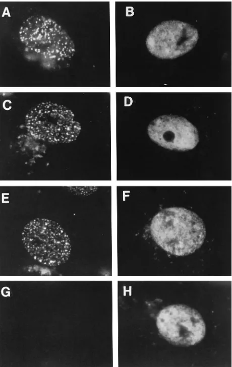
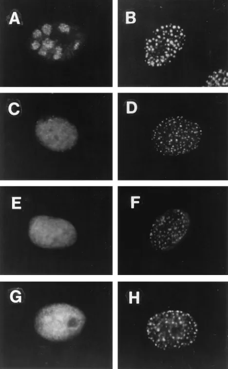
[image:2.612.321.551.288.652.2]Viral replication protein requirements for localization of ICP8 to prereplicative sites. To define the viral replication proteins required for localization of ICP8 to prereplicative sites, we examined the localization of ICP8 in cells infected with viral mutants each defective for one of the other six essential replication proteins. The 3-83 antiserum, which de-tects all forms of ICP8 when used at appropriate dilutions (10), allowed us to detect ICP8 throughout the cell by immunoflu-orescence and to determine which replication proteins are
FIG. 1. Distribution of ICP8 in cells infected with DNA replication-defective mutant viruses. Cells were infected at a multiplicity of infection of 20 with wt virus in the presence of PAA or with the indicated mutant virus without PAA. At 5.5 h postinfection, cells were fixed in formaldehyde and permeabilized in cold acetone. They were then incubated with 3-83 rabbit serum followed by FITC-conjugated goat anti-rabbit immunoglobulin antibody as described in Materials and Methods. (A) wt virus plus PAA. (B) hr99 (UL52). (C) CgalD42 (UL422). (D) hr80 (UL82). (E) HP66 (Pol2). (F) hr114 (UL522). (G) Mock infection. (H)
hr94 (UL92).
on November 9, 2019 by guest
http://jvi.asm.org/
necessary for localization of ICP8 to prereplicative sites. ICP8 localized to prereplicative sites in cells infected with wt virus and treated with PAA (Fig. 1A) and in cells infected with the Pol mutant (Fig. 1E). This is consistent with previous work showing that Pol is not required for ICP8 localization (3, 38). ICP8 also localized to prereplicative sites in cells infected with the UL42 mutant virus (Fig. 1C). In these three situations, the majority of cells expressing ICP8 showed localization to nu-clear punctate prereplicative sites (Table 1). Although the per-centage of UL42 mutant virus-infected cells showing a punc-tate distribution for ICP8 was slightly less than that for wt or Pol mutant virus-infected cells in this experiment, this differ-ence was not observed consistently (29). Therefore, UL42 was also not required for ICP8 localization to these structures. In contrast, in cells infected with the UL5 mutant virus (Fig. 1B), the UL8 mutant virus (Fig. 1D), the UL52 mutant virus (Fig. 1F), or the UL9 mutant virus (Fig. 1H), ICP8 showed a diffuse distribution throughout the nucleus. No cells with prereplica-tive sites were observed in these infections (Table 1). To rule out the possibility that the diffuse ICP8 obscured any prerep-licative sites present, we performed immunofluorescence with the 39S MAb, which recognizes preferentially ICP8 localized to DNA replication structures. Cells infected with UL5, UL8, or UL9 mutant viruses showed no staining with the 39S MAb (44), providing further evidence that no prereplicative sites were formed in these cells. Furthermore, sodium dodecyl sul-fate-polyacrylamide gel electrophoresis analysis showed that ICP8 was expressed at approximately equal levels in all of these infections (29, 44). Therefore, ICP8 was not overex-pressed in the latter infections and was not likely to be obscur-ing prereplicative sites. We conclude from these experiments that the three members of the helicase-primase complex and the origin-binding protein are all necessary for localization of ICP8 to prereplicative sites in infected cells.
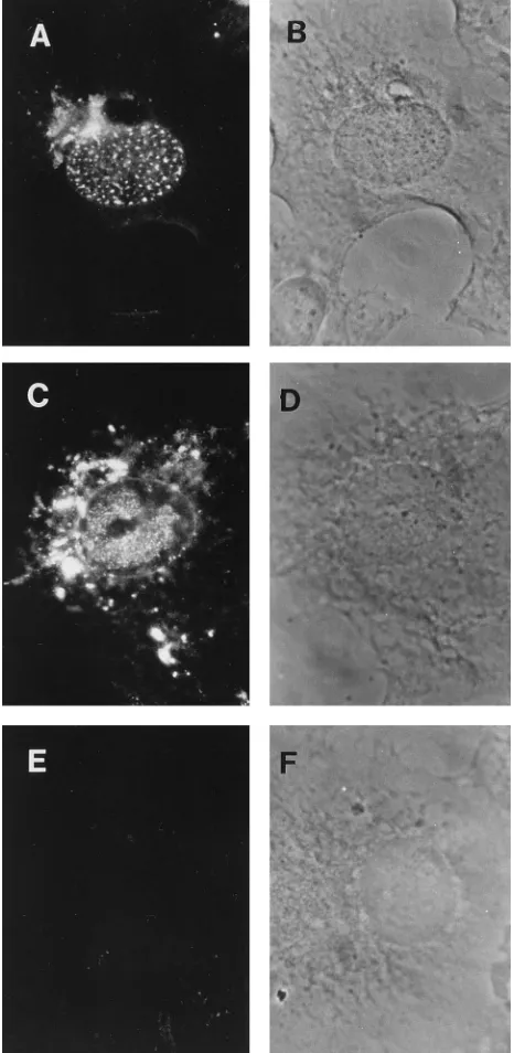
Viral replication protein requirements for localization of UL8 to prereplicative sites.We examined the localization of the UL8 protein as a marker for the intracellular distribution of the helicase-primase complex because UL8 has been shown to localize into the nucleus and promote the nuclear entry of the UL5 and UL52 proteins (4). Using a rabbit anti-UL8 se-rum for immunofluorescence, we detected UL8 in replication compartments in cells infected with wt virus (Fig. 2C) and in punctate nuclear structures in cells infected with wt virus in the presence of PAA (Fig. 2A). Cytoplasmic staining was also observed in virus-infected cells (Fig. 2A and C). Because this staining was greatly reduced in cells treated with PAA (Fig. 2A), it was likely that much of this cytoplasmic staining repre-sented binding of the rabbit immunoglobulin to the viral late Fc receptor in the Golgi apparatus (3). We have therefore
focused on the nuclear staining which is specific for UL8. In addition, the nuclear punctate structures containing UL8 (Fig. 3B) colocalized with sites containing ICP8 (Fig. 3A), demon-strating that UL8 localized to prereplicative sites. To deter-mine which of the other six replication proteins were necessary for the localization of UL8 into prereplicative sites, we
[image:3.612.319.552.73.550.2]exam-FIG. 2. Distribution of UL8 in infected cells. Cells were infected at a multi-plicity of infection of 20 with wt virus in the presence or absence of PAA. At 5.5 h postinfection, cells were fixed in formaldehyde and permeabilized in cold acetone. They were then incubated with the 80K anti-UL8 rabbit serum, followed by incubation with FITC-conjugated goat anti-rabbit immunoglobulin antibody. (A) wt plus PAA, immunofluorescence image. (B) wt plus PAA, phase-contrast image of the field shown in panel A. (C) wt virus, immunofluorescence image. (D) wt virus, phase-contrast image of the field shown in panel C. (E) Mock infection; immunofluorescence image. (F) Phase-contrast image of the field shown in panel E.
TABLE 1. ICP8 distribution in mutant virus-infected cells
Infection
ICP8 distributiona
Diffuse nuclear staining
Punctate sites
wt1PAA 21 79
hr99 (UL52) 100 0
hr8060 (UL82) 100 0
hr114 (UL522) 100 0
hr94 (UL92) 100 0
CgalD42 (UL422) 40 60
HP66 (Pol2) 28 72
a
ICP8 detected with 3-83 antiserum, expressed as a percentage of total cells staining for ICP8.
on November 9, 2019 by guest
http://jvi.asm.org/
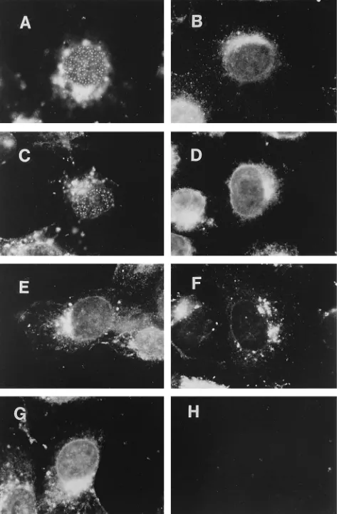
[image:3.612.58.298.85.189.2]ined the localization of UL8 in cells infected with viruses de-fective for each of these proteins. In cells infected with the UL9 mutant (Fig. 4B), the UL5 mutant (Fig. 4E), the UL52 mutant (Fig. 4G), or the ICP8 mutant (Fig. 4D), UL8 exhibited a diffuse nuclear distribution (Table 2). In contrast, in cells infected with the Pol mutant (Fig. 4A) or the UL42 mutant (Fig. 4C), UL8 localized to nuclear punctate structures. Thus, we conclude that the other helicase-primase components, UL5 and UL52, the origin-binding protein, UL9, and the ICP8 single-stranded DNA-binding protein are all necessary for the localization of UL8 to prereplicative sites, whereas Pol and the UL42 accessory protein are not necessary for this localization.
Viral replication protein requirements for localization of Pol to prereplicative sites.We had shown previously that ICP8 is needed for localization of HSV Pol to prereplicative sites (3), but there was no information available on the role of the other viral DNA replication proteins in Pol localization. We therefore used immunofluorescence to examine Pol localiza-tion in cells infected with wt virus or mutant viruses defective for the other replication proteins. The specificity of the Be5 MAb was shown by replication compartment staining in cells infected with wt virus (Fig. 5A) and the absence of staining in cells infected with the Pol2virus (Fig. 5H). In cells infected with wt virus and treated with PAA, approximately 45% of the cells staining for Pol showed localization of Pol to prereplica-tive sites (Fig. 5C and Table 3). However, in cells infected with the UL9 mutant (Fig. 5B), the UL5 mutant (Fig. 5E), the UL8 mutant (Fig. 5G), or the UL52 mutant virus (Fig. 5I), Pol showed a diffuse nuclear distribution in all cells (Table 3). In cells infected with the UL42 mutant virus, Pol was localized to punctate structures in about 13% of the cells that stained (Fig. 5D and Table 3), a level approximately threefold lower than that of the wt. We conclude that UL42 increases the efficiency of Pol localization to prereplicative sites while the other five viral replication proteins are necessary for localization of Pol to prereplicative sites.
Viral protein requirements for ICP8 localization to prerep-licative sites in transfected cells. ICP8 can localize to the nucleus of cells transfected with the ICP8 gene but generally shows a diffuse distribution (17). In cells transfected with the ICP8 gene and the genes encoding UL5, UL8, and UL52, ICP8 localized to punctate structures resembling prereplicative sites (reference 44 and Fig. 6A) in approximately 40% of the cells (Table 4). We therefore transfected various combinations of the helicase-primase genes into cells to determine the minimal requirements for localization of ICP8 to these structures. When expressed alone, ICP8 was distributed diffusely through-out the nucleus (Fig. 6B and Table 4). Transfection of any
[image:4.612.317.555.72.433.2]combination of two of the genes encoding components of the helicase-primase complex was not sufficient for ICP8 localiza-tion to punctate structures (Fig. 6D to F and Table 4). There-fore, all three of the helicase-primase components are neces-sary for ICP8 localization to these prereplicative site-like
FIG. 3. Dual-label immunofluorescence detection of ICP8 and UL8 in in-fected cells. Cells were inin-fected with wt virus in the presence of PAA and at 5.5 h postinfection were fixed in formaldehyde and permeabilized in cold acetone. They were then reacted with the 80K anti-UL8 rabbit serum and the 39S mouse ICP8 mouse MAb followed by incubation with FITC-conjugated goat anti-rabbit immunoglobulin antibody and RITC-conjugated goat anti-mouse immu-noglobulin antibody. (A) ICP8 immunofluorescence image. (B) UL8 immuno-fluorescence image.
FIG. 4. Distribution of UL8 in cells infected with DNA replication-defective mutant viruses. Cells were infected at a multiplicity of infection of 20 with wt virus in the presence of PAA or with the indicated mutant virus. At 5.5 h postinfection, cells were fixed and permeabilized as described in the legend to Fig. 1. They were then incubated with the 80K rabbit serum, followed by incu-bation with FITC-conjugated goat anti-rabbit immunoglobulin antibody. (A) HP66 (Pol2). (B) hr94 (UL92). (C) CgalD42 (UL422). (D) HD2 (ICP82). (E)
[image:4.612.62.295.72.154.2]hr99 (UL52). (F) hr80 (UL82). (G) hr114 (UL522). (H) Mock infection.
TABLE 2. UL8 distribution in mutant virus-infected cells
Infection
UL8 distributiona
Diffuse nuclear staining
Punctate sites
wt1PAA 52 48
hr99 (UL52) 100 0
hr114 (UL522) 100 0
hr94 (UL92) 100 0
CgalD42 (UL422) 53 47
HP66 (UL302) 37 63
HD2 (ICP82) 100 0
a
Expressed as a percentage of the total cells that stained with UL8 antiserum.
on November 9, 2019 by guest
http://jvi.asm.org/
[image:4.612.318.555.611.717.2]structures in transfected cells, but in contrast to the infected cell results, UL9 was not required. This was consistent with the report by Heilbronn and zur Hausen (22) that UL9 is not required for the amplification of cellular DNA by HSV DNA replication proteins in transfected cells. In transfected cells, the HSV DNA replication proteins may be able to interact with a cellular origin-binding protein or other cellular protein to assemble replication structures and replicate cellular DNA.
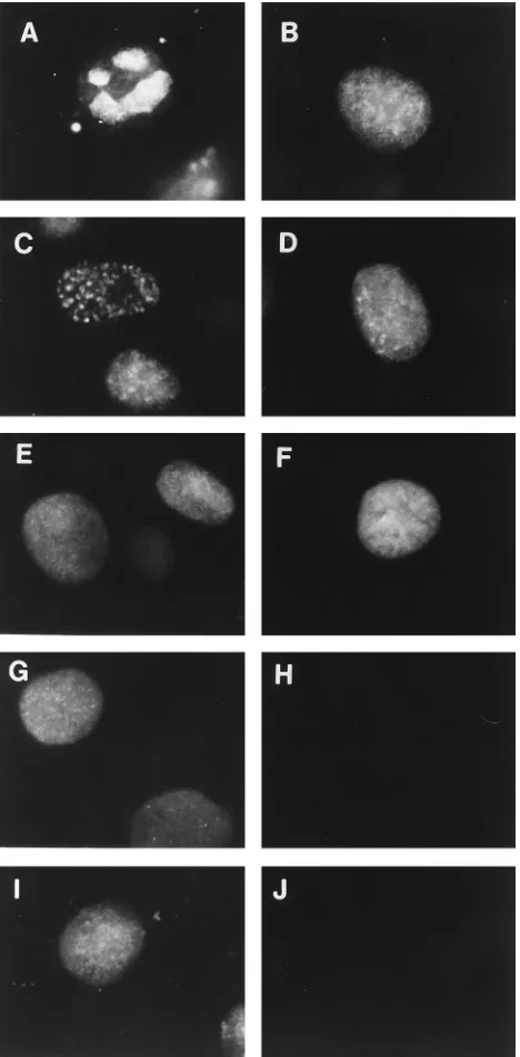
Effects of DNA synthesis inhibitors on the intranuclear lo-calization of ICP8 and Pol.During the course of our experi-ments, we noted that several viral mutants exhibited altered ICP8 staining patterns when PAA was added to the cultures. While cells infected with the UL5 mutant virus normally showed a diffuse ICP8 distribution (Fig. 7C), cultures infected in parallel but maintained in the presence of PAA contained some cells exhibiting a punctate distribution of ICP8 at 5.5 h postinfection (Fig. 7D). Similar results were observed with the UL8 mutant (Fig. 7E and F), the UL52 mutant (Fig. 7G and H), and the UL9 mutant virus (29). Therefore, PAA seemed to reduce the viral gene requirements for localization of ICP8 to punctate structures.
To determine whether this redistribution was specific for PAA or a more general effect of inhibition of DNA synthesis, we employed three other DNA synthesis inhibitory drugs: acy-clovir, hydroxyurea, and aphidicolin. The addition of any of these drugs to cells infected with UL5, UL8, UL52, or UL9 mutant viruses also resulted in the redistribution of ICP8 from a diffuse nuclear pattern to punctate structures (29). There-fore, the effect was not specific for PAA but instead appeared to be common to several DNA synthesis inhibitors.
[image:5.612.62.296.71.547.2]To determine if ICP8 expressed alone would redistribute in the presence of PAA, we transfected the ICP8 gene into cells in the absence or presence of PAA (Fig. 8A and C). We did not detect any change in the localization of ICP8 upon the addition of PAA under these conditions. Therefore, additional viral factors appeared to be necessary for localization of ICP8 to the
[image:5.612.316.557.90.197.2]FIG. 5. Distribution of HSV Pol in cells infected with DNA replication-defective mutant viruses. Vero cells were infected at a multiplicity of infection of 10 with wt or mutant viruses as indicated below. At 5.5 h postinfection, the cells were fixed in formaldehyde and permeabilized in Triton X-100 as described in Materials and Methods. They were then incubated with the Be5 anti-Pol mouse MAb followed by RITC-conjugated goat anti-mouse immunoglobulin antibody. (A) wt virus infection. (B) hr94 (UL92). (C) wt plus PAA. (D) CgalD42. (E) hr99 (UL52). (F) d301 (ICP82). (G) hr80 (UL82). (H) HP66 (Pol2). (I) hr114 (UL522). (J) Mock infection.
[image:5.612.319.552.546.652.2]FIG. 6. Localization of ICP8 in cells transfected with different viral genes. Cells were transfected with the viral genes indicated below or salmon sperm carrier DNA for a total of 16mg of DNA per culture well as described in Materials and Methods. The cells were then processed for immunofluorescence as described in the legend to Fig. 1. (A) Cells transfected with ICP8, UL5, UL8, and UL52 genes. (B) Cells transfected with ICP8 gene alone. (C) Cells, carrier DNA only. (D) Cells transfected with ICP8, UL5, and UL8 genes. (E) Cells transfected with ICP8, UL8, and UL52 genes. (F) Cells transfected with ICP8, UL5, and UL52 genes.
TABLE 3. HSV DNA polymerase distribution in mutant virus-infected cells
Infection
Pol distributiona
Diffuse nuclear staining
Punctate sites
wt1PAA 55 45
hr99 (UL52) 100 0
hr80 (UL82) 100 0
hr114 (UL522) 100 0
hr94 (UL92) 100 0
CgalD42 (UL422) 87 13
d301 (ICP82) 100 0
aExpressed as a percentage of the total cells that stain.
on November 9, 2019 by guest
http://jvi.asm.org/
prereplicative site-like structures induced in the presence of PAA.
The punctate structures formed by the mutant viruses in the presence of PAA showed some properties similar to but some properties different from those of prereplicative sites. There-fore, we wished to further characterize these structures. One feature of prereplicative sites is that residual host cell DNA
synthesis as detected by BrdU labelling of cellular DNA colo-calizes with ICP8 in a subset of prereplicative sites (12). There-fore, we labeled mutant-infected cells for 1 h with BrdU and performed double-label immunofluorescence analysis with an-ti-BrdU and ICP8 antibodies to determine if host cell DNA synthesis occurred at the punctate structures formed in mutant infected cells in the presence of PAA. In cells infected with the UL8 mutant virus, ICP8 localized to punctate structures in a subset of cells (Fig. 9A) which corresponded to the subset of cells labeled with BrdU (Fig. 9B). On the other hand, cells showing diffuse ICP8 (Fig. 9A) did not label with BrdU (Fig. 9B). A similar correlation between punctate ICP8 distribution and BrdU labeling was observed in UL9 mutant-infected cells as well (Fig. 9C and D, respectively). This indicated that cells that were not actively replicating DNA exhibited diffuse nu-clear ICP8, whereas those cells that were replicating DNA contained ICP8 in punctate structures. Therefore, active cel-lular DNA synthesis or some component of the host cell DNA replication apparatus appeared to be necessary for formation of prereplicative sites in the mutant-infected cells. Hence, we conclude that the punctate structures formed in the mutant-infected cells with PAA differ from prereplicative sites in that formation of the punctate structures in the mutant-infected cells has an apparent requirement for cell DNA synthesis or S phase factors. Treatment of cells with DNA synthesis inhibi-tory drugs may make these factors more available for interac-tion with the HSV proteins.
DISCUSSION
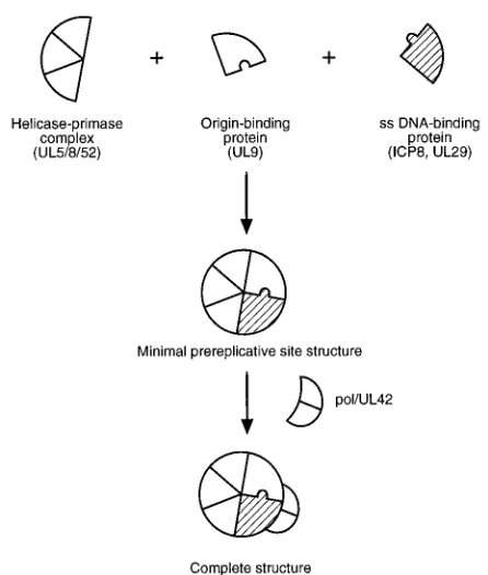
[image:6.612.316.554.69.255.2]The availability of viral mutants defective for each of the seven HSV-encoded gene products essential for viral DNA replication and antibodies or antisera specific for some of these proteins allowed us to define a pathway for assembly of the HSV DNA replication proteins into prereplicative sites in in-fected cells (Fig. 10). Prereplicative sites were originally de-fined as structures in which ICP8 and other viral DNA repli-cation proteins localize in cells when viral DNA replirepli-cation is
FIG. 7. Effect of PAA on localization of ICP8. Vero cells were infected with the viruses listed below in the absence or presence of PAA. At 5.5 h postinfec-tion, the cells were processed for immunofluorescence as described in the legend to Fig. 1. (A) wt virus. (B) wt virus plus PAA. (C) hr99. (D) hr99 plus PAA. (E)
hr80. (F) hr80 plus PAA. (G) hr114. (H) hr114 plus PAA.
[image:6.612.58.298.84.172.2]FIG. 8. Effect of PAA on ICP8 localization in the absence of other viral factors. Cells were transfected with an ICP8 expression vector in the absence or presence of PAA. Cells were harvested and processed for immunofluorescence at 48 h posttransfection and were processed for immunofluorescence as de-scribed in the legend to Fig. 1. (A) Immunofluorescence image for ICP8, no PAA in culture. (B) Phase-contrast image of field shown in panel A. (C) Immunoflu-orescence image for ICP8, PAA added to culture. (D) Phase-contrast image of field shown in panel C.
TABLE 4. ICP8 distribution in transfected cells
Gene(s) transfected
ICP8 distributiona
Diffuse nuclear staining
Punctate sites
ICP8 alone 100 0
ICP81UL51UL8 100 0
ICP81UL51UL52 100 0
ICP81UL81UL52 100 0
ICP81UL51UL81UL52 57 43
a
Expressed as a percentage of the total cells that stained with antibody 3-83.
on November 9, 2019 by guest
http://jvi.asm.org/
[image:6.612.61.295.304.683.2]inhibited by PAA, hydroxyurea, or a Pol defect (38). We pro-posed that these structures might be sites of accumulation of host and viral molecules involved in DNA synthesis. Subse-quently, we observed punctate structures similar to prereplica-tive sites at early times of normal infection which colocalize with BrdU labeling sites and appear to grow into replication compartments (12). Furthermore, release of a block in viral DNA synthesis allows a redistribution of ICP8 from prerepli-cative sites to replication compartments (38). Thus, there are several lines of evidence that prereplicative sites can serve as precursors to replication compartments, making the pathway for formation of prereplicative sites an important step in viral growth.
Under conditions that most closely mimic natural infection, virus-infected cells in the absence of drugs, five viral proteins
are necessary for formation of prereplicative sites: UL5, UL8, UL52, UL9, and ICP8. In contrast, two of the essential DNA replication proteins, Pol and the UL42 accessory protein, are not required for assembly of prereplicative sites. As a result of these observations, we have formulated the model in Fig. 10 showing the five proteins initially coassembling to form pre-replicative site structures in infected cells. Although not shown, cellular proteins likely play an essential role in defining the intranuclear location (13) or structure of these sites. After assembly of the five proteins, Pol and UL42 are illustrated as binding to the structure. Although ample evidence exists for Pol localization to prereplicative sites, there is no evidence for specific staining of prereplicative sites with UL42 antibodies (11, 20, 29). This model shows UL42 as assembling onto pre-replicative sites because we have observed that UL42 increases
FIG. 9. Codistribution of BrdU label and ICP8 in prereplicative sites in cells infected with mutant viruses in the presence of PAA. Cells were infected at a multiplicity of infection of 10 with hr80 or hr99 and labeled with BrdU for 1 h prior to harvesting at 5.5 h postinfection. The cells were then fixed in formaldehyde and permeabilized in cold acetone and then treated with HCl for 10 min and rinsed in water and PBS. The cells were then incubated with a MAb specific for BrdU and 3-83 serum followed by incubation with RITC-conjugated goat anti-mouse immunoglobulin antibody and FITC-conjugated goat anti-rabbit immunoglobulin antibody. (A) ICP8 immunofluorescence image of hr80 mutant-infected cells. (B) BrdU immunofluorescence image of the same field as panel A. (C) Immunofluorescence image of hr99 mutant-infected cells. (D) BrdU immunofluorescence image of the same field as panel C. ICP8 immunofluorescence image of mock-infected cells. (D) BrdU immunofluorescence image of the same field as panel E.
on November 9, 2019 by guest
http://jvi.asm.org/
the localization of Pol to prereplicative sites and because UL42 shows a tight interaction with Pol. The paradoxical lack of UL42 staining in prereplicative sites may be explained by the masking of epitopes on UL42 when it is assembled at prerep-licative sites, as observed for the b subunit or processivity factor for the Escherichia coli Pol III enzyme when it is part of the Pol-template complex (24). Alternatively, the large excess of UL42 not bound to Pol may obscure the small amount localized to prereplicative sites. Further studies are needed to address whether UL42 is localized to prereplicative sites and the nature of the interactions between all of these proteins that promote formation of this nuclear structure in infected cells.
Punctate structures and their relationship to prereplicative sites.Structures similar to prereplicative sites but with slightly different properties form under certain conditions. First, when cells are transfected with the ICP8, UL5, UL8, and UL52 genes, ICP8 localizes to punctate structures resembling pre-replicative sites. While the three components of the helicase-primase complex are required, UL9 is not required. Therefore, the viral protein requirements for formation of these structures are less than those for formation of prereplicative sites in infected cells. Because UL9 is also not needed for HSV DNA replication proteins to amplify cellular DNA sequences in transfected cells (22), it seems likely that in transfected cells, the HSV DNA replication proteins can assemble onto a cel-lular origin-binding protein or other protein to form a func-tional DNA replication complex at specific sites in the cell nucleus. On the basis of these results, we predict that prerep-licative site formation in infected cells involves the binding of UL9 to a viral DNA genome and the concomitant assembly of the other viral proteins. We are currently performing in situ hybridization to determine if the parental HSV genomes are located at prereplicative sites.
Second, the addition of DNA synthesis inhibitory drugs to
cells infected with certain mutant viruses caused a redistribu-tion of ICP8 from a diffuse pattern to punctate structures in some cells. These punctate structures were formed only in cells in S phase. We interpret these results to mean that the drugs could alter the host cell DNA replication machinery or some host cell component present only during S phase to allow interaction of the viral proteins with host cell proteins. By facilitating this virus-host interaction, these drugs appear to bypass a requirement for certain viral proteins in the assembly of DNA replication structures involving ICP8.
In combination, the studies of HSV DNA replication protein localization and induction of cellular DNA amplification argue that HSV replication proteins have an intrinsic ability to inter-act with components of the host cell nucleus and/or DNA replication apparatus. This could be a consequence of virus-cell interactions at several levels. First, this may reflect the normal interactions needed to anchor the HSV DNA replica-tion proteins onto the nuclear framework or nuclear matrix. In this regard, it should be noted that the location of replication compartments is defined by the host cell nuclear architecture, because the sister nuclei of cells that are binucleate prior to infection have similar numbers, shapes, and locations of rep-lication compartments (13). Second, this may reflect the nor-mal interactions between the HSV DNA replication complex and individual cell factors that participate in viral DNA syn-thesis. Finally, this may reflect the interaction between viral and cellular proteins needed to recruit the complete cellular DNA replication apparatus into prereplicative sites (12), which may be used in part or in its entirety in some way for viral DNA synthesis.
Role of ICP8 in prereplicative site formation. There has been some apparent disagreement concerning the role of ICP8 in assembly of viral DNA replication structures in infected cells. At this point, we have accumulated sufficient information to attempt to reconcile some of the differences between these conclusions. We previously concluded (13) that ICP8 ‘‘plays a role in organizing DNA replication proteins within the cell nucleus. . .,’’ but others (20) reworded our conclusion as stating that ICP8 is ‘‘the major organizational protein responsible for attracting other replication proteins to prereplication sites. . .,’’ clearly a different conclusion. We originally proposed (12) that ‘‘ICP8. . .may act in concert with other viral proteins to recruit cellular DNA replication protein complexes to the new loca-tions.’’ Although Goodrich et al. (20) stated that their results disagreed with our conclusions, they in fact stated that ‘‘ICP8 may act in concert with other proteins to direct the replication proteins to sites in preparation for DNA synthesis.’’ Thus, the conclusions from the two studies are actually strikingly similar. It is clear that ICP8 does play a role in assembly of prerepli-cative sites, and furthermore, evidence now shows that ICP8 does act in concert with other viral proteins, namely UL5, UL8, UL52, and UL9, to form prereplicative sites.
The definition of a pathway of assembly for this nuclear structure provides one perspective on the nature of the pro-tein-protein interactions occurring between HSV DNA repli-cation proteins. It will be of interest to examine prereplicative site formation in cells infected with viruses encoding DNA replication proteins with known defects in specific interactions with other proteins from in vitro studies to determine how those interactions contribute to assembly of these structures in infected cells.
ACKNOWLEDGMENTS
[image:8.612.65.288.66.329.2]We thank Sandra Weller, Don Coen, Paul Johnson, Min Gao, and Richard Colonno for providing viral mutant strains, cell lines, and antisera.
FIG. 10. Model of the order of assembly of HSV DNA replication proteins during formation of prereplicative site structures in infected cells. Cellular pro-teins, likely to be a part of the prereplicative sites, are not depicted. ss, single stranded.
on November 9, 2019 by guest
http://jvi.asm.org/
This work was supported by grant CA26345 from the National In-stitutes of Health. Lauren Liptak and Susan Uprichard were supported in part by Public Health Service training grant AI07245.
REFERENCES
1. Boehmer, P. E., M. C. Craigie, N. D. Stow, and I. R. Lehman. 1994. Asso-ciation of origin binding protein and single strand DNA-binding protein, ICP8, during herpes simplex virus type 1 DNA replication in vivo. J. Biol. Chem. 269:29329–29334.
2. Boehmer, P. E., and I. R. Lehman. 1993. Physical interaction between the herpes simplex virus 1 origin-binding protein and single-stranded DNA-binding protein ICP8. Proc. Natl. Acad. Sci. USA 90:8444–8448. 3. Bush, M., D. R. Yager, M. Gao, K. Weisshart, A. I. Marcy, D. M. Coen, and
D. M. Knipe.1991. Correct intranuclear localization of herpes simplex virus DNA polymerase requires the viral ICP8 DNA-binding protein. J. Virol. 65:1082–1089.
4. Calder, J. M., E. C. Stow, and N. D. Stow. 1992. On the cellular local-ization of the components of the herpes simplex virus type 1 helicase-primase complex and the viral origin-binding protein. J. Gen. Virol. 73:531–538.
5. Carmichael, E. P., and S. K. Weller. 1989. Herpes simplex virus type 1 DNA synthesis requires the product of the UL8 gene: isolation and characteriza-tion of an ICP6::lacZ insercharacteriza-tion mutacharacteriza-tion. J. Virol. 63:591–599.
6. Challberg, M. D. 1986. A method for identifying the viral genes required for herpesvirus DNA replication. Proc. Natl. Acad. Sci. USA 83:9094–9098. 7. Chiou, H. C., S. K. Weller, and D. M. Coen. 1985. Mutations in the herpes
simplex virus major DNA-binding protein gene leading to altered sensitivity to DNA polymerase inhibitors. Virology 145:213–226.
8. Crute, J. J., and I. R. Lehman. 1989. Herpes simplex-1 DNA polymerase. Identification of an intrinsic 59339exonuclease with ribonuclease H activity. J. Biol. Chem. 264:19266–19270.
9. Crute, J. J., T. Tsurumi, L. A. Zhu, S. K. Weller, P. D. Olivo, M. D. Challberg, E. S. Mocarski, and I. R. Lehman.1989. Herpes simplex virus 1 helicase-primase: a complex of three herpes-encoded gene products. Proc. Natl. Acad. Sci. USA 86:2186–2189.
10. Curtin, K. D., and D. M. Knipe. 1993. Altered properties of the herpes simplex virus ICP8 DNA-binding protein in cells infected with ICP27 mutant viruses. Virology 196:1–14.
11. de Bruyn Kops, A. 1991. The formation and intranuclear organization of herpes simplex virus DNA replication structures. Ph.D. thesis, Harvard Uni-versity, Cambridge, Mass.
12. de Bruyn Kops, A., and D. M. Knipe. 1988. Formation of DNA replication structures in herpes virus-infected cells requires a viral DNA binding pro-tein. Cell 55:857–868.
13. de Bruyn Kops, A., and D. M. Knipe. 1994. Preexisting nuclear architecture defines the intranuclear location of herpesvirus DNA replication structures. J. Virol. 68:3512–3526.
14. Digard, P., and D. M. Coen. 1990. A novel functional domain of an alpha-like DNA polymerase. The binding site on the herpes simplex virus poly-merase for the viral UL42 protein. J. Biol. Chem. 265:17393–17396. 15. Gao, M., S. F. DiTusa, and M. G. Cordingley. 1993. The C-terminal third of
UL42, a HSV-1 DNA replication protein, is dispensable for viral growth. Virology 194:647–653.
16. Gao, M., and D. M. Knipe. 1989. Genetic evidence for multiple nuclear functions of the herpes simplex virus ICP8 DNA-binding protein. J. Virol. 63:5258–5267.
17. Gao, M., and D. M. Knipe. 1992. Distal protein sequences can affect the function of a nuclear localization signal. Mol. Cell. Biol. 12:1330–1339. 18. Gao, M., and D. M. Knipe. 1993. Intragenic complementation of herpes
simplex virus ICP8 DNA-binding protein mutants. J. Virol. 67:876–885. 19. Goldstein, D. J., and S. K. Weller. 1988. An ICP6::lacZ insertional
mu-tagen is used to demonstrate that the UL52 gene of herpes simplex virus type 1 is required for virus growth and DNA synthesis. J. Virol. 62:2970– 2977.
20. Goodrich, L. D., P. A. Schaffer, D. I. Dorsky, C. S. Crumpacker, and D. S. Parris.1990. Localization of the herpes simplex virus type 1 65-kilodalton DNA-binding protein and DNA polymerase in the presence and absence of viral DNA synthesis. J. Virol. 64:5738–5749.
21. Gottlieb, J., and M. D. Challberg. 1994. Interaction of herpes simplex virus type 1 DNA polymerase and the UL42 accessory protein with a model primer template. J. Virol. 68:4937–4945.
22. Heilbronn, R., and H. zur Hausen. 1989. A subset of herpes simplex virus replication genes induces DNA amplification within the host cell genome. J. Virol. 63:3683–3692.
23. Hernandez, T. R., and I. R. Lehman. 1990. Functional interaction between
the herpes simplex-1 DNA polymerase and UL42 protein. J. Biol. Chem. 265:11227–11232.
24. Johnson, K. O., and C. S. McHenry. 1982. The beta subunit of the DNA polymerase III holoenzyme becomes inaccessible to antibody after formation of an initiation complex with primed DNA. J. Biol. Chem. 257:12310–12315. 25. Johnson, P. A., M. G. Best, T. Friedmann, and D. S. Parris. 1991. Isolation of a herpes simplex virus type 1 mutant deleted for the essential UL42 gene and characterization of its null phenotype. J. Virol. 65:700–710.
26. Knipe, D. M. 1989. The role of viral and cellular nuclear proteins in herpes simplex virus replication. Adv. Virus Res. 37:85–123.
27. Knipe, D. M., D. Senechek, S. A. Rice, and J. L. Smith. 1987. Stages in the nuclear association of the herpes simplex virus transcriptional activator pro-tein ICP4. J. Virol. 61:276–284.
28. Knipe, D. M., and J. L. Smith. 1986. A mutant herpesvirus protein leads to a block in nuclear localization of other viral proteins. Mol. Cell. Biol. 6:2371– 2381.
29. Liptak, L., and D. M. Knipe. Unpublished results.
30. Lukonis, C. J., and S. K. Weller. 1996. Characterization of nuclear structures in cells infected with herpes simplex virus type 1 in the absence of viral DNA replication. J. Virol. 70:1751–1758.
31. Malik, A. K., R. Martinez, L. Muncy, E. P. Carmichael, and S. K. Weller. 1992. Genetic analysis of the herpes simplex virus type 1 UL9 gene: isolation of a LacZ insertion mutant and expression in eukaryotic cells. Virology 190:702–715.
32. Marcy, A. I., D. R. Yager, and D. M. Coen. 1990. Isolation and character-ization of herpes simplex virus mutants containing engineered mutations at the DNA polymerase locus. J. Virol. 64:2208–2216.
33. Marsden, H. S., M. Murphy, G. L. Mcvey, K. A. Maceachran, A. M. Ow-sianka, and N. D. Stow.1994. Role of the carboxy terminus of herpes simplex virus type 1 DNA polymerase in its interaction with UL42. J. Gen. Virol. 75:3127–3135.
34. Mclean, G. W., A. P. Abbotts, M. E. Parry, H. S. Marsden, and N. D. Stow. 1994. The herpes simplex virus type 1 origin-binding protein interacts spe-cifically with the viral UL8 protein. J. Gen. Virol. 75:2699–2706. 35. Monahan, S. J., T. F. Barlam, C. S. Crumpacker, and D. S. Parris. 1993. Two
regions of the herpes simplex virus type 1 UL42 protein are required for its functional interaction with the viral DNA polymerase. J. Virol. 67:5922– 5931.
36. O’Donnell, M. E., P. Elias, B. E. Funnell, and I. R. Lehman. 1987. Interac-tion between the DNA polymerase and single-stranded DNA-binding pro-tein (infected cell propro-tein 8) of herpes simplex virus. J. Biol. Chem. 262: 4260–4266.
37. Olivo, P. D., N. J. Nelson, and M. D. Challberg. 1989. Herpes simplex virus type 1 gene products required for DNA replication: identification and over-expression. J. Virol. 63:196–204.
38. Quinlan, M., L.-B. Chen, and D. Knipe. 1984. The intranuclear location of a herpes simplex virus DNA-binding protein is determined by the status of viral DNA replication. Cell 36:857–868.
39. Randall, R. E., and N. Dinwoodie. 1986. Intranuclear localization of herpes simplex virus immediate-early and delayed-early proteins: evidence that ICP4 is associated with progeny virus DNA. J. Gen. Virol. 67:2163–2177. 40. Rice, S., N. DeLuca, and D. M. Knipe. Unpublished results.
41. Rixon, F. J., M. A. Atkinson, and J. Hay. 1983. Intranuclear distribution of herpes simplex virus type 2 DNA synthesis: examination by light and electron microscopy. J. Gen. Virol. 64:2087–2092.
42. Ruyechan, W. T., and A. C. Weir. 1984. Interaction with nucleic acids and stimulation of the viral DNA polymerase by the herpes simplex virus type 1 major DNA-binding protein. J. Virol. 52:727–733.
43. Showalter, S. D., M. Zweig, and B. Hampar. 1981. Monoclonal antibodies to herpes simplex virus type 1 proteins, including the immediate-early protein ICP 4. Infect. Immun. 34:684–692.
44. Uprichard, S., and D. M. Knipe. Unpublished data.
45. Weisshart, K., K. Morrison, and D. M. Coen. Unpublished results. 46. Weller, S. K. 1991. Genetic analysis of HSV-1 genes required for genome
replication, p. 105–136. In E. K. Wagner (ed.), Herpesvirus transcription and its regulation. CRC Press, Boca Raton, Fla.
47. Wilcock, D., and D. P. Lane. 1991. Localization of p53, retinoblastoma and host replication proteins at sites of viral replication in herpes-infected cells. Nature (London) 349:429–431.
48. Wu, C. A., N. J. Nelson, D. J. McGeoch, and M. D. Challberg. 1988. Iden-tification of herpes simplex virus type 1 genes required for origin-dependent DNA synthesis. J. Virol. 62:435–443.
49. Zhu, L., and S. K. Weller. 1992. The UL5 gene of herpes simplex virus type 1: isolation of a lacZ insertion mutant and association of the UL5 gene product with other members of the helicase-primase complex. J. Virol. 66: 458–468.