STANDARDISATION METHODS FOR ITS FORMULATION
DISSERTATION
Submitted toThe Tamil Nadu Dr. M.G.R. Medical University, Chennai – 32
In partial fulfillment for the award of the Degree of
DOCTOR OF PHILOSOPHY
IN
PHARMACY
Submitted by
S. JAYARAMAN
[EX.II (1) 11428/08]
Under the Guidance of
Dr. K.L. SENTHILKUMAR, M.Pharm., Ph.D.,
April-2012
Padmavathi College of Pharmacy and Research Institute
CERTIFICATE
This is to certify that the work embodied in this thesis entitled
“
PHYTOMEDICINAL AND PHARMACOLOGICAL STUDIES OF
SELECTIVE PLANTS OF KOLLI HILLS AND DEVELOPMENT OF
STANDARDISATION METHODS FOR ITS FORMULATION
”
submitted
to The Tamilnadu Dr. M.G.R Medical University, Chennai - 32 was carried out
by
Mr. S. Jayaraman, [EX.II (1) 11428/08]
at
Padmavathi College of
Pharmacy and Research Institute, Dharmapuri-635205
for the partial
fulfillment of the award of degree of
DOCTOR OF PHILOSOPHY
in
pharmacy.
This work is original and has not been submitted in part or full to any
other degree / diploma or academic award anywhere before.
Place: Dharmapuri
Dr. K.L. SENTHILKUMAR, M.Pharm., Ph.D.,
Date:
Principal
CERTIFICATE
This is to certify that the thesis entitled
“PHYTOMEDICINAL AND
PHARMACOLOGICAL STUDIES OF SELECTIVE PLANTS OF KOLLI
HILLS AND DEVELOPMENT OF STANDARDISATION METHODS
FOR ITS FORMULATION”
is the record of the original work done by
Mr. S. Jayaraman
under my guidance and supervision. The results of the
research presented in this thesis have not previously formed the basis for the
award of any degree, diploma or certificate of this or any other university.
Place: Dharmapuri
Dr. K.L. SENTHILKUMAR, M.Pharm., Ph.D.,
Date:
Professor & Research Guide,
Padmavathi College of Pharmacy and
DECLARATION
I hereby declare that the thesis entitled
“PHYTOMEDICINAL AND
PHARMACOLOGICAL
STUDIES
OF
SELECTIVE
PLANTS
OF KOLLI HILLS AND DEVELOPMENT OF STANDARDIZATION
METHODS FOR ITS FORMULATION”
was carried out by me at
Padmavathi
College
of
Pharmacy
and
Research
Institute,
Dharmapuri
under
the
supervision
and
guidance
of
Prof.
Dr. K.L. SENTHILKUMAR, M.Pharm., Ph.D.,
Padmavathi College
of Pharmacy and Research Institute, Dharmapuri-635205.
This thesis submitted to
The Tamil Nadu Dr. M.G.R. Medical
University, Chennai-32,
as a partial fulfillment for the award of
DOCTOR OF PHILOSOPHY
in Pharmacy.
This work is original and has not been submitted in part or full to any
other degree or diploma of this or any other university.
Place: Dharmapuri
Mr. S. Jayaraman
I feel highly privileged to express my gratitude to my guide,
Prof. Dr. K. L. Senthilkumar, M.Pharm., Ph.D., Principal, Padmavathi College of Pharmacy and Research Institute, Dharmapuri for his valuable guidance, interest, enthusiasm and encouragement throughout the course of this study.
It is a delightful moment for me, to put into words all my deep
sense of gratitude to Prof. K. Senthilkumar, M. Pharm., Padmavathi
College of Pharmacy, for his encouragement and moral support to complete my dissertation work.
I would like to thank sincerely my co-guide, Prof. Dr. Ezhilmuthu,
M.Pharm., Ph.D., Deparment of Pharmaceutics, Padmavathi College of Pharmacy, Dharmapuri, who has been supportive and encouraged me throughout the study.
I deeply grateful to Prof. Dr. G. Arunachalam, M.Pharm., Ph.D.,
FIC., Principal, PGP College of Pharmaceutical Science and Research Institute, Namakkal, for providing me laboratory facilities to carry out my research work and for his constant encouragement given throughout the work.
I would like to thank my dear friend Mr. R. Sivakumar, M.Pharm.,
Asst. Professor., Department of Pharmacognosy, PGP College of
Pharmaceutical Science and Research Institute, Namakkal,
for his constant and valuable help throughout my research study .
My faithful thanks to Prof. Dr. M. Rajkumar, M.Pharm., Ph.D.,
Head, Department of Pharmacognosy, Padmavathi College of Pharmacy,
completing the pharmacological part of the work.
My sincere thanks to Mrs. A. Yasodha, M.Pharm., Professor,
for her valuable suggestions and useful discussions in the structural elucidation of isolated compounds.
I gratefully acknowledge Mr. A. Chandran, M.Pharm., Professor,
Department of Pharmaceutical Chemistry, PGP College of
Pharmaceutical Science and Research Institute, Namakkal for his timely help for the successful completion of this work.
I am grateful to Mr. D. Sakthive, M.Pharm., Assistant Professor,
Department of Pharmaceutics, PGP College of Pharmaceutical Science and Research Institute, Namakkal, for his timely help in the formulation development studies.
I extend my thanks to Mrs. C. Kalaiselvi, M.Pharm., Assistant
Professor, Department of Pharmaceutics, PGP Coll ege of
Pharmaceutical Science and Research Institute, Namakkal for her support throughout my study.
I extended my sincere thanks to Mr. S. Sekar, M.A., MLIS.,
Librarian, PGP College of Pharmaceutical Science and Research Institute, Namakkal, for providing the library facilities and co-operation to complete this work.
A special thanks to Prof. Dr. P.D. Gokulan, M.Pharm.,
Ph.D., Mr. M. Saravanan, M.Pharm., Mr. V. Palanivel, M.Pharm, Mrs. T. Karthiyayini, M.Pharm., Mr. A. Vasanthan, M.Pharm., and
Mr. G. Guru, M.Pharm., faculty members, Padmavathi College of Pharmacy, Dharmapuri for their help and suggestions during the
course of this investigation.
I express my sincere thanks to the Mr. K. Murali, Office
support during this study.
Also I express my sincere thanks to Lab Assistan ts
Mr. J. Ramesh, M.A., B.Ed., and Mrs. K. Nirmala, D.M.E., PGP College of Pharmaceutical Science and Research Institute, Namakkal
for their timely help.
I wish to record my thanks to my father-in-law Mr. A. Ayyavu,
brother-in-law Mr. A. Mohankumar B.A.B.L., and their family for their
encouragement throughout this venture.
I am really thankful to my Brother Mr. S. Jayakumar and
his family for their constant support and encouragement
throughout my study period.
I am indeed grateful to my life companion my Wife
Mrs. J. Tamil Selvi, Son S.J. Harish Kumar and Daughter S.J. Hiruthiksha who gave me their encouragement and lovable support in completing my research work.
Finally, I would like to thank my father Mr. C. Sengottaiyan
and mother Mrs. S. Mathu for their constant encouragement, love
and attention which have made me to accomplish this work.
I INTRODUCTION 1
II REVIEW OF LITERATURE 5
2.1 Review on Justicia gendarussa Burm 6
2.2 Review on Rungia pectinata Linn 8
2.3 Review on Strobilanthes ciliatus Nees 10
2.4 Review on Rhinacanthus nasutus Linn 11
2.5 Herbs with analgesic, antipyretic and anti-inflammatory property 13
2.6 Herbs with antidiabetic property 15
2.7 Herbs with hepatoprotective property 16
2.8 Herbs with antitussive property 17
2.9 Herbs with anticancer property 18
III AIM AND OBJECTIVE OF THE STUDY 20
IV PLAN OF WORK 25
V MATERIALS AND METHODS 27
5.1 Chemicals and Glassware's 27
5.2 Cell line used for in vitro study 30
5.3 List of instruments used for the study 30
5.4 Plant profile of selected plants of Kolli hills 32
5.4.1 Plant profile of Justicia gendarussa Burm 32 5.4.2 Pant profile of Rungia pectinata Linn 35 5.4.3 Plant profile of Strobilanthes ciliatus Nees 38 5.4.4 Plant profile of Rhinacanthus nasutus Linn 41 5.5 Comparative study of selected plants of Kolli hills for further
studies
44
5.5.1 Preparation of extracts for phytochemical screening of selected plants
44
5.5.2 Phytochemical screening of selected plants 45
5.6 Studies on selective plant: Strobilanthes ciliatus Nees 48
5.6.1 Pharmacognostical studies on Strobilanthes ciliatus Nees 48 5.6.2 Determination of physicochemical constants 49
5.6.2.4 Determination of mineral content 52
5.7 Phytochemical studies 53
5.7.1 Preparation of extracts 53
5.7.2 Phytochemical screening of extracts 54
5.7.3 Total content estimation 54
5.7.4 Thin layer chromatographic studies 57
5.7.5 Selection of extract for phytochemical, biological and formulation studies
57
5.7.6 Isolation and characterization of phytoconstituents 58 5.7.7 Spectroscopic analysis for isolated compounds 59
5.8 Pharmacological studies 59
5.8.1 Acute oral toxicity studies 59
5.8.2 Analgesic, antipyretic and anti-inflammatory studies 61 5.8.2.1 Analgesic activity by tail clip method 61 5.8.2.2 Analgesic activity by tail immersion method 62 5.8.2.3 Anti-inflammatory studies by paw edema method 63 5.8.2.4 Antipyretic activity by Brewer’s yeast induced pyrexia 64
5.8.3 Antidiabetic activity 65
5.8.4 Paracetamol induced hepatoprotective studies 67
5.8.5 Antitussive studies 69
5.8.6 In vitro anticancer studies 71
5.9 Formulation development 73
5.9.1 Powder characteristics hydroalcoholic extract 73
5.9.2 Drug polymer compatibility studies 74
5.9.3 Preparation of herbal tablets from hydroalcoholic extract 74 5.9.4 Evaluation of herbal tablet by quality control studies 75 5.9.5 Quantification of marker compound by HPTLC technique 76
VI RESULTS AND ANALYSIS 78
6.1 Comparitive study of selected plants of Kolli hills 78
studies
6.3 Studies on selective plant: Strobilanthes ciliatus Nees 86
6.3.1 Pharmacognsotical studies 86
6.3.2 Determination of physicochemical constants 107
6.3.3 Phytochemical analysis 108
6.3.3.1 Preparation of extracts 108
6.3.3.2 Qualitative phytochemical analysis extracts of Strobilanthes ciliatus Nees
109
6.3.3.4 Total content estimation of selective phytoconstituents 110
6.3.4 TLC studies of extracts 111
6.3.5 Chromatographic fractionation of hydroalcoholic extract of Strobilanthesciliatus Nees
112
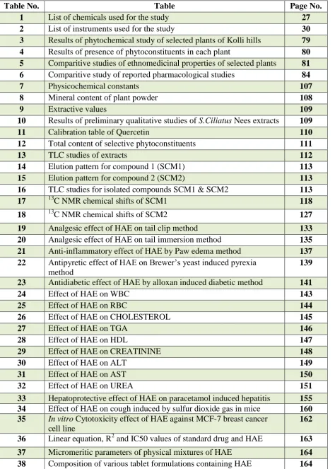
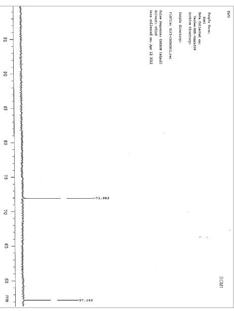
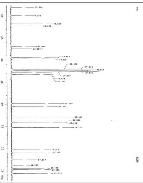
6.3.6 Spectral characterization of isolated compound-I (SCM1) 115 6.3.7 Spectral characterization of isolated compound-II (SCM2) 124
6.4 Pharmacological studies 133
6.4.1 Analgesic activity by tail clip method 133
6.4.2 Analgesic activity by tail immersion method 134 6.4.3 Anti-inflammatory studies by paw edema method 137 6.4.4 Antipyritic activity by Brewer’s yeast induced pyrexia 139 6.4.5 Antidiabetic studies by alloxan induced diabetic method 141 6.4.6 Paracetamol induced hepatoprotective activity 155
6.4.7 Antitussive activity 160
6.4.8 In vitro anticancer studies 162
6.5 Development of formulation 164
6.5.1 Standardization of formulation by HPTLC quantitative analysis using SCM1 marker
167
VI DISCUSSION 175
VII SUMMARY AND CONCLUSION 193
VIII BIBLIOGRAPHY
1 List of chemicals used for the study 27
2 List of instruments used for the study 30
3 Results of phytochemical study of selected plants of Kolli hills 79
4 Results of presence of phytoconstituents in each plant 80
5 Comparitive studies of ethnomedicinal properties of selected plants 81
6 Comparitive study of reported pharmacological studies 84
7 Physicochemical constants 107
8 Mineral content of plant powder 108
9 Extractive values 109
10 Results of preliminary qualitative studies of S.Ciliatus Nees extracts 109
11 Calibration table of Quercetin 110
12 Total content of selective phytoconstituents 111
13 TLC studies of extracts 112
14 Elution pattern for compound 1 (SCM1) 113
15 Elution pattern for compound 2 (SCM2) 113
16 TLC studies for isolated compounds SCM1 & SCM2 113
17 13C NMR chemical shifts of SCM1 118
18 13C NMR chemical shifts of SCM2 127
19 Analgesic effect of HAE on tail clip method 133
20 Analgesic effect of HAE on tail immersion method 135
21 Anti-inflammatory effect of HAE by Paw edema method 137
22 Antipyretic effect of HAE on Brewer’s yeast induced pyrexia method
139
23 Antidiabetic effect of HAE by alloxan induced diabetic method 141
24 Effect of HAE on WBC 143
25 Effect of HAE on RBC 144
26 Effect of HAE on CHOLESTEROL 145
27 Effect of HAE on TGA 146
28 Effect of HAE on HDL 147
29 Effect of HAE on CREATININE 148
30 Effect of HAE on ALT 149
31 Effect of HAE on AST 150
32 Effect of HAE on UREA 151
33 Hepatoprotective effect of HAE on paracetamol induced hepatitis 155
34 Effect of HAE on cough induced by sulfur dioxide gas in mice 160
35 Invitro Cytotoxicity effect of HAE against MCF-7 breast cancer cell line
162
36 Linear equation, R2 and IC50 values of standard drug and HAE 163
37 Micromeritic parameters of physical mixtures of HAE 164
[image:11.595.90.565.99.763.2]42 Peak Rf value, height and area of standard and sample solution 169
43 Concentration of standard and sample applied 172
44 Calibration table 173
45 Recovery study of Stigmasterol 190
46 Intra-day and inter-day precision of the method 190
1 Justicia gendarussa Burm 32
2 Rungia pectinata Nees 35
3 Strobilanthes ciliatus Nees 38
4 Rhinacanthusnasutus Linn 41
5 Apparatus for sulfur dioxide gas production 70
6 Earthen view of Strobilanthesciliatus Nees 86
7 Abaxial view & branching of the leaf system of Strobilanthesciliatus
Nees
87
8 Shoot, leaf and fruiting branches of Strobilanthesciliatus Nees 87
9 Root system Strobilanthesciliatus Nees 88
10 TS of leaf through midrib 93
11 TS of midrib – enlarged 93
12 TS of midrib under polarised light 94
13 TS of midrib-adaxial part-enlarged 94
14 TS of midrib-abaxial part- enlarged (a) 95
15 TS of midrib-abaxial part- enlarged (b) 95
16 TS of lamina 96
17 Lamina of upper part. 96
18 Lamina of lower portion 97
19 Paradermal section to show the venation pattern 97
20 Vein-islets and vein-termination enlarged 98
21 Paradermal section of the abaxial epidermis of the lamina seen under the dark-field microscope
98
22 Stomata-enlarged 99
23 Fragment of the lamina seen in the powder, showing venation. 99
24 Peltate scale 100
25 Peltate scale enlarged 100
26 Peltete scale and stomata in surface view 101
27 Isolated peltate scale 101
28 TS of stem 102
29 TS of stem enlarged 102
30 TS of root (Microscopical characters) 103
31 Secondary phloem & xylem 104
32 Powder microscopy of root 105
33 Powder microscopy of root with vessel elements and fibres 106
34 Calibration curve of Quercetin 110
35 TLC studies of extracts 111
36 TLC studies of compounds SCM1 & SCM2 114
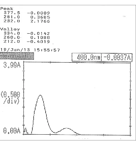
37 UV spectrum of SCM1 115
42 13C NMR of SCM1 (spectra - c) 121
43 13C NMR of SCM1 (spectra - d) 122
44 Mass spectra of SCM1 123
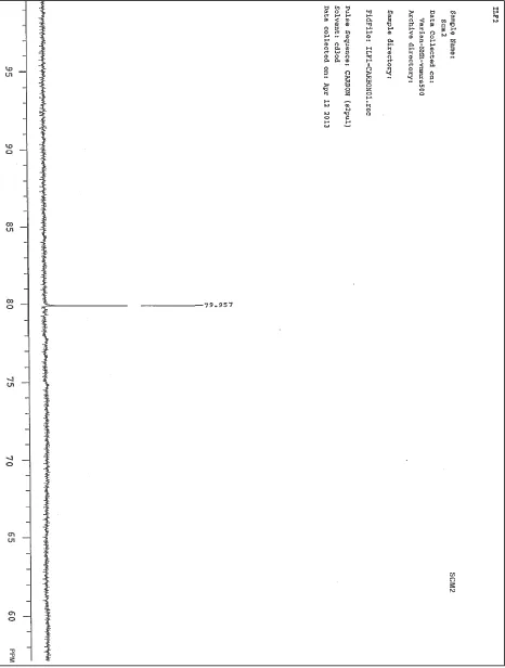
45 UV spectrum of SCM2 124
46 IR spectra of SCM2 125
47 1H NMR spectra of SCM2 126
48 13C NMR of SCM2 (spectra - a) 128
49 13C NMR of SCM2 (spectra - b) 129
50 13C NMR of SCM2 (spectra - c) 130
51 13C NMR of SCM2 (spectra - d) 131
52 Mass spectra of SCM2 132
53 Analgesic effect of HAE on tail clip method 134
54 Analgesic effect of HAE on tail immersion method 136
55 Anti-inflammatory effect of HAE by paw edema method 138
56 Antipyretic effect of HAE on Brewer’s yeast induced pyrexia method 140
57 Dose-response relation of HAE on antipyretic effect 140
58 Antidiabetic effect of HAE by alloxan induced diabetic method 142
59 Effect of HAE on WBC 143
60 Effect of HAE on RBC 144
61 Effect of HAE on CHOLESTEROL 145
62 Effect of HAE on TGA 146
63 Effect of HAE on HDL 147
64 Effect of HAE on CREATININE 148
65 Effect of HAE on ALT 149
66 Effect of HAE on AST 150
67 Effect of HAE on UREA 151
68 Alloxan- induced diabetic with area of necrosis of pancreatic islet cells ( H &E Stained )
152
69 Areas of restored pancreatic islet cells treated with 200mg/kg of HAE 153
70 Areas of restored pancreatic islet cells treated with standard 154
71 Effect of total protein on paracetamol induced hepatoprotective studies 156 72 Effect of liver weight on paracetamol induced hepatoprotective studies 156
73 Effect of AST on paracetamol induced hepatoprotective studies 157
74 Effect of ALT on paracetamol induced hepatoprotective studies 157
75 Normal hepatic cells 158
76 Hepatic cells treated with paracetamol control 158
77 Hepatic cells treated with standard drug 159
81 Dissolution profile of formulation 166
82 Chromatogram after derivatization under visible & UV 160
83 Standard Peak densitogram display (Scanned at 500nm) 170
84 Sample baseline display (Scanned at 500nm) 170
85 3D display of all Tracks 171
86 Track A – HAE of formulation peak densitogram display (Scanned at 500nm)
171
87 Track: SCM1 standard baseline display (Scanned at 500nm) 172
88 Linear curve based on peak area 173
89 Linear curve based on peak height 176
90 Structure of Stigmasterol 181
AdH Adaxial Hump
La Lamina
Lv Lateral vein
MR Midrib
Ads Adaxial strand
Ep Epidermis
GT Ground Tissue
Ph Phloem
Sc Sclerenchyma
X Xylem
Abx Abaxial xylem
AdX Adaxial xylem
Cu Cuticle
AbE Abaxial Epidermis
AdE Adaxial Epidermis
Pa Parenchyma
PM Palisade
SE Sub Epidermal parenchyma cells
VI Vein-islet
VT Vein-termination
EC Epidermal cells
PC Peltate scale
SC subsidiary cells
St Stomata
AW Anticlinal walls
PSe Peltate Scale
TS Transverse section
PP Phloem parenchyma
Sph Secondary phloem
STM Sieve tube Member
Pe Periderm
VE Vessel Elements
FT Fibre Tracheid
Fi Fibre
Lwp Lateral wall pits
P Peroration plate
T Tail
Tri Trichomes
Tn Tannins
CR Cortex
Pi Pith
NAPQI N-acetyl-p-benzoquinone imine
SCM1 Strobilanthes ciliatus marker 1 SCM2 Strobilanthes ciliatus marker 2
NCD's Non Communicable Diseases
RHD Rheumatic heart disease
IHD Ischemic heart disease
CVD Cardiovascular disease
HAE Hydroalcoholic Extract
LOD Limit on Detection
LOQ Limit on Quantification
As Arsenic
Ca Calcium
CCL4 Carbon tetra chloride
Cd Cadmium
CDCL3 Duterated chloroform
CHCl3 Chloroform
Cl Chloride
BDL Below Detection Limit
DL Detection Limit
HCl Hydrochloric acid
Hg Mercury
1
H NMR Proton Nuclear Magnetic Resonance HPLC High performance liquid chromatography HPTLC High performance thin layer chromatography
H2SO4 Sulphuric acid
I Iodine
IR Infera Red
K Pottasium
KI Pottasium iodide
LC50 Median lethal concentration
mg/kg milligram/kilogram
µg/ml microgram/milliliter
MIC Minimum inhibitory concentration
MHz Megahertz
µl Micro liter
Mm Millimeter
MS Mass Spectrum
MTT 3-(4,5 dimethyl thiazol-2-yl)-2,5 diphenyl
tetrazolium
Na Sodium
Na2CO3 Sodium Carbonate
Nm Nanometer
NaOH Sodium Hydroxide
Pb Lead
Ppm Parts per million
p.o Per oral
RSD Relative Standard Deviation
TLC Thin layer chromatography
UV Ultraviolet
ACE Acetone extract
EtOAc Ethyl acetate
MSL Mean sea level
b.wt Body weight
Std Standard
Eth. Ext. Ethanolic extract
Aq. Ext. Aqueous extract
J. gendarussa Justicia gendarussa
R. pectinata Rungia pectinata
S. ciliatus Strobilanthes ciliates
I.
INTRODUCTION
It is a fact that plants and people share a symbiotic relationship. Throughout the ages,
humans have relied on nature for their basic needs for the production of food-stuffs, shelters,
clothing, means of transportation, fertilizers, flavors and fragrances, and, not the least,
medicines. Plants have formed the basis of sophisticated traditional medicinal systems that have
been in existence for thousands of years and continue to provide mankind with new remedies.
Although some of the therapeutic properties attributed to plants have proven to be erroneous,
medicinal plant therapy is based on the empirical findings of hundreds and thousands of years.
India, due to its unique variety of geographical and climatic factors, had a rich, varied
flora of medicinal plants since the Vedic period. Out of a total number of 15,000 plant species in
India about 2,000 are known to have medicinal properties and some of them are used as home
remedies in rural and remotest parts of the country.
India unquestionably occupies the top position in the use of herbal drugs. It is one of the
foremost countries exporting plant drugs and their derivatives, and excels in home consumption
due to the following (1).
Great biodiversity and abundance of flora
Variety of geographical climate conditions, most exotic
Medicinal plants can be grown here
A review on plant drugs
Medicinal plants have important contributions in the health care system of local
communities as the main source of medicine for the majority of the rural population. Plants have
not only nutritional value, but also, in the eyes of the local people, they have medicinal and ritual
or magical values (2). The ethnomedicinal healing systems vary across cultures. According to the
World Health Organization (WHO), more than 3.5 billion people in the developing world rely on
medicinal plants as components of their healthcare (3). The vast majority of people consult
Traditional Medical Practitioners (TMPs) for their health care. Traditional medicine has been
only in India but also, in all countries of the world (4). Thus, medicinal plants are widely used in
the treatment of numerous human and livestock diseases in different parts of the world.
International trade in medicinal plant material for production of pharmaceutical and
neutraceutical preparation is booming. A number of pure phytopharmaceuticals have been
isolated from plants and used in modern system of medicine like reserpine, taxol and ephedrine.
Many of our modern drugs, including digitoxin, atropine and narcotic derivatives have
been developed from plants. Immunomodulatory, antimutagenic, antacids, antinociceptive,
hepatoprotective and anti-diabetic are some activities, which have made the globe to think
seriously regarding herbal medicines. Medicinal plants play a key role in the development and
advancement of modern studies by serving as a starting point for the development of novelties in
drugs (5).
A review of plant drugs in the treatment of human diseases
Medicinal plants or their extracts have been used by humans since time immemorial for
different ailments and have provided valuable drugs such as analgesics (morphine), antitussives
(codeine), antihypertensives (reserpine), cardiotonics (digoxin), antineoplastics (vinblastine and
taxol) and antimalarials (quinine and artemisinin). Some of the plants which continue to be used
from the Mesopotamian civilization to this day are Cedrus spp., Cupressus sempervirens,
Glycirrhiza glabra, Commiphora wightii and Papaver somniferum (6-9).
About two dozen new drugs derived from natural sources were approved by the FDA and
introduced to the market during the period 2000–2005 and include drugs for cancer,
neurological, cardiovascular, metabolic and immunological diseases, and genetic disorders (10).
Seven plant-derived drugs currently used clinically for various types of cancers are taxol from
Taxus species, vinblastine and vincristine from Catharanthus roseus, topotecan and irinotecan
from Camptotheca accuminata, and etoposide and teniposide from Podophyllum peltatum (11).
It is estimated that the worldwide market potential for herbal drugs is around US$ 40
billion. A similar situation also exists for plant-based food additives, fragrances and
biopesticides. Mostly, herbal drugs are collected from the wild, and relatively few species are
cultivated. Over exploitation of plants, particularly when roots, tubers and bark are used for
over exploitation of natural resources and the consequent threats to biodiversity, alternative
biotechnological methods and sustainable practices have been recommended. Several world
organizations and governments have established guidelines for the collection and utilization of
medicinal plants (12,13).
Standardization of herbal drugs
India has produced respectable health care systems such as Ayurveda & Siddha, which
encompasses the entire spectrum of human health and contributes to the positive health of
individuals. Validation of herbal medicines is one of the toughest challenges for the scientists.
There is a need to develop standards to bring this system of medicine in the mainstream of health
services.
“Standardization” expression is used to describe all measures, which are taken during the manufacturing process and quality control leading to a reproducible quality. “Standardization”
expression also encompasses the entire field of study from birth of a plant to its clinical
application. It also means adjusting the herbal drug preparation to a defined content of a
constituent or a group of substances with known therapeutic activity, respectively, by adding
occupants or by mixing herbal drugs or herbal drug preparations (14,15).
Need of standardization
The single and most important factor which stands in the way of wider acceptance of
traditional herbal medicines is the non-availability or inadequate of standards of checking their
quality by chemical or bioassay methods. This also prevents modernization or modification of
the methods of their preparation or production, as there is now to establish the equivalence of the
product made by the modified method with the original product. Thus, standardized drug of
well-defined, consistent quality is needed for reliable clinical trials and therapeutic use. All
major reason advanced for the difficulty in developing quality control standards is that these
products use whole plant or parts of plants or their total extracts. In some cases even a mixture
of a number of plants, it is challenging to develop suitable standards because a vegetable drug or
a preparation there of is not just an analytical operation and it does not end with the identification
and assay of an active principle rather it embodies total information and controls which are
necessary to guarantee composition. Standardization of plant drugs has been stressed by the
Kolli hills
Kolli hills are located in Nammakkal District, Tamil Nadu State, situated in the Southern
end of India and have an area of 490 kilometers. The altitude ranging from 1000 to 1300 m
above MSL and the highest point in Kolli hills is 4663 feet above MSL and the average rainfall is
about 1200 mm per annum (17).
Kolli hills are perhaps better known for its medicinal plants. A wide variety of medicinal
plants and herbs used in Ayurvedic, Siddha and Unani medicine are nurtured, cultivated,
gathered and sent from here. Even the most common medicinal plants, acquire a special value
when grown here, as the medicinal plants from Kolli hills are generally considered to be more
potent and effective (18).
An ethnobotanical survey revealed that total of 108 ethnomedicinal plants used for a
variety of ailments in Kolli hills. Out of the 108 plants were 31 herbs, 26 big trees, three small
trees and 17 shrubs. The medicinal plants from Kolli hills are generally considered to be more
potent and effective (19, 20). For example, the Chitharathai (Galanga lesser), an effective
remedy for cold, Athimaduram (Jamaica liquorice), Karpooravalli (Coleus aromaticus),
Thoothuvalai (Trilobatum), Tulasi (Ocimum sanctum), Kizhanelli (Phyllanthus amarus) and a
host of other herbs, besides a variety of spices are also grown here.
Commonly available most dominant species are Chloroxylon swietenia, Acacia
leucophloea, Dichrostachys cinerea, Rhinacanthus nasutus,Erythroxylum monogynum, Pongamia pinnata, Tamarindus Indica, Dodonaea viscosa, Ziziphus jujube and Memecylon edule.
Macaranga peltata, Holarrhena pubescens, Senna occidentalis, Azima tetracantha,
Lannea coramandelica, Cocculus hirsutus, Chenopodium ambrosioides, Strobilanthes ciliatus
II.
LITERATURE REVIEW
A brief literature survey was done for the planning of present scientific work. In order to
collect the information about the medicinal plants of Kolli hill with regard to their common and
local names, parts used, related ailments and the form of application, many old people and
vaidyas of the study area were interviewed. Subsequently, the information gathered was
confirmed with suitable literatures for authentification and identification of plants.
The ethnopharmacological survey showed more valuable information about medicinal
plants of Kolli hills. The study shows a high degree of ethnobotanical novelty and it reflects the
revival of interest in traditional folk medicine. The plants of Kolli hills have got high reputation
in traditional medicinal practice for its remarkable medicinal properties (18-20).
Acanthaceae (the acanthus family) is a family of dicotyledonous flowering plants
containing almost 250 genera and about 2500 species. Most are tropical herbs, shrubs, or twining
vines; some are epiphytes. The representatives of the family can be found in nearly every habitat,
including dense or open forests, in scrublands, on wet fields and valleys, at the sea coast and
marine areas, and in swamps and as an element of mangrove woods. (21).
The literatures proved that, the plant species belonging to the family Acanthaceae is to
possess medicinal properties against asthma, abscess, anthelmintic, astringent, bronchitis,
bedsores, cancer, cough, diuretic, diarrhea, dysentery, eczema, earache, headache,
inflammations, jaundice, kidney disease, leprosy, paralysis, skin diseases, scabies, toothache,
ulcers, ringworm etc (22).
In the present study, literature review focused the information about folklore claims,
phytochemical, pharmacological studies of plants of Kolli hills such as Justicia gendarussa Burm,
Rungia pectinata Nees, Strobilanthes ciliatus Nees and Rhinacanthus nasutus Linn belonging to the
family Acanthaceae.
Noncommunicable diseases (NCDs) are the leading global causes of death, causing more
deaths than all other causes combined, and they strike hardest at the world’s low- and
middle-income populations. These diseases have reached epidemic proportions, yet they could be
significantly reduced, with millions of lives saved and untold suffering avoided, through
related deaths can be reversed, and gains can be achieved rapidly, if appropriate action is taken.
In view of focusing and treating the non-communicable diseases, the literature study also carried
out for the above selected medicinal plants.
2.1 REVIEW ON JUSTICIA GENDARUSSA BURM
Woradulayapinij et al. (2005) studied and reported the in vitro HIV type-1 reverse transcriptase inhibitory activities of Thai medicinal plants Water and 80% ethanol extracts of 20
Thai medicinal plants including Justicia gendarussa and proved its reverse transciptase
inhibition (24).
Arokiyaraj et al. (2007) investigated the in vitro Immunosuppressive effects medicinal plants of Kolli hills on mitogen stimulated proliferation of the human peripheral blood
mononuclear cells. They proved that, among the plants tested J. gendarussa (100 µg/ml) showed
the highest lymphocyte inhibition (25).
Ratnasooriya et al. (2007) investigated the Antinociceptive and toxicological properties of aqueous leaf extract of Justicia gendarussa Burm. The study proved the moderate and dose
dependent activity of Justicia gendarussa Burm in the hot plate method, but not in the tail flick
test (26).
Senthilkumar et al. (2009) evaluated the larvicidal and mosquitocidal activities against Anopheles stephensi in commonly available medicinal plants, including J gendarussa. All the
extracts were found effective and the results suggest that these extracts are easy to prepare,
inexpensive and safe for mosquito control (27).
Paval et al. (2009) investigated the anti-arthritic potential of the alcoholic extract of the plant Justicia gendarussa using the Freund’s adjuvant-induced and collagen-induced arthritic rat
models. The rats were treated with the ethanolic extract of Justicia gendarussa and with standard
aspirin. The ethanolic extract of Justicia gendarussa showed significant anti-arthritic activity that
was statistically similar to that of aspirin (28).
Mpiana et al. (2010) evaluated the Antisickling activity in the species of J.tenella, J.gendarussa and J.insularis. They carried out the tests with anthocyanins extract and showed a
87% for Justicia gendarussa, 92% for Justicia insularis and 80% for Justicia tenella. The results
obtained from these three species of Justicia confirm those already obtained with species:
Justicia secunda. That indicates similarity of these species in their phytochemical composition
and biological activity (29).
Sonala et al. (2011) carried out the morphological, qualitative and quantitative
microscopy, powder study, ash values, extractive value and loss on drying studies on leaves of
“Justicia gendarussa Burm” (30).
Sonala et al. (2011) studied the pharmacognostical properties on root of Justicia gendarussa. They studied the phytochemical, physico-chemical, TLC and preliminary HPTLC
analysis of different extracts of the roots of J. gendarussa (31).
Jothimanivannan et al. (2010) studied the Anti-inflammatory and analgesic activities of the ethanol extract of aerial parts of Justicia gendarussa (EJG) in animal models were evaluated
by using carrageenan-induced rat paw edema and cotton pellet granuloma method. The study
was carried out in two different dose levels of 250 and 500 mg kg-1 orally. The pharmacological
screening of the extract showed significant (p<0.001-0.01) dose-dependent anti-inflammatory
activity with good analgesic profile when compared with reference standard (32)
Basah et al. (2011) studied the effect of Justicia gendarussa leaves extract on oxonate-induced hyperuricemic male albino rats. In this experiment the results showed that all extracts
could reduce uric acid levels of rats. The decreasing potency of uric acid level was equal to doses
increase, so the best result that can reduce uric acid level was J. gandarussa extract at a dose 5.2
g/kg bw. The results indicated that J. gendarussa leaves extract may be effective for the
prevention and the treatment of hyperuricemia (33).
Uddin et al. (2011) studied the antioxidant, antimicrobial and cytotoxic activities on methanol, petroleum ether, carbon tetrachloride and chloroform extract of whole plant of Justica
gendarussa. In brine shrimp lethality bioassay, the petroleum ether soluble materials demonstrated the highest toxicity with LC50 of 1.27μg /ml (34).
Verma et al. (2011) investigated the in vitro anti-inflammatory property on an ethanol extract of Justicia gendarussa leaves. An ethanol extract of Justicia gendarussa inhibits
macrophage. The studies proved the scientific evidence to support the anti-inflammatory
properties of Justicia gendarussa (35).
Subramanian et al. (2012) evaluated the phytochemical and antimicrobial properties of ethanolic and aqueous extracts of stem and leaves (Justicia gendarussa) against 12 human
pathogens. Based on the present result, stem extracts (aqueous and ethanolic) showed significant
antimicrobial activity against most of the human pathogens in both methods. The result revealed
that the antimicrobial properties of stem and leaves of J. gendarussa moreover associated with
the presence of phenolic compounds, flavonoids, terpenoids, glycosides and tannins (36).
Saha et al. (2012) evaluated the anthelmintic activities of crude methanolic extract of dried leaves and stems of Justicia gendarussa. Methanolic extract of leaves (50 mg/ml) caused
paralysis of the worms at 35.33 min and death at 70.67 min while methanolic extract of stem of
J. gendarussa caused paralysis at 41.33 min and death at 89.33 min The study confirms the
significant anthelmintic activities of leaves and stems extract of J. gendarussa (37).
Subhashini et al. 2013 studied the detailed microscopic parameters and reported the detailed description of an anatomical structure of both aerial and underground organs of G.
vulgaris Nees (38).
Subramanian et al. (2013) studied the preliminary anti-anxiety effect on Justicia gendarussa Burm. The plant extract at the dose of 250 and 500 mg kg-1 were evaluated for
anti-anxiety screening studies, with a view to ascertain the verity of its traditional use as an
anxiolytic. The effect was comparable to that of the benzodiazepine diazepam at the dose of 2.0
mg kg-1 (39).
2.2 REVIEW ON RUNGIA PCTINATA LINN
Sagoo et al. (1982) investigated the Cytological properties on 19 species of Acanthaceae including Rungia pectinata Nees from Pachmarhi hills in Central India. The study revealed the
first count of chromosome numbers of four species, namely, Dyschoriste depressa Nees, n=30;
Lepidagathis fasciculata Nees, n=10; L. hyalina Nees, n=10 and Justicia diffusa Willd.
var.prostrata Roxb, n=9. New cytotypes have been located in three species as Hemigraphis
Diploid cytotypes of three species, viz., Blepharis maderaspatensis (Linn.) Roth, n=15; Justicia
betonica Linn. n=17 and Thunbergia alata Bojer ex. Sims, n=9 have been detected for the first
time from India (40).
Swain et al. (2008) investigated the acute and subchronic toxicity studies on Rungia pectinata leaves in albino mice and rats. They reported that no significant changes in both the
absolute and relative organ weights between the control and the test groups. The liver enzymes
and haematological parameters were statistically equal in all the groups and the hydroalcoholic
leaf extract was non-toxic in albino rats (41).
Swain et al. (2008) investigated the anti-inflammatory and diuretic activities of hydroalcoholic extracts of leaves of Rungia pectinata and Rungia repens in wistar rats. The
results obtained were compared with that of standard drug aspirin and frusemide to their
anti-inflammatory and diuretic activity respectively. R. pectinata showed better anti-inflammatory
activity than R. repens. The antimicrobial potency of the aerial parts of Rungia pectinata and
Rungia repens has been studied using the petroleum ether, benzene, chloroform, acetone and
ethanol extract against a wide number of bacteria and fungi by disc diffusion method. The
ethanol extract at a concentration of 30 to 60 μg/disc showed significant activity against the
bacteria and fungus investigated (42).
Krishna et al. (2011) studied and reported the wound healing, anti-inflammatory, analgesic and antibacterial property of Rungia pectinata Linn, Rubia cordifolia Linn and
Scoparia dulcis Linn. The formulations containing extracts of the above mentioned herbs were
formulated and their wound healing activity was studied on experimentally induced open wounds
in albino rats through topical route. They proved that, wound healing property of polyherbal
formulation appears to be due to the presence of its active principles, which accelerates the
healing process and confers breaking strength to the healed wound (43).
Dipa et al. (2011) studied the preliminary micromorphological characters in 13 plants of the family Acanthaceae including Rungia pectinata. They reported the presences of glandular
and non-glandular trichomes and concluded that these characters can serve as pharmacognostic
2.3 REVIEW ON STROBILANTHES CILIATUS NEES
Thomas et al. (1998) studied the agronomic practices for aromatic and medicinal plants of kerala including S.ciliatus. The plnat details such as importance, distribution, botany,
cultivation collecticion, agrotechnology and properties of S.ciliatus were reported from their
studies (45).
Reneela et al. (2010) studied that the petroleum ether extract of the roots of S.ciliatus yielded three compounds lupeol, stigmasterol and betulin by silica gel chromatography and she
identified by spectral techniques. Further, they isolated and identified lupeol and stigmasterol
glycoside from the acetone extract of root.
The column chromatographic analysis of the acetone extract of the stem of S.ciliatus
yielded five compounds namely, lupeol, stigmasterol, betulin, taraxerol and
4-acetyl-2,7-dihydroxy-1,4,8-triphenyloctane-3,5-dione. From their study, they identified as the Lupeol was
the major constituent with a yield (0.18%) from the root of S.ciliatus(46).
Reneela et al. (2010) carried out in vitro studies for the different extracts of root and stem of S.ciliatus. The acetone and ethanolic extracts of S.ciliatus have been evaluated for its
antimicrobial, antioxidant and cytotoxic properties. Both the stem and root extracts of S.ciliatus
showed moderate antibacterial activity against the three species of bacteria, namely
Staphylococcus aureus, Klebsiella Sp., Pseudomonas Sp. and one species of fungi i.e.,
Aspergillus Sp. Further, they reported that the acetone and ethanolic extracts of S.ciliatus
inhibited the formation of superoxide, hydroxyl and DPPH radicals and lipid peroxidation. The
extract also showed good cytotoxicity against Daltons Lymphoma Ascites tumor cells (DLA)
and Ehrlish Ascites Tumor Cells (46).
Raiby Paul (2011) reported that the wide use of S.ciliatus in Ayurveda and detailed the medicinal uses ofS.ciliatusin different ancient traditional medicinal preparations. The plant was
used as active ingredient in Sahacharadi thailam, Sahacharadi kashayam, Varanadi kashayam,
Bhonaga thailam, Shtavargam kashayam, Maharasnadi kashayam, Sathavaryadi kashayam,
Balasahacharadi kashayam, Balaaireyakadi kashayam, Balakulathhadi kashayam,
Raiby Paul P. (2011) performed a brief comparative clinical study on the efficacy of fresh and dry Sahachara (Strobilanthes ciliatus Nees) and he identified that, fresh kasaya (extract) of the plant was more effective than the kasaya of dry plant against sciatica (47).
Venkatachalapathy and Subban. (2012) isolated Lupeol from the petroleum ether extract of aerial parts of S.ciliatusby column chromatography and identified by IR, NMR, and
MS spectral data. Followed by lupeol was quantified in the petroleum ether extract by HPTLC
method (48).
Venkatachalapathi and Subban. (2013) documented that the antimicrobial activity of the petroleum ether and methanolic extracts from the S.ciliatus. They were evaluated the extract
against three gram positive (Bacillus subtilis, Corynebacterium, Micrococcus luteus) and three
gram negative (Escherichia coli, Salmonella paratyphi, Klebsiella pneumonia) bacterial strains
and three fungal strains (Trichophyton rubrum, Microsporum gypseum, Monascus
purpureus).They concluded that the presence of lupeol from the petroleum ether and methanol
extract of S.ciliatus is to possess effective natural antifungal and antibacterial property (49).
2.4 REVIEW ON RHINACANTHUS NASUTUS LINN
Nanthakumar et al. (2004) reported the antibacterial and antifungal activities of ethanolic leaf extracts of R.nasutus. They reported that the extract was more effective against
bacterial strains of Bacillus subtilis and Salmonella paratyphi. The same extracts had exhibited
highest inhibitory activity against the fungal strains Candida albicans and Aspergillus flavus
(50).
Gotoh et al. (2004) studied the ethanolic extract of root and aqueous extract of leaves of
Rhinacanthus nasutus and the active moiety rhinacanthin C was assessed for its in vitro and in
vivo property on several cancer cell lines. Results showed rhinacanthin C exhibited in vitro
antiproliferative activity, comparable to or slightly weaker than 5FU. Both extracts showed in
vivo antiproliferative activity (51).
dependent potent antifungal activity against Candidaalbicans and Trichophytonmentagorphytes
(52).
Rongsriyam et al. (2006) investigated mosquito larvicidal effect of tablet formulations made from the methanol extract of dried root powder of R nasutus and it showed potential
potential property against Aedes aegypti and Culex quinquefasciatus larvae (53).
Siripong et al. (2006) isolated three main naphthoquinone esters - Rhinacanthins C, N, and Q - from the roots of R nasutus that induced apoptosis of human cervical carcinoma HeLaS3
cells. The study demonstrated that rhinacanthin-N suppressed tumor growth in vivo, and
suggested that liposomes are useful for preparing the injectable formulation of hydrophobic
drugs (54).
Sadasivan et al 2008 investigated the wound healing profile of Rhinacanthus nasutus Linn. (Kurz) on different wound models in wistar rats. Study of the roots of in an incision wound
model showed potent wound healing promoting activity (55).
Tewtrakal et al. (2009) studied the Antiallergic principles on Rhinacanthus nasutus
leaves. Three naphthoquinone derivatives, rhinacanthin-C, D and N were isolated from the leaf
extract. The study indicated that all the three compounds possessed very potent anti-allergic
activity against antigen-induced B-hexosaminidase release as a marker of degranulation in
RBL-2H3 cell with the IC50 values of 6.9, 8.9 and 6.4 µM respectively (56).
Puttarak et al. (2010) investigated the antimicrobial activities of Rhinacanthus nasutus extracts as well as rhinacanthin-C against Streptococcus mutans, Propionib acteriumacnes,
Helicobacter pylori, Staphylococcus aureus, S.epidermidis and Candida albicans were evaluated
by microdilution assay. It was found that the Rhinacanthus nasutus extract exhibited potent
bactericidal activity against S.mutans with MIC and MBC of 4 mg/ml, and potent bacteriostatic
activity against S.epidermidis, P.acnes and S.aureus with the MICs of 8–16 mg/ml (57).
Shyamal et al. (2010) investigated the hepatoprotective effect of R. nasutus root extract in rats treated with aflatoxin B1. Aflatoxin B1 causes its hepatotoxic effects in liver cells by
oxidative stress, which causes damage to DNA, proteins and lipids. They suggested that R.
Basha et al. (2012) studied the antitubercular effect of n-hexane, chloroform and ethanol extracts on leaves of Rhinacanthus nasutus using Microplate Alamar Blue assay (MABA). The
results showed that ethanolic extract has more significant Antitubercular activity as compared to
n-hexane, chloroform extracts. Pyrazinamide and Streptomycin are taken as standard drugs (59).
Desu et al. (2013) investigated the anti-hyperlipidemic effect of methanolic extract of whole plant of Rhinacanthus nasutus on fat diet induced hyperlipidemic rat models. The results
demonstrated that the methanolic extract of whole plant of Rhinacanthus nasutus possessed
significant antihyperlipidemic activity (60).
Rao et al. (2013) evaluated the therapeutic efficacy of Rhinacanthus nasutus (R. nasutus) on mitochondrial and cytosolic enzymes in streptozotocin-induced diabetic rats.The result of the
study indicated the administration of R.nasutus altered the activities of oxidative enzymes in a
positive manner. Also, the R. nasutus improves mitochondrial energy production (61).
2.5 HERBS WITH ANALGESIC, ANTIPYRETIC AND ANTI-INFLAMMATORY PROPERTIES
Adedapo1 et al. (2000) carried out an investigation to evaluate the anti-inflammatory and analgesic activities in animal models using an aqueous extract of the stem bark of Margaritaria
discoidea. They proved that the plant extract significantly reduced the formation of edema
induced by carrageenan and histamine, as well as in acetic acid induced writhing models (62).
Palmeiro et al. (2002) have reported that the anti-inflammatory and analgesic properties of hydroalcoholic extracts of leaves, roots, and fruits of Plantago australis Lam.
(Plantaginaceae). Carrageenan-induced rat hind paw edema was significantly inhibited by oral
administration of these extracts. All studied extracts also reduced the total number of writhes
induced by acetic acid. They concluded that the hydroalcoholic extracts of leaves, roots, and
fruits of P. australis are endowed with anti-inflammatory and analgesic activities (63).
Netoa et al. (2005) have studied and reported that the anti-inflammatory and antinociceptive effects of the crude hydroalcoholic extract of Pfaffia glomerata roots. They
assessed by using carrageenan-induced rat paw edema method and acetic acid-induced writhing
anti-inflammatory and analgesic effects, similar to those observed for non-steroidal drugs, such
as indomethacin and they suggested that the mechanism of action might be associated with the
inhibition of prostaglandin synthesis, as observed for most non-steroidal drugs (64).
Saha and Ahmed. (2009) have studied and reported the analgesic and anti-inflammatory activities of the extract of Albizia Lebbeck. In the carrageenan-induced rat paw edema test for
acute inflammation, the extract showed 36.68% and 27.51% inhibition of edema respectively. In
the acetic acid induced writhing test the extract showed a significant reduction in the number of
writhes with 39.9 % and 52.4 % of inhibition, respectively. In radiant heat tail-flick test the crude
extract produced 40.74% and 61.48% elongation of tail flicking time 30 minutes after oral doses
(65).
Anju K et al. 2010 evaluated the analgesic activity of the methanolic extract of the T. bispinosa root against the standard drug pentazocine in adult Swiss albino mice by tail flick &
tail immersion method. The both doses of T. bispinosa roots methanolic extract were found to
produce significant analgesic activity in both tail flick method and in tail immersion method
(66).
Patil et al. (2010) studied the analgesic activity of the bark extract of Ficus bengalensis
using hot-plate method and tail-immersion method in rats. The antipyretic activity of bark of
Ficus bengalensis was studied in Brewer’s yeast-induced pyrexia in rats. The ethanolic extract
showed the effect was similar to aspirin while the pet ether, chloroform, and water was weaker
than the aspirin in analgesic as well as antipyretic activity of the bark of Ficus bengalensis (67).
Yadav et al. (2011) studied the anti-pyretic and analgesic activity of leaf extracts of
Ficus bengalensis Linn in rats. The ethanolic and water extract showed significant analgesic
activity by hot plate and tail immersion analgesic activity respectively. Water and Chloroform
extract showed significant decrease in elevated body temperature. They concluded that the
extracts of leaves of Ficus bengalensis showed analgesic and antipyretic effects, similar to those
observed for non-steroidal drug such as aspirin (68).
Ismaila et al. (2012) reported the antinociceptive effect of the aqueous root extract of
Alchornea cordifolia in mice. The protective activitywas assessed using the acetic acid-induced
mouse writhing reflex, formalin-induced paw licking, hot plate and tail clip models of pain. The
reflex as well as significant dose-dependent inhibition of the neurogenic and inflammatory pains
associated with the formalin test. The formalin test result showed that the extract (100-400
mg/kg, p.o) increased pain threshold in a dose dependent manner in both phases, but the effect
was more prominent against the inflammatory phase (69).
2.6 HERBS WITH ANTIDIABETIC PROPERTY
Masirkar et al. (2008) studied the antidiabetic activity of ethanolic extract of the root powder of Pseudarthria viscida against alloxan-induced diabetes in albino rats. The ethanolic
extracts showed significant activity as compared to standard glibenclamide (70).
Ahmed et al. (2010) carried out to evaluate the antidiabetic activity of Vinca rosea
methanolic whole plant extracts in alloxan induced diabetic rats. The methanolic whole plant
extract at high dose (500 mg/kg) exhibited significant antihyperglycemic activity. The
methanolic extracts also showed improvement in parameters like body weight and lipid profile as
well as regeneration of β-cells of the pancreas in diabetic rats. They concluded that alcoholic
whole plant extracts of the Vinca rosea at high dose (500 mg/kg) exhibited significant
antihyperglycemic activity than whole plant extract at low dose (300 mg/kg) in alloxan-induced
diabetic rats (71).
Ramakrishna et al. (2011) investigated the in-vivo hypoglycemic activity of ethanolic extract of Triumfetta pilosa Roth using Streptozotocin induced diabetic rats. Different
biochemical parameters were used to determine the blood glucose levels using streptozotocin
induced diabetes and analyzed its effect in kidneys after 21 days treatment. Ethanol extract had
shown significant protection and lowered the blood glucose levels when compared to normal in
glucose tolerance tests (72).
Chaulya et al. (2011) screened the Hyperglycemic effect on methanolic extract of
Cyperus tegetum Roxb against alloxan monohydrate induced diabetes on Wistar strain albino
rats. It was significantly lowered blood glucose level. From their study it was concluded that the
methanol extract may be considered to have good antihyperglycemic active principles without
Prakash et al. (2012) evaluated the antihyperglycemic activity effects of ethanol extract of Exacum wightianum Arn (Gentianaceae) in streptozotocin induced diabetic rats. In diabetic
induced rats fed with E. wightianum ethanol extract at 100 and 200 mg/kg body weight, the
fasting plasma glucose levels were reduced to normal body and liver weight were found to be
increased. Whereas blood glucose, protein, albumin and creatinine levels were estimated after
two weeks. The extract significantly inhibited the induction of albuminuria, proteinemia and
uremia (74).
2.7 HERBS WITH HEPATOPROTECTIVE PROPERTY
Jamshidzadeh et al. (2006) have studied the effects of different concentrations of the hydroalcoholic extract of dried, powdered leaves of Cichorium intybus, on CCl4-induced
hepatotoxicity in vivo in rats. Their study showed that the Cichorium intybus extract could
protect the liver from CCl4-induced damages with doses of 50 and 100 mg/kg, but
concentrations higher than 200 mg/kg were less effective (75).
Setty et al. (2007) observed the hepatoprotective effect of Hydro-ethanolic extract (70%) of Calotropis procera flowers against paracetamol induced hepatitis in rats. They studied the
levels of biochemical markers of hepatic damage like SGPT, SGOT, ALP, bilirubin, cholesterol,
HDL and tissue GSH in both treated and untreated groups. Paracetamol (2 g/kg) has enhanced
the SGPT, SGOT, ALP, bilirubin and cholesterol levels and reduced the serum levels of HDL
and tissue level of GSH. Treatment with hydro-ethanolic extract of C.procera flowers (200
mg/kg and 400 mg/kg) has brought back the altered levels of biochemical markers to the near
normal levels in the dose dependent manner (76).
Uthandi and Ramasamy (2011) have studied and reported the hepatoprotective activity of sesame meal (Sesamum indicum) on high fat fed wistar rats. They proved that that sesame
meal (Sesamum indicum) has antihepatotoxic effects on serum, hepatic marker enzyme activities
and blood bilirubin level as well as improving the protein level from a high fat diet induced
hepatotoxicity (77).
used as standard drug. Levels of various biochemical parameters in serum and the histopathology
of liver were assessed to study the hepatoprotective effect of the extract. The extract showed
significant reduction in the D-Galactosamine induced liver damage and symptoms of liver injury.
Histopathology of the liver sections confirmed that the extract prevented hepatic damage induced
by D-Galactosamine (78).
2.8 HERBS WITH ANTITUSSIVE PROPERTY
Saraswathy et al. (2008) investigated for its anti-tussive effect on benzene, petroleum ether and ethanol extracts of leaves of Oldenlandia umbellata using cough model induced by
sulfur dioxide gas in mice. The ethanol extract at doses of 250 and 500 mg/ kg showed maximum
inhibition of cough reflex at 3 hours after drug administration and the anti-tussive activity was
comparable to that of codeine phosphate, a standard anti-tussive agent (79).
Pattanayak et al. (2009) tested the antitussive effect of methanol extract of C.grandis
fruits by counting the numbers of coughs produced due to aerosols of citric acid. In another set of
experiment they studied the cough activity of the methanol extract using a cough model induced
by sulfur dioxide gas in mice. The results showed significant reduction of cough number
obtained in the presence of both concentrations of methanol extract as that of the prototype
antitussive agent codeine phosphate. Also, methanol extract exhibited the significant antitussive
effect at 100, 200 and 400 mg/kg, per orally by inhibiting the cough within 90 min of performing
the experiment (80).
Gupta et al. (2009) studied and reported the anti-tussive activity of the combination of herbal drugs as formulations using sulphur dioxide (SO2) induced cough model in mice. The
mice were exposed to sulphur dioxide for 45 sec and placed in an observation chamber for the
counting of cough bouts, by two independent observers, for five minutes. All the formulations
showed significant antitussive activity in sulphur dioxide induced cough model (81).
Kaushik et al (2011) has investigated in vivo antitussive activity of root extracts (methanol and water) of Tectona grandis Linn. (Verbenaceae) using a cough model induced by
sulfur dioxide gas in rats. Treatment with aqueous extract and the methanol extract at 500 mg/kg
p.o. Both the extracts (methanol and water) significantly (P<0.05) suppressed the cough at the
dose level of 500 mg/kg (82).
Sankar et al. (2011) evaluated the potential antitussive activity in a mouse model induced by sulphur dioxide gas of ethanolic extract of “Rosa centifolia”. The ethanol extract of
Rosa centifolia (250mg/kg 500 mg/g p.o) showed significant anti-tussive activity, the cough
inhibition (35% and 57% respectively) and being comparable to that of 10-20mg/kg of codeine
phosphate (83).
2.9 HERBS WITH ANTICANCER PROPERTY
Ahmed et al. (2003) reported the chemotherapeutic and chemoprotective effects of extract of Nigella sativa which is containing anticancer principles of quinones that include
thymoquinone (TQ) and dithymoquinone. TQ-induced cytotoxicity was investigated using
canine osteosarcoma (COS31), its cisplatin-resistant variant (COS31/rCDDP), human breast
adenocarcinoma (MCF7), human ovarian adenocarcinoma (BG-1) and Madin-Darby canine