0022-538X/93/073703-09$02.00/0
Copyright ©1993, American Society forMicrobiology
Genetic
Analysis
of
the
Cofactor
Requirement for Human
Immunodeficiency
Virus Type 1 Tat
Function
STEVENJ. MADOREANDBRYAN R. CULLEN*
HowardHughes Medical Institute, Section of Genetics and Department of Microbiology, Duke
University
MedicalCenter, Durham,
North
Carolina 27710Received 27January1993/Accepted23March 1993
The Tat protein of humanimmunodeficiency virus type 1is a potenttranscriptionaltransactivator ofthe viral long terminal repeat promoter element. Tat function requires the direct interaction of Tat with a
cis-actingviral RNA target sequence termed the trans-activationresponse (TAR) element and hasalsobeen
proposedtorequireatleastonecellularcofactor. We have usedagenetic approachtoattempttoexperimentally definethe role of the cellular cofactor in Tat function and TARbinding.Ourdata suggest thatneither Tatnor
thecellular cofactor binds to TAR alone in vivo andindicate, instead,thatthe interaction ofTatwith itscellular
cofactor is aprerequisitefor TARbinding.The knownspeciestropism oflentivirus Tatproteinsappears to arise from the fact that notonlyTatbutalso the cellular cofactorcanmarkedly influencethe RNAsequence
specificity
of the resultantprotein complex. These dataalso suggest thattheTat cofactor is likelya cellulartranscription factor that has been highly conserved during vertebrate evolution. We hypothesize that the primary functionof Tatistoredirect this cellularfactortoanovelviralRNAtargetsiteandtotherebyinduce activation of viralgeneexpression.
Replication of thepathogenic retrovirus human
immuno-deficiencyvirustype1(HIV-1)iscritically dependentonthe functionalexpressionof the viral nuclearregulatory proteins
Tat and Rev(reviewedinreference12). TheTatproteinisa
transcriptional trans activator of the HIV-1 long terminal repeat (LTR) promoter element, while Rev acts posttran-scriptionally to induce the cytoplasmic expression of mRNAs that encode the viral structuralproteins.The mech-anisms of action of Tat and Rev, while therefore clearly distinct, are nevertheless each dependent on their direct
interaction with cis-acting viral RNA target sites termed, respectively, the trans-activation response (TAR) element
(14, 40, 49)and the Revresponse element(RRE) (29, 51). Although many DNA and RNA tumor viruses encode
sequence-specific transcriptionaltransactivators,the HIV-1 Tat protein ishighly unusual in actingvia an RNA, rather than a DNA, target sequence. Indeed, similar RNA
se-quence-specific regulatory proteins have as yet been
ob-served onlyin the otherprimate immunodeficiencyviruses and in a subset of the more distantly related ungulate lentiviruses, including equine infectious anemia virus
(EIAV) and bovine immunodeficiency virus (12, 13, 27).
While theprecisemechanismofaction oftheselentivirus Tat proteins has remained uncertain, activation ofviral RNA expressionatboth theleveloftranscriptioninitiation and the level ofelongationhasbeenproposed(15, 19, 23, 24, 26, 31,
44).
Many eukaryotic transcription factors display amodular domain structure featuring discrete protein sequences
in-volved, respectively, in conferring nucleic acid sequence specificity and in activating transcription from the bound
template (32, 37). Similarly, mutational analysis has led to
the definition oftwodomains within the 86-amino-acid Tat protein thatappear to fulfill comparable functions (Fig. 1).
The arginine-rich basic domain of Tat not only acts as a
nuclear localizationsignal(41)butalsoisbothnecessaryand
*Correspondingauthor.
fullysufficient forspecificbindingtoTARinvitro (6, 40, 49). ThecoremotifofTat ishighly conservedamongalllentiviral
Tat proteins, while the cysteine motif is conserved in all primate lentiviruses (8). Mutation of either of these two
motifs can inactivate HIV-1 Tat function in vivo without
affecting the ability of the proteintobind TAR in vitro (17, 22, 41, 47). These sequenceshavetherefore been proposed toformacellular cofactorbinding domain (8, 9, 13, 42, 47).
While less well conserved sequences located N terminal to
theseTat motifs are essential for fullbiological activity in
vivo, the more C-terminalsequences, including the second
coding exon of Tat, appear dispensable for efficient trans
activation of the HIV-1 LTR(8, 13, 17, 25, 39, 44, 47) (Fig.
1).
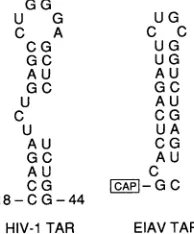
The cis-acting RNA target site for HIV-1 Tat is a
59-nucleotide (nt) RNA stem-loop structure located immedi-ately proximaltothestartsiteforviralmRNAtranscription (Fig. 2) (4, 16). Mutations that disrupt the helicalstructureof
the 27-ntapical regionofTAR,orthat affectthe
pyrimidine-rich 3-ntbulge,preventTARfunction invivoandalso inhibit
the in vitro interaction of Tat with TAR (3, 14, 40). In contrast, mutation of the6-nt terminal loop of TAR hasno
detectable affect on the in vitro Tat-TAR interaction yet
effectively blocks TAR function in vivo (14, 16). It has
therefore been proposedthat trans activation of the HIV-1
LTRbyTatmight requirethespecificinteraction ofnotonly
Tat but also a cellular cofactor(s) with TAR (40). It has
further been suggested that a specific interaction of this
TAR-binding cellular cofactor(s) with Tat might well be
criticaltotheformation of thishypotheticalternarycomplex (46). Althoughseveralcellularproteins thatbindto TARor
Tat have been reported, the identification of a cellular
cofactorrequired for Tatfunction and the definition ofthe
role ofthis cofactor in the mechanismofactionof Tathave
sofarremained elusive (30, 34, 50).
Inthisstudy,wehave usedarangeofgenetic approaches toaddress this latterquestion.Our data supportthe
hypoth-esis that the primary role of Tat is to recruit a cellular transcription factortothe HIV-1 LTRTAR element.These
3703
on November 9, 2019 by guest
http://jvi.asm.org/
AN C22S C37S K41A ARK 72 86
Basic l |
Co-Factor TAR
Binding Binding
Domain Domnain
FIG. 1. Domain organization of the Tat protein of HIV-1. The functional domain organization of the 86-amino-acid Tat protein presented is based on reports from a number of laboratories, including, particularly, the work of Kuppuswamy et al. (25) and Derse and coworkers (8, 13). The core motif, marked by the
consensus amino acid sequenceN'-KXLGIXY-C', is conserved in all lentivirus Tat proteins. In contrast, five of the seven cysteine residues thatconstitute the essential Cys motif of HIV-1 Tat are lackingin the eTatprotein (8).
datafurther suggest that neither Tatnorthe cellular cofactor iscapable ofinteractingwithTAR on itsown invivo.
MATERIALSAND METHODS
Construction of molecular clones. We have previously described the cytomegalovirus immediate-early promoter-basedexpressionvectorpBC12/CMVaswellas derivatives containing cDNAforms of the HIV-1 tatgene (pcTat) and
rev gene (pcRev) (48). Expression plasmids containing the
mutant
AN,
C22S,C37S, and K41A forms of HIV-1 Tatwere derived from pcTat as previously described (41, 47). The ARK Tat mutant contains an extensive substitution mutation within theTatbasic motif thatchangesTatamino acids 50to56 from N'-KKRRQRR-C' to N'-YVQILLY-C'. The ARK mutation was generated in the pcTat context by using the polymerase chain reaction (33)withoverlapping oligonucle-otideprimers encodingtheappropriateamino acidchanges. Theexpression plasmidpcTat/Rev encodesafusion protein consisting of
the
full-length HIV-1 Tat protein attached tothe N terminus of the full-length HIV-1 Rev protein (48). Plasmids encoding mutant forms of Tat in this Tat-Rev fusion context were derived as previously described (48). Reporter plasmids containing thecat indicator gene under the control of the wild-type HIV-1 LTR (pTAR/CAT) or
under the control ofanHIV-1LTRcontainingthestem-loop IIB
(SLIIB)
domain of the RRE in place of TAR (pSLIIB/ CAT) have been described previously, as has the Rev function reporterplasmid pDM128/CMV (21, 28, 48).All EIAV expression plasmids were derived from the pMA-1 proviral clone (1). A full-length cDNA copy of the
G G
U G
C A
CG
GC
AU
G C U C
U A U
GC
AU
C G
18- C G-44
U G
C C
U G U G AU GC A U
CG U A C G AU
C
ICAP-G C HIV-1TAR EIAV TAR
FIG. 2. Comparisonof theHIV-1 and EIAV TARelements. The primarysequenceandproposed secondarystructure of the critical
27-nt apical regionof the 59-ntHIV-1 TAR elementare compared
with those of thecomplete 25-nt EIAV TARelement.
EIAV tat gene was derived by the polymerase chain
reac-tion, using oligonucleotide primers that permitted isolation of both coding exons of EIAVtat. The 5' primer used to isolate the first exon substituted anNcoI site anda
consen-sustranslation initiation codon (5'-CCAUGG-3')inplace of the leucine codon that normally serves as the initiation codon for EIAV Tat (eTat) (8). The 3' primer inserted an EagIsite atthe end of the firstexon.Oligonucleotideprimers used to isolate the second eTatexoninsertedanEagI site at
the beginningofthisexonandintroducedaunique XhoI site immediately 3' to the Tat translation termination codon. A cDNAgenerated by ligationofthese two genomicEIAV tat
gene fragments at the introduced EagI site encodes the
Correct eTat amino acidsequence, althoughthe codon usage
forarginine 32(AGA--CGG) and proline 33 (CCC--CCG) is modified. The resultant NcoI-to-XhoI fragment, containing the full-lengthEIAV tatgene,wasinserted into the pBC12/ CMV expression plasmid to generate peTat. Similarly, pcRev/eTat contains thisfull-length eTat cDNA fusedtothe Cterminus of theHIV-1 Revprotein. The pR43G derivative of peTat contains a mutation ofarginine 43 to glycine that was introduced into the EIAV tat gene by the polymerase chain reaction. This criticalarginine residue is locatedatthe equivalent position in the Tat core consensus sequence to
lysine41of HIV-1Tat(Fig. 1) (8). The pEIAV/CAT plasmid containsEIAV LTRsequences,extendingfrom -212 to+40
relative to the cap site, substituted in place of the HIV-1
LTR of pTAR/CAT. The pEIAV/CAT plasmid therefore
contains the entire EIAV TAR element (10).
Cell culture and DNA transfection. The cell lines COS, HeLa, L, and QC1-3 were maintained as previously de-scribed (28). HeLa and L-cell cultureswere transfected by the calciumphosphate procedure, using a total of 5.1 ,ugof DNAper35-mm-diameterplate. COS and QC1-3 cellswere
transfected byusing DEAE-dextran and chloroquine with a total of 500 ng ofDNAper35-mm-diameter plate (11). For
assaysoftransdominance,HeLacellsweretransfected with
2 ,ug of the relevant chloramphenicol acetyltransferase (CAT)reporterplasmid, 0.1 ,ugof the effectorplasmid, 2.0
,ug
of the competitor plasmid, and 1 ,ug of salmon sperm carrier DNA. At -48 h after transfection, cultures wereharvested and relative levels of CAT enzyme activitywere
determinedby the diffusion method (35). All values reported
wereobtained during the linear phase of this kinetic assay for CAT activity and have been adjusted for any minor variability in the level of total protein in each extract, as
determinedby the method of Bradford(5). Expressionof the various Tatmutantsin transfected COS cellswasmonitored by radiolabeling with [35S]cysteinefollowedby immunopre-cipitation with a rabbit polyclonal anti-Tat antiserum as
previouslydescribed (11, 47, 48). RESULTS
Many eukaryotic transcription factors contain discrete
functionaldomains that confer nucleic acid sequence speci-ficity or transcription activation potential (32, 37). It has therefore proven possible to construct functional chimeric
trans activators consisting of the nucleic acid binding do-main ofone protein attached to the effector domain of a
second protein (37). Similarly, we and others have shown that fusion proteins consistingof Tat linked to a heterolo-gous RNA binding domain can activate gene expression fromanHIV-1 LTRinwhichTARhas beenreplaced by the appropriateRNAtarget(42, 43, 48).Inparticular,ithasbeen shownthat aTat-Revfusionprotein canefficiently activate
on November 9, 2019 by guest
http://jvi.asm.org/
[image:2.612.86.265.77.135.2] [image:2.612.128.225.568.685.2]1 2 3 4 9 6 7
-14.3
_6.2
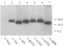
FIG. 3. Immunoprecipitation analysis ofmutantHIV-1Tat pro-teins.Expression plasmids encodingthe indicated Tatproteinswere
transfectedintoCOScellsaspreviouslydescribed(11,47).At48h aftertransfection, cellswerelabeled with[35S]cysteine(11),andthe resultant extract wassubjectedtoimmunoprecipitationwitharabbit polyclonal anti-Tat antiserum (11, 47). Precipitated proteins were
resolvedbyelectrophoresisandvisualizedbyautoradiography.The wild-type Tat protein is predicted to migrate at -16 kDa (47). Positionsofprotein molecularweightmarkers areindicated atthe right inkilodaltons. Neg, negativecontrol.
gene expression from anHIV-1 LTR in which critical TAR
sequences have beenreplaced by the RRE SLIIB primary Revbinding site(48).
Inthe contextof such aTat-Revfusion,Revservesas an autonomousnucleic acidbindingdomain while Tat
contrib-utestheeffector domain. Mutations introduced into Tat that specifically inactivate the Tat RNA binding domain, while highly deleterious in the context of the wild-type protein, should therefore have no effect on the activity of the chimeric protein. However, mutations that inactivate the effectordomain ofTatshouldbe equally inhibitory in both assaysystems.
To examine this issue, we introduced five distinct, well-characterized mutations into the Tat-Rev fusion protein context(Fig. 1) (25, 41, 47). IntheANmutation, Tat amino acids 2 to 9 inclusively have been deleted. The cysteine 22-to-serine(C22S)mutation is in theTatcysteine motif,the cysteine 37-to-serine(C37S)mutation isatthe border of the
Tat coremotif,and thelysine41-to-alanine(K41A)mutation lies within the core motif. A fourth missense mutation, termedARK,introduces six missense mutations into the Tat basicdomain (50-KKRRQRR-56 changedto 50-YVQILLY-56). The AN deletion mutant of Tat has been reported to retain a low level of Tat activity (47), while the four Tat
missense mutations have been reportedto beentirely inac-tive (25, 41, 47). With the exception of the ARK mutant, which lacks a functional nuclear localization signal, these Tat mutants are predicted to show the nuclear/nucleolar subcellularlocalization characteristic of Tat (25,41,47). To confirm that these mutations also donotsignificantly effect the level ofsynthesisandexpression ofTat,wedetermined the level of expression of these mutant proteins in
trans-fected tissue culture cells by using immunoprecipitation analysis(11, 47, 48).Aspredictedfrom earlier work(25, 41, 47),thesemutationswereshowntohavenodetectableeffect
on thelevel of Tatproteinexpression (Fig. 3).
As expected, analysis of the biological activity of these fusionproteinsin transfected HeLa cells demonstrated that all four Tat missense mutations abrogated the abilityof the
Tat-Revfusionproteintoactivateexpressionofanindicator gene linkedtothewild-typeHIV-1LTR,while the
AN/Rev
deletionmutantretainedaminimal level ofactivity (Fig. 4). Similarly, the three mutations introduced into the cysteine and core motifs of Tat also abolished the ability of the
*0 lOOx
0~~
FIG. 4. Biological activity of wild-type and mutant Tat-Rev fusion proteins. The relative abilities of the indicated proteins to trans activate the HIV-1 LTR via the wild-type TAR element present inthe indicator plasmid pTAR/CAT (dotted bars) or via the RRE-derived RNA target sequence present in the indicator plasmid pSLIIB/CAT (greybars)arecompared.Also shownaretheabilities of these same proteins to posttranscriptionally regulate cat gene expression by interacting with the RRE target present in the indicator constructpDM128/CMV (cross-hatched bars). The paren-talexpression plasmidpBC12/CMV served as a negative control. These data arerepresentative ofaseries of transfection experiments performed with the human cell line HeLa.
Tat-Rev fusion protein to activate HIV-1 LTR-dependent gene expression via theRRE SLIIB RNA target, while the AN/Rev mutant displayed a barely detectable level of Tat function (Fig. 4). Inmarked contrast, the ARK/Revmutant
remainedfully activeon thisheterologous RNAtargetsite. The pDM128/CMV indicator construct contains the cat indicator gene withinanRRE-containing intron and is unable toexpresssignificant levels of cytoplasmic cat mRNA in the absence of HIV-1 Rev function (21, 28). Both Rev and the
Tat-Rev fusion protein efficiently activate CAT expression from the pDM128/CMV construct upon cotransfection into HeLacells(Fig. 4). Similarly, allfivemutantTat-Revfusion proteins retained the ability to induce catgene expression from this Rev indicator construct (Fig. 4). We therefore conclude that all of these fusionproteinsarefully capable of functionallyinteractingwith theRREelement presentonthe DM128/CMV-encoded cat transcript. The inability of the C22S, C37S,and K41A andANfusionproteins to effectively activate HIV-1 LTR gene expression via the RRE-derived SLIIB RNA target therefore doesnotresultfrominstability
or from an inabilitytobind the RRE.
The Tat RNAbinding domain is not autonomous in vivo.
The datapresented inFig. 4 areconsistent with the hypoth-esis, enunciated perhapsmostclearly byDerseand
cowork-ers (8, 13), that the Tat cysteine and core motifs form the minimal Tat activation domain while the N-terminal
se-quences of Tat contribute strongly to full effector domain function. Importantly, these data alsoclearly show that this activation domain remainsactive in thepresence ofa
defec-tive TAR RNA binding motif, i.e., is fully autonomous.
However, they do notaddress the question of whetherthe Tat RNA binding domain is also independent of effector domain function invivo.Toaddress thisissue,weexamined whether any ofthese Tat mutants could act ascompetitive inhibitors of either Tat orTat-Rev. The rationale for these
experiments derivesfromthe observationinseveral systems
that an inactive but stable trans activator that retains a
on November 9, 2019 by guest
http://jvi.asm.org/
[image:3.612.120.251.79.175.2] [image:3.612.342.544.79.226.2]TABLE 1. Analysis of transinhibition of Tat function by various Tat and Revderivatives
Challenge Mean relative residual CAT activity +
SD'
plasmid Tat onTAR Tat-RevonSLIIB
pcTat 1.10 ± 0.12 0.17 ± 0.06
pcRev 0.75 ± 0.26 0.24 ± 0.08
pAN 0.63 ± 0.18 0.53 ± 0.05
pC22S 0.97± 0.06 1.07± 0.15
pC37S 0.70± 0.06 1.02± 0.14
pK41A 1.09 ± 0.34 0.98 ± 0.24
pARK 0.63 ± 0.07 0.27 ± 0.07
aThe relative abilities ofan -20-foldexcessoftheindicatedproteinsto
blocktransactivationof thewild-type pTAT/CAT reporter plasmidby Tat or
of thepSLIIB/CAT reporter plasmid byTat-Rev werecomparedby
transfec-tionintoHeLacells.The data arederivedfrom threedistinct experimentsand aregivenrelative tothevalueforaculture inwhich theparental pBC12/CMV vectorwasusedasa negative control, whichwasarbitrarilyset at 1.00.
functional nucleic acid binding motif but lacks an effector domain will compete withthewild-type protein for binding
to the target sequence and, hence, inhibit trans activation (20,37). As theefficiencyof thiscompetition shouldsimply reflect the ratio of wild-type to mutant protein, such a
trans-dominant negative phenotype should be a defining characteristic of stable, inactive regulatory proteins that retain full nucleic acid binding potential. A second class of trans-dominant negative mutants is observed when the de-fectiveregulatory proteinlackstheabilitytobind the nucleic acid target but retains theabilitytobindtoand sequestera
limiting cellular cofactor, a phenomenon generally known as squelching (37).
To ascertain whether any of these mutantproteins could display a dominant negative phenotype, we examined the ability of Tat to activate the wild-type HIV-1 LTR or of Tat-Revtoactivate the pSLIIB/CATindicator construct in the presenceofa20-fold molarexcessof therelevantmutant
protein expression plasmid. As shown in Table 1, several proteins were able to significantly inhibit Tat-Rev function
on pSLIIB/CAT. In particular, the wild-type Tat protein inhibited Tat-Rev function by -83%, while Rev inhibited Tat-Rev activity by -76%. These controls in fact serve as
examples of the two distinct classes of trans-dominant inhibitors predicted above. TheRevproteinis incapableof interacting with the Tat cofactor but can inhibit Tat-Rev function bycompetingforbindingtothe RRE-derived SLIIB RNA target.Tat, ontheotherhand, is unabletobindtothe SLIIB RNA target yet is capable of interacting with the cellular Tat cofactor. Inhibition of Tat-Rev function byTat
musttherefore result from cofactorsequestrationor squelch-ing.Aspredicted by this hypothesis, the three Tatmutants
C22S, C37S, and K41A, which lack a functional effector domain, have also lost theabilitytoinhibit Tat-Rev function via SLIIB. Incontrast,theARKmutantofTat, which lacks
a functional RNA binding motif but retains the effector domain (Fig. 1), also retains the ability to inhibit Tat-Rev function via the SLIIB target (Table 1). The somewhat reduced effectiveness of this inhibition, compared with the inhibition of Tatitself, mayreflect the inappropriate subcel-lularlocalizationof the ARK mutant. TheANmutantof Tat, which retains a low but detectable level of Tat function(47), also retained a low but apparently significant capacity to
inhibit Tat-Revfunction on SLIIB in trans.
In contrast to the observations with Tat-Rev on SLIIB, none of the various proteins testedwere ableto effectively
[image:4.612.358.501.75.222.2] [image:4.612.55.295.98.197.2]TATTAT yB+
:~~~~:~~:~:ilI~
Clular Complex | Co-Factor FormationJ%: B
I TAR Binding
A:Co-FatorActivation
Donain
B:Tat andCo-Factor RNA BindingDomains
C: Co-Factor Recruitment Transactivation TAR DomainofTat
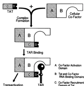
FIG. 5. Model for thetemporal assembly of the HIV-1 Tat-TAR activation complex. It is proposed that the interaction of the viral Tatproteinwithacellular cofactor represents the firststepin the mechanism of action of Tat. This protein-protein interaction is predictedtobedependentontheintegrityof thecore,cysteine,and,
to a lesser extent, N-terminal domains of HIV-1 Tat. This model predicts that neither Tatnorthe cellular cofactor is abletobind TAR alone invivo. The secondstepis the interaction of the Tat-cofactor complex with TAR to form an active ternary complex. This is dependent both on the specific interaction ofTat with the TAR pyrimidine bulge and on the specific interaction of the cellular cofactor with the TAR loop. It is further hypothesized that the cellular cofactor is directly responsible for the trans activation of HIV-1 LTR-dependent gene expressionthat results from this pro-tein-RNAinteraction.Although Carrolletal.(9) have also proposed a model in which Tat conveys a cellular cofactor to the TAR element, the presented model differsinthat the cellular cofactor is predicted to beessential for TARbinding byTat.
inhibit Tat function via the wild-type TAR element (Table1). It is therefore apparent that the C22S, C37S, and K41A proteins, despiteanapparently intactTARbinding motif,are
nevertheless incapable of competing with Tat for bindingto
TAR. Of interest, the ARKmutant also exhibited a signifi-cantly reduced ability to squelch trans activation of the wild-type HIV-1 LTR by Tat compared with its ability to inhibit Tat-Rev function via the SLIIB RNA target. While this may suggest that the interaction of Tatwith its cellular cofactor is stabilized by binding to the viral TARelement,
the data presented in Table 1 also clearly demonstrate that the interaction of Tat with a limiting cellular cofactor can
readily occurin the absence ofbindingtoTAR. Inaddition, these observations argue that mutant Tat proteins that are
unable to bind this cellular cofactor are also unable to
effectively compete for binding to TAR regardless of whether they retain afully intact basicdomain. Thesimplest interpretation of these data is that the interaction of Tat with
a cellular cofactor precedes, and is a prerequisite for, binding to TAR in vivo. A model for the in vivo Tat-TAR
interaction that incorporates this prediction is presented in Fig. 5.
Tat species specificity results from inefficient binding to TAR. Several groups have demonstrated that Tat is only minimally active in murinecells and have further shownthat this low activity can becomplemented inhybridmousecells containing human chromosome 12 (2, 18, 36). Indeed, these data provide one ofthe strongest arguments for the impor-tance of a human cellular cofactor in the mechanism of action of HIV-1 Tat. Ifthe model for the temporal assembly of the Tat activation complexproposed in Fig. 5 is accurate, we can then hypothesize either that mouse cells do not
on November 9, 2019 by guest
http://jvi.asm.org/
TABLE 2. Speciesspecificityof trans activation by lentivirus Tatproteins
pEffector
Fold transactivationaReporterplasmid plsi
plasmid
HeLa L QC1-3Part A
pTAR/CAT pcTat 257 9 7
pcTat/Rev 117 2 1
peTat 1 1 1
pSLIIB/CAT pcTat 3 2 3
pcTat/Rev 110 35 146
Part B
pEIAV/CAT peTat 11 15 6
pcRev/eTat 3 7 8
pcTat 2 1 2
pSLIIB/CAT peTat 1 1 1
pcRev/eTat 103 21 19
a Thedata comparetherelative abilities of the listed effectorplasmids to
trans activate CAT expression directed by the three indicated reporter
constructsafter transfection into cells ofhuman(HeLa),mouse(L),orquail
(QC1-3) origin andrepresent the averagesofthreetransfectionexperiments.
provideacellular cofactor that iscapable of interacting with Tatorthat suchacomplexindeed forms but is then unable
to interact with TAR. If the former is correct, then it is predicted that Tat-Rev function via SLIIB should also be inefficient inmousecells. Conversely, ifTARbinding is the rate-limiting step, then Tat-Rev should be fully active. It should be noted, however, that these two possibilities are
notmutually exclusive.
Toexaminethisissue,weintroduced appropriate HIV-1-based indicator and effector plasmids into human HeLa cells, intomouseLcells, and into thequailcell line QC1-3. As shown in Table 2, part A, these data reproduce the previous observation that neither TatnorTat-Revcan effec-tively trans activate the wild-type HIV-1 LTR in cells derived from theseother species. In contrast, Tat-Revwas
observedtobeaveryeffectivetrans activator of the HIV-1 LTRvia the introduced SLIIB RNA target in both murine and avian cells. We therefore conclude that it is the interac-tion ofTatwithTAR, rather than the recruitmentbyTatof
an appropriate cellular cofactor, that is inefficient in these nonhuman cell lines.
Although RNAsequence-specifictransactivation of tran-scriptionwasfirst described forHIV-1, comparable regula-tory proteins have subsequently been described in other primate lentiviruses and in theungulate lentiviruses EIAV and bovine immunodeficiency virus (8, 13, 27). The eTat protein has beenextensively studiedbyDerse and
cowork-ers and hasanumberofinteresting properties. EIAVTAR, like HIV-1TAR,hasbeen showntoformanRNAstem-loop structure (10). However, EIAV TAR lacks both the bulge and the loop sequences that are known to be critical for HIV-1 TAR function(Fig. 2). Further,while eTat contains sequences thataresimilarto the HIV-1 Tatbasic and core
motifs, it lacks anyequivalentof theequallycriticalcysteine motif(Fig.1) (8). Finally, althougheTatfunctionseffectively in cells ofequine orcanineorigin,it isonlyminimally active in human cells and cannot functionally interact with the
HIV-1 TAR element (Table2, partA) (8).
Toexamine whether the mechanistic basis for thespecies tropism of the eTat protein was similar to that seen with
HIV-1Tat,wederived effectorplasmidsthat encodedeither
aneTat cDNA (peTat) or afusion proteinofRev and eTat
Rev
MMM
Tat
K41Am
eTat
R43G
0 20 40 60 80 100
[image:5.612.375.510.79.247.2]%Inhibition ofTrans-Activation
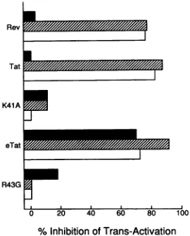
FIG. 6. Competition between the Tat proteins of HIV-1 and EIAVforthe samecellular cofactor.Theabilitiesof an excessofthe indicated proteins to block trans activation of the pTAR/CAT indicator construct by Tat (solid bars) or of the pSLIIB/CAT indicatorconstruct byHIV-1 Tat-Rev (hatched bars)orRev-eTat (open bars) arecompared. These data are representativeofthree separatetransfectionexperiments in HeLa cells.
(pcRev/eTat). Inaddition,weconstructedanindicator plas-mid, termed pEIAV/CAT, that placed thecatgeneunder the control of EIAV LTR sequences previously shown to be fully responsivetoeTat(10).Analysis of theseconstructsin transfected HeLa cellsconfirmed the modest activity of eTat in human cells (Table 2, part B). However, the Rev-eTat fusion protein proved as effective as the HIV-1 Tat-Rev fusion protein in activating the HIV-1 LTR via the SLIIB RNA target. Enhanced activity on the SLIIB RNA target
was also observed with the Rev-eTat fusion protein in both
mouse and quail cells. We therefore conclude that the species tropism of eTat again primarily reflects species-specific differences in the efficiency of the interaction
be-tween eTat and EIAV TAR rather than differences in eTat activation potential.
HIV-1 Tat andeTatinteract withthe same cellularcofactor. The markedly different RNA sequence specificities and primarysequencesof theTatproteinsofHIV-1andEIAV, when combined with their dissimilarspecies tropism, could be viewed as inconsistent with the hypothesis that these distantly related lentivirusregulatory proteinsinteract with the same cellular cofactor. However, the observation that these proteins can each trans activate the HIV-1 LTR by >100-fold when targetedto theintroduced SLIIB target by fusion to Rev (Table 2) could also indicate similar mecha-nisms of action.
To address whether HIV-1 Tat and eTat indeed interact with thesamecofactor in humancells,weexamined whether Tatand eTat could inhibit theactivity of Taton HIV-1TAR, Tat/Revon SLIIB, or Rev/eTat on SLIIB (Fig. 6) in
trans-fectedHeLa cells.Specificitycontrols wereprovided bythe
effector domain-minus K41A mutant ofTat and the similar R43Gmutantof eTat.Asshown inFig. 6,both Tat andeTat, but not the K41A or R43G derivative, were able to very effectively inhibit the activity of either Tat-Rev and Rev-eTat on the SLIIB target. Remarkably, eTat, which is not active on the HIV-1 TAR element (Table 2, part A), also proved able toinhibitHIV-1 Tat functionvia thewild-type HIV-1TARelementby-70%under these assayconditions. These datatherefore support thehypothesisthatHIV-1 Tat
.
on November 9, 2019 by guest
http://jvi.asm.org/
[image:5.612.63.302.100.251.2]and eTat interactwith, and compete for, the same cellular cofactor in transfected HeLa cells.
DISCUSSION
Theexperimentsreported in this articleweredesignedto
address the importanceof cellularcofactor(s) inthe mecha-nism of action of Tat andtoshedlightonhow the molecular interactionbetweenTat, TAR, and thiscofactor(s) canlead
totransactivation of the HIV-1 LTR.Astheidentityofthe cellularcofactor(s)for Tat remainsunknown,wehavebeen forced to use an entirely genetic approach to these ques-tions. Although the full verification of the experimental conclusions derived from this workmustthereforeawait the future biochemical dissection of the mechanism of action of Tat, these data neverthelessprovideclear evidence in favor of the model for Tat function presented in Fig. 5. In particular,these data supportthe following hypotheses.
(i) The interaction of Tat withan essential cellular cofactor
isindependentof TARbinding.This hypothesisissupported by two observations. First, the Tat-Rev fusion protein is fully functional when targeted to the heterologous SLIIB RNA target sequence, and this activity is not affected by mutation of theTatbasicdomain(Fig.4). Second, both Tat and a Tat mutant
(ARK)
lacking afunctional basic domainare abletoeffectively squelchtheactivityof Tat-Rev via the SLIIB target sequence (Table 1). In contrast, theactivityof Tat-Revon the SLIIB RNA target and the abilityof Tat to
inhibit thisactivityarebothdependentontheintegrityof the Tat cysteine and core motifs and, to a lesser extent, the N-terminaldomain. These observations confirm and extend previously publishedresults obtainedby usingTat-MS2coat protein fusions andanHIV-1 LTR constructcontainingthe MS2 operator RNA in place of TAR
(9,
42) or by using aGAL4-Tat fusionprotein targeted to aGAL4 DNA
binding
siteintroduced into the HIV-1 LTR U3region(44). Overall, these data demonstrate that the arginine-rich RNAbinding domain of Tat plays at most aminor role in mediating the interaction of Tat with a cellular cofactor(s) and further suggest that the Tat cofactor bindingdomain is coincident with the first -48amino acids ofTat, including,inparticular, the Tat coreand cysteinemotifs(8, 13, 44).
(ii) The interaction of Tat with a cellular cofactor is a
prerequisite for TAR binding in vivo. Several groups have demonstrated that the Tat basic domain is not
only
neces-sarybut alsofully sufficient for TARbindingin vitro(6, 40, 49). In particular, neither the Tat core motif nor the Tat cysteine motif appears to playany detectable role in medi-ating this interaction in vitro (22). Tat mutants lacking a
functional
cysteine
orcore motif,such asC22S, C37S, and K41A, should therefore compete effectivelywithwild-type Tatfor TARbinding
invivo. However, noneof these three inactive mutants proved capableof exertingany inhibitory effectonTat intransfected cells(Table 1). Incontrast, Rev, which could be viewed as a Tat-Rev derivative lacking afunctional effector domain, was able to markedly inhibit Tat-Rev function via SLIIB bycompeting forSLIIB RNA binding. Itshould beemphasized thattheC22S, C37S, and
K41A Tatproteinsareallfully stableinvivo andalsodisplay anapparentlywild-type subcellularlocalization (Fig. 3) (25, 41,47).Thesimplest interpretationof thisresultistherefore that the cofactorbindingdomainofTatplaysacritical role in mediatingTARbindinginvivo.
(iii) Tat may directly interact with only a single cellular cofactor. As shown inFig. 1, Tatappearstocontainatleast threemotifs thatareessential for invivofunction.The basic
domain's sole purpose lies inmediating the interaction of Tat with TAR, while both the cysteine and core motifs are
required for Tat effector function. The question then be-comes whether these motifs bind one or more cellular cofactors. Clearly, genetic experiments such as those de-scribed in this reportcannotdistinguish between binding of a single polypeptide, binding of a preformed multiprotein complex, or the highly cooperativebinding oftwo ormore cofactors to Tat. However, these data donotappear consis-tentwith thehypothesis that Tat bindstwo or morecellular cofactors independently. For example, if thecore and cys-teine motifs each boundadistinct cellular cofactor thatwas
independently required for activation, thenonewould pre-dict that mutation ofonemotifwould leave the other freeto
bind, and hence sequester, the other cellular cofactor. How-ever,noneofthese pointmutantsproved ableto exertsuch a squelching phenotype (Table 1). Further, if a distinct sequencelocatedwithin either the Tatcore orcysteinemotif was involved in effector function while a second was in-volved inrecruitingacofactorrequiredfor TARbinding,we
would thenpredict that mutation of the former wouldgivea
protein that could compete for TARbinding,whilemutation of the latter wouldgiveaprotein thatwasactive when fused
to Rev, i.e., the phenotype seen with ARK. However, all core andcysteine motifmutantsprovedinactive in the Rev fusion context. The simplest hypothesis to explain these observations is therefore that the N-terminal -48 amino acids of Tat, including, inparticular, the cysteineand core
motifs, likely bind asinglecellular cofactor.
(iv) Tat does notcontain atranscription activation domain per se. Several classes of transcription activation domains have been defined. Ofthese, the mostintensivelystudied is clearly the acidic activation motif first defined in the yeast protein GAL4 and seenin a particularly active form in the herpes simplex virustrans activator VP16 (32, 37). Rappa-portetal. (39) have proposed that the Tatproteincontainsa comparable acidic activation motif toward the protein N terminus. However, the observation that point mutations within the Tat core andcysteine motifs block trans activa-tionby the Tat-Rev fusionprotein demonstrate that the Tat
N terminus does not contain an autonomous activation motif. In fact, the observation that the samemutations that block Tattrans activation in theTat-Rev contextalso block squelchingby Tatstrongly suggests that Tat doesnotcontain
a transcription activation motif perse but insteadrecruits a
cellular cofactor that contains sequences able to fulfill this function; i.e., it is the cofactor that is recruited to TARby Tat that is eitherdirectly orindirectly responsiblefortrans
activation of HIV-1 LTR-dependent gene expression. (v) The species tropism ofTat reflects differences in the
efficiencyof the Tat-TAR interaction in vivo.The Tatprotein of HIV-1 is highlyactive in human cells but displaysmodest activity in cells of rodent or avian origin. Similarly, the distantly related eTat protein is active in equine andcanine cells butonlyminimallyactive in human cells(8).Inthecase ofHIV-1Tat, it has been demonstratedthatacellular factor encodedonhuman chromosome12markedlyenhancestrans activation of the HIV-1 LTR in rodent cells (18,
36),
thus leading to the suggestion that an equivalent cofactor might be lacking in cells of murine origin. Here, we demonstrate that thespeciestropismof HIV-1 and eTatcanbeessentially overcome ifTatistargetedtotheheterologous SLIIB RNA target site by fusion to Rev (Table 1). In particular, trans activation of the HIV-1 LTRbyTat-Revwas -18-foldmoreefficient on the SLIIB RNA target in mouse cells and -100-fold more efficient in avian cells. Similarly, the
on November 9, 2019 by guest
http://jvi.asm.org/
eTat fusion proved far more active on the SLIIB RNA target than on the homologous EIAV TAR element when tested in human cells. It is therefore apparent that species tropism does not result from the inability of HIV-1 or eTat to interact with the appropriate cellular cofactor. It is, rather, the efficiency of the subsequent interaction of the Tat-cofactor complex with TAR that primarily determines the level of trans activation in cells of the species in question. We note thatcomparable data demonstrating efficient trans activation of the HIV-1 LTR in mouse cells by a Tat-MS2 coat protein fusion have recently been presented by Alonso et al. (2).
(vi) Lentivirus Tat proteins interact with the same cellular
cofactor. Among lentivirus Tat proteins, that of EIAV is the mostdissimilar to that of HIV-1. The eTat protein is unique among Tat proteins in lacking a cysteine motif and is also unusual in that it shows no ability to trans activate the HIV-1 LTR(8, 13). Remarkably, however, the eTat protein proved as effective as HIV-1 Tat in trans activating the HIV-1 LTR when both were targeted to the SLIIB RNA sequence by fusion to Rev (Table 1). The equivalent, -100-fold trans activation seen with these two distinct Tat fusion proteins suggested that they might be recruiting the same transcrip-tion factor to the SLIIB RNA target. To address this issue, we examined whether these proteins could inhibit SLIIB RNA-dependent activation of the HIV-1 LTR by the Tat-Rev or Rev-eTat fusion protein. Remarkably, both Tat and eTat proved able to efficiently inhibit trans activation by both fusion proteins. Indeed, eTat proved able to exert a significant inhibitory effect on wild-type Tat function via the HIV-1 TARelement. This inhibition was entirely dependent
ontheintegrity of the conserved effector domain core motif of these two Tat proteins. These data therefore indicate that HIV-1 Tat and eTat compete with each other for the same limiting cellular cofactor in transfected human cells. Prior to completion of this work, Carroll et al. (9) also reported that EIAVTatcouldinhibit the activity of a HIV-1 Tat-MS2 coat protein hybrid, but not of HIV-1 Tat itself, in transformed HeLa cells. Although these recent data therefore differ at least in part from the results presented in this report, Carroll
etal.(9) nevertheless also proposed that HIV-1 Tat and eTat interactwith the same cellular cofactor prior to binding their homologous TAR element (Fig. 5).
(vii) The cellular cofactor modulates the RNA sequence specificityoftheTat-cofactor complex. The TARelements of
the lentiviruses HIV-1 and EIAV are quite distinct (Fig. 2). In particular, the EIAV TAR element clearly lacks any equivalent of the pyrimidine-rich bulge and conserved hex-anucleotide loop that are critical for HIV-1 TAR function in vivo (10). It is, therefore, not surprising that HIV-1 Tat is inactive on the EIAVTAR element and vice versa (Table 2). However, the evidence presented above clearly indicates that the cellularcofactors recruited by Tat and eTat are the same, at least in human cells. We have argued that Tat interacts with a single cofactor and that this cofactor affects theefficiency of the Tat-TAR interaction. This view implies that the same cellular cofactor must be involved in mediating the interaction of both HIV-1 Tat and eTat with their respective, highly divergent, TAR elements. It is, therefore, not surprising that these interactions are not equivalently efficient in cells from different species (Table 2). The human cofactoris, according to this hypothesis, highly effective at mediating the Tat-TAR interaction of the human virus HIV-1 but is far less competent to mediate the Tat-TAR interaction of theequine virus EIAV. The high activity of eTat in equine and caninecells (8, 13) implies that these species express an analogous protein that can effectively mediate this latter
binding event. The equivalent protein expressed by mouse and quail cells, in contrast, is not proficient at mediating either of these Tat-TAR interactions but could, presumably, be effective on yet a third RNA sequence.
Of interest, the observation that the HIV-1 and EIAV Tat proteins can work effectively in a range of species, including nonmammalian cells, when targeted to a heterologous RNA binding domain (Table 2) indicates that the Tat cofactor has been conserved since the evolutionary divergence of mam-mals and birds and has also retained the structural features that allow it to interact with Tat. However, it also appears that the RNA sequence specificity of this cofactor has evolved significantly over time, thus generating the species tropisms that are now characteristic of the various lentivirus Tat proteins.
A model for the temporal assembly of a
ternary
complex onTAR. In this report, we have presented a genetic analysis of the role of the cellular cofactor in the mechanism of action of Tat. These data, in combination with recent observations from other groups (2, 9, 42, 46), have suggested a specific model for the genesis of the Tat transcription activation complex (Fig. 5). The primary, and most novel, feature of this model is that neither Tat nor the cellular Tat cofactor binds TAR alone in vivo. Instead, we propose that these proteins must first form a protein complex. This complex then binds to TAR with a sequence specificity that is determined both by Tat and by the cellular cofactor (Fig. 5). This hypothesis is therefore inconsistent with the conten-tion, based entirely on in vitro analysis, that Tat alone can bind to TAR in vivo.
Although the binding of TAR by the Tat basic motif in vitro occurs with high affinity, it displays a relatively low level of specificity. Thus, Tat proteins in which the basic motif has been substituted by other natural or artificial stretches of basic amino acids have been reported to retain full in vivo activity (6, 45), while the ability of the basic domain of Tat to selectively bind TAR appears to be specified by a single arginine residue within the basic motif (7). Indeed, the amino acid arginine itself seems to retain much of the same RNA specificity (38). It has therefore seemed improbable that the sequence information present in the basic motif of Tat could alone be sufficient to direct the specific binding of Tat to TAR under in vivo conditions. The hypothesis that a cellular cofactor might be involved in mediating the Tat-TAR interaction was first suggested by the observation that the ability of a TAR RNA decoy to seques-ter the Tat protein in vivo, and hence inhibit HIV-1 LTR trans activation, was critically dependent on the integrity of terminal loop RNA sequences that have no effect on Tat binding in vitro (46). As noted above, the data presented in this report strongly support the proposal that the interaction of Tat with a cellular cofactor is, in fact, an essential prerequisite for TAR binding in vivo.
The TAR element cannot bind an essential cellular cofac-tor distinct from the one that binds to Tat, because it would then be impossible to functionally substitute a heterologous RNAtarget sequence for TAR. Given, then, that the cellular cofactors that bind Tat and TAR are one and the same, what is the evidence that the cofactor does not bind TAR alone? The most compelling evidence for this hypothesis derives from the observation that the interaction of TAR with a cellular cofactor is necessary but not sufficient to direct Tat toTAR. The evidence for necessity includes the observation that TAR sequences that are not directly involved in binding to Tat are nevertheless required for TAR function in vivo (14, 40) as well as the finding that the species origin of the Tat
on November 9, 2019 by guest
http://jvi.asm.org/
cofactor markedly affects the level oftrans activation ob-tained with a particular TAR element (Table 2). The evi-dence that the TAR-cofactorinteraction is not sufficient to
recruit Tat to TARderives from thefindingthat the Tatbasic domain is critical for TARbinding, butnoteffectorfunction, in vivo (Fig. 4) (42, 44) and that complexes of the same
cellular Tat cofactor with either HIV-1 Tator eTat display entirely different TAR RNA sequence specificities in vivo (Table 2). Indeed, the data presented in this report arefully consistent with thehypothesis, firstsuggested byCarrollet
al. (9), that the entire purpose of Tat issimplytorecruit the apparently ubiquitous cellular transcription factor referred
to here as the Tat cofactor to a novel, viral RNA target sequence and to thereby activate viral LTR-specific gene expression. Tat could therefore be viewed more as a trans
specifier ofapreexistingcellulartranscriptionfactor thanas a transcriptionaltrans activatorin itsown right.
Animportantpredictionof the modelpresentedinFig.Sis that the cellular Tat cofactor is unlikely to bind TAR with significantaffinityin the absenceofTat.Effortstodefine the Tat cofactor onthe basis of itsability tobind TAR in vitro maytherefore be doomedtofailure.Instead, ourdata would suggestthat it is the affinityof the cofactor for Tat itself that should provide the best biochemical or genetictool for the identification of this interesting cellularregulatoryprotein.
ACKNOWLEDGMENTS
This researchwas fundedbythe Howard Hughes Medical
Insti-tute.
We thank SusanCarpenterfor the pMA-1EIAVproviralclone, Laurence Tiley for many helpful discussions, Pamela Brown for technicalassistance, andSharon Goodwinfor secretarialexpertise.
REFERENCES
1. Alexandersen, S., and S. Carpenter. 1991. Characterizationof variable regionsin theenvelope and S3 openreadingframe of equineinfectious anemia virus. J. Virol. 65:4255-4262. 2. Alonso, A., D. Derse, and B. M. Peterlin. 1992. Human
chromo-some 12 is required foroptimal interactions between Tat and TARofhumanimmunodeficiencyvirus type 1 in rodent cells. J. Virol.66:4617-4621.
3. Berkhout, B., and K.-T.Jeang. 1989. trans-activationofhuman immunodeficiencyvirus type 1 is sequencespecificfor both the single-stranded bulge and loop of the trans-acting-responsive
hairpin: aquantitative analysis. J. Virol. 63:5501-5504. 4. Berkhout, B., R. H. Silverman, and K.-T. Jeang. 1989. Tat
trans-activates the human immunodeficiency virus through a
nascentRNAtarget. Cell 59:273-282.
5. Bradford, M. M. 1976. A rapid and sensitive method for the quantitation of microgram quantities of protein utilizing the principleofprotein-dyebinding. Anal. Biochem.72:248-254. 6. Calnan, B.J., S. Biancalana, D. Hudson, and A. D. Frankel.
1991. Analysis of arginine-rich peptides from the HIV Tat protein reveals unusual features of RNA-protein recognition. Genes Dev. 5:201-210.
7. Calnan, B.J., B.Tidor, S. Biancalana, D.Hudson, and A. D. Frankel. 1991.Arginine-mediated RNArecognition: the argin-inefork. Science 252:1167-1171.
8. Carroll, R., L. Martarano, and D. Derse. 1991. Identification of lentivirus Tat functional domains throughgenerationofequine infectious anemiavirus/human immunodeficiencyvirus type 1
tatgenechimeras. J. Virol. 65:3460-3467.
9. Carroll, R., B. M. Peterlin, and D. Derse. 1992. Inhibition of humanimmunodeficiencyvirus type 1 Tatactivity by coexpres-sion ofheterologoustrans-activators. J. Virol. 66:2000-2007. 10. Carvalho, M., andD. Derse. 1991. Mutational analysis of the
equineinfectious anemia virusTat-responsiveelement. J. Virol. 65:3468-3474.
11. Cullen,B. R.1987. Use ofeukaryotic expression technologyin
the functional analysis of cloned genes. Methods
Enzymol.
152:684-704.
12. Cullen,B. R. 1992.Mechanismofactionofregulatory
proteins
encodedbycomplexretroviruses.Microbiol.Rev.56:375-394. 13. Derse,D.,M.Carvalho,R.Carroll,and B. M. Peterlin. 1991. A
minimallentivirus Tat. J. Virol.65:7012-7015.
14. Dingwall,C.,I.Ernberg,M.J.Gait,S. M.Green,S.Heaphy,J. Karn,A. D.Lowe,M.Singh,and M. A.Skinner. 1990. HIV-1tat proteinstimulatestranscription bybindingtoaU-rich
bulge
inthestemoftheTAR RNAstructure.EMBO J.9:4145-4153. 15. Feinberg,M.B.,D.Baltimore,andA. D. Frankel.1991. Therole
ofTat in the humanimmunodeficiencyvirus life
cycle
indicatesaprimaryeffectontranscriptional
elongation.
Proc.Natl.Acad. Sci. USA88:4045-4049.16. Feng, S., and E. C. Holland. 1988. HIV-1 tattrans-activation
requires the loop sequence within tar. Nature
(London)
334: 165-167.17. Garcia,J.A.,D.Harrich,L.Pearson,R.Mitsuyasu,and R. B. Gaynor. 1988. Functional domains
required
for tat-induced transcriptional activation of the HIV-1 long terminal repeat. EMBO J.7:3143-3147.18. Hart,C.E.,C.Ou,J.C.Galphin,J.Moore,L. T.Bacheler,J. J.
Wasmuth, S. R. Petteway, and G. Schochetman. 1989. Human chromosome 12 is required for elevated HIV-1
expression
in human-hamsterhybrid cells.Science246:488-491.19. Hauber, J.,A. Perkins, E. P.Heimer, and B. R. Cullen. 1987. Trans-activation of human
immunodeficiency
virus gene expres-sionis mediatedbynuclearevents.Proc.Natl.Acad.Sci. USA 84:6364-6368.20. Herskowitz,I. 1987. Functional inactivationof genes
by
domi-nantnegativemutations. Nature(London)
329:219-222. 21. Hope,T.J.,X.Huang,D.McDonald,and T. G. Parslow.1990.Steroid-receptorfusion of the human
immunodeficiency
virus type 1 Rev transactivator: mappingcryptic
functions of the arginine-richmotif. Proc. Natl. Acad. Sci.USA87:7787-7791. 22. Kamine, J.,P. Loewenstein,and M. Green. 1991.Mapping
ofHIV-1 Tatproteinsequencesrequiredfor
binding
toTARRNA. Virology182:570-577.23. Kao,S.-Y.,A. F.Calman,P.A.Luciw,and B. M.Peterlin. 1987. Anti-termination oftranscriptionwithin thelongterminalrepeat of HIV-1bytatgeneproduct. Nature
(London)
330:489-493. 24. Kato, H.,H.Sumimoto,P.Pognonec,C.-H.Chen,C. A.Rosen,and R. G. Roeder.1992. HIV-1 Tatactsas a
processivity
factor in vitro inconjunctionwith cellularelongation
factors. Genes Dev. 6:655-666.25. Kuppuswamy, M., T. Subramanian, A. Srinivasan, and G. Chinnadurai. 1989. Multiple functional domains of Tat, the trans-activator of HIV-1 definedby mutational
analysis.
Nu-cleic AcidsRes. 17:3551-3561.26. Laspia,M.F.,A. P.Rice,and M.B.Mathews.1989. HIV-1 Tat proteinincreasestranscriptionalinitiationandstabilizes
elonga-tion. Cell 59:283-292.
27. Liu,Z.-Q.,D. Sheridan,andC. Wood. 1992. Identification and characterization of the bovine
immunodeficiency-like
virustat gene. J. Virol.66:5137-5140.28. Malim, M. H., D. F. McCarn, L. S. Tiley, and B. R. Cullen. 1991. Mutational definition of the human
immunodeficiency
virustype 1 Rev activationdomain. J. Virol. 65:4248-4254. 29. Malim, M. H., L. S. Tiley, D. F. McCarn, J. R. Rusche, J.Hauber,and B. R. Cullen. 1990. HIV-1structuralgene expres-sion requires binding of the Revtrans-activator to its RNA targetsequence. Cell 60:675-683.
30. Marciniak,R.A.,M. A.Garcia-Blanco,andP. A.Sharp.1990. Identification and characterization of a HeLa nuclear
protein
that specifically binds tothe
trans-activation-response
(TAR)
element of human
immunodeficiency
virus. Proc. Natl. Acad. Sci.USA87:3624-3628.31. Marciniak, R. A., and P. A. Sharp. 1991. HIV-1 Tat
protein
promotesformation of
more-processive elongation
complexes.
EMBO J. 10:4189-4196.
32. Mitchell,P.J.,andR.Tjian.1989.
Transcriptional
regulation
in mammalian cellsbysequence-specific
DNAbinding
proteins.
Science245:371-378.
on November 9, 2019 by guest
http://jvi.asm.org/
33. Mullis, K. B., and F. A. Faloona. 1987. Specific synthesis of DNA invitro via apolymerase-catalyzed chain reaction. Meth-ods Enzymol. 155:335-350.
34. Nelbock, P., P. J. Dillon, A. Perkins, and C. A. Rosen. 1990. A cDNAfor a protein thatinteracts with the human immunodefi-ciency virus Tat transactivator. Science 248:1650-1653. 35. Neumann, J. R., C. A. Morency, and K. 0. Russian. 1987. A
novel rapid assay for chloramphenicol acetyltransferase gene expression. BioTechniques 5:444-447.
36. Newstein, M., E. J. Stanbridge, G. Casey, and P. R. Shank. 1990. Human chromosome 12 encodes a species-specific factor which increases human immunodeficiency virus type 1 tat-mediated trans-activationinrodentcells. J. Virol. 64:4565-4567. 37. Ptashne, M. 1988. How eukaryotic transcriptional activators
work. Nature (London) 335:683-689.
38. Puglisi, J. D., R. Tan, B. J. Calnan,A. D. Frankel, and J. R. Williamson. 1992. Conformation of the TAR RNA-arginine complex by NMR spectroscopy. Science 257:76-80.
39. Rappaport, J., S.-J. Lee, K. Khalili, andF.Wong-Staal. 1989. The acidic amino-terminal region of the HIV-1 Tat protein constitutes an essentialactivatingdomain. New Biol. 1:101-110. 40. Roy, S., U. Delling, C.-H. Chen, C. A. Rosen, and N. Sonenberg. 1990. A bulge structure in HIV-1 TAR RNA is required for Tat binding and Tat-mediated trans-activation. Genes Dev. 4:1365-1373.
41. Ruben, S., A.Perkins, R. Purcell, K. Joung, R. Sia, R.Burghoff, W. A. Haseltine, andC. A. Rosen. 1989. Structural and func-tional characterization of human immunodeficiency virus tat protein. J. Virol. 63:1-8.
42. Selby, M. J., and B. M. Peterlin. 1990. Trans-activation by HIV-1Tatvia aheterologous RNAbinding protein.Cell 62:769-776.
43. Southgate, C., M. L.Zapp, and M. R. Green. 1990.Activation of transcription by HIV-1 Tat protein tethered to nascent RNA through another protein. Nature (London) 345:640-642. 44. Southgate, C. D.,and M.R.Green. 1991. The HIV-1 Tatprotein
activates transcription from an upstream DNA-binding site: implicationsfor Tatfunction. Genes Dev. 5:2496-2507. 45. Subramanian, T., M. Kuppuswamy, L. Venkatesh, A.
Srini-vasan, and G.Chinnadurai. 1990. Functional substitution of the basicdomain of the HIV-1trans-activator,Tat, with the basic domain ofthe functionally heterologous Rev. Virology 176:178-183.
46. Sullenger, B. A., H. F. Gallardo, G. E. Ungers, and E. Gilboa. 1991. Analysis of trans-acting response decoy RNA-mediated inhibitionof humanimmunodeficiency virus type 1 transactiva-tion. J.Virol. 65:6811-6816.
47. Tiley, L. S., P. H. Brown, and B. R. Cullen. 1990. Does the human immunodeficiency virus Tat trans-activator contain a discrete activation domain?Virology 178:560-567.
48. Tiley, L. S., S. J.Madore, M. H. Malim, and B. R. Cullen. 1992. The VP16 transcription activation domain is functional when targeted to apromoter-proximal RNA sequence. Genes Dev. 6:2077-2087.
49. Weeks, K.M.,C. Ampe, S. C. Schultz, T. A.Steitz, and D. M. Crothers. 1990.Fragments of the HIV-1 Tat protein specifically bind TARRNA. Science 249:1281-1285.
50. Wu, F., J. Garcia, D. Sigman, and R. Gaynor. 1991. Tat regulates binding of the humanimmunodeficiency virus trans-activating region RNA loop-binding protein TRP-185. Genes Dev. 5:2128-2140.
51. Zapp, M.L., and M. R. Green. 1989. Sequence-specific RNA binding by the HIV-1 Rev protein. Nature(London) 342:714-716.