R E S E A R C H
Open Access
Molecular identification and transmission studies
of X-cell parasites from Atlantic cod
Gadus
morhua
(Gadiformes: Gadidae) and the northern
black flounder
Pseudopleuronectes obscurus
(Pleuronectiformes: Pleuronectidae)
MA Freeman
1,2*†, M Eydal
3†, M Yoshimizu
5, K Watanabe
6, AP Shinn
2, K Miura
7, K Ogawa
4†Abstract
Background:Epidermal pseudotumours fromHippoglossoides dubiusandAcanthogobius flavimanusin Japan and gill lesions inLimanda limandafrom the UK have been shown to be caused by phylogenetically related protozoan parasites, known collectively as X-cells. However, the phylogenetic position of the X-cell group is not well supported within any of the existing protozoan phyla and they are currently thought to be members of the Alveolata.
Ultrastructural features of X-cells in fish pseudotumours are somewhat limited and no typical environmental stages, such as spores or flagellated cells, have been observed. The life cycles for these parasites have not been
demonstrated and it remains unknown how transmission to a new host occurs.
In the present study, pseudobranchial pseudotumours from Atlantic cod,Gadus morhua, in Iceland and epidermal pseudotumours from the northern black flounder,Pseudopleuronectes obscurus, in Japan were used in experimental transmission studies to establish whether direct transmission of the parasite is achievable. In addition, X-cells from Atlantic cod were sequenced to confirm whether they are phylogenetically related to other X-cells and epidermal pseudotumours from the northern black flounder were analysed to establish whether the same parasite is responsible for infecting different flatfish species in Japan.
Results:Phylogenetic analyses of small subunit ribosomal DNA (SSU rDNA) sequence data from Atlantic cod X-cells show that they are a related parasite that occupies a basal position to the clade containing other X-cell parasites. The X-cell parasite causing epidermal pseudotumours inP. obscurusis the same parasite that causes
pseudotumours inH. dubius. Direct, fish to fish, transmission of the X-cell parasites used in this study, via oral feeding or injection, was not achieved. Non-amoeboid X-cells are contained within discrete sac-like structures that are loosely attached to epidermal pseudotumours in flatfish; these X-cells are able to tolerate exposure to seawater. A sensitive nested PCR assay was developed for the sub clinical detection of both parasites and to assist in future life cycle studies. PCR revealed that the parasite inP. obscurus was detectable in non-pseudotumourous areas of fish that had pseudotumours present in other areas of the body.
Conclusions:The inability to successfully transmit both parasites in this study suggests that either host detachment combined with a period of independent development or an alternate host is required to complete the life cycle for X-cell parasites. Phylogenetic analyses of SSU rDNA confirm a monophyletic grouping for all sequenced X-cell parasites, but do not robustly support their placement within any established protist phylum. Analysis of SSU rDNA from X-cells in Japanese flatfish reveals that the same parasite can infect more than one species of fish.
* Correspondence: [email protected] †Contributed equally
1
Institute of Ocean and Earth Sciences & Institute of Biological Sciences, University of Malaya, Kuala Lumpur, Malaysia
Full list of author information is available at the end of the article
Background
X-cell disease in fish typically develops either as epider-mal pseudotumours, gill filament lesions or pseudobran-chial swellings in various marine species [1]. X-cells associated with epidermal pseudotumours in the flat-head flounder, Hippoglossoides dubiusSchmidt, 1904 and the yellowfin gobyAcanthogobius flavimanus (Tem-minck et Schlegel, 1845) from northern Japan, have been shown, using small subunit ribosomal DNA (SSU rDNA) sequence data, to be related protozoan parasites that have an unresolved taxonomic identity [2]. Freeman [1] further demonstrated that the X-cell parasite causing gill filament lesions in the European dab, Limanda limanda(L., 1758), is related to the two Japanese X-cell parasites, and suggested they belong in the alveolate group and that they are basal members of the Myzozoa. Pseudobranchial X-cell pseudotumours occur in gadoid fish from the Pacific and Atlantic Oceans [3], but thus far have not been studied phylogenetically.
In the coastal waters of Hokkaido, seven species of pleuronectid flatfish have been reported to have epider-mal pseudotumours containing X-cells [4]. Of these seven species, only X-cells from H. dubius have been characterised using SSU rDNA analyses [5], and it is not known how host specific X-cell parasites are, and whether the same X-cell parasite is responsible for caus-ing epidermal pseudotumours in more than one flatfish species.
Experimental transmission of X-cell disease between fish has been attempted, but has never convincingly been achieved. However, most transmission studies were based on the assumption, at the time, that the X-cell condition had a viral aetiology and some studies may not have been suitable for the successful experimental transmission of protozoan parasites. A cell-free homoge-nate of epidermal pseudotumour tissue from the yellow-fin goby,A. flavimanus, was subcutaneously inoculated into uninfected individuals, but no pseudotumour growth was observed during the trial [6]. Gill lesion regression was observed in European dab, L. limanda, that were being maintained in the laboratory, and subse-quent attempts to transmit the X-cell condition to unin-fected fish using an inoculum derived from X-cell material were not successful [7]. Cohabitation experi-ments with Atlantic cod,Gadus morhua L. 1758, were conducted by Morrisonet al. [3], but were inconclusive due to the high mortalities of wild-caught X-cell infected fish under experimental conditions and the uncertainty that visibly uninfected wild-caught cod were truly naïve at the start of the experiment. However, a single uninfected fish did develop a large unilateral lesion after two months cohabitation with an X-cell infected cod.
In the present study we sampled pseudobranchial pseudotumours from Atlantic cod, G. morhua, from Iceland and epidermal pseudotumours from the north-ern black flounder, Pseudopleuronectes obscurus, (Her-zenstein, 1890) from northern Japan. Experimental transmissions of X-cell parasites from Atlantic cod and northern black flounder to naïve fish were attempted and a sensitive PCR assay was developed for their sub-clinical detection. In addition, SSU rDNA analyses were utilised in order to confirm whether the same parasite is responsible for infecting different flatfish species in northern Japan, and whether the X-cell parasite causing pseudobranchial pseudotumours in Atlantic cod is phylogenetically related to those caus-ing epidermal tumours and gill filament lesions in flat-fish and gobies.
Methods
Sampling and transmission studies of X-cell parasites from cod in Iceland
Pseudobranchial pseudotumours were excised from naturally infected young Atlantic cod selected from a land based cod farm in North West Iceland that is stocked with a local population of wild juvenile cod caught in an adjacent fjord for on-rearing. A sub-sample of pseudotumour tissue from each fish was fixed in 10% buffered formalin and 95% ethanol for histology and DNA analyses respectively; the remainder was briefly stored at 4°C until required. Pseudobranchial tissues from cod with no pseudobranchial swellings were fixed in 95% ethanol for use as negative control tissue in the DNA study. The pseudotumour tissue was gently homo-genised in PBS with a sterile pestle and mortar and diluted with PBS to allow sufficient homogenate for the experiment. The homogenate was viewed under a com-pound microscope to ensure that intact X-cells were present. Disease free hatchery-reared juvenile cod, used in the experiment, came from the hatchery station of the Marine Research Institute in Iceland at Staður, Grin-davík and the trial was conducted at the Sandgerði Mar-ine Centre, Iceland. Fish were maintaMar-ined in 1.5 m diameter rearing tanks and supplied with clean bore-hole sea water at a constant temperature of 9°C.
Three fish from each group were sacrificed and exam-ined for signs of pseudotumour development nine weeks into the trial, and the remaining fish were examined on week 17 at the end of the trial (oral group, n = 10; IC group, n = 4; control group, n = 6). All pseudobranchial tissues were removed, longitudinally bisected and fixed in either 95% ethanol for DNA analyses or 10% buffered formalin for histology. Nine fish that died during the trial (weeks 1-9) were frozen until week 17 and exam-ined with those that survived the duration of the experiment.
Sampling and transmission studies of X-cell parasites from flatfish in Japan
Pseudopleuronectes obscurus with typical X-cell epider-mal pseudotumours were taken as a by-catch by shrimp fishermen during the summer shrimp fishing season in Notsuke bay in Eastern Hokkaido, Japan. Infected fish were maintained in tanks until required for the experi-mental infections. Pseudotumours were dissected from the underlying tissues in recently culled fish. Some pseudotumour tissue was fixed in 10% buffered formalin and 95% ethanol for histology and DNA analyses respec-tively, the remainder was minced with scissors, half being set aside for the feeding experiment and the rest gently homogenised in physiological saline using a pestle and mortar and a ground-glass tissue homogeniser. The resulting homogenate was passed through a fine gauze cloth to remove large pieces of connective tissue and centrifuged at 1800 g for 5 min. The pellet was resus-pended in 3 ml of saline, examined microscopically for the presence of intact X-cells and used immediately as an inoculum in the fin base injection experiment. Nega-tive control skin samples, for the DNA study, were also taken from uninfected areas of X-cell infected fish and from fish with no visible signs of pseudotumours.
X-cells from discrete sac-like structures, found at the extremities of epidermal pseudotumours, were also examined microscopically and placed in seawater in 24-well tissue culture plates and maintained at 15°C to observe any amoeboid forms and monitor their ability to adhere to plastic over a 24 hour period.
Fish used in the transmission experiments were hatch-ery reared and assumed to be free from disease. Two species of flatfish were used in the experimental infec-tions: barfin flounder, Verasper moseri (Jordan et Gilbert, 1898), family Pleuronectidae (righteye flounders); and Japanese flounder,Paralichthys olivaceus(Temminck et Schlegel, 1846), a lefteye flounder, family Paralichthyi-dae (Large-tooth flounders). In total, 150 fish of each spe-cies were used; fifty as control fish, fifty for the feeding of infected material and fifty for the fin base injection experiment.
For the feeding experiment, small pieces of pseudotu-mour tissue approximately 1 mm3 in size were fed directly to fish that had been starved for two days and their feeding behaviour was observed to ensure that fish fed uniformly. For the subcutaneous fin base injection experiment, fish were anaesthetised and 25μl of inocu-lum injected with a fine needle at the base of the dorsal fin rays. 25 μl was considered the largest volume to safely inject into fish smaller than 50 mm total length. Control fish received no treatment. Experimental fish were kept in six separate tanks in a flow-through system at 15°C in full strength seawater, and observed for three months. Total length measurements of fish at the begin-ning of the experiment were: barfin flounder 35-49 mm (mean 41.9 mm; n = 20) and Japanese flounder 23-37 mm (mean 30.8 mm; n = 20). At the end of the experiment, fish were examined using a dissecting micro-scope for the presence of epithelial pseudotumours on the skin and fins. PCR was not performed on tissue sam-ples from the fin base injection sites for the flatfish group.
Histological examination of pseudotumours
Fresh pseudotumours were fixed in 10% buffered forma-lin for 48 h and transferred to 70% ethanol for proces-sing in an automatic tissue processor. After embedding in wax, blocks were trimmed and sections of 5 μm were cut on a Reichert-Jung Biocut microtome before being stretched on a water-bath at 45°C and floated on to slides. Slides were dried overnight in an oven at 60°C prior to staining with haematoxylin and eosin. The sac-like structures fromP. obscurus were carefully removed, from live fish, using forceps, and fixed in 2.5% glutaral-dehyde in 0.1 M Sorensen’s phosphate buffer (pH 7.4) at 4°C for 4 h. Fixed tissues were then rinsed in 0.1 M Sorensen’s phosphate buffer (pH 7.4) at 4°C overnight before being post-fixed in 1% osmium tetroxide for 1 h, dehydrated through an ethanol series, embedded in Spurr’s resin and polymerised at 60°C for 48 h. Semi-thin sections of 0.5μm thickness were cut using a glass knife on a Reichert Ultracut E ultramicrotome and stained with 1% Azur II followed by 1% methylene blue in 1% borax (50:50).
DNA extraction, PCR amplification and SSU rDNA sequencing of X-cell parasites
Partial SSU rDNA for both X-cell parasites was first amplified using the primer pair: H-F1 M 5’ gttctttcttgattc-tatrag 3’and H-R3 M 5’taggaattcctcgttcaagacg 3’that were modified from degenerate haplosporidian primers [8], and require a 48°C annealing temperature. Universal primers 18e [9] and 606f/r and 18gM [10] were used in combination with the above primers and with more speci-fic X-cell primers designed from initial sequence reads (Table 1). All PCRs were performed in 20μl volumes con-taining ~10 ng of genomic DNA, 15 pmol of each primer, 0.25 mM of dNTP, PCR buffer with a final MgCl2
concen-tration of 2 mM and 0.5 units of Taq DNA polymerase. After an initial denaturation at 95°C for 5 min, samples were subjected to 35 cycles of amplification (denaturation at 95°C for 30 s, primer annealing at 55°C for 30 s (unless otherwise stated), and extension at 72°C for 1 min), fol-lowed by a 7 min terminal extension at 72°C. PCR ampli-cons were purified using a PCR purification kit (QIAGEN Inc) and used directly in sequencing reactions. The sequencing reactions were performed using BigDye® Ter-minator Cycle Sequencing chemistry utilising the same oligonucleotide primers that were used for the PCRs. DNA sequencing was completed on amplicons from four infected fish for each species. Sense and anti-sense strands were sequenced for all PCR products and contiguous sequences constructed manually using CLUSTAL_X [11] and BioEdit [12]. CLUSTAL_X was used for the sequence alignments with the settings for gap opening/extension penalties being adjusted manually to achieve optimum alignments. Regions of ambiguous sequence alignments were manually edited using the BioEdit sequence align-ment editor and final alignalign-ments of specific taxa were gen-erated using CLUSTAL_X.
Appropriate taxa were chosen for the phylogenetic analyses by performing nucleotide BLAST searches with the X-cell sequences [13] and reviewing previous phylo-genetic analyses that included X-cell sequence data [1,2,5]. Additional taxa were chosen to further represent the major protist phyla (see Additional file 1).
Phylogenetic analyses were conducted using maximum parsimony (MP) methodologies in PAUP*4.0 beta10 [14]. MP analysis was done using a heuristic search with
tree bisection-reconnection (TBR) branch swapping, 10 random taxon addition replicates, using the accelerated transformation (ACCTRAN) option. Gaps were treated as missing data and clade support was assessed using bootstrapping with 1000 replicates. Bayesian inference (BI) analyses were conducted using MrBayes v. 3.0 [15]. Models of nucleotide substitution were evaluated for the data using MrModeltest v. 2.2 [16]. The most para-meter-rich evolutionary model based on the AIC was the general time-reversible, GTR+I+G model of evolu-tion. Therefore, the settings used for the analysis were nst = 6, with the gamma-distributed rate variation across sites and a proportion of invariable sites (rates = invgamma). The priors on state frequency were left at the default setting (Prset statefreqpr = dirichlet (1,1,1,1)). Posterior probability distributions were gener-ated using the Markov Chain Monte Carlo (MCMC) method with four chains being run simultaneously for 1,000,000 generations. Burn in was set at 2500 and trees were sampled every 100 generations making a total of 7500 trees used to compile the consensus trees.
Specific nested PCR detection system
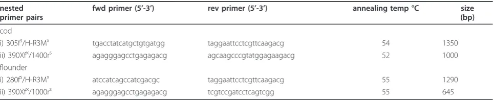
A highly sensitive and specific nested PCR detection assay was developed for the X-cell parasites from Atlan-tic cod and the northern black flounder, using the newly acquired SSU rDNA sequence data. Oligonucleotide pri-mers were designed that were universal for all currently known X-cell sequences and specific primers were also designed for each new X-cell parasite sequence. These primers were then used in the (universal/specific) com-binations described in Table 1. Nested PCR reactions were conducted using the same PCR conditions described above, but with adjusted annealing tempera-tures (Table 1), the products of the first round reaction were used as template DNA for the second reaction.
[image:4.595.57.540.614.712.2]In accordance with section 8.6 of the ICZN’s Interna-tional Code of Zoological Nomenclature, copies of this article are deposited at the following five publicly acces-sible libraries: Natural History Museum, London, UK; American Museum of Natural History, New York, USA; Museum National d’Histoire Naturelle, Paris,
Table 1 PCR primers used for the nested amplification of X-cell SSU rDNA
nested primer pairs
fwd primer (5’-3’) rev primer (5’-3’) annealing temp °C size
(bp)
cod
i) 305fs/H-R3Mx tgacctatcatgctgtgatgg taggaattcctcgttcaagacg 54 1350
ii) 390Xfx/1400rs agagggagcctgagagacg agcaagcccgtatggagaagacg 52 1000
flounder
i) 280fs/H-R3Mx atccatcagccatcgacgc taggaattcctcgttcaagacg 55 1290
ii) 390Xfx/1000rs agagggagcctgagagacg tcgtccgatcctcagtcgg 55 645
s
X-cell species-specific primer. x
France; Russian Academy of Sciences, Moscow, Russia; Academia Sinica, Taipei, Taiwan.
Results
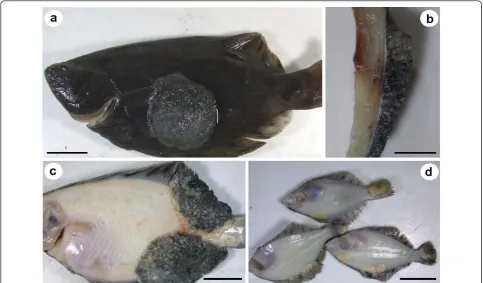
Atlantic cod with pseudobranchial pseudotumours were hand-selected from schooling juvenile fish in large indoor tanks. Infected cod appeared to be darker in col-our and generally smaller than uninfected ones from the same year class (Figure 1a). Infected pseudobranchs were significantly enlarged and had a creamy-whitish appearance, whereas uninfected pseudobranchs had a normal gill-red colour (Figure 1b &1c). The excised pseudobranchial pseudotumours were approximately 0.8 cm in length from fish that measured 10-12 cm in total length. The pseudobranchial pseudotumours from six bilaterally infected fish were used in the transmission experiment.
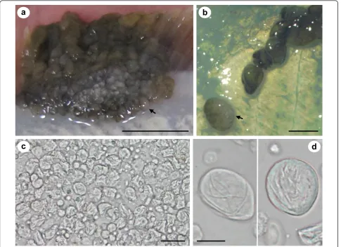
Pseudopleuronectes obscuruswith epithelial pseudotu-mours caused by X-cell infection were caught in Notsuke Bay, Hokkaido (Figure 2a-d). Epithelial pseudo-tumours were typical of those described from numerous flatfish species from Hokkaido and were often large and significantly raised from the body surface, but did not penetrate the underlying musculature (Figure 2a &2b), making them easy to remove from the fish to prepare the homogenate for the transmission experiment. Pseu-dotumours were always pigmented on the ocular (eyed) side, but could be either pigmented or non-pigmented on the abocular (non-eyed) side (Figure 2c &2d) and
when sampled, juvenile fish were also sometimes infected (Figure 2d). Pseudotumours often extended to the edge of the fins (Figure 2c & Figure 3a). Here, and at other margins of the pseudotumour, sac-like struc-tures were observed that could be detached, intact, with mild pressure (Figure 3b). The sacs were relatively fra-gile, easy to rupture and contained X-cells and other cellular debris (Figure 3c). X-cells from ruptured sacs that had been maintained in seawater for 24 hrs showed no signs of adhering to the plastic tissue culture plates, had no pseudopodia and were not amoeboid in form. After 24 hrs in seawater they appeared similar to ones that were freshly removed from sacs and had not suf-fered any noticeable shrinkage (Figure 3d).
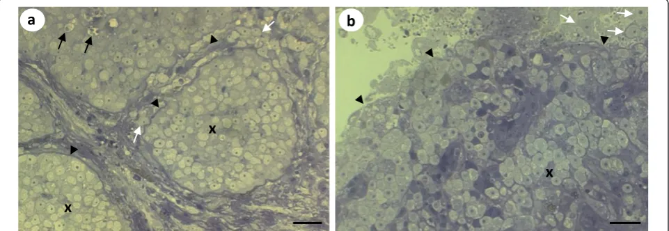
Histological analyses of the pseudotumours of cod and northern black flounder revealed that X-cells were pre-sent in large numbers in both pseudotumour types (Fig-ure 4a-d). InP. obscurus, the X-cells were limited to the epidermal skin layer and were arranged in a regular pat-tern of folded tissues that contained numerous melano-macrophages and other unidentifiable host cells (Figure 4a &4b). In cod the X-cells appeared to be adjacent to, but not intermingled with, pseudobranchial tissue (Fig-ure 4c). However, pseudobranchial cartilages were also seen surrounded by X-cells deeper into the pseudotu-mour lesions (Figure 4d).
Semi-thin sections of the sac-like structures show that in the central region of the sac, X-cells are found to be mostly restricted to large masses within the host
a
b
[image:5.595.56.540.452.692.2]c
epidermal tissue contained by a thick basal lamina-like membrane with underlying connective tissues. Some X-cells inside these large masses appear to be degener-ate, whilst others were observed amongst the connective tissues outside of the membrane that contains the majority of the parasites (Figure 5a). At the border of the sac there is a less organised tissue structure observed. Aggregations of X-cells are no longer con-tained within membranous structures and they are less densely packed together with numerous X-cells being present at the peripheral margin of the sac and some appearing to be external to the sac (Figure 5b).
Visual inspection of cod pseudobranchial tissues and microscopic inspection of both flatfish species at the end of the transmission experiments failed to detect the presence of abnormal tissue growth or signs of pseudo-tumours developing. Furthermore, PCR analyses of pseudobranchial DNA from experimental cod did not detect the presence of cod X-cell parasite DNA. PCR was not performed for the infection trials of the flatfish group.
Nested PCRs were successfully developed for both X-cell parasite species (Table 1), and no cross reactivity occurred between the two types of X-cells during PCR analyses. During the development of the nested PCR
assay, negative control tissue from infectedP. obscurus (skin samples taken >2 cm from pseudotumour sites), were sometimes found to be positive for the presence of X-cell DNA in the second round of the PCR; whereas skin samples from uninfected P. obscurus remained negative during both rounds of the PCR. Negative con-trol tissues from visibly uninfected cod were not found to contain X-cell DNA with the nested PCR.
Partial SSU rDNA sequence data from the X-cell para-sites infecting cod,G. morhua, and the northern black flounder,P. obscurus, were successfully obtained and have been deposited in GenBank (G. morhua X-cell, 1728 bp GU296508; P. obscurus X-cell, 1712 bp GU296509). A BLAST search with the contiguous X-cell sequence from P. obscurus showed a very high homol-ogy (> 99%) with the X-cell sequence reported from H. dubius [5]. A more detailed comparison with the X-cell sequence fromH. dubius revealed a 99.76% simi-larity over 1692 bases of comparable data, confirming that the two isolates from different flatfish hosts should be considered conspecific.
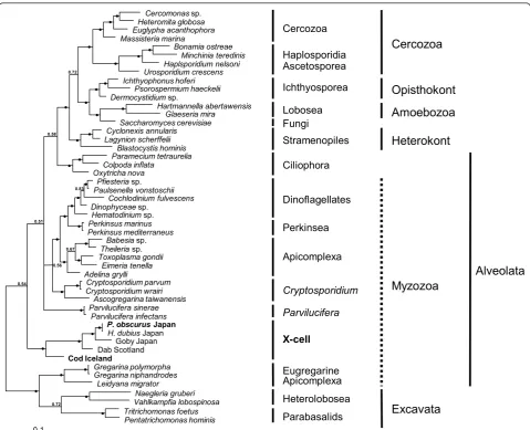
[image:6.595.56.540.89.372.2]Maximum parsimony and Bayesian phylogenetic ana-lyses produced trees with a similar topology. The Baye-sian inference tree (Figure 6) shows that all known X-cell sequences group together to form a monophyletic clade
that is within the alveolates and is basal to the majority of the myzozoan taxa. The sequence from P. obscurus groups with the sequence fromH. dubiusand collectively they form a clade with the species infecting the yellowfin goby from Japan. More basal to the Japanese/Pacific grouping is the X-cell parasite from European dab and basal to this main X-cell group is the sequence for the Atlantic cod pseudobranchial parasite. This monophy-letic grouping for the X-cell parasites was robustly sup-ported with internal probabilities of 1.0 at all nodes within the group. However, it was less well supported from the main spine of the tree with a probability of 0.54 and did not group with other protist phyla.
Discussion
The inability to successfully transmit both X-cell para-sites in this study indicates that either the experimental design or conditions were not suitable for transmission
to take place or that the X-cell stages seen in fish pseu-dotumours are not infective to new fish hosts. As com-prehensive precautions, described in the methods, were taken in preparation for and throughout the transmis-sion trials, we conclude that fish to fish transmistransmis-sion of X-cells is not readily achievable using feeding of infected tissues or by injection of an X-cell inoculum. Ideally we would have been able to use more fish and run the experiments for a longer period. However, the availabil-ity of infected tissues for feeding and homogenate pre-parations was limited.
We have shown that non-flagellate and non-amoeboid X-cells are easily shed from mature epidermal pseudotu-mours in discrete sac-like structures. These X-cells appear to lack a suitable apparatus with which to suc-cessfully infect a new fish host, unless direct transmis-sion is via ingestion of the sacs. However, if simple ingestion of sloughed off sacs were sufficient then
a
b
[image:7.595.57.543.89.441.2]c
d
feeding of freshly minced tumour tissue should have resulted in transmission of the parasite. Therefore, we believe that it is more likely that the X-cells undergo further development in the environment or in an inter-mediate host before reinfection of the fish host can occur. The continued support for X-cells being phylo-genetically related to the Myzozoa would also indicate that a flagellated life stage is present at some point in the life cycle which would be more capable of finding and infecting a new host fish.
Amoeboid X-cell forms
As no environmental spore-like or flagellated stages have been described from X-cell pseudotumours in fish, in spite of numerous thorough histopathological and ultastructural studies [1], it is not immediately obvious how transmission to a new host might take place
naturally. However, there have been several descriptions of amoeboid-like forms in advanced and ruptured X-cell pseudotumours which may represent the infective agent for a new host or a free-living stage leaving the fish host. Small amoeboid forms of X-cells have been described at the boundaries of epidermal pseudotu-mours in flatfish from Japan [17], free-floating amoeboid X-cells have been observed in advanced pseudobranchial pseudotumours in Atlantic cod [18] and free X-cells seen in the interlamellar spaces of ruptured gill lesions in the American plaiceH. platessoides[19]. Freeman [1] recently suggested that transmission of X-cells from the fish host could be via these amoeboid stages and that this amoeboid phase to their life cycle would most likely be present in marine sediments and indeed most fish species known to become infected with X-cells are clo-sely associated with the benthic environment [1].
a
c
b
d
m
d
x
x
ch
ch
ps
ps
x
[image:8.595.56.544.88.424.2]x
However, recent molecular phylogenetic studies [[1,2,5], present study] do not support the hypothesis that X-cells are amphizoic amoebae having a free-living stage in coastal marine sediments. Furthermore, in the present study we have clearly demonstrated that the X-cells con-tained in sac-like structures are not amoeboid in form, do not adhere to plastic culture plates and do not form pseudopodia. We consider this sac stage to be the most likely form to naturally detach from the host fish as they are delicately attached to the main pseudotumour and they are seemingly a discrete unit. During the histologi-cal analysis of these structures, the margins of the sacs were not found to contain X-cells in the typical orga-nised pattern that is observed in other parts of the pseu-dotumour. Rather, these X-cells are less densely packed together and some appear outside the sac membrane. This could signify that the sacs ultimately degenerate and release the X-cells into the environment. In addi-tion, X-cells from the sacs appear to be able to with-stand full salinity seawater, indicating that they are capable of surviving in the marine environment.
During the development of the diagnostic PCR, visibly uninfected areas of skin from infectedP. obscuruswere found to contain X-cell DNA. This supports the findings by Yamazaki et al. [17] who described the presence of very small amoeboid-like ‘wandering’ X-cells, only 3-4 μm in diameter, at the boundaries of epidermal pseudotumours in flatfish from northern Japan. These data suggest that amoeboid forms of X-cells do exist in fish tissues and are probably responsible for autoinfec-tion within the host, causing new pseudotumour growth
once host invasion and initial development has occurred.
Experimental design
It is possible that the methods used in the present study were not suitable for parasite transmission to occur between fish. Nevertheless, the transmission of fish para-sites with monoxenous life cycles between fish using similar methods to the ones used in this study has been achieved for various fish parasites [20,21]. However, suc-cessful parasite transmission can also be affected by environmental conditions such as salinity and tempera-ture. Therefore, in order to reduce the likelihood of this occurring, in our experiments we endeavoured to use seawater whose temperature and salinity were the same as those of the seawater of the natural environment from where infected fish were taken.
If the release of X-cells from mature or ruptured pseudotumours is required for transmission to a new host, and transmission between fish is possible without an alternate host, then long-term cohabitation experi-ments in tanks with suitable substrate cover, allowing time for the development of an environmental stage, could prove to be more successful in future X-cell trans-mission studies. Furthermore, using fish with advanced pseudotumours, close to rupture, could be important for successful transmission to occur between experimental fish. However, both gadoid and pleuronectid fish with X-cell pseudotumours have very poor survival rates after capture [3,22] which may hinder such long-term cohabi-tation experiments. In addition, lesion regression has
a
b
x
x
[image:9.595.58.539.91.257.2]x
also been observed in captive X-cell infected dab [22], suggesting that environmental conditions, such as tem-perature, may also be critical for the experimental trans-mission of X-cell parasites to take place and for pseudotumours to develop.
Geographical and environmental niches
X-cell pseudotumours in numerous wild fish popula-tions have been reported to occur within extremely nar-row geographic locations [4,23], being found in high numbers at one location and almost absent in adjacent localities or bays. This irregular distribution pattern ori-ginally led to suggestions that the X-cell condition was caused by localised coastal pollution [24,25]. A compre-hensive study by Katsura et al. [4] of the distribution of X-cell infected fish in Hokkaido, however, revealed that
the presence of X-cell pseudotumours was not related to coastal pollution, but had a strong correlation to the substrate type and amount of tidal water exchange. They found a very low frequency of infected fish (0.02-0.27%) on the north side of the Notsuke peninsula where a strong tide flows from north to south, but south of the peninsula, where the bay is protected from the strong current and the waters are‘stagnated and the bottom quite muddy’, fish had a 12.3% prevalence of X-cell pseudotumours. Similar findings for the distribu-tion patterns of populadistribu-tions of X-cell infected fish in Europe [23] suggest that certain environmental condi-tions such as substrate type and tidal flow potentially favour the survival and development of a free-living X-cell stage or can maintain an obligate alternate host that has a restricted zone for habitation.
Cercomonassp. Heteromita globosa Euglypha acanthophora Massisteria marina Bonamia ostreae Minchinia teredinis Haplsporidium nelsoni Urosporidium crescens Ichthyophonus hoferi Psorospermium haeckelii Dermocystidiumsp. Hartmannella abertawensis Glaeseria mira Saccharomyces cerevisiae Cyclonexis annularis Lagynion scherffelii Blastocystis hominis Paramecium tetraurelia Colpoda inflata Oxytricha nova Pfiesteriasp. Paulsenella vonstoschii Cochlodinium fulvescens Dinophyceaesp. Hematodiniumsp. Perkinsus marinus Perkinsus mediterraneus Babesiasp.
Theileria sp.
Toxoplasma gondii Eimeria tenella Adelina grylli Cryptosporidium parvum Cryptosporidium wrairi Ascogregarina taiwanensis Parvilucifera sinerae Parvilucifera infectans
P. obscurus Japan H. dubius Japan
[image:10.595.58.538.87.476.2]Goby Japan Dab Scotland Cod Iceland Naegleria gruberi Vahlkampfia lobospinosa Tritrichomonas foetus Pentatrichomonas hominis Gregarina polymorpha Gregarina niphandrodes Leidyana migrator 0.1 z z 0.72 0.54 z z z z z z 0.58 z z z z z z z z z z z z z z 0.83 0.56 0.51 0.67 z z z z z z z z z z z z z z 0.72 z z Fungi Cercozoa Haplosporidia Ascetosporea Stramenopiles Lobosea Ciliophora X-cell Eugregarine Apicomplexa Heterolobosea Parvilucifera Parabasalids
Cercozoa
Ichthyosporea ApicomplexaOpisthokont
Excavata
Heterokont
Amoebozoa
Myzozoa
Alveolata
Dinoflagellates Perkinsea CryptosporidiumSpecies specificity
Katsura et al. [4] suggested that species specificity was present among X-cell infections in flatfish from Hok-kaido, as some pleuronectid fish (Liopsetta pinnifasciata and Limanda punctatissimus) were collected in large numbers from numerous sites with muddy substrates with other X-cell infected pleuronectid fish being pre-sent, but they were never found to be infected with X-cells themselves. During this study we have shown that the same parasite causes X-cell pseudotumours in both H. dubius and P. obscurus from geographically distant locations in northern Japan, demonstrating that X-cell parasites are not species specific in some pleuro-nectid fish. It would be interesting to expand on this study and investigate whether the same parasite infects all seven pleuronectid fish known to be susceptible to epidermal X-cells infections in northern Japan. In our experimental transmission study, we used two fish spe-cies due to their availability from hatcheries; Verasper moseri (family Pleuronectidae) andParalichthys oliva-ceus(family Paralichthyidae).Verasper moseri, sampled from the same location we sampledP. obscurusfor this study, have been shown to be susceptible to infection with X-cell parasites causing characteristic epidermal pseudotumours [4], however it remains possible that the X-cells fromP. obscurusare a different species to those in V. moseri. The Japanese flounder, P. olivaceus, has not previously been reported to be susceptible to infec-tion with X-cell parasites and was included in this study due to its availability from hatcheries and its economic importance. We believe that P. olivaceusis probably not susceptible to the X-cell parasite infecting other flatfish species in northern Japan, as no reports are found in the literature and they are not pleuronectid fish. Its inclu-sion in this experiment could also have been valuable as a negative control had transmission been successful for V. moseri. In the gadoid transmission experiment we were able to use the same species for both donor and experimental fish, hence eliminating any species specifi-city concerns for the experiment.
Phylogenetic relationships
Using SSU rDNA alone, Miwa et al. [2,5] were able to demonstrate that X-cell parasites infecting different fish hosts (goby and flathead flounder) were clearly related, but were not able to demonstrate a stable phylogenetic relationship between the X-cell group and the other taxa used in their analyses. In a more comprehensive phylogenetic analysis, again limited to SSU rDNA, Free-man [1] confirmed monophyly for the X-cell group, but could only place the X-cell clade within the superphy-lum Alveolata and suggested that they were possibly related to both apicomplexans and dinoflagellates, and referred to them as basal myzozoans. The present study
is the first to include a SSU rDNA sequence for the cod X-cell parasite, and our phylogenetic analyses confirm monophyly for the X-cell group, but again fail to locate the X-cell clade convincingly within any of the recog-nised alveolate groupings. However, the inclusion of the sequence from cod X-cells has altered the expected topology of the tree for the alveolate group.Parvilucifera spp. that normally group with other perkinsids at the base of the dinoflagellates clade now occupy an unre-solved branch in the tree. It is evident from recent mole-cular studies [[1,2,5], present study] that SSU rDNA sequence data alone is not sufficiently informative to robustly place the X-cell group in phylogenetic studies and that additional more conserved gene regions should be studied to clarify their phylogenetic relationship with other alveolate taxa.
Suggested life cycle for X-cell parasites
To date, numerous histopathological and ultrastructural studies of X-cell parasites in fish have failed to reveal a developmental stage beyond the familiar X-cell, either locked in host tissue or as a free form in advanced pseu-dotumours. Here, we demonstrate that non-amoeboid X-cells are contained within discrete sac-like structures that are loosely attached to the fish host and that the X-cells from these sacs are able to tolerate seawater conditions. We believe that this represents good evi-dence that detachment from fish occurs naturally. Sac-like structures have not been observed or reported from pseudobranchial pseudotumours in gadoids or in X-cell gill lesions. However, free-floating X-cells have been observed in advanced pseudobranchial pseudotumours in Atlantic cod [18] and ruptured pseudotumours have been reported from X-cell gill lesions [19], which may serve as alternative release mechanisms for non-epidermal X-cells.
We do not believe that fish represent a dead-end or incidental host for X-cells. X-cells have evolved very specific tissue tropisms in the fish species they infect and have been reported from 5 teleost orders globally [1], suggesting a long-term and well-established host parasite relationship. Therefore, we can conclude that X-cell development in the fish host is purely a prolifera-tive phase that leads to further development either out-side of the fish host or in an alternate host. The lack of direct transmission for the X-cell parasites in this study supports this theory. Figure 7 shows four proposed stages in the life cycle for the X-cell parasite infecting flatfish in northern Japan.
developed during a free-living environmental phase in the life cycle.
II. A proliferative cycle starts in the fish which devel-ops into large epidermal pseudotumours, each con-taining large quantities of X-cells.
III. As the pseudotumours mature, discrete sac-like structures are lost from the host fish and fall to the seabed where the X-cells are released into the substrate.
IV. If the substrate type is suitable, either free-living environmental development occurs or the X-cells infect and develop within an alternate host to pro-duce infective stages for new fish hosts.
Understanding the life cycle of parasites that can infect commercially valuable fish species is important. In Europe, cod farming is an emerging industry and is still in the rela-tively early stages of development. However, pseudobran-chial X-cell infections in Atlantic cod have already been shown to cause serious pathology and mortalities in farmed fish [26]. Identifying farm-sites that might favour the propagation of or allow X-cells parasites to become established in surrounding sediments will assist future aquaculture endeavours, such as gadoid farming.
Conclusions
The presence of discrete sac-like structures, filled with non-amoeboid X-cells, which are readily detached from the host fish and the lack of fish to fish transmission for
both parasites in this study, suggests that other stages exist in the X-cell life cycle. This type of developmental cycle for X-cells may explain the lack of typical alveolate features seen in fish X-cells, which may be present in other stages of the life cycle. We suggest that X-cells have a second development phase either as free-living environmental organisms in the marine sediments or in an alternate host.
Analysis of SSU rDNA from X-cells infecting flatfish in Japan confirmed that the same X-cell parasite can infect more than one species of fish. Phylogenetic ana-lyses of X-cell SSU rDNA places them as a monophy-letic group in the alveolates. PCR analysis of infected flatfish showed that X-cell DNA is detectable in non-pseudotumour tissues, suggesting that some X-cells are motile in fish and may be responsible for autoinfection in the fish host.
Additional material
Additional file 1: Supplementary data. Additional small subunit ribosomal DNA sequences used in the phylogenetic analyses.
Acknowledgements
The Royal Society of Edinburgh (2× RSE International Exchange Programme: IEP Open - Outgoing Fellowships), The Sasakawa Foundation (grant No: 3034) and The Daiwa Foundation (grant No: 6211/6466) all provided initial funding for international travel with associated costs from the UK to Japan and Iceland and funding for work conducted at the University of Stirling. The University of Tokyo funded a visiting Associate Professorship for MAF to finalise the research in Japan during 2010.
Author details 1
Institute of Ocean and Earth Sciences & Institute of Biological Sciences, University of Malaya, Kuala Lumpur, Malaysia.2Institute of Aquaculture, University of Stirling, FK9 4LA, Scotland, UK.3Institute for Experimental Pathology, University of Iceland, Keldur, Reykjavík, Iceland.4Laboratory of Fish Diseases, The University of Tokyo, Tokyo 113-8657 Japan.5Laboratory of Microbiology, Hokkaido University, Hakodate 041-8611, Japan.6Department of Aquatic Bioscience and Industry Faculty of Bio-Industry, Tokyo University of Agriculture, Abashiri, Hokkaido 099-2493, Japan.7Hokkaido Central Fisheries Experiment Station, Yoichi 046-8555, Japan.
Authors’contributions
ME and MAF collected material in Iceland and set up the transmission experiment, ME maintained the experiment for the duration. MY and KW organised sample collection in Japan, KO and MF set up the transmission experiment and KM maintained the experiment for the duration. MAF performed wax histology and DNA analyses, developed the nested PCR and drafted the manuscript. APS cut and viewed semi-thin plastic sections of the sac-like structures.
Competing interests
The authors declare that they have no competing interests.
Received: 2 December 2010 Accepted: 8 February 2011 Published: 8 February 2011
References
1. Freeman MA:X-cell parasites in the European dabLimanda limandaare related to other X-cell organisms: a discussion on the potential identity of this new group of parasites.Parasitology2009,10:151-170.
muddy substrate suitable temperature, salinity
and tidal flow
I
II
III
IV
Adv
an
ced
lesions,
loss of
p
seudotumour
ex
tr
em
ities
In
fectious stag
e
proliferative cycle
[image:12.595.57.290.88.248.2]unknown developmental cycle
2. Miwa S, Kamaishi T:Protistan X-cells in pseudotumors of yellowfin goby
Acanthogobius flavimanusare a distinct organism from those in flathead flounderHippoglossoides dubius.Dis Aquat Org2009,85:53-57.
3. Morrison CM, Shum G, Appy RG, Odense P, Annand C:Histology and prevalence of X-cells lesions in Atlantic cod (Gadus morhua).Can J Fish Aquat Sci1982,39:1519-1530.
4. Katsura K, Yamazaki F, Hamada K, Oishi K, Harada T, Shinkawa T:
Geographic distribution and frequency of pseudotumourous fishes collected from the coastal waters of Hokkaido, Japan.B Jpn Soc Sci Fish
1984,50:979-984.
5. Miwa S, Nakayasu C, Kamaishi T, Yoshiura Y:X-cells in fish
pseudopseudotumours are parasitic protozoans.Dis Aquat Org2004,
58:165-170.
6. Ito Y, Kimura I, Miyake T:Histopathological and virological investigations of papillomas in soles and gobies in coastal waters of Japan.Prog Exp Pseudotumour Res1976,20:86-93.
7. Diamant A, Smail DA, McFarlane L, Thomson AM:An infectious pancreatic necrosis virus isolated from common dabLimanda limandapreviously affected with X-cell disease, a disease apparently unrelated to the presence of the virus.Dis Aquat Org1988,4:223-227.
8. Renault T, Stokes NA, Chollet B, Cochennec N, Berthe F, Gerard A, Burreso EM:Haplosporidiosis in the Pacific oysterCrassostrea gigasfrom the French Atlantic coast.Dis Aquat Org2000,42:207-214.
9. Hillis DM, Dixon MT:Ribosomal DNA: molecular evolution and phylogenetic inference.Quart Rev Biol1991,66:411-453.
10. Freeman MA, Yokoyama H, Ogawa K:Description and phylogeny of
Ceratomyxa ankosp. n. andZschokkella lophiisp. n. from the Japanese anglerfish,Lophius litulon(Jordan).J Fish Dis2008,31:921-930. 11. Thompson JD, Gibson TJ, Plewniak F, Jeanmougin F, Higgins DG:The
CLUSTAL-X windows interface: flexible strategies for multiple sequence alignment aided by quality analysis tools.Nucl Acids Res1997,
24:4876-4882.
12. Hall TA:BioEdit: a user-friendly biological sequence alignment editor and analysis program for Windows 95/98/NT.Nucleic Acids Symp Ser1999,
41:95-98.
13. Altschul SF, Gish W, Miller W, Myers EW, Lipman DJ:Basic local alignment search tool.J Mol Biol1990,215:403-410.
14. Swofford DL:PAUP* Phylogenetic analysis using parsimony (*and other methods), v. 4.0 beta10.Sinauer Associates, Sunderland, MA, USA; 2002. 15. Ronquist F, Huelsenbeck JP:MrBayes 3 Bayesian phylogenetic inference
under mixed models.Bioinformatics2003,19:1572-1574.
16. Nylander JAA, Ronquist F, Huelsenbeck JP, Nieves-Aldrey JL:Bayesian phylogenetic analysis of combined data.Syst Biol2004,53:47-67. 17. Yamazaki F, Hibino T, Oishi K, Harada T, Stich HF, Acton AB:X-cell morphology in the epidermal papillomas of flatfish collected from coastal waters of Hokkaido, Japan.B Jpn Soc Sci Fish1978,44:407-413. 18. Watermann B, Dethlefsen V:Histology of pseudobranchial
pseudotumours in Atlantic cod (Gadus morhua) from the North Sea and the Baltic Sea.Helgolander Meeresun1982,35:231-242.
19. Maclean SA, Despres-Patanjo LI:Gill X-cell lesion in American plaice (Hippoglossoides platessoides) concurrent with lymphocystis infection.J Fish Biol1993,43:947-950.
20. Redondo MJ, Palenzuela O, Riaza A, Ángeles A, Álvarez-Pellitero P:
Experimental transmission ofEnteromyxum scophthalmi(Myxozoa) an enteric parasite of turbotScophthalmus maximus.J Parasitol2002,
88:482-488.
21. Lee SJ, Yokoyama H, Ogawa K:Modes of transmission ofGlugea plecoglossi(Microspora) via the skin and digestive tract in an experimental infection model using rainbow trout,Oncorhynchus mykiss
(Walbaum).J Fish Dis2004,27:435-444.
22. Diamant A, McVicar AH:The effect of internal and external X-cell lesions on common dab,Limanda limandaL.Aquaculture1987,67:127-133. 23. Diamant A, McVicar AH:Distribution of X-cell disease in common dab,
Limanda limandaL., in the North Sea, and ultrastructural observations of previously undescribed developmental stages.J Fish Dis1989,12:25-37. 24. Stich HF, Acton AB, Forrester CR:Fish tumors and sub-lethal effects of
pollutants.J Fish Res Board Can1976,33:1993-2001.
25. Wellings SR, McCain BB, Miller BS:Epidermal papillomas in Pleuronectidae of Puget Sound, Washington.Prog Exp Tumor Res1976,20:55-74.
26. Eydal M, Kristmundsson A, Bambir SH:Pseudobranchial X-cell
pseudotumors in young wild and farmed Atlantic codGadus morhuain Iceland.Dis Aquat Org2010,91:83-88.
doi:10.1186/1756-3305-4-15
Cite this article as:Freemanet al.:Molecular identification and transmission studies of X-cell parasites from Atlantic codGadus morhua (Gadiformes: Gadidae) and the northern black flounder
Pseudopleuronectes obscurus(Pleuronectiformes: Pleuronectidae). Parasites & Vectors20114:15.
Submit your next manuscript to BioMed Central and take full advantage of:
• Convenient online submission
• Thorough peer review
• No space constraints or color figure charges
• Immediate publication on acceptance
• Inclusion in PubMed, CAS, Scopus and Google Scholar
• Research which is freely available for redistribution