The Effect of Fixed Orthodontic
Brackets on the Bacterial Composition
of Dental Plaque in Adolescents
Thesis submitted in accordance with the requirements of the
University of Liverpool for the degree of Doctor of Dental Science
(Orthodontics)
Amal Sadeq
1
Acknowledgements
I would like to thank Professor Neil Pender, Professor Susan Higham and Dr Sabeel Valappil for their supervision and continued support over the last three years.
I would like to thank Dr Janet Risk for her help and advice with all the molecular techniques used and Dr Gleb Komarov for his assistance with the QLF equipment.
2
Table of contents
!"#$%&'()*(+($,-.////////////////////////////////////////////////////////////////////////////////////////////////////////.0!
,!1'(.%2."%$,($,-.////////////////////////////////////////////////////////////////////////////////////////////////////////////.3!
'4-,.%2.24*56(-.////////////////////////////////////////////////////////////////////////////////////////////////////////////////////.7!
'4-,.%2.,!1'(-.////////////////////////////////////////////////////////////////////////////////////////////////////////////////////.00!
!1-,6!",.//////////////////////////////////////////////////////////////////////////////////////////////////////////////////////////////.03!
3
!"O"!$! I&0&373&*+'$*B$3F4$=/KC%PPQ$043F*8$""""""""""""""""""""""""""""""""""""""""""""""""""""""""""""""""""$$ ..! "#A! %55%*'!25!2+'12$2&',*!(00),(&*%-!2&!'1%!0)(<=%!7,*+25)2+(!##########################!44! "#">! $%'%*',2&!25!,&,',()!*(+,%-!)%-,2&-!BC1,'%!-02'!)%-,2&-D!##################################!4@! "#""! 0+%;%&',2&!(&$!'+%('7%&'!25!C1,'%!-02'!)%-,2&-!###############################################!:/!
"8!9,(6.3:.!4+-./////////////////////////////////////////////////////////////////////////////////////////////////////////////////.;<! /#"! (,7-!#############################################################################################################################################!:9! /#/! &=))!1802'1%-%-!###################################################################################################################!:9!
"8!9,(6.=:.-,5)>.)(-4*$./////////////////////////////////////////////////////////////////////////////////////////////.;7! 4#"! -'=$8!$%-,3&!(&$!0(+',*,0(&'-!########################################################################################!:?! ."!"!!!!!!!R328:$84'&,+$""""""""""""""""""""""""""""""""""""""""""""""""""""""""""""""""""""""""""""""""""""""""""""""""""""""""""""""""""""$$ AJ! ."!"-!!!!!!=753&(&17+3'$"""""""""""""""""""""""""""""""""""""""""""""""""""""""""""""""""""""""""""""""""""""""""""""""""""""""""""""""""""""$$ AJ! ."!".$! QM()2'&*+$(5&345&7$"""""""""""""""""""""""""""""""""""""""""""""""""""""""""""""""""""""""""""""""""""""""""""""""""""""""""""$$ AO! 4#/! %'1,*()!(00+2;()!##################################################################################################################!:@!
"8!9,(6.;:.4)($,424"!,4%$.!$)."%''(",4%$.%2.6().2'5%6(-"($,.
9'!?5(.//////////////////////////////////////////////////////////////////////////////////////////////////////////////////////////////////////////.;@! :#"!!!!!!!7%'12$!#####################################################################################################################################!:A! A"!"!$! >84+3&B&(73&*+$*B$548$B)2*54'(4+3$1)7924$""""""""""""""""""""""""""""""""""""""""""""""""""""""""""""""$$ AS! A"!"-$! /*))4(3&*+$*B$548$B)2*54'(4+3$1)7924$""""""""""""""""""""""""""""""""""""""""""""""""""""""""""""""""""""""$$ D!! :#/! +%-=)'-!######################################################################################################################################!."!
"8!9,(6.A:.96('4+4$!6>.)($,!'.9'!?5(.!$!'>-4-.////////////////////////////////////////.AA!
4
D"-".$! >84+3&B&(73&*+$*B$,4+*0&($%T?$"""""""""""""""""""""""""""""""""""""""""""""""""""""""""""""""""""""""""""""""""$$ DJ! .#/! (70),5,*(',2&!25!6(*'%+,()!$&(!68!0*+!#####################################################################!.@! D"-"!$! =/K$547(3&*+$""""""""""""""""""""""""""""""""""""""""""""""""""""""""""""""""""""""""""""""""""""""""""""""""""""""""""""""""""""$$ DO! D"-"-!!!!!!>84+3&B&(73&*+$*B$=/K$15*82(3'$""""""""""""""""""""""""""""""""""""""""""""""""""""""""""""""""""""""""""""""""$$ DO! .#4! 3%&%+(',2&!25!$33%!0+25,)%!###########################################################################################!.A! D"."!!!!!!!R&)G45$'37&+&+,$"""""""""""""""""""""""""""""""""""""""""""""""""""""""""""""""""""""""""""""""""""""""""""""""""""""""""""""""""$$ #!! 44/.6(-5',-./////////////////////////////////////////////////////////////////////////////////////////////////////////////////////////////.<0!
"8!9,(6.<:.B!'4)!,4%$.%2.*"C"'!+9.9"6.964+(6-./////////////////////////////////////////.<7! 9#"! 0+,7%+!$%-,3&!#########################################################################################################################!9?! 9#/! $33%!*2&$,',2&-!20',7,-(',2&!########################################################################################!9A!
5
?#/#"#"4! 0GHIJKJLGMI!"4!#####################################################################################################################################!@9! ?#/#"#":! 0GHIJKJLGMI!":!#####################################################################################################################################!@?!
"8!9,(6.D:.4)($,424"!,4%$.!$).+%$4,%64$*.%2.&84,(.-9%,.'(-4%$-./.@0!
@#"! 7%'12$!######################################################################################################################################!A"! O"!"!!!!!!!I4'&*+$&84+3&B&(73&*+$""""""""""""""""""""""""""""""""""""""""""""""""""""""""""""""""""""""""""""""""""""""""""""""""""""$$ S!! O"!"-$! I4'&*+$927+3&B&(73&*+$"""""""""""""""""""""""""""""""""""""""""""""""""""""""""""""""""""""""""""""""""""""""""""""""""""$$ S!! @#/! +%-=)'-!######################################################################################################################################!A/!
"8!9,(6.@:.)4-"5--4%$.///////////////////////////////////////////////////////////////////////////////////////////////////.@7! A#"!!!!!,$%&',5,*(',2&!25!+%$!5)=2+%-*%&'!0)(<=%!#################################################################!A?! A#/!!!!!,$%&',5,*(',2&!25!+%$!5)=2+%-*%&'!0)(<=%!6(*'%+,()!*2702-,',2&!##################!AA! A#4!!!!!,$%&',5,*(',2&!(&$!72&,'2+,&3!25!C1,'%!-02'!)%-,2&-!########################################!">:! A#:!!!!!),7,'(',2&-!25!'1%!-'=$8!#################################################################################################!">.! S"A"!$$$$$R701)4VVVVVVVVVVVVVVVVVVVVVVVVVVVVVVVVVVV""!WD! S"A"-$$$$$?+7):'&'$*B$548$B)2*54'(4+3$1)7924VVVVVVVVVVVVVVVVVVVV"!W#! S"A".$$$$$X637&+&+,$HI;$&07,4'VVVVVVVVVVVVVVVVVVVVVVVVVVV!WO! S"A"A$$$$$R373&'3&(7)$7+7):'&'VVVVVVVVVVVVVVVVVVVVVVVVVVVVV!WS! "8!9,(6.0E:."%$"'5-4%$-./////////////////////////////////////////////////////////////////////////////////////////.00E!
"8!9,(6.00:.6("%++($)!,4%$-.2%6.25,56(.&%6#.///////////////////////////////////.000!
6(2(6($"(-.///////////////////////////////////////////////////////////////////////////////////////////////////////////////////////.00=!
!99($)4F.!.////////////////////////////////////////////////////////////////////////////////////////////////////////////////////////.03;! ,&52+7(',2&!-1%%'!########################################################################################################################!"/:!
!99($)4F.1.////////////////////////////////////////////////////////////////////////////////////////////////////////////////////////.03<! =&$%+!"9-!,&52+7(',2&!-1%%'!##################################################################################################!"/9!
6
!99($)4F.".////////////////////////////////////////////////////////////////////////////////////////////////////////////////////////.03D! 0(+%&'N3=(+$,(&!,&52+7(',2&!-1%%'!###################################################################################!"/@!
!99($)4F.).////////////////////////////////////////////////////////////////////////////////////////////////////////////////////////.03@! *2&-%&'!52+7!##################################################################################################################################!"4>!
!99($)4F.(.////////////////////////////////////////////////////////////////////////////////////////////////////////////////////////.0=0! (--%&'!52+7!#####################################################################################################################################!"4"!
!99($)4F.2./////////////////////////////////////////////////////////////////////////////////////////////////////////////////////////.0=3! ?(5:)70&84$84+7325&+,$'3*(Y$'*)23&*+$15417573&*+VVVVVVVVVVVVVVV!.-!
.>O!'(%!6PQQRH!B")D!#############################################################################################################################################!"4/! $RMGIPHJMS!TUVPIJUM!B">>!WVD!##########################################################################################################################!"4/! $RMGIPHJMS!TUVPIJUM!LRH!">>!WV!TUVPIJUM!###################################################################################################!"44! ">X!(WWUMJPW!0RHTPVQGIR!B"!WVD!###############################################################################################################!"44!
!99($)4F.*.////////////////////////////////////////////////////////////////////////////////////////////////////////////////////////.0=;! -,);%+!-'(,&,&3!+%(3%&'-!(&$!0+2'2*2)!###############################################################################!"4:! 0HUIUKUV!#####################################################################################################################################################################!"4:!
7
List of figures
Figure 1.1: PCR-DGGE profiles ... 29!
Figure 1.2: The clinical appearance of white spot lesions seen at the gingival
margins of the upper incisors. ... 39!
Figure 1.3: Tooth image with QLF where the contrast between sound and
demineralised enamel is clear. ... 40
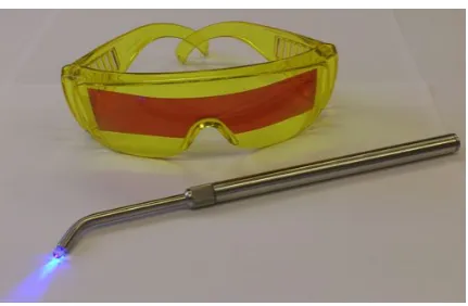
Figure 4.1: The ToothcareTM devoce.. ... 50!
Figure 4.2: The chart used to note the postiion of the red fluorescent plaque once
identified with Toothcare TM. ... 50!
Figure 4.3: the distribution of red fuorescent plaque collected from each tooth. 53
Figure 4.4: The relationship between red fluorescent plaque and white spot lesions
as a percentage of the total number of tooth surfaces with red fluorescent plaque. ... 54!
Figure 4.5: The relationship between red fluorescent plaque and white spot lesions
as a percentage of the total number of tooth surface. ... 54!
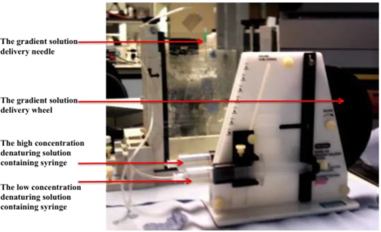
Figure 5.1: The gradient delivery apparatus. ... 59
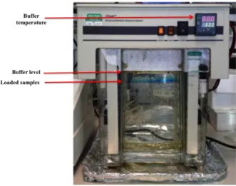
Figure 5.2: Fully assembled DGGE apparatus with the samples loaded and seen as
blue horizontal lines.. ... 60!
Figure 5.3: The agarose gel (0.8%) profile of genomic DNA stained with ethidium
bromide.. ... 62!
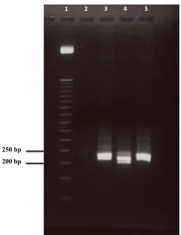
Figure 5.4: The agarose gel (2.5%) profile of 16s rRNA gene-PCR product stained
8
Figure 5.5: PCR-DGGE profiles from red fluorescent dental plaque samples and
laboratory strains of oral bacteria stained with silver staining………....64
Figure 6.1: Agarose gel (2.5%) profile of 16s rRNA gene GC-Clamp PCR product
stained with ethidium bromide.. ... 688!
Figure 6.2: Calibration graph of bp size and the corresponding distance travelled for
each band of the 50 bp DNA Marker. ... 69!
Figure 6.3: DGGE acrylamide gel (7.5%) with a denaturant concentration range of
30% to 60%. ………..……...70
Figure 6.4: DGGE acrylamide gel (7.5%) with a denaturant concentration range of
25% to 45%.. ... 71!
Figure 6.5: S.mutans colonies after 72 hours of aerobic incubation at 37oC on BHI
agar. ... 72!
Figure 7.1: agarose gel (2.5%) profile of GC-clamp 16s rRNA gene-PCR product
stained with ethidium bromide.. ... 75!
Figure 7.2: agarose gel (2.5%) profile of GC-clamp 16s rRNA gene-PCR product
stained with ethidium bromide. ... 75!
Figure 7.3: agarose gel (2.5%) profile of GC-clamp 16s rRNA gene-PCR product
stained with ethidium bromide. ... 76!
Figure 7.4: agarose gel (2.5%) profile of GC-clamp 16s rRNA gene-PCR product
stained with ethidium bromide.. ... 76!
Figure 7.5: agarose gel (2.5%) profile of GC-clamp 16s rRNA gene-PCR product
9
Figure 7.6: DGGE profiles from red fluorescent dental plaque samples and
laboratory strains of oral bacteria stained with ethidium bromide.. ... 78!
Figure 7.7: DGGE profiles from red fluorescent dental plaque samples and
laboratory strains of oral bacteria stained with ethidium bromide.. ... 79!
Figure 7.8: DGGE profiles from red fluorescent dental plaque samples and
laboratory strains of oral bacteria stained with ethidium bromide. ... 80
Figure 7.9: DGGE profiles from red fluorescent dental plaque samples and
laboratory strains of oral bacteria stained with ethidium bromide.. ... 81!
Figure 7.10: An example of DGGE analysis performed with TotalLab Quant
software ... 87!
Figure 8.1: QLF images of an upper right lateral incisor at baseline (A) and 5
months after bracket placement (B)... ... 94!
Figure 8.2: A QLF image of an upper right lateral incisor for participant number 6
where demineralisation was detected………...96!
Figure H-1: DGGE profiles and TotalLab Quant software analysis of red fluorescent
dental plaque samples and laboratory strains of oral bacteria stained with ethidium bromide… ... 87!
Figure H-2: DGGE profiles and TotalLab Quant software analysis of red fluorescent
dental plaque samples and laboratory strains of oral bacteria stained with ethidium bromide. ... 87!
Figure H-3: DGGE profiles and TotalLab Quant software analysis of red fluorescent
10
Figure H-4: DGGE profiles TotalLab Quant software analysis of red fluorescent
11
List of tables
Table 4.1: Number of visits and red fluorescent plaque samples collected. ... 52!
Table 7.1: The number and time (T) of visits for each participant. ... 74!
Table 7.2: The distribution of bands and their corresponding microorganisms for
participants 1-5. ... 80!
Table 7.3: The distribution of bands and their corresponding microorganisms for
participants 6-10 ... 89!
Table 7.4: The distribution of bands and their corresponding microorganisms for
participants 11-14. ... 90!
Table 8.1: Number of visits, QLF images collected and white spot lesiosn identified
per participant. ... 92!
Table 8.2: The distribution of white spot lesions detected with QLF, their average
12
Abstract
BACKGROUND: Demineralisation around orthodontic brackets is a
considerable risk of orthodontic treatment. It can occur very early during treatment and involve a large number of teeth with aesthetic and dental health consequences. Early detection of demineralisation is very important in terms of treatment and prevention.
The presence of bacteria is one of the requirements for demineralisation to take place and it is unclear whether the type of bacteria in the plaque or the quantity of certain types of bacteria are significant influences on demineralisation.
AIM: To identify any general changes in the bacterial composition of dental
plaque in adolescents undergoing fixed orthodontic appliance therapy. A secondary aim is to determine the incidence of white spot lesion development and if this was related to the identified red fluorescent plaque.
STUDY DESIGN: Prospective longitudinal cohort study.
METHODS: Fourteen 11 to 23 year old consecutive patients attending the
13
RESULTS: The incidence of white spot lesions was recorded as 4.2% of the
total surfaces of the teeth included as detected with QLF, the development of white spot lesions was not associated with the presence of red fluorescent plaque. There were differences in the bacterial composition of red fluorescent supragingival plaque in terms of P. gingivalis, S. mutans and S. gordonii between different participants, although changes in the plaque composition between visits for the same participant was not significantly associated with the development of white spot lesions in adolescents.
CONCLUSIONS: With the limitations of this study, the placement of fixed
14
Chapter 1
Introduction
15
1.1
Dental caries
According to the World Health Organisation dental caries is one of the most prevalent chronic diseases in the world and is the primary cause of oral pain and tooth loss. An estimated five billion people worldwide had experienced dental caries, and its treatment is estimated to account for 5-10% of health costs in industrialised countries (Petersen 2003).
Dental caries is the localised destruction of susceptible dental hard tissues by acidic by-products from bacterial fermentation of dietary carbohydrates. It is a multifactorial disease that starts with microbiological shifts within the complex biofilm and is affected by salivary flow and composition, exposure to fluoride, consumption of dietary sugars, and by preventive behaviours such as teeth cleaning (Selwitz et al. 2007). It was described as a chronic disease that progresses slowly in most individuals and in the absence of treatment can progress from initial loss of mineral at the ultra structural level to total destruction of tooth structure. Caries lesion development is not continuous, but a highly dynamic series of alternating periods of progression and arrest/regression (Takahashi and Nyvad 2008).
1.2
Pathogenesis
16
disturbed by mechanical wear (mastication, attrition, abrasion from brushing, flossing or toothpicks).
The plaque biofilm is a prerequisite for caries lesions to occur. It is characterised by continued microbial activity resulting in continued metabolic events in the form of minute pH fluctuations. Dental caries lesions are the product of innumerable metabolic events in biofilms, which have covered the tooth surface. When this outcome results in a cumulative loss of mineral from the tooth of such a magnitude, the porosity in the enamel gives rise to a decrease in enamel translucency. This early stage in enamel lesion formation will manifest itself as a white-spot lesion. The shape of the lesion reflects where the biofilm has been allowed to grow and remain for prolonged periods.
1.3
The role of dental biofilm in the aetiology of
dental caries
Dental plaque is a general term for the complex but organised cooperative community of microorganisms that develops on the tooth surface, embedded in a matrix of polymers of bacterial and salivary origin. It forms via an ordered sequence of events, resulting in a structurally and functionally organised species rich microbial community (Marsh 2004).
17
microflora, only a single or very small number of species were actively involved in the disease (Marsh 1994).
Many studies have suggested that Streptococcus mutans are the major group of bacteria associated with human dental caries. That is mainly due to their frequent isolation from cavitated carious lesions, their highly acidogenic and aciduric nature, their ability to produce water insoluble glucans and their capability of inducing caries formation in animals when fed a sucrose rich diet (Takahashi and Nyvad 2008). It was believed that dental caries is a specific treatable infection and that mutans streptococci and lactobacilli are responsible for the majority of human dental decay (Loesche 1986). Streptococcus mutans colonise the fissures of the teeth soon after they erupt and that inevitably causes decay unless other bacteria occupy the fissure depths, and then there is a possibility that decay will not take place or at least be greatly reduced.
A systematic review by Tanzer examined the evidence concerning microbial causes and associations with dental caries in humans. The included studies comprised of 25 interventional randomised clinical trials and 79 prospective and retrospective longitudinal and case control studies. The majority of these studies support a strong positive statistical association of mutans streptococci with carious lesions. The overall conclusion of the review suggested a central role of mutans streptococci in the initiation of caries on smooth surfaces and crown fissures of teeth (Tanzer et al., 2001).
18
consume lactate are involved in the carious process. Thus, a heterogeneous mixture of microorganisms could play a role in disease (Thomas and Nakaishi 2006).
Some recent studies indicate that the relationship between mutans streptococci and caries is not absolute. Whereas high proportions of mutans streptococci may persist on the teeth surfaces without leading to lesion development, caries can develop in the absence of these species. A cross-sectional study showed that 10% of subjects with rampant caries in the permanent dentition did not have detectable levels of S. mutans (Aas et al., 2005) and suggested that health and disease related bacterial species other than S. mutans are likely to play an important role in caries progression.
In addition to the specific and non-specific plaque hypotheses, an alternative hypothesis has been proposed (Marsh 1994; Takahashi and Nyvad 2008). In brief, it proposes that the organisms associated with disease may also be present at sound sites, but at levels too low to be clinically relevant. Disease is then a result of a shift in the balance of the resident microflora driven by a change in local environmental conditions.
19
fermentable carbohydrates intake increases and/or salivary flow is impaired, then the biofilm spends more time below the critical pH for enamel demineralisation (approximately pH 5.5). The effect of this on the microbial ecology would be twofold. Firstly, conditions of low pH favour the proliferation of aciduric and acidogenic bacteria especially mutans streptococci and lactobacilli, but not exclusively so (Van et al., 1994), and tips the balance towards demineralisation. Secondly, greater numbers of aciduric bacteria such as mutans streptococci and lactobacilli in plaque would result in more acid being produced at even faster rates, thereby enhancing demineralisation further.
Bacteria other than mutans streptococci and lactobacilli could also make acid under similar conditions, but at a slower rate, or could initiate lesions in the absence of other aciduric species in a more susceptible host. If highly aciduric species were not present initially, then the repeated conditions of low pH coupled with the inhibition of competing organisms might increase the likelihood of colonisation of mutans streptococci or lactobacilli.
20
1.4
Development of dental plaque biofilm
Dental plaque is a general term for the complex but organised cooperative community of microorganisms that develops on the tooth surface, embedded in a matrix of polymers of bacterial and salivary origin (Overman 2000). Dental plaque forms via an ordered sequence of events, resulting in a structurally and functionally organised species rich microbial community. According to Marsh, the development of plaque biofilm can be subdivided into several stages (Marsh 2004).
a. Acquired pellicle formation: the formation of plaque begins with the rapid non selective deposition of several salivary proteins onto all exposed intraoral surfaces, that takes place as soon as a tooth surface is cleaned (Nassar et al., 1995). The thickness of the pellicle ranges from 50-150nm after 24 hours and is influenced by the shear forces acting at the site of formation (Hannig and Balz 1999).
21
c. Pioneer microbial colonisers and irreversible attachment: within a short time, initial weak physiochemical interactions may become irreversible due to adhesins on the microbial cell surface becoming involved in specific, short-range interactions with complementary receptors in the acquired pellicle. The initial colonisers constitute of a highly selected part of the oral microflora. They are organisms able to withstand the high oxygen concentrations and to resist the various removal mechanisms of the oral cavity such as swallowing, chewing and salivary and crevicular fluid outflow (Sbordone and Bortolaia 2003). Within minutes coccal bacterial appear, and they are mainly S. sanguinis, S. oralis and S. mitis. Once attached, these pioneer populations start to divide and form microcolonies. These early colonisers become embedded in bacterial extracellular slimes and polysaccharides together with additional layers of adsorbed salivary proteins and glycoproteins. The irreversible attachment of cells to the tooth involves specific short range stereochemical interactions between components on the microbial cell surface (adhesins) and complementary receptors in the acquired pellicle. d. Coaggregation/coadhesion and microbial succession: over time, the plaque
22
e. Mature biofilm formation: the microbial diversity of plaque will increase over time due to successive waves of microbial succession and subsequent growth. The growth rate of individual bacteria within plaque slows as the biofilm matures. Confluent growth on the tooth surface produces a biofilm with 3-dimensional structure. Some of the adherent bacteria synthesise extracellular polymers, which will make a major contribution to the plaque matrix (Rosan and Lamont, 2000).
Microorganisns in the mature biofilm can adhere firmly to tooth surfaces unless subjected to shear forces such the ones resulting from brushing or chewing. Some bacteria can actively detach themselves from within the biofilm so as to be able to colonize elsewhere. Streptococcus mutans can synthesise an enzyme that can cleave proteins from its own surface and thereby detach itself from a mono-species biofilm.
1.5
Importance of plaque identification
23
which are pathogenic, it is more important to detect the virulent bacterial species that are linked to oral diseases.
1.6
Plaque quantification techniques
1.6.1
Disclosing
If plaque is present in large amounts it can be clearly seen without any aid. But in small quantities, and because it is generally colourless, plaque is usually stained prior to assessment. Common disclosing agents used include erythrosine (E 127 red). In certain products this erythrosine is usually combined with a blue dye (E 133). Disclosing is a very effective method for patients’ oral hygiene improvement. But for the researcher, it lacks precision, objectivity, sensitivity, specificity and reliability that are required for clinical trials (Pretty et al., 2005).
1.6.2
Computer based plaque analysis
24
1.6.3
Fluorescein disclosing and digital plaque image analysis
(DPIA)
This technique relies on the use of fluorescein as a disclosing agent (Lang et al., 1972) and subsequent digital measurement of the fluorescent component. Under UV light, fluorescein disclosed plaque would appear yellow green in comparison to the dark oral tissues. Image analysis is then carried out for each tissue. On the computer, plaque appears red, dental hard tissues are blue and soft tissues are green making the separation of various oral tissues clear and unambiguous.
The technique permits smaller amounts of plaque to be measured and smaller changes between treatments to be detected due to its increased sensitivity (Smith et al., 2001). But the patient will be at risk of demineralisation, because in order to achieve good fluorescence, the pH of the system has to be low. In addition, the system is expensive and the plaque quantification is limited to the facial surfaces of the anterior teeth (Sagel et al., 2000).
1.6.4
Plaque quantification using 3D co-ordinate data
25
For purposes of superimposition, the plaque located around the mid-line of the coronal half has to be removed. This coud have a negative impact on caries studies. The other down fall of this system is its reliance on direct impressions of the teeth because the laser scanner cannot be used within the mouth.
1.6.5
Plaque detection with quantitative light-induced
fluorescence (QLF)
This technique relies on the ability of plaque bacteria to produce red fluorescing components. The phenomenon of red fluorescence in carious dental hard tissues and in dental plaque and calculus was first reported in the 1920’s (Lennon et al., 2006). When plaque is present in large amounts on tooth surfaces, it fluoresces deep red or bright orange (Pretty et al., 2005; van der Veen and de Josselin de Jong 2000). Red auto fluorescence is presumed to originate from bacterial products that are chromophores of porphyrins (Konig et al., 1998), but the exact nature of the fluorescing chromophores is still not known.
Studies on the relation between red auto fluorescence of plaque and red fluorescence of isolated bacteria from that plaque using QLF found that red-fluorescing plaque comprised only about 62% of the total plaque that was found around the gingival margin corresponding to initial plaque formation (van der Veen et al., 2006).
26
green, lactobacilli fluoresced orange-red, the actinomycetes fluoresced red and P. intermedia fluoresced bright red.
A different study (Coulthwaite et al., 2006) looked at the microbiological origins of green, orange and red fluorescence in dental plaque to evaluate the relationship between plaque age and fluorescence. The study utilised the QLF technique to identify fluorescence from denture plaque rather than plaque on natural teeth. Based on culture methods; it was reported that red fluorescent plaque contained both red and green fluorescent organisms. Moreover, obligate anaerobic bacteria were the source of red fluorescence; C. albicans were the source of orange fluorescence, cariogenic streptococci were the source of green fluorescence and lactobacilli had no typical fluorescence (Theilade et al., 1983; Verran and Maryan 1997; Verran and Whitehead 2005).
1.7
Methods of isolation and identification of dental
plaque bacteria
27
The rapid development of molecular biological techniques during the past 15 years have lead to an increased use of culture-independent methods to supplement or replace culture based detection, identification and typing of bacteria. Molecular techniques involve the study of cellular macromolecules such as DNA, RNA and proteins.
1.7.1
PCR based methods (conventional, multiplex, real time)
Polymerase chain reaction (PCR) is a technique used to amplify a single or few copies of a piece of DNA across several orders of magnitude generating millions or more copies of a particular DNA sequence. The method relies on thermal cycling, that is cycles of repeated heating and cooling of the reaction for DNA melting and enzymatic replication of the DNA.
28
1.7.2
Hybridisation-based techniques
These techniques are based on the spontaneous pairing of two strands of nucleic acid double helix. DNA probes, usually oligonucleotides, have been used to probe bacterial DNA from the oral cavity. Oligonucleotide probes are short single stranded DNA sequences of 15 to 30 bases complementary to their target sequence. Soon after their development, they were used for identifying and detecting oral microorganisms (Dix et al., 1990; Moncla et al., 1990; Tsai et al., 2003).
In most studies using the 16s rRNA gene cloning and sequencing technique to investigate oral bacterial community diversity, plaque samples were collected form distinct oral sites. These include subgingival dental surfaces (Kumar et al., 2003; Kumar et al., 2005b; Kumar et al., 2006; Muyzer et al., 1993; Zijnge et al., 2006), supra gingival dental surfaces (Becker and Marcus 1971; Li et al., 2007), tongue surfaces (Kazor et al., 2003; Riggio et al., 2008) and from root canals (Siqueira et al., 2007; Siqueira and Rocas 2005). It should be noted that sampling from distinct oral anatomical sites reflects the microbial community related to that distinct site and cannot be generalized to represent the whole of the oral microbiota.
1.7.3
Denaturing gradient gel electrophoresis (DGGE)
29
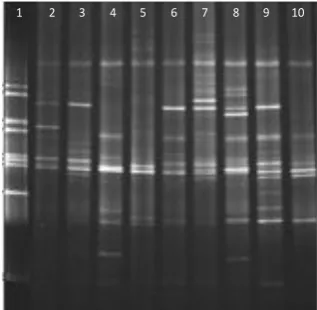
[image:30.595.245.404.443.598.2]DGGE has been one of the most commonly used techniques to study bacterial communities from diverse ecosystems (Muyzer et al., 1993). It is an electrophoretic method that can detect differences between DNA fragments of the same size but with different sequences. Since PCR products from a given reaction are of similar basepair size, conventional separation by agarose gel electrophoresis results only in a single DNA band that is largely non-descriptive. During DGGE, the PCR products encounter increasingly higher concentrations of chemical denaturants (formamide and urea) as they migrate through the polyacrylamide gel and PCR products will begin to denature at a threshold denaturant concentration. When DNA denatures, it opens in a domain which is anchored by a higher melting domain or domains that have not melted. At this point the migration rate is slowed down. When the fragments completely denature, the migration becomes a function of size. This technique is capable of identifying a single base change in a segment of DNA (Figure 1.1).
Figure 1.1: PCR-DGGE profiles of PCR-amplified bacterial 16s rDNA segments.
Lane 1: reference marker created from 16s rDNA of a number of bacterial
species, lanes 2-10 samples collected from the stimulated saliva of caries-free subjects where each band corresponds to a different bacteria, adapted from Li et al., 2005
30
1.8
DGGE and dental plaque
Traditional studies of oral microorganisms relied heavily upon cultivation methods. More recently a number of studies used the PCR-DGGE method to determine the microbial diversity in the oral environment. The microbial diversity in saliva and dental plaque for caries risk assessment and caries ecology were studied using DGGE and compared to the migration pattern of 25 known bacterial strains (Li et al., 2005). The 20 subjects were defined as caries free or caries active. The study found that the total cultivable bacterial counts and the total number of detectable bands on the DGGE was significantly higher in the caries-free group. However, there was no significant difference in the S.mutans, a prominent caries associated bacterium, levels between the two groups. The study suggested that a portion of the oral microbiota of the caries-active individuals may be absent, suppressed or replaced.
In another study that characterised the microflora of dental carious lesions by a combined cultural and molecular analysis (Munson et al., 2004). Five subjects whose carious dentine was removed and collected from the teeth were studied. The cultural analysis involved incubation of the samples on blood agar, Rogosa agar, and fastidious agar. The molecular analysis involved 16s rRNA gene isolation and sequencing. The results confirm the diversity of the bacterial community found in dental caries with 44 identified taxa out of the total of 95 taxa identified, 31 of which were not described previously.
31
plaque in a clinical single-blind study. Plaque samples from the interproximal area of molar teeth from twelve individuals with a mean age of 41.6 years were collected. A total of three samples were collected from each individual; baseline, 24 hours after prophylaxis and four days after tooth brushing. Samples from each individual were pooled and subjected to PCR-DGGE analysis. The results showed that the mean number of detected PCR amplicons was significantly different between the three groups, with samples collected from sites after brushing showing less amplicons, confirming the effect of fluoride on microbial growth (Li et al., 2006).
The PCR-DGGE approach was employed to identify as much of the endodontic bacterial community in the root canals of teeth with evidence of chronic apical periodontal lesions (Machado de Oliveira et al., 2007). Samples collected from eight single-rooted teeth were subjected to PCR-DGGE analysis and bands (total of 12) were removed from the gel and sequenced. The results of the sequencing analysis revealed the presence of both cultivable and uncultivable species. Six bands were closest to Fusobacterium species. Two bands were 97-99% matching to Bacteroides species. Two bands had high similarities to Prevotella. Three bands were identified as Escheirchia coli. Representatives of the genera Dialister, Synergistes, Eubacterium and Peptostrepococcus were also identified.
32
sequenced. A total of 92 distinct amplicons were detected from the overall DGGE profiles. On average, the total number of detectable bands was significantly higher in the caries-free group. The diversity of bacteria in the caries-free group was greater than in the caries-group. The overall conclusion was that the microbial diversity and complexity of the microbiota in dental plaque are significantly less in severe early-childhood caries than in caries-free children.
33
1.8.1
Limitations of the PCR-DGGE method
PCR-DGGE is a molecular method that has been shown to be reliable, reproducible, rapid and inexpensive (Muyzer 1999) but as with any other method it has its limitations. These limitations can be related to the PCR or the DGGE stages of analysis. It is limited to its resolution and sensitivity. Bias during DNA extraction and amplification of the 16s rRNA gene is another factor as it is based on the assumption that DNA is equally extracted from all bacterial species (Siqueira, Jr. and Rocas 2005; Suzuki and Giovannoni 1996). It relies on the quality and reproducibility of bacterial sample processing and DNA extraction. Also, only microorganisms that are present in relatively high concentrations are represented on the gel (Muyzer and Smalla 1998), therefore not all of the microbial population within a given habitat appear on the DGGE profile. Another limitation is the fact that single species with multiple rRNA gene copies can display a DGGE profile with multiple bands (Machado de Oliveira et al., 2007).
1.9
Effect of orthodontic appliances on the plaque
microflora
34
patients had significantly more S.mutans infected sample sites than did non-banded caries free subjects.
A longitudinal clinical study to investigate the influence of orthodontic brackets on the relative number of S.mutans in dental plaque where a number of plaque samples were collected before and during fixed orthodontic treatment over a period of one month. The percentage of S.mutans continued to increase from the last pre-bracket sample to the last pre-bracket sample (Mattingly et al., 1983).
Another study found that the levels of S.mutans and lactobacilli increase after the insertion of fixed orthodontic appliances and the use of chlorhexidine significantly reduces the numbers of lactobacilli. A difference in caries incidence on the buccal surfaces of bonded teeth in the treated group compared to the untreated controls was found, although it was not statistically significant (Lundstrom and Krasse 1987a; Lundstrom and Krasse 1987b).
The effect of orthodontic bands on the gingival tissues and the microbial composition of dental plaque was investigated on ten subjects undergoing orthodontic treatment using dark-field microscopy. The results showed a significant increase in the percentage of spirochetes, motile rods, filaments and fusiform and a decrease in cocci after banding accompanied by signs of gingivitis (Huser et al., 1990).
35
significantly in saliva after the insertion of fixed appliances. In plaque a greater number of microorganisms were found on the lateral incisor, which was attached to the arch-wire with an elastomeric ring compared to the incisor ligated with stainless steel wire (Forsberg et al., 1991).
A cross sectional study aimed to evaluate the levels of S.mutans in the saliva of patients before, during and after orthodontic treatment. Saliva samples were collected from 75 subjects at different stages of orthodontic treatment. The results showed that subjects in active orthodontic treatment had significantly higher total number and percentage of S.mutans compared to the subjects in retention, at post retention and the untreated age matched controls. The overall conclusion was that orthodontic treatment does not result in any long-term elevation of S.mutans levels (Rosenbloom & Tinanoff 1991).
The effect of certain orthodontic bonding composites and a glass ionomer cement on the adhesion of a strain of Streptococcus mutans was examined. Four different composites and one glass ionomer cement were tested for microbial growth. In vitro analysis showed that materials with rough surfaces attached more bacteria than ones with smooth surfaces. A stronger correlation was found between surface free energy of the material and the amount of S.mutans growth suggesting that surface free energy has a greater influence than surface roughness on bacterial colonisation (Blunden et al., 1994).
36
of in vitro supragingival plaque is not affected by the type of bonding material used, but biofilms formed over freshly made bonding materials known to release fluoride did not contain S.mutans (Badawi et al., 2003).
The prevalence of Candida and Enterobacteriaceae in a group of 50 adolescents during fixed orthodontic appliance therapy over only a 3 month follow-up period was measured. The results showed a significant increase in plaque index after the insertion of fixed orthodontic appliance and a significant increase in candidal number mainly C.albicans detected by the imprint technique (Hagg et al., 2004).
The short-term effect of the placement of orthodontic brackets on the subgingival micribiota and periodontal parameters was studied. The study lasted for three months only and included two groups of subjects: 30 patients who were about to receive orthodontic treatment and 30 untreated controls. Microbiological analysis of subgingival samples showed that scores for bleeding on probing, plaque index and gingival plaque index increased after bracket placement. The number of periodontopathic species was elevated following bracket placement (Naranjo et al., 2006).
37
The influence of orthodontic treatment on the numbers of opportunistic bacteria and fungi in the oral cavity of 42 patients was examined. It was found that the isolation frequencies of opportunistic bacteria and fungi increase during orthodontic treatment (Kitada et al., 2009).
A scanning electron microscope technique was used to quantitatively analyse the formation of bacteria on supra- and subgingival surfaces of orthodontic bands on 10 patients. The results showed that despite the presence of supragingival biofilm, no mature subgingival biofilm was found on the orthodontic bands. A demarcation line was found on all bands corresponding to a supragingival biofilm and the absence of a mature subgingival biofilm (Demling et al., 2009).
DNA probe analysis was used to describe the distribution of various levels of putative periodontal pathogens in child and adolescent population, and to monitor changes in the levels these pathogens during and after fixed orthodontic treatment. The results showed that the count of 6 of the pathogens increased significantly after 6 months of treatment but returned to pre-treatment levels by 12 months (Thornberg et al., 2009).
The caries risk factors in children undergoing orthodontic treatment with sectional brackets were investigated. The results showed that the levels of mutans streptococcus remained unchanged during and after active orthodontic treatment while the levels of lactobacilli in the caries high risk group were significantly increased (Sanpei et al., 2010).
38
disproportionate increase in S.mutans that would fall back to normal levels on removal of the appliance, and that materials used have some effect on bacterial levels where bands have similar effects as bonds.
1.10
Detection of initial caries lesions (white spot
lesions)
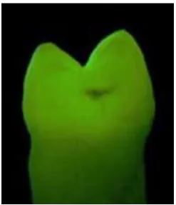
Due to the high prevalence of white spot lesion development in orthodontic patients, many clinical and laboratorial studies focussed on investigating this phenomenon using different techniques. For clinical examination, visual inspection is a non-invasive method of caries detection on accessible smooth surfaces. Figure 1.2 shows an example of white spot lesions as a result of poor oral hygiene and plaque accumulation at the gingival margin, whereas orthodontic white spot lesions are seen around the orthodontic bracket at the labial surface of the tooth.
39
Figure 1.2: The atypical clinical appearance of white spot lesions seen at the gingival margins of the upper incisors.
Conventional diagnostic techniques lack sufficient sensitivity and specificity for early detection of lesions and cannot provide information on caries activity (Choo-Smith et al., 2008).Various optical methods have been developed to quantify enamel demineralisation and remineralisation. These include non-fluorescent methods of photographs and optical caries monitor, and fluorescent methods relying on the use of ultraviolet or laser light, which can be a particularly dangerous form of radiation especially to the eyes (Angmar-Mansson and ten Bosch 1993).
40
The device produces light from an arc lamp (with a peak intensity of 370 nm) that passes through a blue filter a long a liquid light guide to a hand piece that can be directed at the tooth surface. Enamel auto-fluorescence is then detected using an intra oral camera. The reflected light passes through a yellow high pass-filter of 520 nm in front of the camera. The images are then stored, processed and analysed with a customised software (Inspektor Research System BV Amsterdam, The Netherlands) (Al-Khateeb et al., 1997) .
[image:41.595.260.384.80.224.2]Images obtained from digitally converted photographic slides were compared with those of QLF to measure enamel demineralisation surrounding an orthodontic bracket in vitro (Benson et al., 2003). It was found that both the area of demineralisation and a relative assessment of mineral loss of a white spot lesion surrounding an orthodontic bracket can be recorded and quantified reproducibly by either of the two techniques. The repeatability of using image analysis to measure
41
demineralisation from a photographic image is very similar to the technique of analysing a fluorescent image of the tooth using the customised software with QLF.
The prevalence and severity of white spot lesions in orthodontic patients after fixed appliance therapy were studied using QLF in comparison to visual inspection (Boersma et al., 2005). The results showed that the same distribution pattern of white spot lesions was detected with QLF and visual inspection. However, more lesions were found with QLF than with visual inspection and the difference between the percentages of caries affected surfaces in boys (40%) and girls (22%) were significant. Other factors such as age, treatment period or eating frequency did not show a significant correlation.
Longitudinal monitoring of the behaviour of white spot lesion directly after fixed appliance treatment as well as 6 weeks, 6 months and 2 years later by means of QLF in 51 subjects showed that many patients develop white spot lesions during fixed appliance treatment. A total of 351 carious surfaces were detected that did not resolve after de-bonding of the appliance. Out of those, 171 lesions remained stable, 145 lesions showed some improvement and 35 lesions got worse (Mattousch et al., 2007).
42
1.11
Prevention and treatment of white spot lesions
Enamel demineralisation prevention starts with tooth brushing instructions and diet advice to ensure that the patients have and are able to maintain an adequate level of oral hygiene throughout treatment. Other preventative methods include topical fluorides, oral irrigation systems, electric toothbrushes and sonic and ultrasonic toothbrushes. Boyd and co-workers (Boyd et al., 1989) studied the difference between a rotary electric tooth brush and commercial tooth brushes on the periodontal status during fixed orthodontic treatment. The results supported the fact that rotary electric toothbrushes can be more effective in maintaining the periodontal health of adolescents undergoing fixed appliance therapy.
Another factor that plays a role in prevention is the fluoride-releasing cementing agents. Glass ionomer cements and resin-modified glass ionomer cements have been shown to have a remineralisation effect on demineralised enamel adjacent to orthodontic bands and brackets (Basdra et al., 1996; Donly et al., 1995; Sadowsky et al., 1981). Despite these preventative measures white spot lesions still occur. In this case efforts should be focussed on remineralising these early lesions and preventing their progression.
43
stannous fluoride. Another agent that can have a remineralisation effect is casein phosphopeptide-amorphous calcium phosphate (CPP-ACP).
When enamel remineralises fluoride, calcium and phosphate ions penetrate the surface zone and precipitate on sound enamel at the margins of the subsurface demineralised lesion (Silverstone et al., 1988). The remineralisation process does not recreate enamel prisms and the remineralised enamel has a higher calcium, phosphate and fluoride content. For that reason it appears whiter than natural enamel.
A number of clinical and laboratorial studies were conducted to investigate the effect of professional topical treatments on the remineralisation potential of white spot lesions. Buchalla and co-workers (Buchalla et al., 2002) evaluated the fluoride uptake, retention and remineralising efficacy of a single dose application of highly concentrated fluoride solution (10,000 ppm) in a double blind, placebo controlled, randomised crossover in situ study and concluded that a single application of this highly concentrated fluoride solution increases enamel remineralisation.
Altenburger and colleagues (Altenburger et al., 2009) aimed to test whether 1.0% amine fluoride fluid is better than a 0.5% solution in terms of fluoride retention and mineral change in initial caries enamel lesion over a period of 28 days using a double-blind, placebo-controlled, randomised cross over in situ study. The results suggested that the use of 1% fluoride resulted in a significantly higher remineralisation compared with the use of 0.5% fluoride.
44
The results showed no significant difference between the three regimes on the remineralisation of early enamel carious lesions.
It is important to realise that fluoride is only effective as a remineralising agent in the early stages of lesion development and cannot achieve full remineralisation even with high concentrations. One difficulty with enamel remineralisation is the preferential remineralisation of the outer enamel surface which in turn can slow or prevent the complete remineralisation by restricting mineral ion diffusion into deeper regions of the lesion (Larsen and Fejerskov 1989; ten Cate et al., 1996; ten Cate and Duijsters 1983). ACP-CPP has been shown to slow the progression of caries significantly and to promote the regression of early lesions (Reynolds et al., 2003). More recent studies by Lata et al. (Lata et al., 2010) aimed to investigate the remineralisation potential of fluoride, ACP-CPP and the combination of both on early enamel lesions. The results showed that fluoride varnish is more effective than ACP-CPP cream in remineralising the early enamel lesion at the surface level and the combination of both does not provide any additive remineralisation potential. All three treatments were not effective in remineralising the lesion at the subsurface level.
45
46
Chapter 2
Aims
2.1
Aims
The aims of this study were to make use of novel diagnostic methods including Quantitative Light-Induced Fluorescence (QLF) and DNA amplification to identify any general changes in the bacterial composition of supragingival red fluorescent dental plaque in adolescents undergoing fixed orthodontic appliance therapy. A secondary aim was to determine the incidence of white spot lesions and if there was any relationship with the identified red fluorescent plaque.
2.2
Null Hypotheses
1. There is no difference in the general bacterial composition of red fluorescent supragingival dental plaque before and after the placement of fixed orthodontic appliances in adolescents undergoing fixed orthodontic appliance therapy.
47
Chapter 3
Study design
3.1
Study design and participants
3.1.1
Study design
A prospective non-interventional 12-month longitudinal cohort investigation of potential changes in the general composition of supragingival red fluorescent dental plaque and the incidence of white spot lesions in adolescents undergoing orthodontic treatment using fixed brackets.
3.1.2
Participants
A sample of thirty 11 to 23 year old patients scheduled to have fixed orthodontic appliance therapy at the Orthodontic Department of Liverpool University Dental hospital was to be recruited for this study. Based on a recommendation from a statistician, the sample size was selected on the basis of the probability of identifying plaque constituents, which are common in the target population. A sample size of 30 will give a 95% probability of identifying plaque constituents, which occur in at least 10% of the target population.
48
3.1.3
Exclusion criteria
Due to the effect of antibiotics on plaque bacterial microflora, any participant who had received antibiotic therapy in a period less than eight weeks prior to their participation in the study, or at any time during the study, were excluded.
3.2
Ethical approval
Ethical approval was granted by the North West Ethics Committee, Liverpool; a representative of the National Research Ethics Committee (reference 09/H1005/63). Approval from the research and development department of the Royal Liverpool and Broadgreen University Hospitals Trust was also obtained (reference 3822). The University of Liverpool acted as a co-sponsor with the Royal Liverpool and Broadgreen Hospitals NHS Trust (reference UoL000454).
49
Chapter 4
Identification and collection of red fluorescent
plaque
Red fluorescence of plaque is thought to originate from porphyrins, which are by-products of bacterial metabolism (Coulthwaite et al., 2006, Pretty et al., 2005). When plaque is present in large amounts on tooth surface, it fluoresces deep red or bright orange (Pretty et al., 2005, van der Veen and de Josselin de Jong 2000). Obligate anaerobes are the source of red fluorescence and cariogenic streptococci are the source of green fluorescence (Verran and Whitehead 2005; Coulthwaite et al., 2006), section 1.6.5.
4.1 Method
4.1.1
Identification of red fluorescent plaque
50
[image:51.595.211.427.69.210.2]The site from which the dental plaque was collected from was noted on a chart (Figure 4.2). When no red fluorescent plaque could be identified, none of the plaque was collected, even if gross plaque deposits were clearly seen without the use of ToothcareTM.
Figure 4.2: The chart used to note the position of the red fluorescent plaque once identified with ToothcareTM.
[image:51.595.213.429.417.538.2]51
4.1.2
Collection of red fluorescent plaque
Once the red fluorescent plaque was identified on a single tooth’s surface, a sterile dental instrument was used to remove the plaque from the tooth surface. The collected plaque was transferred into a sterile Eppendorf® 1.5 ml microcentrifuge tube. The plaque sample was kept on ice on clinic until it was ready to be transferred and kept at -20o C for further analysis.
4.2
Results
52
Table 4.1: Number of visits and red fluorescent plaque samples collected per participant if identified with ToothcareTM.
Participant 1 2 3 4 5 6 7 8 9 10 11 12 13 14
Number of visits 7 8 9 9 5 7 6 5 9 7 8 9 8 7
Total number of red fluorescent plaque samples
collected.
7 7 2 9 4 5 6 1 9 6 8 8 7 1
53
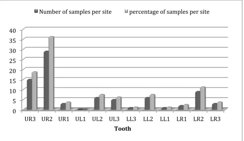
Figure 4.3: The distribution of red fluorescent plaque collected from each tooth.
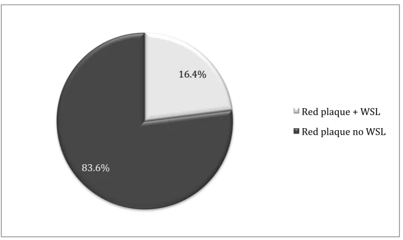
16.4% of the sites where red fluorescent plaque was detected (n=80) showed signs of demineralisation identified with QLF, and 83.6% of the sites where red fluorescent plaque was detected did not show any signs of demineralisation when assessed using QLF (Figure 4.4). The overall incidence of white spot lesions was not related to the presence of red fluorescent plaque when analysed using chi square test, resulting in a p value >0.05 (x2= 3.3) (Figure 4.5).
>! .! ">! ".! />! /.! 4>! 4.! :>!
=+4! =+/! =+"! =)"! =)/! =)4! ))4! ))/! ))"! )+"! )+/! )+4! ,GGHI.
54
"9#:X!
@4#9X!
+RZ!LVG[PR!\!C-)! +RZ!LVG[PR!MU!C-)!
@#AX!
/:#:X!
""#:X!
..#4X!
+RZ!LVG[PR!\!C-)!
+RZ!LVG[PR!MU!C-)! C-)!MU!HRZ!LVG[PR!
-UPMZ!!
Figure 4.4: The relationship between red fluorescent plaque and white spot lesions as a percentage of the total number of tooth surfaces with red fluorescent plaque.
[image:55.595.112.508.73.309.2] [image:55.595.108.496.407.637.2]55
Chapter 5
Preliminary dental plaque analysis
PCR amplification of conserved regions of the 16s rRNA gene followed by DGGE analysis has been widely used as molecular biology methods of isolation and identification of dental plaque bacteria. Molecular techniques are used to overcome the limitations encountered when using cultivation-based techniques (section 1.7).
I. Methods
5.1
Total bacterial DNA extraction
5.2.1
Isolation of genomic DNA
To isolate the bacterial genomic DNA, MasterPureTM Complete DNA and RNA Purification Kit (Epicentre Biotechnologies) was used. The following protocol that was provided with the purification kit (Epicentre Biotechnologies) was modified and then followed to lyse the cells and release the DNA from the nuclei:
1. The collected frozen plaque sample was thawed and re-suspended in 1ml of DNAse free water.
2. Five µl of Proteinase K solution (0.18 mg/ml, [50 mM Tris-Hcl (pH 7.5)], 100 mM NaCl, 0.1 mM EDTA, 10 mM CaCl2, 0.1% Triton® X-100, 1 mM
dithiothreotol in 50% glycerol solution), and 30µl of 10% sodium dodecyl sulphate (SDS, 0.5%) were added to the sample and mixed by inversion. 3. The sample was then incubated in a water bath at 55o C for 1 hour and
56
4. The cells were then pelleted by centrifugation; the supernatant discarded leaving approximately 25µl of liquid.
5. The pelleted cells were then re-suspended by vortexing for 10 seconds. 6. For each sample, 3µl of Proteinase K was diluted into 300µl of Tissue and
cell lysis solution. The solution was added to the sample and mixed thoroughly.
7. The sample was then incubated in a water bath at 65o C for 15 minutes, mixed every 5 minutes by vortexing.
8. The sample was then cooled to 37o C, by placing it in a water bath, for 10 minutes.
9. Three µl of 5µg/µl RNase A was then added to the sample and mixed thoroughly.
10.The sample was incubated at 37o C for 30 minutes.
11.The sample was then left on ice for 3-5 minutes before proceeding with total DNA precipitation.
5.2.2
Total DNA precipitation
Following the isolation of the bacterial genomic DNA the following was undertaken to precipitate the DNA and remove any bacterial protein from the sample:
57
2. The debris (denatured protein) was pelleted by centrifugation at 4oC for 10 minutes at !10,000 x g in a micro-centrifuge. If the resultant pellet was clear, small or loose, an additional 25µl of MPC protein precipitating reagent was added to the sample, mixed vigorously and centrifuged to pellet the protein again.
3. The pellet was then discarded, and the clear supernatant transferred to a clean micro-centrifuge tube.
4. 500µl of cold isopropanol was added to the recovered supernatant. The tube was then inverted 30-40 times.
5. The DNA was pelleted by centrifugation at 4oC for 10 minutes at ! 10,000 x
g in a micro-centrifuge.
6. The isopropanol was carefully discarded without disturbing the DNA pellet. The DNA pellet was then left to air dry.
7. Finally the pellet was re-suspended in 10µl of TE (10 mM Tris-HCl [pH 7.5], 1 mM EDTA) buffer and stored at 4oC (if required) for further analysis.